Embed Size (px)
Citation preview
601
Myelopathy and Radiculopathy Due to Cervical Spondylosis: Myelographic-CT Correlations Giuseppe Scotti, 1 Giuseppe Scialfa, Sandra Pieralli , Edoardo Boccardi , Frieda Valsecchi , and Cristina Tonon
Forty patients with symptoms and signs of radicular disease or spinal cord involvement secondary to cervical spondylosis were studied with myelography (using non ionic water-soluble contrast medium) followed by computed tomographic (CT) myelography. In 17 patients CT was also performed before myelography. CT myelography adds useful information to the myelographic findings. Cord compression is better evaluated and osteophytes can be differentiated from disk herniation. Plain CT can demonstrate a herniated disk but with less accuracy than CT myelography. Cord and root compression are not seen directly on plain CT; for this reason myelography should be the first procedure in patients with myelopathy or myeloradiculopathy, which may be followed by CT myelography.
Computed tomography (CT) has substantially modified the neuroradiologic approach to the diagnosis of herniated disk and spondylosis in the lumbar region [1 -5]. But examination of the cervical spine by CT requires a diffe rent approach than in the lumbar region . Thinner intervertebral disks, different anatomic landmarks (less or no epidural fat) , and a greater frequency of osteophytes and narrowing of the canal th an in disk hern iations are some of the distinguishing concerns of the cervical spine.
Clinical presentation and differential diagnosis are also different. Pure cervical radiculopathy (n eurologic defi c it due to nerve root compression) is less common and occurs in younger patients . Myelopathy (neurologic defi c it due to spinal cord compression) of different deg rees of severity , ranging from simple hyperreflexia to severe progressive paraparesis, is more common and may be associated with signs of radicular involvement. Clinical signs of spinal cord involvement may be mistaken as indicating the presence of chronic degenerative disease of the spinal cord (e.g., amyotrophic lateral sclerosis) or of a tumor in or adjacent to the cord.
With cervical spine CT, there is a need not only to visualize the bony and soft-tissue structures surrounding the spinal canal and to study more levels than in the lumbar reg ion, but also to recognize the effects of spondylosis on the spinal cord and nerve roots. CT slices must be thinner than in the lumbar reg ion in order to visualize the intervertebral disk and neural foramina.
This study c'ompares the myelog raphic and CT findings in patients with cervical spondylosis in order to assess the values and lim itations of CT performed wi thout and with subarachnoid contrast enhancement, and to define the role of CT in the d iagnostic evaluation of patients with cervical myelopathy and / or radiculopathy.
Materials and Methods
Forty patients (30 men and 10 women, mean age 56 years, range 30-74 years) with symptoms and signs of radicular or spinal cord disease secondary to cervical spondylosis were examined . The clinical presentat ion was that of radicular pain in 13 patients. In the other 27 patients signs of myelopathy (u sually slowly progressive) were the major complaint. These were associated with radiculopathy in 13 patients.
All patients were investigated by means of myelograph y, using 15 ml of the non ionic water-soluble contrast medium iopamidol (iopamiro 300, Bracco , Milan, lIaly) in a concentration of 300 mg 1/ ml introduced via lumbar puncture. CT was pe rfo rmed with in 2 hr after myelography in all patients . In 17 patients , plain CT was also performed before myelog raphy. A Siemens Somatom 2N scanner or a GE CT / T 8800 unit was used for the CT stud ies. An average of three levels were examined, selected on th e basis of c linica l signs or myelographic findings . Eight to 12 contiguous slices (1.5 or 2 mm th ick ness) per level were done. The levels were always localized by means of a preliminary dig ital radiograph in the lateral projection .
Resu lts
Myelograph ic evidence of nerve root compression and / or of anterior dural sac compression by herniated disk or osteophytes was demonstrated in all patients. A single level was involved in 13 patients (C3- C4, one; C4-C5, three; C5-C6, six; C6-C7 , two; C7 -T1 , one). In the other 27 patients, mu ltip le levels were involved (C3- C4, seven; C4-C5, 14; C5-C6, 22; C6-C7, nine; C7 - T1 , none).
When CT is performed after myelography with the subarachnoid spaces opacified by contrast medium (CT myelography), anterior compression of the dural sac can be c learl y visualized. If there is no space between the posterior margin of the vertebral body and the anterior surface of the sac , the compression is due either to osteophyte (fi g . 1) or to ossif ication of the posterior long itudinal
ligament. Otherwise, a herniated disk can be suspected. At CT myelography, anterior compression of the dural sac by osteophytes was found in 27 patients and by d isk herniation in 18. The spinal cord outlined by contrast medium can be c learly seen. Compression and deform ity of the cord can be demonstrated and may be severe, with reduct ion of the anteroposterior diameter of the cord even to
I All auth0rs: Ospedale Niguarda, Cil Granda, Department o f Neuroradiology, Piazza Ospedale Maggiore 3, 20162 Milan, Italy. Address reprin t requests to G. Scotti.
AJNR 4 :601-603, May/ June 1983 01 95-61 08 / 83 / 0403-0601 $ 00.00 © American Roen tgen Ray Society
602 SPINE AND INTERVERTEBRAL DISK AJNR:4, May/ June 1983
A B
Fig. 1 .-Cervical spine of 55-year-old woman with progressive paraparesis in last 2 years. A, Plain CT. Large osteophyte protruding into spinal canal. B, CT myelogram demonstrates severe degree of cord compression by impinging osteophyte.
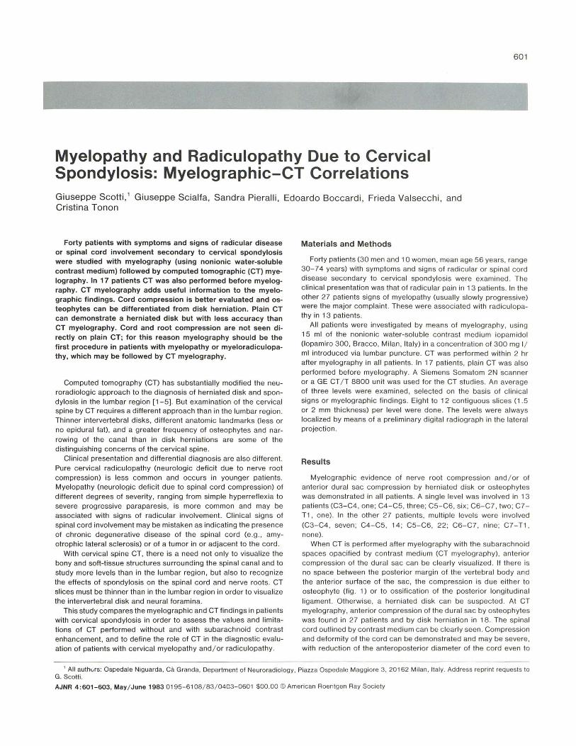
A B
c D Fig. 2. - CT myelographic images. A, Normal spinal cord and nerve roots
with in neural foramina. B, Spinal cord compression by hern iated disk. C, Osteophyte impinging on right anterior aspect of opacified sac. D, Narrow canal and hypertrophy of ligaments.
one-third of normal in some cases (fig. 2) . Clear evidence of cord compression was found in 24 cases. Nerve root sheaths opacified by contrast medium can be easi ly followed to the neural foramina, and compression or amputation was found in 19 cases.
In the 17 patients examined by plain CT before myelography, th ere was c lose correspondence between pre- and postcontrast CT in demonstrating narrowing of the spinal canal and neural foramina
by osteophytes, seen in 12 cases by both methods. Disk herniation was recognized in eight cases on CT with intrathecal contrast medium, but on ly in five by plain CT. Cord compression was visualized in 10 patients on CT images with subarachnoid contrast med ium, but could not be demonstrated on plain CT. The same was
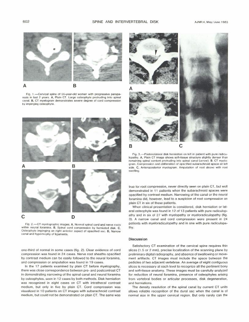
B c Fig. 3.-Posterolateral disk herniation on left in patient with pure radicu
lopathy. A, Plain CT image shows soft-tissue structure slightly denser than remaining spinal content protruding into spinal canal (arrow). B , CT myelogram. Compression and obliteration of opac:ified subarachnoid space on left side. C, Anteroposterior myelogram. Amputation of root sleeve with root swelling.
true for root compression, never directly seen on plain CT, but well demonstrated in 11 patients when the subarachnoid spaces were opacified by contrast medium. Narrowing of the canal or the neural foramina did , however, lead to a suspicion of root compression on plain CT in six of these patients.
When c linical presentation is considered, disk herniation or lateral osteophyte was found in 12 of 13 patients with pure radiculopathy and in six of 27 with myelopathy or myeloradiculopathy (fig. 3). A narrow canal and cord compression were present in 24 patients with myeloradiculopathy and in one with pure radiculopathy.
Discussion
Satisfactory CT examinat ion of the cervical spine requires thin slices (1.5 or 2 mm), precise localization of the scanning plane by preliminary digital radiography , and absence of swallowing or movement artifacts. CT images must include the space between the pedicles of two adjacent vertebrae. An average of eight contiguous slices is necessary at each level to recognize all the pertinent bony and soft-t issue anatomy. These images must be carefu lly analyzed for reduction of neural foramina, presence of osteophytes arising from vertebral bodies or articular processes, disk degeneration, and herniations.
The density resolution of the spinal canal by current CT units allows reliable recognition of the dural sac when the canal is of normal size in the upper cervical region. But only rarely can the
AJNR:4, May / June 1983 SPINE AND INTERVERTEBRAL DISK 603
spinal cord be visualized on plain CT below the level of C2 . Disk herniation may be defined as a soft-t issue structure protruding posteriorly from the disk with a density higher than the remaining structures within the spinal canal.
When a prominent osteophyte is demonstrated with resu ltant narrowing of the spinal canal, the dural sac is seen less well on plain CT, and spinal cord compression can only be suspected, not proven . CT myelography is far superior to plain CT in demonstrating spinal cord and root compression. The spinal cord is sometimes severely narrowed, and the cross-sectional ax ial plane is best to show the extent of compression and the relation of the dural sac to the wal ls of the spinal canal.
The presence of subarachnoid contrast medium allows differentiation between compression due to osteophyte or to disk herniation . In a given slice, when the dural sac is displaced posteriorly and no osteophytes are seen at that level or at the levels above and below,
one may assume the comporession is due to a soft hernia . Ossification of the posterior longitudinal ligament was identified
in three of our patients. In th ese cases, CT was crucial in demonstrating the thickness and extent of the ossificat ion behind the vertebral body, as it was with a thin line of separation between the ossification and the posterior surface of the vertebral body. This information was essential to the neurosurgeon in planning an op
erative approach to decompress the affected portion of the spinal cord.
Myelography plays a basic role in patients with cerv ical myelopathy and / or radicu lopathy in showing the spine and spinal cord in its full extent and in demonstrating multiple levels of involvement, dynamic changes with movement, and partial block with the head hyperextended due to posterior compression by infolding of the ligamenta !lava. Water-soluble contrast medium is well tolerated
and provides excellent visualization of the nerve roots and root sleeves.
We believe the diagnostic protocol shou ld be tailored accord ing to the patient 's clinical presentat ion. In patients with isolated signs of pure radicular compression and with pain and sensory defi c it at a well defined level, plain CT should be the first examination . Digital radiography in the lateral projection for localizing purposes could
replace plain x-ray films. The CT examination shou ld inc lude the intervertebral space cl inically involved and one level above and below. If a disk hern iation impinging on the neural foramen and compressing the nerve root or a narrowing of the neural foramen by a lateral osteophyte is seen, myelography can be avoided .
In patients with myelopathy or myelorad iculopathy a myelogram must always be obtained since th e pathological levels may be
multiple. Identical symptomatology can be due to other causes including tumors and chronic degenerative diseases of the spinal cord . CT myelog raphy provides useful complementary information on the exact location and extent of osteophytes and the degree of cord com pression. CT before myelog raphy in th ese cases does not seem at present to provide suffic ient information to avoid myelography.
REFERENCES
1. Haughton VM , Williams AL. Computed tomography of the spine. SI. Louis: Mosby, 1982
2. Willi ams AL, Haughton VM . CT appearance of bulging annulus. Radiology 1982; 142 : 403-408
3. Will iams AL, Haughton VM , Syvertsen A. CT in the d iagnosis of herniated nuc leus pulposus. Radiology 1980; 135: 95-100
4. Carrera GF, Williams AL , Haughton VM. Computed tomography in sciatica. Radiology 1980;137: 433-437
5 . Federle MP, Moss AA, Marjohn FL. Role of computed tomography in patients with sciatica. J Comput Assist Tomogr 1980;4: 335-341