Embed Size (px)
Citation preview
LUND UNIVERSITY
PO Box 117221 00 Lund+46 46-222 00 00
Mycotoxins in indoor environments. Determination using mass spectrometry.
Bloom, Erica
2008
Link to publication
Citation for published version (APA):Bloom, E. (2008). Mycotoxins in indoor environments. Determination using mass spectrometry. Lund University.
Total number of authors:1
General rightsUnless other specific re-use rights are stated the following general rights apply:Copyright and moral rights for the publications made accessible in the public portal are retained by the authorsand/or other copyright owners and it is a condition of accessing publications that users recognise and abide by thelegal requirements associated with these rights. • Users may download and print one copy of any publication from the public portal for the purpose of private studyor research. • You may not further distribute the material or use it for any profit-making activity or commercial gain • You may freely distribute the URL identifying the publication in the public portal
Read more about Creative commons licenses: https://creativecommons.org/licenses/Take down policyIf you believe that this document breaches copyright please contact us providing details, and we will removeaccess to the work immediately and investigate your claim.
Download date: 02. Apr. 2022
Mycotoxins in Indoor Environments Determination using Mass Spectrometry
Erica Bloom
2008
Doctoral Thesis
Department of Laboratory Medicine Division of Medical Microbiology
Lund University Sweden

Cover picture by Eric Carleman and Erica Bloom. Scanning electron microscope picture of Stachybotrys chartarum. © Erica Bloom 2008 Mycotoxins in indoor environments. Determination using Mass Spectrometry. ISBN: 978-91-86059-65-1 ISSN: 1652-8220 Lund University. Faculty of Medicine Doctoral Dissertation Series 2008:112. Printed by Media-Tryck, Lund 2008.
- 2 -

“The pure and simple truth is rarely pure and never simple.”
Oscar Wilde

TABLE OF CONTENTS
LIST OF PAPERS..............................................................................................- 6 - ABSTRACT........................................................................................................- 7 - POPULÄRVETENSKAPLIG SAMMANFATTNING ....................................- 8 - ABBREVIATIONS.......................................................................................... - 10 - INTRODUCTION.......................................................................................... - 13 -
INDOOR AIR QUALITY AND HEALTH.........................................................................- 13 - MICROORGANISMS IN WATER DAMAGED INDOOR ENVIRONMENTS .........................- 15 - EXPOSURE ASSESSMENT METHODS FOR MOLD ........................................................- 16 - MOLDS AND THEIR BIOLOGICALLY ACTIVE METABOLITES.......................................- 18 - DETECTION OF MYCOTOXINS................................................................................. - 26 -
Mass Spectrometry........................................................................................................ - 27 - GC-MS ............................................................................................................................ - 29 - HPLC-MS........................................................................................................................ - 31 - MS detection of mycotoxins .................................................................................................... - 33 -
AIMS OF THE STUDY................................................................................... - 35 - MATERIAL AND METHODS ...................................................................... - 37 -
SAMPLES ................................................................................................................. - 37 - SAMPLING AND CULTIVATION ................................................................................ - 38 - MICROSCOPY .......................................................................................................... - 38 - QPCR ..................................................................................................................... - 39 - CHEMICAL EXPERIMENTS....................................................................................... - 39 -
Extraction .................................................................................................................. - 39 - Purification and hydrolysis ............................................................................................. - 40 - Derivatization (for GC-MS).......................................................................................... - 41 - GC-MS ..................................................................................................................... - 42 - HPLC-MS ................................................................................................................ - 45 -
SUMMARY OF RESULTS.............................................................................. - 49 - MASS SPECTROMETRY ............................................................................................ - 49 - OVERVIEW OF ALL ANALYZED SAMPLES .................................................................. - 50 - ADDITIONAL COMMENTS........................................................................................ - 52 -
DISCUSSION .................................................................................................. - 53 - MOLD IN BUILDINGS - GUIDELINES AND REGULATIONS......................................... - 53 - THIS STUDY IN CONTEXT........................................................................................ - 56 -
CONCLUSIONS.............................................................................................. - 59 - ACKNOWLEDGEMENTS............................................................................. - 61 - REFERENCES................................................................................................ - 63 - PAPERS I-IV.................................................................................................... - 81 -
- 5 -
LIST OF PAPERS
This thesis is based on the following publications, which are referred to in the text by
their Roman numerals:
I. Bloom, E., K. Bal, E. Nyman, and L. Larsson. (2007). Optimizing a GC-MS
method for screening of Stachybotrys mycotoxins in indoor environments.
J Environ Monit. 9(2): 151-156.
II. Bloom, E., K. Bal, E. Nyman, A. Must, and L. Larsson. (2007). A mass
spectrometry-based strategy for direct detection and quantification of some
mycotoxins produced by Stachybotrys spp. and Aspergillus spp. in indoor
environments. Appl Environ Microbiol. 73(13): 4211-7.
III. Bloom, E., L. F. Grimsley, C. Pehrson, J. Lewis, and L. Larsson. (2008).
Molds and mycotoxins in dust from water-damaged homes in New Orleans
after Hurricane Katrina. Indoor Air. In press.
IV. Bloom, E., E. Nyman, A. Must, C. Pehrson, and L. Larsson. (2008). Molds
and mycotoxins in indoor environments. A survey in water-damaged buildings.
Submitted.
Paper I is reproduced by permission of The Royal Society of Chemistry.
Paper II is reproduced by permission of The American Society for Microbiology.
Paper III is reproduced by permission of Wiley-Blackwell Publishing.
- 6 -
ABSTRACT
Dampness in indoor environments may cause various health problems. The specific
causative agent(s) are unknown but may originate from degradation processes in damp
materials, microbial growth, or a combination of these phenomena. The health impact
of dampness in buildings is a politically, legally, and economically important question.
Scientists at the U.S. EPA and Lawrence Berkeley National Laboratory have estimated
that the symptoms of 4.6 of the total of 21.8 million asthmatics in the USA are caused
by indoor dampness and mold to an annual cost of 3.5 billion dollars.
Mycotoxins are secondary metabolites produced by molds which may be e. g.
cytotoxic (e. g. macrocyclic trichothecenes produced by Stachybotrys chartarum), genotoxic
(e. g. sterigmatocystin and aflatoxins produced mainly by Aspergillus spp. including A.
versicolor and A. flavus), or immunosuppressive and neurotoxic (e. g. gliotoxin produced
by Penicillium spp. and Aspergillus spp. e. g. A. fumigatus). Airborne mycotoxins have been
demonstrated in water-damaged buildings using both ELISA and mass spectrometry.
However, whether mycotoxins at the concentrations found in mold-damaged
environments represent a health risk upon inhalation is not known. The mechanisms
for mycotoxin uptake, metabolism, and interaction e. g. with other fungal constituents
such as proteins and (1→3)-β-D-glucan (a fungal cell membrane constituent) are poorly
understood.
In this project analytical methods for the detection and determination of selected
mycotoxins using GC-MS and HPLC-MS were developed. The methods were applied
to 167 mold-contaminated building material samples, of which 67 % were mycotoxin
positive. Thus, many molds not only posess the genetic capacity to produce
mycotoxins but do it regularly in water damaged indoor environments. In addition, we
demonstrated mycotoxins in dust settled in the breathing zone in indoor environments
where severe mold-contamination was identified on building materials. We thereby
confirm that mycotoxins on such materials can become airborne and thus inhalable.
This project is an example of fruitful national and international inter-disciplinary
collaboration between the building industry, companies specialized in remediation
measures, and universities.
- 7 -
POPULÄRVETENSKAPLIG SAMMANFATTNING
Dålig ventilation, fukt, hög förekomst av partiklar i luften samt damm i inomhusmiljö
kan relateras till en rad luftvägsbesvär. Framförallt är fukt i byggnader sammankopplad
med ohälsa och obehagssymptom. Fukt kan reagera med olika byggmaterial och bilda
irriterande flyktiga produkter samt även resultera i mikrobiell tillväxt, framför allt av
mögel. Amerikanska forskare har uppskattat att symptomen hos 4,6 av USA:s 21,8
miljoner astmatiker orsakas av fukt och mögel och att detta kostar det amerikanska
samhället årligen 3,5 miljarder dollar. Trots att det finns nästintill otaliga rapporter om
samband mellan mögel i inomhusmiljöer och ohälsa har man ännu inte på ett
vetenskapligt sätt definitivt lyckats knyta mätbara exakta mikrobiologiska parametrar t.
ex. astma/allergi. Det är alltså tyvärr ännu oklart vilka ämnen, som bildas vid förhöjd
fukthalt, som ger besvär, bl. a. för att det saknas tillgång till pålitlig detektionsmetodik.
I Sverige har en långlivad debatt pågått om mögel, mögelgifter (s. k. mykotoxiner) och
dess betydelse för vår hälsa, men det är få i världen som i praktiken forskar på just
mögelgifter.
Man vet att mögelsvampar som kan växa till inomhus har en förmåga att producera
mykotoxiner, ämnen som är oerhört toxiska (bl. a. cancerframkallande, vävnads-
förstörande och inflammationsinducerande). Dock har man ansett att mängden
mykotoxin som vi exponeras för (baserat på antal sporer man funnit i mögelskadade
hus) är så liten att den rimligen inte kan ha någon inverkan på vår hälsa. Bakgrunden
till detta projekt är att nya, omvälvande data har visat att mykotoxiner – förutom att de
är direkt toxiska - även påverkar immunceller i en riktning som innebär ökad risk för
allergibenägenhet och att det krävs oerhört små mängder (pikogramnivå) för att
framkalla dessa reaktioner. Dessutom har det visats att mögelsvampar frigör mycket
små partiklar (små hyf-fragment) som aldrig sedimenterar. Dessa partiklar är mycket
mindre -och deponeras mycket effektivare i lungorna - än sporer. Därmed kan vår
exponering för mögel vara flera hundra gånger större än vad som tidigare beräknats.
Denna exponering pågår konstant under en stor del av dygnets timmar (vi vistas
inomhus ca 90 % av vår tid), något som inte alltid tagits hänsyn till i tidigare studier.
Målsättningen med detta projekt har varit att utveckla nya analytiska metoder för
- 8 -
bestämning av några utvalda mykotoxiner och att tillämpa dem på en mängd olika
prover från mögelskadade inomhusmiljöer. Delar av byggnadsmaterial (synbart
angripna lister, trösklar, prov av gipsplattor, tapet etc.) samt dammprover, främst från
golvytor och hyllor, insamlades från mögelskadade hus via professionella skadeutredare
samt andra universitet som vi samarbetat med, i exempelvis New Orleans (USA).
Extrakt av dessa prover renades, separerades med vätske- (HPLC) eller
gaskromatografi (GC) och analyserads med masspektrometri (MS), en analytisk kemisk
metod som bl a också används för dopinganalyser och för kriminaltekniska ändamål.
I HPLC-MS-analyserna detekterades sterigmatocystin och alfatoxin B1 (som bildas av
Aspergillus-arter), gliotoxin (som bildas av Aspergillus- och Penicillium-arter), samt de
makrocykliska trikotecenerna satratoxin G och H (som bildas av Stachybotrys chartarum).
I GC-MS-analyserna utnyttjades det faktum att de makrocykliska trikotecenerna, som
produceras av S. chartarum kemotyp S, vid hydrolys bildar verrukarol medan det
inflammationsinitierande mykotoxinet trikodermin, som produceras av S. chartarum
kemotyp A, bildar trikodermol. Genom att bestämma både verrukarol samt
trikodermol kan man således ”screena” prover för mykotoxiner producerade av S.
chartarum. Mängden svampbiomassa mättes också genom att bestämma en
svampmarkör, ergosterol, en molekyl som i naturen unikt återfinns i svampars
cellmembran.
Våra analyser visar att de mögelsvampar som projektet främst fokuserat på, och som
ofta återfinns i inomhusmiljöer i samband med fuktskada, inte bara har förmågan att
producera mögelgifter –de gör det regelmässigt. Dessutom har vi bekräftat att
mögelgifter som härrör från synliga mögelfläckar på angripna byggmaterial kan bli
luftburna och att vi därmed inandas dem. De metoder som utvecklats i projektet
kommer i framtiden att kunna användas som verktyg i studier bl. a. syftande till att
utröna mögelgifternas eventuella inverkan på vår hälsa. Det tvärvetenskapliga
samarbetet inom projektet har varit mycket givande och illustrerar nyttan av kontakter
mellan byggbranschen, skadeutredare samt forskare inom olika discipliner i
inomhusmiljöforskningen.
- 9 -
ABBREVIATIONS
AFLAB Aflatoxin B1
BSTFA N, O-Bis(trimethylsilyl)trifluoroacetamide CDC Center for disease control and prevention CFU Colony forming units CI Chemical ionization CID Collision induced dissociation DNA Deoxyribonucleic acid EI Electron ionization ELISA Enzyme linked immunosorbent assay EPA Environmental protection agency ERG Ergosterol ESI Electrospray ionization GC Gas chromatography GLIO Gliotoxin HFB Heptafluorobutyryl HFBI Heptafluorobutyrylimidazole HPLC High pressure liquid chromatography Ig Immunoglobulin INF-γ Interferon gamma IL- Interleukine LPS Lipopolysaccharide MEA Malt extract agar MS Mass spectrometry MSMS Tandem mass spectrometry MSTFA N-Methyl-N-trimethylsilyltrifluoroacetamide MTR Macrocyclic trichothecene MVOCs Microbial volatile organic compounds NICI Negative ion chemical ionization PBMCs Periferal blood mononuclear cells (q)PCR (quantitative/real-time) Polymerase chain reaction PICI Positive ion chemical ionization RCS Reuter centrifugal sampler RESP Reserpine RNA Ribonucleic acid SATG Satratoxin G SATH Satratoxin H
- 10 -
SIM Selected ion monitoring sp. / spp. Species (singular form/plural form) STRG Sterigmatocystin TCMS Trimethylchlorosilane TFA Trifluoroacetic acid Th cell T-helper cell TMS Trimethylsilyl TNF-α Tumour necrosis factor alpha TRID Trichodermol TSIM N-trimethylsilylimidazole VER Verrucarol q / Q Quad
- 11 -

- 12 -
Introduction
INTRODUCTION
Indoor air quality and health
We spend up to 90% of our time indoors. The indoor air quality has been recognized
to have a great impact on our health and well-being. During the energy crisis in the
70’s construction procedures changed and buildings were tightly sealed, ventilation
rates reduced, and new materials introduced to minimize energy, and financial, loss.
The resulting changes in indoor air quality, together with other characteristics of our
western lifestyle, have been associated with the significant increase of allergic diseases
in Sweden (Aberg 1989, Aberg et al 1995) and the world (Beasely et al 2003) over the
last decades.
In buildings, moisture and dampness (Bornehag et al 2001), poor ventilation, dust,
and high concentration of particles in the indoor air is associated with a number of
respiratory problems (Mommers et al 2005). Still, the specific causes of the adverse
health effects are unknown. However, it is likely that the components involved in
health related symptoms originate from:
• chemical processes catalyzed by water causing degradation of indoor materials
resulting in production of potentially irritating volatile organic compounds.
• growth of microorganisms which contain, or produce, bioactive compounds.
• a combination of the above.
Dampness, and the combination of water leakage and PVC flooring in the bedroom,
was shown to be associated with increased prevalence of symptoms in airway, nose and
skin among over 10,000 preschool children in Sweden (Bornehag et al 2005). Further,
there are studies linking chemical exposures such as formaldehyde, molds and bacteria,
and furry pet allergens (Daisy et al 2003, Tranter et al 2005) to health effects including
asthma and allergic symptoms, airway infections, and impaired learning ability (Daisy et
al 2003, Mendell and Heath 2005). Also, phthalate plasticizers in dwellings have been
associated with asthma and allergic symptoms in children (Bornehag et al 2004, Kim et
al 2006).
- 13 -
Introduction
The hygiene hypothesis speculates that a lack of early childhood exposure to
infectious agents, symbiotic microorganisms, and parasites increases susceptibility to
allergic diseases by impairing immune system development. Factors as pet keeping,
day-care attendance, family size, geographical differences, home building construction,
urban vs. rural home environment etc, may thus influence health status later in life.
Some studies support this hypothesis as pet exposure during the first year of life seems
to be health protective (Hesselmar et al 1999). On the other hand, a study on 10,851 1-
6 year-old children in Sweden showed that day-care attendance was not protective
against any classical allergic symptoms, e. g. wheezing, cough, asthma, rhinitis, eczema
etc (Hägerhed-Engman et al 2006). Thus, some studies tend to support the hygiene
hypothesis and some seem to contradict it. One explanation may be that the hygiene
hypothesis is valid or not depending on the microbial flora that we are exposed to.
Interestingly, short chain lengths of 3-hydroxy fatty acids from LPS (of Gram-negative
bacteria) have related to protective effects for asthmatic symptoms while longer chain
lengths have related in the opposite way. Short carbon chain lengths of 3-hydroxy fatty
acids (C10, C12 and C14) are found in, e.g. Escherischia coli and Pseudomonas spp., while
long carbon chain lengths (C16 and C18) are found in Actinobacteria (Hyvärinen et al
2006, Zhao et al 2008). However, to the best of the author’s knowledge there is no
support for anything equivalent to the hygiene hypothesis concerning molds or
mycotoxins since there are no studies demonstrating that any level of mold exposure
could be to advantage in health development.
In 2004 the Institute of Medicine (in the USA) published a review article –Damp
Indoor Spaces and Health- summarizing the evidence of associations between damp
indoor environments and mold and health outcomes. According to the Institute there
is sufficient evidence for an association between exposure to dampness and mold or
other agents in indoor environments and upper respiratory tract symptoms (including
nasal congestion, rhinitis, allergic rhinitis, sneezing, runny or itchy nose, sinusitis, and
sore throat), cough, wheeze, chest tightness, shortness of breath, and asthma
symptoms in sensitized persons with asthma. Moreover, there is sufficient evidence for
an association between such exposure and hypersensitivity pneumonitis in susceptible
persons as well as severe respiratory infections in immunocompromised patients (e.g.
- 14 -
Introduction
persons undergoing high-dose cancer chemotherapy, recent recipients of solid-organ
transplants) and colonization and potential lung infection in patients with chronic
pulmonary disorders (e. g. cystic fibrosis, asthma, and chronic obstructive pulmonary
disease). However, there is limited evidence for associations with respiratory illness in
otherwise healthy children, and inadequate or insufficient evidence for associations
with respiratory illness in healthy adults. There is also inadequate or insufficient
evidence for associations between dampness and mold exposure and pulmonary
hemorrhage, neurological effects, or cancer (Institute of Medicine 2004).
Microorganisms in water damaged indoor environments
The specific features of a moisture intrusion problem vary according to building
construction practices and climate differences. Moisture and dampness indoors mainly
result from construction flaws and inadequate maintenance of the building. Sources of
moisture can be intrusion of rain, flood or snow in a leaky basement, wall or attic, leaky
waste or water pipes or condensation problems such as faulty window framing and
inappropriate location of the vapor barrier in the wall cavity. Another example is lack
of ventilation in attics and crawl spaces or behind closets and drawers. Occupant
behavior may also contribute to the problem. Bad practices are, e. g. not venting dryers
to the outside, and not maintaining plumbing, heating, ventilation or air/conditioning
systems (Dillon et al 1999).
We are daily exposed to various microorganisms. However, there are some specific
microorganisms that especially thrive, and thus have found their niche, in moist and
water damaged indoor environments, viz. the indoor microflora shift when a building is
damaged by water or moisture intrusion. A variety of microorganisms has been found
in water-damaged indoor environments, e. g. amoebas (Yli-Pirilä et al 2006),
mycobacteria (Torvinen et al 2006), actinomycetes (Hyvärinen et al 2006), other
opportunistic bacteria, and a diversity of fungi (Dillon et al 1999, Hyvärinen et al 2002).
Nevalainen and Seuri (2005) suggest that the microflora of each moisture-damaged
building may be unique. Fungal growth mainly relates to building dampness (Meyer et
al 2004) while the inhabitant themselves can be a major source of gram-positive
bacterial exposure (Fox et al 2005).
- 15 -
Introduction
Exposure assessment methods for mold
Fungi are prevalent indoors as well as outdoors and culture is the most common
method for detection and identification of the mycoflora. The culture media most
commonly used vary between laboratories and may not be optimal for water-damaged
building related fungi as they were originally designed for fast-growing food-borne
fungi (Andersen and Nissen 2000). Typing is generally performed to genus level and
more seldom to species level. The latter analysis is demanding to the mycologist, e. g.
the Penicillium family consists of hundreds of species, with the subgenus Penicillium
alone constituting at present of 58 species (Samson and Frisvad 2004). Fungal
identification can also be performed chemically based on metabolic profiles (Nielsen et
al 2003).
In one strategy for the interpretation of culture results it is assumed that it is
primarily soil fungi that grow on building materials, thus the ratio of the sum of the
concentrations of soil fungi vs. phylloplane fungi should be near 1 if the mycoflora is
not being amplified indoors (Dillon et al 1999). The extent of fungal damage on
surfaces indoors has been well correlated to the number of cultivable spores in the
indoor air (Hunter et al 1988, Miller et al 2000, Rao et al 2007, Schwab et al 2007).
One weakness of using culture is that it merely illustrates which live mold spores
grow on a specific medium used under the conditions provided during a certain
amount of time (generally 5-7 days). Only a small fraction of the mold spores in indoor
environmental samples are alive and cultivable. Yet, dead microbial material can still
contain toxic metabolites, allergens etc (Green et al 2006, Wilson et al 2004). In a
culture medium or a water-damaged building material the secondary metabolite
production, even within a single isolate (Jarvis 1995), may vary over time due to
fluctuations in water-activity, nutrition and coexisting microbial flora. Slow growing
species like Stachybotrys spp. are frequently highly underrepresented using culture (Miller
et al 2000). Pietarinen et al (2008) found that qPCR determination of fungi and
Streptomyces generally showed higher concentrations and prevalence than culture.
PCR has been used to identify microorganisms in indoor environments. This method
can also determine if an organism has the genetic ability to produce a toxic metabolite,
e.g. by detecting the Tri5 gene encoding trichodiene synthase essential in the early
- 16 -

Introduction
synthesis pathway of trichothecene production in Stachybotrys spp. (Land et al 2003,
Peltola et al 2002), without showing if the genes in question are actually expressed.
Also, there are concerns of cross-reactivity, e.g. the primers and probes used by
Haugland (1999) did not differentiate S. chartarum from S. chlorohalonata and S.
yunnanensis (Li and Yang 2005).
Other PCR methods use primers directed at the nuclear ribosomal RNA operon, e.g.
the Mold Specific Quantitative PCR method (MSQPCR) developed by Haugland and
Vesper (2002, US patent 6,387,652) which was partly funded by (and approved as an
analytical tool at) the U. S. Environmental Protection Agency (EPA). This method was
developed to be used for indoor environmental samples, e. g. airborne particles (Chew
et al 2006), house dust, and other bulk samples (Paper III, Vesper et al 2004 and 2007).
Over 130 different indoor fungi can be identified and quantified by this technology,
and detection sensitivity is high, e. g. for S. chartarum 2 fungal genome copies (Haugland
et al 1999, Roe et al 2001). It is recommended to include a reference sequence and to
dilute the DNA extract prior performing the analysis (samples should not contain >0.2
mg of dust/ml) for monitoring and preventing PCR inhibition (Haugland et al 1999,
Keswani et al 2005, Roe et al 2001). The MSQPCR method has been found to be
reproducible and more robust than conventional PCR in overcoming inhibition
(Keswani et al 2005). Using MSQPCR analysis of house dust, asthmatic children in
North Carolina has showed higher indoor mold burdens than US median homes
(Vesper et al 2007).
By using ELISA, in a study comprising of 831 housing units and 2,456 individuals in
the USA, exposure to Alternaria alternata was associated with active asthma symptoms.
In this study specific A. alternata -antigens in vacuumed house dust were measured
using a polyclonal anti- A. alternata assay (Salo et al 2006). Other ELISA assays for
mold antigens in indoor environmental samples are being developed, e. g. based on
human monoclonal antibodies to an extracellular protein (SchS) of S. chartarum (Xu et
al 2008). Furthermore there are also immunoassays using antibodies directed against e.g.
stachyhemolysin and stachyrase-A (Vojdani 2005), and the spore-wall antigen SchS34
of S. chartarum (Rand and Miller 2008). In ELISA, however, possible cross-reactivity
among fungi has to be taken into account (Schmechel et al 2006).
- 17 -

Introduction
The endotoxin of Gram-negative bacteria and the cell wall component (1-3)-β-D-
glucan of fungi can be measured by Limulus Amebocyte lysate assays. (1-3)-β-D-glucan
has been used as a measure of fungal exposure. However, the (1-3)-β-D-glucan content
in fungal spores differs widely between species, e. g. Cladosporium spp. and Aspergillus
spp. produce relatively large amounts whereas A. alternata produce very low levels
(Iossifova et al 2008). In addition, there are also other sources of (1-3)-β-D-glucan, such
as pollen, plant material and soil bacteria (Lee et al 2006). Therefore, the use of (1-3)-β-
D-glucan as an exposure marker for fungi has been questioned (Iossifova et al 2008).
Nevertheless, airborne (1-3)-β-D-glucan, indicated to be of fungal origin using size
exclusion chromatography, has been strongly correlated to ergosterol (ERG) and
visible mold. ERG on the other hand was more highly correlated to visible mold than
(1-3)-β-D-glucan (Foto et al 2005).
ERG is a sterol exclusively found in fungal cell membranes and can be a measure of
fungal biomass (Larsson and Saraf 1997, Sebastian and Larsson 2003). Spore content
of ERG is generally about 1±0.25 µg/mg among common indoor fungi (Miller and
Young 1997) although it may vary between species (Bermingham et al 1995, Robine et
al 2005). ERG concentrations in solid materials has been related to active biomass
(Bjurman 1994), and been shown to be in agreement with viable fungal concentrations
in building material samples (Pasanen et al 1999). Interestingly, the indoor air exposure
to microorganisms, measured by determination of microbial chemical markers in
airborne dust using gas chromatography- mass spectrometry (GC-MS), vary markedly
between countries, seasons, and between urban and rural regions (Wady et al 2004).
Molds and their biologically active metabolites.
Hundreds of fungal taxa have been observed indoors (Scott 2001). Normally, the most
common airborne fungal genera found in indoor environments are Cladosporium spp.,
Penicillium spp, Aspergillus spp. and non-sporulating molds (Hunter et al 1988, Shelton
et al 2002). If the normal indoor mycoflora is shifted however, as in case of water-
damage, a relatively small number of species commonly dominate as fungi are
predominately restricted by water-activity. The building materials most prone to mold
- 18 -
Introduction
contamination in general are water-damaged and aged organic materials such as
wooden materials, jute, wallpaper, and cardboard (Gravesen et al 1999).
In water-saturated building materials S. chartarum, C. globosum, and M. echinata are
associated with materials containing cellulose (most often the paper layers of
gypsumboard), Penicillium spp., yeasts, and rot fungi occur in wood (including
particleboard and plywood), and ceramic products, paints and glue seem to favor
Acremonium spp. and A. versicolor (Dillon et al 1999, Hyvärinen et al 2002). The xerohilic
mold A. versicolor (as well as P. aurantiogriseum, P. viridicatum, P. brevicompactum, and
Paecilomyces variotti) may also contaminate wetted or damp wallboard if water activity
decreases. An additional 20 species (approximately) have been regarded as important
indoor contaminants of water-damaged housing (Dillon et al 1999).
The biomechanics of mold spore dispersal varies with type of substrate and mold
species. In general S. chartarum spores are not easily released at airspeeds common in
indoor environments (Tucker et al 2007), although more spores are dispersed from
building materials than from culture media. In contrast, the aerosolization of particles
from A. versicolor cultured on malt extract agar (MEA), ceiling tiles, and gypsumboard is
fairly constant (Seo et al 2008). Spore emissions at low air velocity flow have shown to
be directly proportional to airflow and indirectly proportional to relative humidity
(Menentrez and Foarde 2004). Thus, spore aerosolization may increase when mold
contaminated material is dried. Even after long periods of water deprivation molds
may still be cultivable and toxic (Wilson et al 2004), and both spores and fragments,
also from previously overlooked genera, have been identified as sources of allergens
(Green et al 2006).
Health effects of exposure to molds have been reviewed in literature, e. g. (Curtis et al
2004, McGinnis 2004, Mazur et al 2006, Simon-Nobbe et al 2008). Both single
substances and synergistic effects contribute to the toxicity (Shulz et al 2004), e.g.
bacterial LPS significantly increases the toxic effects of trichothecenes (Zhou et al
2000). The biological activity of microbial spores may vary depending on type of
growth substrate; viz. inflammatory response and cytotoxicity in cell cultures have
shown to differ with building material on which the microorganisms grow (Murtoniemi
et al 2001, 2002, 2003a and b, Roponen et al 2001).
- 19 -
Introduction
Mold levels in dust were found to be associated with new-onset of asthma in office
employees in damp indoor environments (Park et al 2008) and the severity of asthma
was associated with sensitization to mold (Zureik et al 2002). The risk of developing
asthma in young children has shown to increase with the severity of moisture damage
and visible mold in the main living quarters (Pekkanen et al 2007), and mold odor was
associated with eye symptoms in day-care centers (Ruotsalainen et al 1995).
Furthermore, chronic indoor exposure to mold was associated with inflammatory
markers in nasal lavage fluid and high prevalence of respiratory symptoms in school
employees (Hirvonen et al 1999). In the Leipzig Allergy Risk children Study (LARS) a
significant association was found between the incidence of respiratory tract infections
and exposure to Penicillium spp. spores. In addition, exposure to Aspergillus spp. was
associated with allergic rhinitis and related symptoms, and significantly lower numbers
of Th1 cytokine-producing (IFN-γ, TNF-α, IL-2) T-cells (Müller et al 2002).
(1-3)-β-D-glucan appers to affect the immune system, e.g. in non-atopic individuals a
dose-response relationship was found between levels of (1-3)-β-D-glucan in house dust
and the IFN-γ/IL-4 ratio in serum of the exposed residents (Beijer et al 2003).
Immediate toxicological effects in cell cultures on the other hand, have predominately
been induced by the growth medium, thus exometabolites/toxins are likely to be
responsible for lung damage than e. g. fungal cell wall components (Piecková et al 2006).
Many molds that thrive in damp indoor environments are potent mycotoxin
producers and may play a role in the reported adverse health effects (American
Academy of Pediatrics: Committee on Environmental Health 1998, Bush et al 2006,
Müller et al 2002, Nevalainen and Seuri 2005, Robbins et al 2000, Salo et al 2006).
Mycotoxins have been extensively reviewed in literature (Bennett and Klich 2003,
Jarvis 2002, Jarvis and Miller 2005, Miller 1992, Nielsen 2003, Samson 1992), along
with their e.g. immunomodulatory and neurotoxic effects (Bondy and Pestka 2000,
Campbell et al 2004, Kilburn 2004, Kuhn and Ghannoum 2003). Experiments in
animal models show that inhalation of mycotoxin is several times more toxic than
ingestion (Cresia 1987 and 1990). However, the most important question - if airborne
mycotoxins at concentrations found in mold damaged indoor environments make us sick - is yet
to be answered.
- 20 -
Introduction
Mycotoxins are secondary metabolites produced by molds, e.g. to gain strategic
advantages over encroaching organisms, and have been suspected of being used in
chemical warfare (in “Yellow rain” in Laos, Watson et al 1984). The genes for synthesis
of mycotoxins, e.g aflatoxin and sterigmatocystin (STRG) among Aspergilli, are well
conserved. Mycotoxin production in Aspergillus spp. is coupled to the sporulation
process and is influenced by water activity, temperature, pH, carbon and nitrogen
source (Calvo et al 2002). The higher the water activity is in a substrate the higher the
secondary metabolite and mycotoxin production (Nielsen et al 2004). A given
mycotoxin may be produced by different molds and one single mold species can have
the ability to produce several mycotoxins. For example, in A. nidulans, penicillin and
STRG production are oppositely regulated by pH (penicillin is favored in alkali and
STRG in acidic environments, reviewed by Calvo et al 2002).
Based on spore counts, the airborne mycotoxin concentrations found in damp
buildings have been speculated to be insufficient for causing adverse health effects
(Bush et al 2006, Kelman et al 2004). However, indoor molds may fragment into very
small airborne mycotoxin-containing particles, resulting in up to a 500-fold larger
exposure than assumed previously (Brasel et al 2005, Górny et al 2002, Kildesø et al
2003, Sørenson et al 1987). In addition, Cho et al (2005) showed that the respiratory
deposition of S. chartarum fragments was over 200-fold higher than of spores in adults
and an additional 4 to 5 times higher in infants.
Stachybotrys grows on materials rich in cellulose such as bedding straw for
domesticated animals (Harrach et al 1983) and on the paper lining of gypsum boards
(paper I, II and IV, Gravesen et al 1999, Nielsen et al 1998a and b). This mold needs
high water activity to be established, viz. if a gypsum board is wetted Stachybotrys will
likely start to grow (Menentrez et al 2004) even without artificial inoculation (Price and
Ahearn 1999).
Stachybotrys spp. produces a vaste array of toxic compounds, e.g. atranones (Hinkley et
al 1999 and 2003), spirocyclic drimanes (Jarvis et al 1995), MVOCs (Gao and Martin
2002), proteinases (Yike et al 2007), siderophore and hemolysins (Vesper et a 2000),
MTRs (El-Maghraby et al 1991, Grove 1993, Jarvis et al 1995), and simple
- 21 -

Introduction
trichothecenes such as trichoverrol A, trichoverrol B, trichodermin and trichodermol
(Nielsen 2002).
S. chartarum (Figure 1 and 2) has been involved in disease outbreaks (Croft et al 1986,
Hodgson et al 1998, Johanning et al 1996) and linked to pulmonary hemorrhage in
animals and humans (CDC 1995, Elidemir et al 1999, Flappan et al 1999, Jarvis et al
1998). This mold -the “toxic black mold”- has attracted media interest (New York
Times 2001) and also been extensively reviewed in scientific literature (Hintikka 2004,
Jarvis 2003, Masten 2004, Pestka et al 2008). The pulmonary effects have been
reviewed by Yike and Dearborn (2004). The effects of airway exposure to S. chartarum
and its mycotoxins are dose-dependent (Flemming et al 2004, Leino et al 2003, Rand et
Figure 1. Light and phase-contrast
microscope photographs of S. chartarum.
- 22 -
Introduction
al 2006, Rao et al 2000) even at low spore doses (1 to 1.5 x 10⁴ spores/kg bodyweight,
Flemming et al 2004). The effects seem related to alcohol-soluble toxins in the spores
(Rao et al 2000, Yike et al 2001). Exposure to Stachybotrys combined with other atypical
fungi in occupational environments is associated with lower respiratory-,
dermatological-, eye-, constitutional-, and chronic fatigue symptoms, as well as fewer
T-cells and dysfunction (Johanning et al 1996). There has been no correlation found
between IgE or IgG antibodies against Stachybotrys and disease (Johanning et al 1996,
Hodgson et al 1998, Savilahti et al 2002). Antibodies to mold and satratoxins in
individuals exposed in water-damaged indoor environments have been reviewed by
Vojdani et al (2005).
There are at least 3 different species of Stachybotrys based on metabolite production: S.
chlorohalonata, S. chartarum chemotype S, and S. chartarum chemotype A (Andersen et al
2002 and 2003). There seem to be no correlation between these species and geographic
area (Andersen et al 2002, Cruse et al 2002, Elanskii et al 2004). S. chartarum chemotype
A produces inflammatory atranones and simple thrichothecenes such as trichodermin
(Rand et al 2006), whereas chemotype S produces cytotoxic MTRs which have been
detected in sera from individuals and pets exposed to S. chartarum in water-damaged
indoor environments (Brasel et al 2004, Mader et al 2007, Yike et al 2006). The MTR
SATG can also, at low levels, specifically target olfactory sensory neurons in mice
initiating inflammatory response (rhinitis) in the nose which further extends into the
brain resulting in mild focal encephalitis (Islam et al 2006).
The toxicological properties are different between Stachybotrys strains (Hudson et al
2005, Nielsen et al 2002, Nikulin et al 1996). Murine lung responses generated by
spores from S. chartarum evoke rapid inflammation which is sustained 4 times longer
for chemotype A than for chemotype S (Flemming et al 2004). The cytotoxic effects
observed seem to derive from the MTRs of S. chartarum chemotype S (Nielsen et
al2002), although the liquid culture medium of indoor-originated S. chartarum
chemotype A has also been observed to be cytotoxic when intratracheally instilled in
Wistar rats (Piecková et al 2006).
- 23 -
Introduction
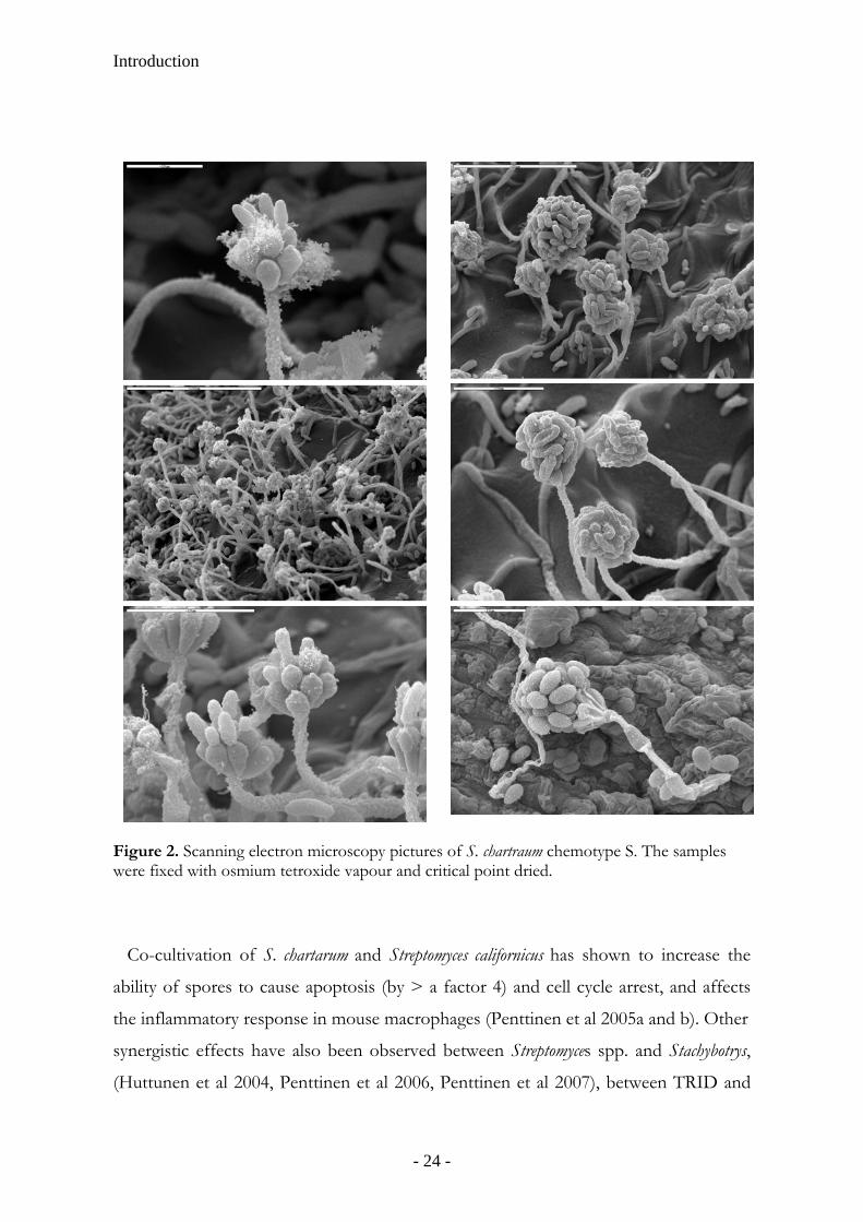
Figure 2. Scanning electron microscopy pictures of S. chartraum chemotype S. The samples were fixed with osmium tetroxide vapour and critical point dried.
Co-cultivation of S. chartarum and Streptomyces californicus has shown to increase the
ability of spores to cause apoptosis (by > a factor 4) and cell cycle arrest, and affects
the inflammatory response in mouse macrophages (Penttinen et al 2005a and b). Other
synergistic effects have also been observed between Streptomyces spp. and Stachybotrys,
(Huttunen et al 2004, Penttinen et al 2006, Penttinen et al 2007), between TRID and
- 24 -
Introduction
the bacterium Streptomyces spp. (Huttunen et al 2004), and between Stachybotrys sp and A.
versicolor (Murtoniemi et al 2005).
The actinomycete Streptomyces is a gram-positive soil bacterium often isolated from
moisture-damaged buildings. Spores of S. californicus has been shown to provoke toxic
effects in the lungs of mice at the same level of exposure as spores of S. chartarum after
repeated intranasal instillations (Nikulin et al 1997). The toxic effects of S. californicus
seem not however be limited to the lungs of mice but also include exposure-induced
effects in spleen and lymph nodes (to where intense recruitment of neutrophils,
macrophages and lymphocytes is seen which suggests that both adaptive and non-
adaptive immunological responses are involved). The immunostimulation in the lungs
and the systemic immunotoxicity, especially in the spleen are effects which resemble
those caused by chemotherapeutic agents (Jussila et al 2003).
Well-known mycotoxin producers in the Aspergillus family are A. flavus and A.
parasiticus who produce the highly carcinogenic aflatoxins (IARC 2002) via the
precursors STRG (aflatoxin B1, AFLAB, aflatoxin G1) and dihydro-STRG (aflatoxin B2,
aflatoxin G2). AFLAB-production by A. flavus may be inhibited though antagonistic
interactions with a variety of other mold strains (including non-AFLAB-producing A.
flavus strains, Cvetnic´ and Pepeljnjak 2007). Aflatoxigenic strains of A. flavus and A.
fumigatus who produce mycotoxins in culture may not do so on building materials even
without competition (Ren et al 1999). A. versicolor on the other hand, one of the most
commonly encountered molds in water-damaged indoor environments, produces
STRG and 5-methoxy-STRG (Gravesen et al 1999). A. versicolor lacks the enzymatic
pathway necessary to convert these precursors to the corresponding aflatoxins; hence,
it emits large amounts of the carcinogenic compound STRG (Frisvad 1989). The
biological activity of STRG, measured as its ability to initiate bile duct hyperplasia in
ducklings, is found to be 125 times lower than of AFLAB (Lillehoj and Ciegler 1968).
In the study of Sumi et al (1994) inhalation of A. versicolor spores over a period of 6
months resulted in granulomatous lesions in the lungs of exposed germ-free rats.
Another member of the Aspergillus family is A. fumigatus, often the cause of
aspergilliosis in immunocompromised patients. The mycotoxin GLIO is suspected to
be a virulence factor of A. fumigatus and has been determined in the serum of patients
- 25 -
Introduction
suffering from aspergilliosis (Lewis et al 2005). GLIO is also produced by species of
the Penicillium genus, including P. citrinum, and by other Aspergillus spp and Monascus spp
(Kupfahl et al 2008). The toxicity of GLIO has been shown to be due to the presence
of a disulphide bridge (Müllbacher et al 1986), capable of inactivating proteins via
reaction of thiol groups and of generating reactive oxygen species (Gardiner et al 2005).
GLIO was first considered as an antibiotic (Timonin 1942) but was later excluded
from medical use as it was found to be neurotoxic (Axelsson 2006) and
immunosuppressive (Orciuolo et al 2007), e.g. genotoxic to human lymphocytes
(Dönmez-Altuntas et al 2007). In addition, GLIO has, along with the mycotoxins
citrinin and patulin (also produced by Aspergillus and Penicillium spp), been shown to be
immunomodulatory at picogram levels in CD3-CD28-stimulated human peripheral
blood mononuclear cells (PBMCs). IFN-γ production was inhibited, an effect caused
by a reduction of the number of IFN-γ-producing T lymphocytes rather than by a
reduced functional capacity of the individual cells, thus, causing a T-cell polarization
towards a Th2 phenotype (Wichmann et al 2002). This Th2-polarization has been
shown to be accompanied by the up-regulation of IgE-syntesis (Wichmann et al 2003).
When exposing human monocytic cells to low doses of GLIO (100 ng/ml) and citrinin
(less than 10 µg/ml) a cytokine imbalance was observed with a reduction of Il-10
concentrations compared to those of TNF-α and IL-6, potentially resulting in an
increased risk of an inflammatory response (Johannessen et al 2005). Citrinin exposure
to human alveolar epithelial cells, at nontoxic concentrations, also causes depletion of
intracellular glutathione suggesting an increased susceptibility to inflammatory trigger
agents in the environments, such as LPS or other microbial components (Johannessen
et al 2007)
Detection of mycotoxins
Today, standardized methods for determination of microbial constituents and products
are lacking. There are over 200 mycotoxins identified to date, as well as fungal
proteinases (Yike et al 2007) including ribotoxins (Lacadena et al 2007) and hemolysins
(Van Emon et al 2003, Vesper et al 2001, Vesper and Vesper 2002), and toxins and
uncharacterized biologically active compounds from bacteria, e. g. Streptomyces
- 26 -
Introduction
(Andersson et al 1998). The biologically active mold components determined so far are
few and merely the tip of the iceberg.
One approach of measuring mycotoxins indoors is to determine their toxic activity,
e.g. as in the luciferase translation bioassay which is based on trichothecenes inhibition
of firefly luciferase in cell culture (Yike et al 1999). However, the toxic effects of S.
chartarum seem to be related to methanol-soluble toxins in the spores (Rao et al 2000a)
and in the in vitro luciferase protein translation assay the presence of alcohol is not
desirable as it has been shown to inhibit the reaction in a dose dependant manner
(Black et al 2006). Toxic substances may however be removed from the spore surface
by washing in aqueous solution (Karunasena et al 2004) and the luciferase translation
bioassay has shown to be a convenient method giving reproducible results.
Nevertheless is does not demonstrate the toxin composition of environmental samples
(Yike et al 1999).
ELISA incorporated with macrocyclic trichothecene-specific antibodies has been
used in detecting MTRs in airborne dust (Charpin-Kadouch et al 2006, Brasel et al
2005) and in serum samples from individuals exposed to mold (primarily Stachybotrys
spp., Brasel et al 2004). In addition, antibodies produced against the hemolytic agent
stachylysin (produced by Stachybotrys spp.) were successfully used in an ELISA analysis
of serum samples from human adults working in water-damaged buildings with
Stachybotrys-contamination (Van Emon et al 2003).
Mass Spectrometry
Mass spectrometry (MS) is a chemical analytical technique which has both qualitative
and quantitative uses, such as identifying unknown compounds, determining a
compounds’ isotopic element, structure (by observing its fragmentation), amount in a
sample, or other physical, chemical, or biological properties. In MS the composition
of a compound or sample is identified on the basis of the m/z ratio of charged particles.
A MS system has three essential modules: an ion source which transforms the
molecules in a sample into ionized fragments; a mass analyzer, which sorts the ions by
applying electric and magnetic fields; and a detector, which measures the value of an
indicator quantity and thus provides data for calculating the abundances each ion
- 27 -

Introduction
Sample
Mass analyserIon source Detector
Data analysis
Figure 3. The principle construction of a mass spectrometer.
fragment present (http://wikipedia.org/) (Figure 3). There are different types of mass
analyzers such as magnetic sector analyzers, quadrupole mass filters, quadrupole ion
traps, time-of-flight analyzers, and ion cyclotron resonance instruments (Chapman
1993).
In the ion trap all analyses events may be separated in time but not in space. Ions
may be ejected using resonance excitation, whereby a supplemental oscillatory
excitation voltage is applied to the end cap electrodes and the trapping voltage
amplitude and/or excitation voltage frequency is varied to bring ions into a resonance
condition in order of their m/z ratio. Also a non-resonance mode may be applied
(Chapman 1993).
Quadrupole mass analyzers use radio frequency and oscillating electrical fields
between four parallel rods to selectively stabilize or destabilize ions passing through to
the detector, i. e. it acts as a mass selective filter (Odham and Larsson 1984). A
common variation of the quadrupole is the triple quadrupole (papers I-IV), in which a
linear series of three separate quadrupoles is used. The first (Q1) and third (Q3)
quadrupoles act as mass filters, and are separated by the middle non-mass filtering (q2)
quadrupole, which acts as a collision cell (using argon gas, paper I-IV). When collecting
data in the full scan mode, a target range of mass fragments is determined, a method
useful in determining unknown compounds in a sample. During instrument method
development chemical standards are first analyzed in full scan mode to determine the
retention time and the mass fragment fingerprint before moving to a SIM method.
SIM only monitors selected peaks associated with a specific substance entered into the
- 28 -
Introduction
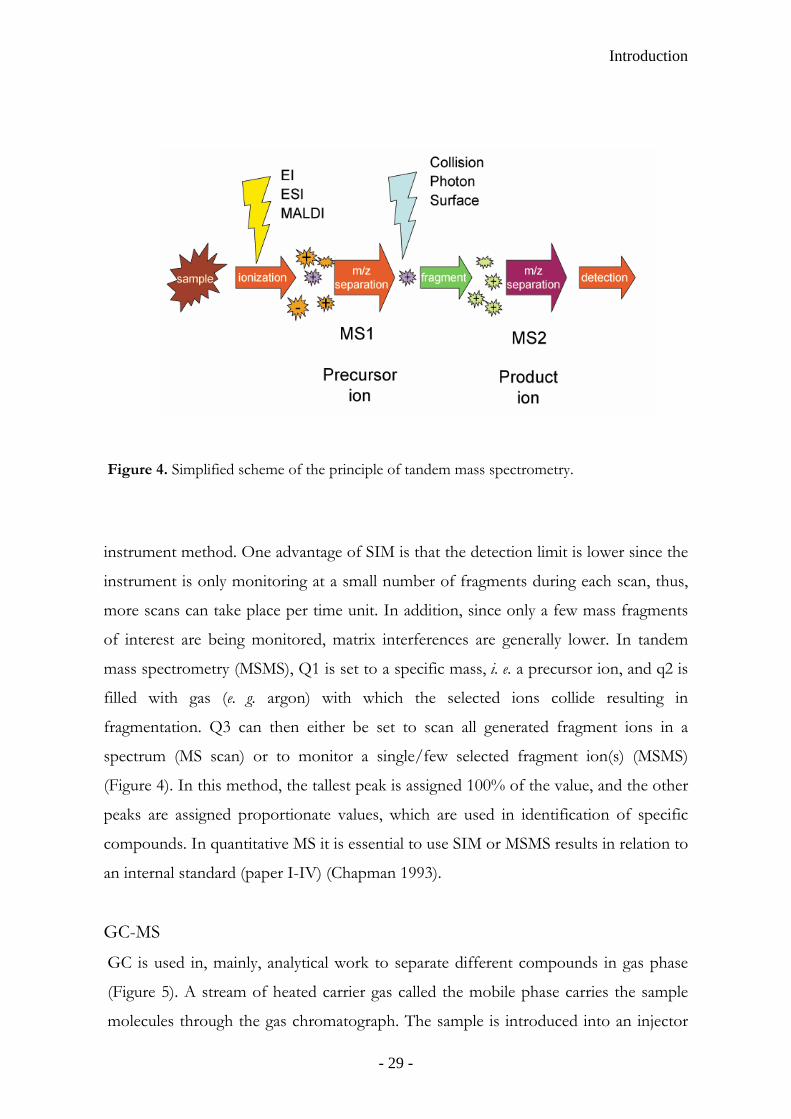
Figure 4. Simplified scheme of the principle of tandem mass spectrometry.
instrument method. One advantage of SIM is that the detection limit is lower since the
instrument is only monitoring at a small number of fragments during each scan, thus,
more scans can take place per time unit. In addition, since only a few mass fragments
of interest are being monitored, matrix interferences are generally lower. In tandem
mass spectrometry (MSMS), Q1 is set to a specific mass, i. e. a precursor ion, and q2 is
filled with gas (e. g. argon) with which the selected ions collide resulting in
fragmentation. Q3 can then either be set to scan all generated fragment ions in a
spectrum (MS scan) or to monitor a single/few selected fragment ion(s) (MSMS)
(Figure 4). In this method, the tallest peak is assigned 100% of the value, and the other
peaks are assigned proportionate values, which are used in identification of specific
compounds. In quantitative MS it is essential to use SIM or MSMS results in relation to
an internal standard (paper I-IV) (Chapman 1993).
GC-MS
GC is used in, mainly, analytical work to separate different compounds in gas phase
(Figure 5). A stream of heated carrier gas called the mobile phase carries the sample
molecules through the gas chromatograph. The sample is introduced into an injector
- 29 -
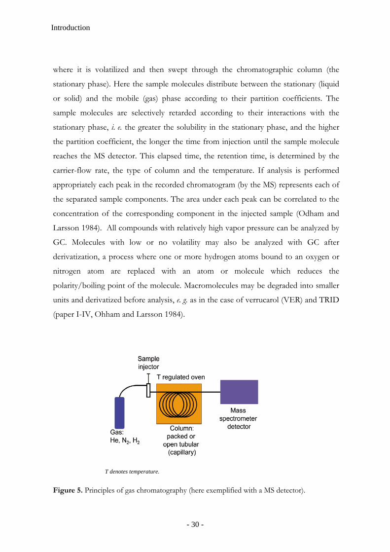
Introduction
where it is volatilized and then swept through the chromatographic column (the
stationary phase). Here the sample molecules distribute between the stationary (liquid
or solid) and the mobile (gas) phase according to their partition coefficients. The
sample molecules are selectively retarded according to their interactions with the
stationary phase, i. e. the greater the solubility in the stationary phase, and the higher
the partition coefficient, the longer the time from injection until the sample molecule
reaches the MS detector. This elapsed time, the retention time, is determined by the
carrier-flow rate, the type of column and the temperature. If analysis is performed
appropriately each peak in the recorded chromatogram (by the MS) represents each of
the separated sample components. The area under each peak can be correlated to the
concentration of the corresponding component in the injected sample (Odham and
Larsson 1984). All compounds with relatively high vapor pressure can be analyzed by
GC. Molecules with low or no volatility may also be analyzed with GC after
derivatization, a process where one or more hydrogen atoms bound to an oxygen or
nitrogen atom are replaced with an atom or molecule which reduces the
polarity/boiling point of the molecule. Macromolecules may be degraded into smaller
units and derivatized before analysis, e. g. as in the case of verrucarol (VER) and TRID
(paper I-IV, Ohham and Larsson 1984).
Figure 5. Principles of gas chromatography (here exemplified with a MS detector).
T denotes temperature.
- 30 -
Introduction
The most common form of ionization in GC-MS is electron ionization (EI).
Separated gaseous compounds are fed online from the GC into the MS ion source
where they are bombarded with free electrons emitted from a charged metallic filament,
causing an ionization that fragments the molecules. In chemical ionization (CI)
(positive ion CI, PICI, or negative ion CI, NICI) a reagent gas, e. g. methane (paper I)
or ammonia (paper II-IV), is introduced into the ion source. The molecules of the
reagent gas are ionized by the electrons omitted by the filament and the formed ions
then react with the introduced sample molecules causing an ionization of the analyte
molecules. CI is n technique than EI. One of the main benefits of
using CI is that a mass fragm
e analyte, stationary phase and mobile
hase composition. The use of high pressure (high pressure LC, HPLC) increases the
ty which gives the sample molecules less time to diffuse within the column,
and the analyte.
a softer ionizatio
ent closely corresponding to the molecular weight of the
analyte of interest is produced (Chapman 1993).
HPLC-MS
Similar to GC-MS liquid chromatography MS (LC-MS) separates compounds
chromatographically, however, the mobile phase is liquid, usually a mixture of water
and organic solvents, e. g. methanol or acetonitrile. The column holds a
chromatographic packing material (the stationary phase), and a pump moves the
mobile phase(s) through the column. The sample analyzed is introduced into the
stream of mobile phase and is retarded by specific chemical or physical interactions
with the stationary phase as it traverses through the column. The amount of
retardation generally depends on the nature of th
p
linear veloci
leading to improved resolution in the resulting chromatogram. A refinement in HPLC
is to vary the mobile phase composition during the analysis, i. e. to create a gradient
elution. The gradient separates the analyte molecules as a function of the affinity of the
analyte for the current mobile phase composition relative to the stationary phase. The
mobile phases may contain buffers or salts to assist in the separation of the analyte
components or compounds such as TFA which acts as an ion pairing agent. The
choice of solvents, additives and gradient depend on the nature of the stationary phase
- 31 -
Introduction
The most commonly used form of HPLC is reversed phase HPLC in which a non-
polar stationary phase (e. g. silica treated with RMe2SiCl, where R is a straight chain
alkyl group such as C18H37 or C8H17) and an aqueous, moderately polar mobile phase
are used. With this type of stationary phase, retention time is longer for molecules
which are non-polar (e. g. C-H, C-C), while polar molecules (-OH, -NH2, COO- or -
NH3+) elute more readily. Due to the overall decrease in surface area branched chain
compounds (or organic compounds with single C-C-bonds) elute more rapidly than
their linear isomers. Organic compounds containing a C=C or C-C-triple bonds also
tend to have relatively shorter retention times. The retention time is increased for an
analyte by adding a polar solvent to the mobile phase, or decreased by adding a more
hydrophobic solvent. The pH value of the mobile phase(s) changes the hydrophobicity
of the analyte, and thus influence the separation process. To control the pH a buffering
agent, such as sodium phosphate, formic acid or TFA is often added to the mobile
phase. The buffers also neutralize charges on any residual exposed silica on the
stationary phase and, in addition, act as ion pairing agents to neutralize charge on the
analyte (http://wikipedia.org/).
In electrospray ionization (ESI) mode the analyte and mobile phase mixture is
pushed, at a flow rate of a few microlites per minute, through a very small needle
capillary together with a nebulizing gas (nitrogen for positive ESI and air for negative
ESI). The outlet of the capillary, the capillary tip, has a charged potential (typically 3-5
kV) and when the analyte and mobile phase mix enters through the capillary tip into
the ion source (spray chamber) the liquid flow convert into a mist of small droplets.
Heated nitrogen (drying gas) is often used to facilitate the nebulizing and evaporation
process, e. g. to minimize vacuum chamber contamination by solvent molecules. This
produces a stable analyte ion current which communicates between atmosphere
pressure and the first vacuum stage of the MS guided by a charged shield and passed
through a long metal capillary tube that (Champman 1993). A schematic overview of
the HLPC-MS interface is provided in Figure 6.
- 32 -
Introduction
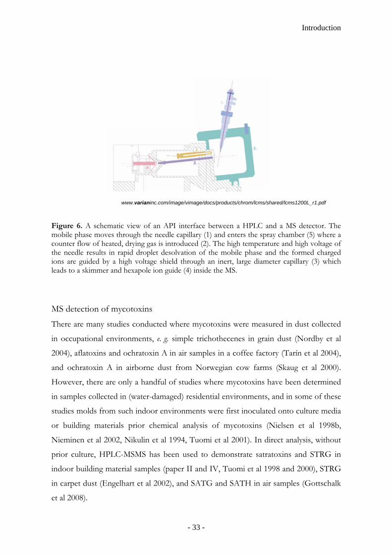
Figure 6. A schematic view of an API interface between a HPLC and a MS detector. The mobile phase moves through the needle capillary (1) and enters the spray chamber (5) where a counter flow of heated, drying gas is introduced (2). The high temperature and high voltage of the needle results in rapid droplet desolvation of the mobile phase and the formed charged ions are guided by a high voltage shield through an inert, large diameter capillary (3) whichleads to a skimmer and hexapole ion guide (4) inside the MS.
MS detection of mycotoxins
There are many studies conducted where mycotoxins were measured in dust collected
www.varianinc.com/image/vimage/docs/products/chrom/lcms/shared/lcms1200L_r1.pdf
samples collected in (water-damaged) residential environments, and in some of these
udies molds from such indoor environments were first inoculated onto culture media
emical analysis of mycotoxins (Nielsen et al 1998b,
in occupational environments, e. g. simple trichothecenes in grain dust (Nordby et al
2004), aflatoxins and ochratoxin A in air samples in a coffee factory (Tarín et al 2004),
and ochratoxin A in airborne dust from Norwegian cow farms (Skaug et al 2000).
However, there are only a handful of studies where mycotoxins have been determined
in
st
or building materials prior ch
Nieminen et al 2002, Nikulin et al 1994, Tuomi et al 2001). In direct analysis, without
prior culture, HPLC-MSMS has been used to demonstrate satratoxins and STRG in
indoor building material samples (paper II and IV, Tuomi et al 1998 and 2000), STRG
in carpet dust (Engelhart et al 2002), and SATG and SATH in air samples (Gottschalk
et al 2008).
- 33 -
Introduction
For Stachybotrys trichothecenes a convenient GC-MS screening method was applied
(paper I, II, and IV, Croft et al 1986, Gravesen et al 1999, Nielsen et al 1998a). This
method provides information of the total trichothecenes content derived from
Stachybotrys spp. (Hinkley and Jarvis 2001, Nielsen et al 1998a and b). In this analysis
VER and TRID, hydrolysis products of S. chartarum MTRs and trichodermin
respectively, are determined (Bata et al 1985, Harrach et al 1981, Jarvis et al 1984,
Krishnamurthy et al 1986, Szathmary et al 1976). The deacetylation process of
trichodermin also occurs naturally when entering, e. g. Mucor cells (Fonzi and Sypherd
1986).
Interestingly, SATG covalently bound to lysine, cysteine, and histidine has also been
detected by MS as adducts in serum samples of human and cat residents in S. chartarum
-contaminated buildings (Mader et al 2007, Yike et al 2006).
- 34 -

AIMS OF THE STUDY
The aims of this thesis were:
1. To develop GC-MS and HPLC-MS analytical methods for determination of
selected mycotoxins typically produced by molds encountered in water-
damaged buildings.
2. To apply these methods for direct mycotoxin analysis of building materials
and dust samples from authentic water-damaged environments.
3. To study the prevalence of selected potent mycotoxins in samples from
indoor environments with verified mold damage.
4. To initiate studies on potential associations between mycotoxins and fungi as
analysed by culture, PCR, and determination of ergosterol. .
- 35 -

- 36 -

Material and Methods
MATERIAL AND METHODS
Samples
All studied samples were collected in dwellings with a history of water damage; the
types of buildings/locations (for samples analysed in paper I, II and IV) are
summarized in Table 1. Pieces of gypsum board, linoleum flooring, wall paper, tile,
concrete, isolation, wooden skirting boards etc were collected by company personell
specialized in remediation measures. In total 167 building material samples were
analyzed for mycotoxins (paper I, II, IV); one hundred of these building materials were
also analyzed for ERG (paper IV).
Settled dust samples (n=32) were collected on cotton swabs (n=4) and ALK filters
(ALK-ABELLÒ A/S, Denmark) (n=28) in Swedish water-damaged dwellings (paper I,
II, and IV), viz. in single-family houses (n=3), offices (n=3), apartments (n=2),
churches (n=2), schools (n=2), and care facilities (n=2). Eighteen of these samples
were analyzed for ERG (paper IV). In addition, bulk dust samples (n=7) were collected
in 5 flood-damaged houses in New Orleans using a brush and pan (paper III).
Table 1. Houses/locations (n=88, n=14, and n=30) where samples (building materials, settled dust, and RCS cultures respectively) were collected.
Type of collected sample Location/house where samples were collected Building material3 Settled dust RCS cultureGovermental building 2 0 1 Office building 19 3 6 Shop 0 0 1 Care facility (e. g. hospital) 2 2 3 School building 19 2 4 Apartment building 18 2 6 Singel family house 11 3 1 Children’s daycare center 10 0 3 Sport facility1 3 0 2 Church 0 2 0 Others2 4 0 3 1Sport facility denotes for building materials: a swimming hall and two gyms; and for RCS cultures: an indoor icehockey rink and a swimming hall. 2Others denotes for building materials: two cinemas, an atelier, and a hotel; and for RCS cultures: an emergency exit tunnel, a public transporation vehicle, and a greenhouse. 3The 4 building materials in paper I are not included.
- 37 -
Material and Methods
Cultures (n=45) of airborne fungal particles were collected using a Reuter Centrifugal
Sampler (RCS) during a sampling time of 4 min (paper II and IV) in apartments (n=6),
offices (n=6), schools (n=4), care facilities (n=3), kindergartens (n=3), and a single
family house, a shop, a municipal hall, an indoor ice rink, a swimming hall, a green
house, a public transportation vehicle, and an emergency exit tunnel.
Sampling and Cultivation
Building material samples and dust were cultured on MEA (a nutrient rich agar
medium) (paper I-IV). Aliquots from serial dilutions of dust collected i New Orleans
(paper III) were also plated on czapek yeast extract agar (CYA, a medium which
promotes sporulation of aflatoxigenic fungi) and dichloran 18% glycerol agar (DG18,
high water content medium). All plates were incubated at 23 °C for a minimum of 5-7
days and the colony forming units (CFUs) were identified to genus level according to
Samson et al (2004).
Airborne dust samples were collected using an RCS (from Biotest Diagnostics, NJ,
USA), an air sampler which collects airborne microorganisms quantitatively onto a
culture medium according to the impaction principle (paper II and IV). Air (40 l/min)
was sucked into the sampler from a distance of at least 40 cm by means of an impeller,
entering the impeller drum, and set in rotation (average rotational speed of 4096 rpm ±
2 %). The contained particles in the air were impacted by centrifugal force onto a
plastic strip containing Rose-bengal agar. After sample collection, the agar strips were
incubated at 23 °C for 5-7 days, and colonies were counted as CFU/m3 and identified
according to Samson et al (2004).
Microscopy
Phase-contrast microscopy was used to study mold cultures and tape-lift samples
(paper I-IV). Tape-lifts were collected by pressing tape onto a moldy surface. A drop
of lactic phenol was then put on a microscope slide, the tape was pressed onto the slide,
and another drop of lactic phenol was added. Finally a coverslip was overlaid and the
slide was put under the microscope. The mold spores were examined and identified to
- 38 -
Material and Methods
genus level according to Samson et al (2004). In absence of spores the mycelium was
denoted mycelia sterila.
QPCR
Dust samples (paper III) were sent to Anozona (Uppsala, Sweden), contract partner of
Fugenex (Yorkshire, UK), who are licensed to use DNA extraction and MSQPCR
analysis according to Haugland and Vesper (2002, US patent 6,387,652). In summary,
dust sample extracts were diluted, and the DNA was extracted by a rapid bead-milling
method (Dneasy Plant Mini Kit; tungsten carbide bead, Qiagen, Hilden, Germany).
DNA sequences were amplified using fluorescent labeled probes (TaqMan™ system)
producing real time detection of PCR products (Haugland et al 2004) on a 7300 Real-
time PCR Instrument (Applied Biosystems, Foster City, USA). The amplified DNA
sequences were then counted, compare to DNA standard curves, and quantified as
number of DNA-sequences (gene copies) per milligram dust. Detailed information on
the primer and probe sequences (i. e .directed at the nuclear ribosomal RNA operon,
internal spacer regions, ITS1 or ITS2) is provided at
http://www.epa.gov/microbes/moldtech.htm.
Chemical experiments
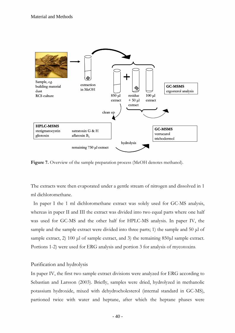
The sample preparation process is illustrated in Figure 7.
Extraction
The samples were weighted, covered with methanol in glass test tubes with Teflon-
lined screw caps, and stored in the dark, overnight or for 72 h, at room temperature.
After extraction samples were centrifuged and the supernatants were decanted into
new tubes. The extraction method was then elaborated with to improve recovery of
the analytes, e. g. sterile water were added and the mixtures were extracted twice with 2
ml heptane (paper I-II), or the extraction was repeated with dichloromethane after
which the methanolic and dichloromethane phases were pooled (and heptane-
extraction omitted, paper III-IV).
- 39 -
Material and Methods
Sample, e.g.building materialdustRCS culture
extraction in MeOH
GC-MSMSergosterol analysis
HPLC-MSMS sterigmatocystin satratoxin G & Hgliotoxin aflatoxin B1
clean up
GC-MSMSverrucaroltrichodermol
remaining 750 µl extracthydrolysis
100 µl extract
850 µl extract
residue + 50 µl extract
Sample, e.g.building materialdustRCS culture
extraction in MeOH
GC-MSMSergosterol analysis
HPLC-MSMS sterigmatocystin satratoxin G & Hgliotoxin aflatoxin B1
clean up
GC-MSMSverrucaroltrichodermol
remaining 750 µl extracthydrolysis
100 µl extract
850 µl extract
residue + 50 µl extract
Figure 7. Overview of the sample preparation process (MeOH denotes methanol).
The extracts were then evaporated under a gentle stream of nitrogen and dissolved in 1
ml dichloromethane.
In paper I the 1 ml dichloromethane extract was solely used for GC-MS analysis,
whereas in paper II and III the extract was divided into two equal parts where one half
was used for GC-MS and the other half for HPLC-MS analysis. In paper IV, the
sample and the sample extract were divided into three parts; 1) the sample and 50 µl of
sample extract, 2) 100 µl of sample extract, and 3) the remaining 850µl sample extract.
Portions 1-2) were used for ERG analysis and portion 3 for analysis of mycotoxins.
Purification and hydrolysis
In paper IV, the first two sample extract divisions were analyzed for ERG according to
Sebastian and Larsson (2003). Briefly, samples were dried, hydrolyzed in methanolic
potassium hydroxide, mixed with dehydrocholesterol (internal standard in GC-MS),
partioned twice with water and heptane, after which the heptane phases were
- 40 -
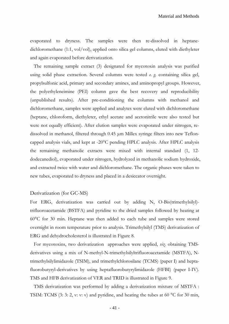
Material and Methods
evaporated to dryness. The samples were then re-dissolved in heptane-
dichloromethane (1:1, vol/vol), applied onto silica gel columns, eluted with diethyleter
and again evaporated before derivatization.
The remaining sample extract (3) designated for mycotoxin analysis was purified
using solid phase extraction. Several columns were tested e. g. containing silica gel,
propylsulfonic acid, primary and secondary amines, and aminopropyl groups. However,
the polyethyleneimine (PEI) column gave the best recovery and reproducibility
(unpublished results). After pre-conditioning the columns with methanol and
dichloromethane, samples were applied and analytes were eluted with dichloromethane
(heptane, chloroform, diethyleter, ethyl acetate and acetonitrile were also tested but
were not equally efficient). After elution samples were evaporated under nitrogen, re-
dissolved in methanol, filtered through 0.45 µm Millex syringe filters into new Teflon-
capped analysis vials, and kept at -20°C pending HPLC analysis. After HPLC analysis
the remaining methanolic extracts were mixed with internal standard (1, 12-
dodecanediol), evaporated under nitrogen, hydrolyzed in methanolic sodium hydroxide,
and extracted twice with water and dichloromethane. The organic phases were taken to
new tubes, evaporated to dryness and placed in a desiccator overnight.
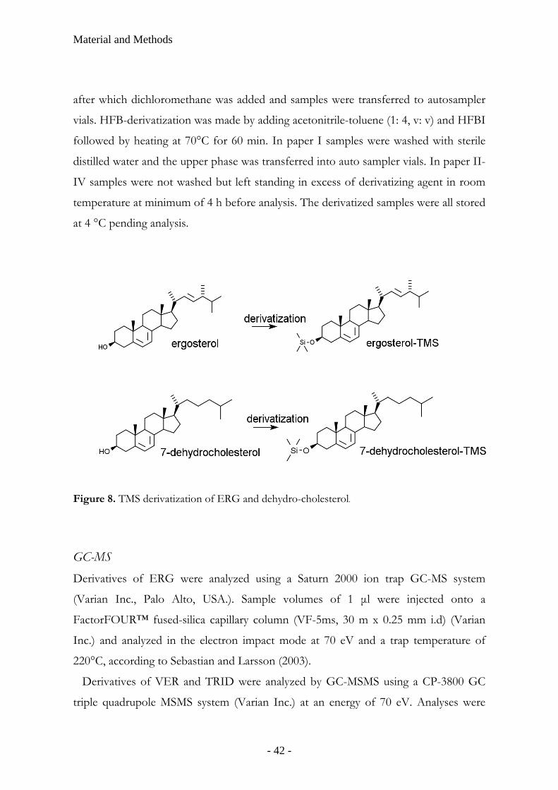
Derivatization (for GC-MS)
For ERG, derivatization was carried out by adding N, O-Bis(trimethylsilyl)-
trifluoroacetamide (BSTFA) and pyridine to the dried samples followed by heating at
60°C for 30 min. Heptane was then added to each tube and samples were stored
overnight in room temperature prior to analysis. Trimethylsilyl (TMS) derivatization of
ERG and dehydrocholesterol is illustrated in Figure 8.
For mycotoxins, two derivatization approaches were applied, viz. obtaining TMS-
derivatives using a mix of N-methyl-N-trimethylsilyltrifluoroacetamide (MSTFA), N-
trimethylsilylimidazole (TSIM), and trimethylchlorosilane (TCMS) (paper I) and hepta-
fluorobutyryl-derivatives by using heptafluorobutyrylimidazole (HFBI) (paper I-IV).
TMS and HFB derivatization of VER and TRID is illustrated in Figure 9.
TMS derivatization was performed by adding a derivatization mixture of MSTFA :
TSIM: TCMS (3: 3: 2, v: v: v) and pyridine, and heating the tubes at 60 °C for 30 min,
- 41 -
Material and Methods
after which dichloromethane was added and samples were transferred to autosampler
vials. HFB-derivatization was made by adding acetonitrile-toluene (1: 4, v: v) and HFBI
followed by heating at 70°C for 60 min. In paper I samples were washed with sterile
distilled water and the upper phase was transferred into auto sampler vials. In paper II-
IV samples were not washed but left standing in excess of derivatizing agent in room
temperature at minimum of 4 h before analysis. The derivatized samples were all stored
at 4 °C pending analysis.
Figure 8. TMS derivatization of ERG and dehydro-cholesterol.
GC-MS Derivatives of ERG were analyzed using a Saturn 2000 ion trap GC-MS system
(Varian Inc., Palo Alto, USA.). Sample volumes of 1 µl were injected onto a
FactorFOUR™ fused-silica capillary column (VF-5ms, 30 m x 0.25 mm i.d) (Varian
Inc.) and analyzed in the electron impact mode at 70 eV and a trap temperature of
220°C, according to Sebastian and Larsson (2003).
Derivatives of VER and TRID were analyzed by GC-MSMS using a CP-3800 GC
triple quadrupole MSMS system (Varian Inc.) at an energy of 70 eV. Analyses were
- 42 -

Material and Methods
performed in EI mode with an ion source temperature of 250°C (TMS-derivatives) and
200°C (HFB-derivatives) (paper I), or in negative CI mode at an ion source
temperature of 200°C or 150°C using methane (0.4 kPa, paper I) or ammonia (0.8 kPa,
paper II-IV), respectively, as ionization gas. Sample volumes of 1-2 µl were injected
onto a FactorFOUR™ fused-silica capillary column (VF-5ms, 30 m x 0.25 mm i.d) in
the splitless mode with a helium carrier gas pressure of 69 kPa, using a CombiPAL
autosampler (CTC Analytics, Zwingen, Switzerland). The injector syringe was washed 5
times with acetone and toluene, respectively, before and after each sample injection.
A mix of HFBI and acetone (1:3, v:v) was injected in between samples to eliminate any
trace of un- or semi-derivatized VER/TRID. The temperature of the column was
programmed from 90 to 280°C at 20°C per minute (the injector and transfer line
temperature was 280°C). The MSMS conditions were optimized by repeatedly injecting
0.1–1 ng amounts of standards at different collision energy, ion source
temperature,and argon pressure in the collision cell. The parameters that gave the
largest product ion peak area were selected. Detection sensitivity, defined as amount of
standards injected with a signal-to-noise ratio ≥4 (software calculated peak-to-peak
values), was determined by analyzing derivatized standard preparations diluted in
dichloromethane (TMS- derivatives) or acetonitrile–toluene (1:4, v:v) (HFB-
derivatives). The instrument performance was ensured by including TRID/VER
standards and 1,12-dodecanediol (internal standard) in each batch of samples analyzed.
Two calibration curves were constructed for VER/TRID (n=3) (0, 25, 50, 100, 250,
500, 1000 pg and 0.5, 1, 2.5, 5, 10, 25 ng respectively) together with internal standard
(250 pg and 2.5 ng respectively). The coefficient of variation was calculated by dividing
the standard deviation by the mean peak area ratio of VER/TRID standard to the
internal standard, and the recovery was calculated by dividing the mean peak area from
1-ng injections of VER/TRID standards that had passed the sample preparation
procedure with corresponding VER/TRID standards that did not pass this procedure.
Ions used in SIM analysis of VER–TMS2 were m/z 320 (probably representing M–
TMSOH), m/z 307 (M–CH2OTMS), m/z 277 (M–CH2OTMS–2CH3), and m/z 217
- 43 -
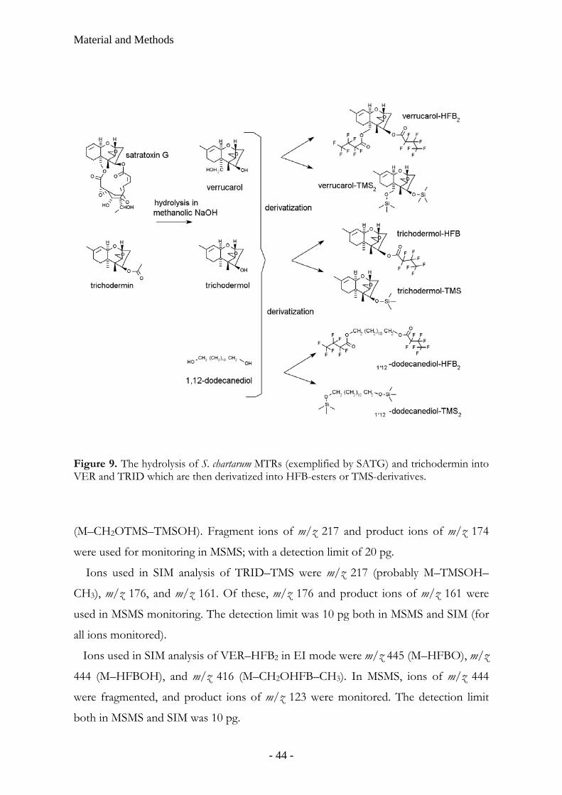
Material and Methods
Figure 9. The hydrolysis of S. chartarum MTRs (exemplified by SATG) and trichodermin into VER and TRID which are then derivatized into HFB-esters or TMS-derivatives.
(M–CH2OTMS–TMSOH). Fragment ions of m/z 217 and product ions of m/z 174
were used for monitoring in MSMS; with a detection limit of 20 pg.
Ions used in SIM analysis of TRID–TMS were m/z 217 (probably M–TMSOH–
CH3), m/z 176, and m/z 161. Of these, m/z 176 and product ions of m/z 161 were
used in MSMS monitoring. The detection limit was 10 pg both in MSMS and SIM (for
all ions monitored).
Ions used in SIM analysis of VER–HFB2 in EI mode were m/z 445 (M–HFBO), m/z
444 (M–HFBOH), and m/z 416 (M–CH2OHFB–CH3). In MSMS, ions of m/z 444
were fragmented, and product ions of m/z 123 were monitored. The detection limit
both in MSMS and SIM was 10 pg.
- 44 -
Material and Methods
In TRID–HFB-analysis ions of m/z 446 (M) and m/z 431 (M–CH3) were used in
SIM. In MSMS, fragment m/z 446 and product ions of m/z 431 were monitored. The
detection limit both in SIM and MSMS was 2 pg.
In NICI mode, VER–HFB2 SIM analysis was perfomed monitoring ions m/z 638,
m/z 302, and m/z 213, and parent ion m/z 638 (M–HF) and product ion m/z 213
(HFBO) were used for MSMS monitoring.
In case of TRID–HFB, ions of m/z 426 (M–HF) were used both in MSMS
monitoring product ions of m/z 159 (C3F5CO) and SIM. The detection limits both of
VER and TRID were 0.1 and 0.2 pg in, respectively, MSMS and SIM.
HPLC-MS In paper II-IV, a ProStar HPLC/1200L triple quadrupole MSMS system (Varian Inc.,
Walnut Creek, CA, USA) was used. 20 µl of sample was injected using an autosampler
(Varian, model 410) into a Polaris 5µM C18-A 150x2.0 mm RP-18 column equipped
with a MetaGuard 2.0mm Polaris 5µM C18-A pre-column (Varian). The column was
maintained at 25 °C and the flow rate was 0.2 ml/min. A supplement of 10 mM
ammonium acetate and 20 µM sodium acetate was added to the methanol-aqueous
buffer for increasing cationization in the ESI mode. A variety of modifications in the
mobile phase program were applied (paper II-IV). Finally, an initial methanol
concentration of 20% was held for 1 min, after which it was raised linearly (9 min) first
to 70% and then to 100% (10 min) and held for 9 min before it was lowered (2 min)
again to 20% and kept 9 min for stabilization (paper IV). 10 µl of methanol was
injected in between samples to minimize cross-contamination. Nitrogen from a
nitrogen generator (Domnick Hunter Ltd, Tyne & Wear, UK) was used both as
nebulizing gas (50 psi) and as drying gas (20 psi), and argonium (0.23 mPa) was used
for collision induced dissociation (CID). The capillary temperature was 310°C, the
capillary voltage 40 V, the needle voltage 5000 V, and the electron multiplier voltage
2000V. MS spectra were collected with a scan time of 0.5 s and a scan width of 0.7 s.
All these parameters were elaborated with and tested to achieve the highest possible
signal in the mycotoxin MSMS- analysis.
- 45 -
Material and Methods
The MS was tuned through direct injection of PPG tuning solution with a syringe
according to the manufacturer’s protocol. Mycotoxin standards and the internal
standard reserpine (RESP) were included in each batch of samples analyzed in order to
ensure instrument performance. Calibration curves were constructed by injecting
mycotoxin together with internal standard at a minimum of seven concentration points.
The coefficient of variation was calculated by dividing the standard deviation with the
mean peak area ratio of mycotoxin standard to internal standard, and the recovery was
calculated by dividing the mean peak area from injections of mycotoxin standards that
had passed the sample preparation procedure with corresponding standards that did
not pass this procedure.
In STRG-analysis ions of m/z 325 (M+H)+ were used for fragmentation in MSMS,
and the product ions of m/z 310 and m/z 281 were monitored and used for
identification purposes; the detection limit was 0.2 pg (injected amount monitoring
m/z 310, signal-to-noise ratio [peak-to-peak value] ≥4).
For SATG ions of m/z 567 (M+Na)+ were used for fragmentation in MSMS, and its
product ions m/z 263 and m/z 231 were monitored.
Figure 10. The chemical structure of the internal standard RESP and selected mycotoxins.
- 46 -

Material and Methods
In the SATH analysis m/z 551 (M+Na)+ was used for fragmentation in MSMS, and
its product ions of m/z 321 and m/z 303 were monitored. The detection limits for
SATG and SATH were not set as the crude standards used were unadequate for
quantification purposes.
For reserpine, m/z 609 (M)+ was used as the parent ion in MSMS and its product ion
m/z 195 was monitored.
For AFLAB, ions of m/z 313 (M+Na)+ were used in MSMS to produce product ions
of m/z 241. The detection limit for AFLAB was 5 pg.
In positive ESI analysis of GLIO m/z 349 (M+Na)+ were used in MSMS and
product ions of m/z 285 were monitored. The detection limit was 5 pg. Chemical
structures of selected mycotoxins analyzed in HPLC-MS is provided in Figure 10.
- 47 -

- 48 -
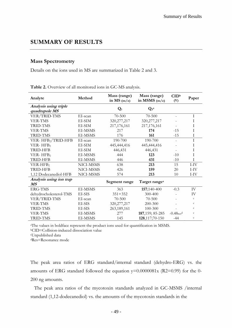
Summary of Results
SUMMARY OF RESULTS
Mass Spectrometry
Details on the ions used in MS are summarized in Table 2 and 3.
Table 2. Overview of all monitored ions in GC-MS analysis.
Analyte Method Mass (range) in MS (m/z)
Mass (range) in MSMS (m/z)
CIDb (V) Paper
Analysis using triple quadrupole MS
Q1 Q3a
VER/TRID-TMS EI-scan 70-500 70-500 - I VER-TMS EI-SIM 320,277,217 320,277,217 - I TRID-TMS EI-SIM 217,176,161 217,176,161 - I VER-TMS EI-MSMS 217 174 -15 I TRID-TMS EI-MSMS 176 161 -15 I VER- HFB2/TRID-HFB EI-scan 190-700 190-700 - I VER- HFB2 EI-SIM 445,444,416 445,444,416 - I TRID-HFB EI-SIM 446,431 446,431 - I VER- HFB2 EI-MSMS 444 123 -10 I TRID-HFB EI-MSMS 446 431 -10 I VER-HFB2 NICI-MSMS 638 213 15 I-IV TRID-HFB NICI-MSMS 426 159 20 I-IV 1,12 Dodecanediol-HFB NICI-MSMS 574 213 10 I-IV Analysis using ion trap MS Segment range Target rangea
ERG-TMS EI-MSMS 363 157;140-400 -0.3 IV dehydrocholesterol-TMS EI-SIS 351+352 300-400 - IV VER/TRID-TMS EI-scan 70-500 70-500 - c
VER-TMS EI-SIS 320,277,217 200-300 - c
TRID-TMS EI-SIS 263,189,161 100-300 - c
VER-TMS EI-MSMS 277 187,159; 85-285 -0.48resd c
TRID-TMS EI-MSMS 145 128,117;70-150 -44 c
aThe values in boldface represent the product ions used for quantification in MSMS. bCID=Collision-induced dissociation value cUnpublished data dRes=Resonance mode
The peak area ratios of ERG standard/internal standard (dehydro-ERG) vs. the
amounts of ERG standard followed the equation y=0.0000081x (R2=0.99) for the 0-
200 ng amounts.
The peak area ratios of the mycotoxin standards analyzed in GC-MSMS /internal
standard (1,12-dodecanediol) vs. the amounts of the mycotoxin standards in the
- 49 -
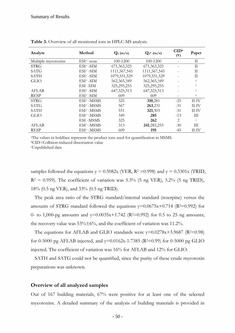
Summary of Results
Table 3. Overview of all monitored ions in HPLC-MS analysis.
Analyte Method Q1 (m/z) Q3a (m/z) CIDb (V) Paper
Multiple mycotoxins ESI+ -scan 100-1200 100-1200 - II STRG ESI+ -SIM 671,363,325 671,363,325 - II SATG ESI+ -SIM 1111,567,545 1111,567,545 - II SATH ESI+ -SIM 1079,551,529 1079,551,529 - II GLIO ESI+ -SIM 562,365,349 562,365,349 - c
ESI- -SIM 325,295,255 325,295,255 - c
AFLAB ESI+ -SIM 647,325,313 647,325,313 - c
RESP ESI+ -SIM 609 609 - c
STRG ESI+ -MSMS 325 310,281 -25 II-IV SATG ESI+ -MSMS 567 263,231 -31 II-IV SATH ESI+ -MSMS 551 321,303 -31 II-IV GLIO ESI+ -MSMS 349 285 -13 III ESI- -MSMS 325 262 2 c
AFLAB ESI+ -MSMS 313 241,285,253 -30 III RESP ESI+ -MSMS 609 195 -45 II-IV aThe values in boldface represent the product ions used for quantification in MSMS. bCID=Collision-induced dissociation value cUnpublished data
samples followed the equations y = 0.5082x (VER, R2 =0.998) and y = 0.3301x (TRID,
R2 = 0.959). The coefficient of variation was 5.3% (5 ng VER), 3.2% (5 ng TRID),
18% (0.5 ng VER), and 33% (0.5 ng TRID).
The peak area ratio of the STRG standard/internal standard (reserpine) versus the
amounts of STRG standard followed the equations y=0.0675x+0.714 (R2=0.992) for
0- to 1,000-pg amounts and y=0.0035x+1.742 (R2=0.992) for 0.5 to 25 ng amounts;
the recovery value was 53%±6%, and the coefficient of variation was 11.2%.
The equations for AFLAB and GLIO standards were y=0.0278x+3.9687 (R2=0.98)
for 0-5000 pg AFLAB injected, and y=0.0162x-1.7385 (R2=0.99) for 0-5000 pg GLIO
injected. The coefficient of variation was 16% for AFLAB and 12% for GLIO.
SATH and SATG could not be quantified, since the purity of these crude mycotoxin
preparations was unknown.
Overview of all analyzed samples
Out of 167 building materials, 67% were positive for at least one of the selected
mycotoxins. A detailed summary of the analysis of building materials is provided in
- 50 -
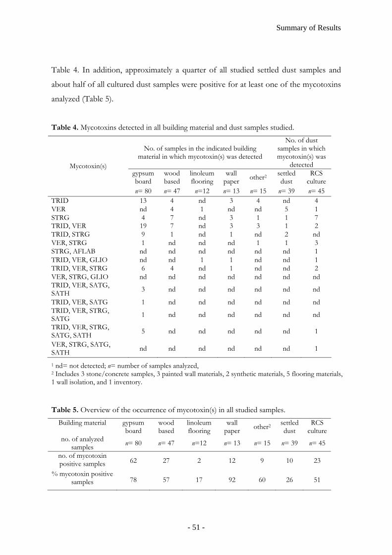
Summary of Results
Table 4. In addition, approximately a quarter of all studied settled dust samples and
about half of all cultured dust samples were positive for at least one of the mycotoxins
analyzed (Table 5).
Table 4. Mycotoxins detected in all building material and dust samples studied.
No. of samples in the indicated building material in which mycotoxin(s) was detected
No. of dust samples in which mycotoxin(s) was
detected gypsum board
wood based
linoleum flooring
wall paper other2 settled
dust RCS
culture
Mycotoxin(s)
n= 80 n= 47 n=12 n= 13 n= 15 n= 39 n= 45 TRID 13 4 nd 3 4 nd 4 VER nd 4 1 nd nd 5 1 STRG 4 7 nd 3 1 1 7 TRID, VER 19 7 nd 3 3 1 2 TRID, STRG 9 1 nd 1 nd 2 nd VER, STRG 1 nd nd nd 1 1 3 STRG, AFLAB nd nd nd nd nd nd 1 TRID, VER, GLIO nd nd 1 1 nd nd 1 TRID, VER, STRG 6 4 nd 1 nd nd 2 VER, STRG, GLIO nd nd nd nd nd nd nd TRID, VER, SATG, SATH 3 nd nd nd nd nd nd
TRID, VER, SATG 1 nd nd nd nd nd nd TRID, VER, STRG, SATG 1 nd nd nd nd nd nd
TRID, VER, STRG, SATG, SATH 5 nd nd nd nd nd 1
VER, STRG, SATG, SATH nd nd nd nd nd nd 1
1 nd= not detected; n= number of samples analyzed, 2 Includes 3 stone/concrete samples, 3 painted wall materials, 2 synthetic materials, 5 flooring materials, 1 wall isolation, and 1 inventory. Table 5. Overview of the occurrence of mycotoxin(s) in all studied samples.
Building material gypsum board
wood based
linoleum flooring
wall paper other2 settled
dust RCS
cultureno. of analyzed
samples n= 80 n= 47 n=12 n= 13 n= 15 n= 39 n= 45
no. of mycotoxin positive samples 62 27 2 12 9 10 23
% mycotoxin positive samples 78 57 17 92 60 26 51
- 51 -
Summary of Results
Additional comments
The MS method development for TRID and VER was started by analyzing TMS-
derivatives (Langseth and Rundberget 1998) on a GC ion trap MS instrument.
Sensitivity improved considerably when changing to a triple-quadrupole MS instrument,
HFB-derivatives (Langseth and Rundberget 1998, Nielsen et al 1998a and b), and NICI
(Kostiainen et al 1989, Kostiainen and Rizzo 1988). The reduction in background noise
when using MSMS analysis increased sensitivity by a factor of 100 compared to results
reported in previous studies (Nielsen et al 1998a).
In HPLC-MS it may seem controversial to use sodium as additive in the mobile
phase since formic acid (Wilkes and Lay 2001), TFA (Smedsgaard 1997) or ammonium
formate (Gottschalk et al 2008) are commonly used. However, sodium as an additive
has been tested in other studies (Demulle et al 2006, Tuomi et al 1998 and 2000) and
has been recommended to increase cationization in ESI-MS (Schnieder et al 1991).
- 52 -
Discussion
DISCUSSION
Mold in buildings - guidelines and regulations
Regardless of the incomplete knowledge we have of the specific agents and
mechanisms that are responsible for indoor air related health problems most
institutions, agencies and organisations agree that mold exposure in general should be
avoided. However, this advice is not new. The noun tsara'at mentioned in Leviticus
14:33-45 (3rd book of the Old Testament) has been interpreted as mold (Heller et al
2003). In this text the Lord tells Moses and Aaron how to remediate a mold-affected
house. First a priest should be asked to inspect it. Then the mold should be scraped off
the building material and dumped outside of town in an “unclean place”. The building
should then be sealed before the priest returns for inspection after one week. If the
mold damage has spread it is advised to tear down the entire building.
Today mold exposure is regarded as a health concern mainly where the exposure is
very high, e. g. in dusty agricultural occupational environments (Lacey and Dutkiewicz
1994) and during remediation procedures after water- and mold-damage e. g. in the
aftermath of natural disasters. Recently, the Federal Emergency Management Agency
(FEMA), part of the U.S. Department of Homeland Security, issued a warning to
residents cleaning homes and buildings damaged by e. g. flood and storms, encouraging
the use of protective equipment and recommending everyone to avoid unnecessary
exposure to mold. Moreover, specific groups, e. g. immunocompromised patients and
children younger than 12 years are recommended to avoid any exposure to high
concentrations of mold in flooded areas (FEMA 2008).
The FEMA warning applies to e. g. cleaning management after hurricane Katrina, one
of the costliest and deadliest natural disasters in the history of United States. This
hurricane struck Louisiana, Mississippi, and parts of Florida, Georgia, and Alabama in
August of 2005. In New Orleans, the city levee system catastrophically failed; about
80% of the city flooded to varying depths up to about 6 m, causing a large loss of lives
and property damage (Knabb et al 2005). Due to the floods, ca 80% of all houses and
their contents were subsequently contaminated by mold growth. The situation and
- 53 -
Discussion
associated health risks were described e. g. in popular science reporting from a
mycologist’s own home (Bennett 2006), and in reports from the U. S. Center for
Disease Control and prevention (CDC) and the National Institute for Occupatinal
Safety and Health (NIOSH). The living conditions and health-related needs of
returning residents were assessed (CDC 2006) and evaluations of health hazards were
made (West et al 2006). Additional quality tests were conducted by the Natural
Resources Defense Council (NRDC 2005). In scientific press there were several
reports, e. g. review papers (Manuel 2006) and cross-sectional studies on chronic
conditions among children and adolescents (Rath et al 2007). Several studies were also
made to assess bioaerosols with respect e. g. to molds and endotoxins (Chew et al 2006,
Rao et al 2007, Solomon et al 2006), as well as glucans (Rao et al 2007) in New Orleans.
We showed that the molds present in this subtropical climate zone produce
mycotoxins (paper III).
Exposure to mold is not only to be avoided in high level exposure environments like
in the aftermath of natural disasters. Immunocompromised, allergic, asthmatic patients
or people with breathing problems in general are regarded as being especially sensitive
to mold (CDC Disaster Safety 2006). The Committee on Environmental Health, in the
American Academy of Pediatrics, has released a policy statement which describes
guidelines for pediatricians to help reduce infants’ exposure to mold (American
Academy of Pediatrics: Committee on Environmental Health 1998). Scientists at the
U.S. EPA and Lawrence Berkeley National Laboratory have estimated that the
symptoms of 4.6 of the 21.8 million asthmatics in the USA are caused by indoor
dampness and mold to an annual cost of 3.5 billion dollars (Mudarri and Fisk 2007).
The U.S. EPA and CDC provide guidelines for mold cleanup online. Here residents
are advised to resolve the problem which originally caused the water-damage and to
clean the moldy surface (if less than ca 0.9 m2 area) with detergent or bleach while
avoiding exposure to the mold (EPA 2008, CDC Disaster Safety 2006). There are also
other, more local guidelines in the USA, e. g. on assessment and remediation of fungi in
indoor environments in New York (The New York City Department of Health and the
New York City Human Resources Administration 2008).
- 54 -
Discussion
In the Canadian Environmental Protection Act 1999 (Government of Canada 2007)
Health Canada simply recommends “to control humidity and diligently repair any
water damage in residences to prevent mold growth; and to clean thoroughly any
visible or concealed mold growing in the residential buildings”.
The Swedish National Board of Health and Welfare (1999, The Environmental Code,
chapter 9, § 3) recommends to remediate and prevent water damage and not to tolerate
visible mold or smell of mold in residential buildings. It specifically mentions to take
special consideration to sensitive residents/occupants and advocates investigations of
the buildings including both microbial and chemical analyses. If concealed mold is
suspected the authorities can demand a more comprehensive investigation of the
building according to The Environmental Code (chapter 26, § 22). Section 6:24 of the
BFS 2006:22 amendments of the Swedish Board of Housing, Building and Planning
states that “buildings and their installations shall be designed in such a way that
microorganisms cannot affect indoor air to the extent where they are detrimental to
human health or cause unpleasant smell” (BFS 2006).
The World Health Organisation (WHO) has through a working group developed
guidelines for indoor air quality taking into consideration pollutants, indoor
combustion and biological agents (WHO 2006) including the issues of dampness and
mold (WHO 2007). These guidelines mainly recommend prevention of microbial
growth through control of moisture and water-damage. If problems of dampness and
microbial exposure occur remediation should be performed, not least among low-
income populations to prevent additional burdens of poor health on this vulnerable
group. Interestingly, the WHO working group also states that “Building owners are
responsible for providing healthful workplaces or living environments free of excessive
moisture and mold problems by ensuring proper building construction and
maintenance. Occupants are responsible for managing water use, heating, ventilation,
appliances etc. in a proper manner that does not lead to dampness and mold growth”.
In conclusion, guidelines from the Old Testament to the WHO all agree that mold
and moisture should be avoided regardless of what causative agent(s) lay behind the
reported health problems in damp or water-damaged indoor environments. This
statement is clear and provides a general strategy how to handle these problems while
- 55 -
Discussion
further research is conducted to bring more detailed clarity to this complex
phenomenon.
This study in context
In the mid 80s and 90s, mold problems indoors came in focus as several disease
outbreaks were detected (Croft et al 1986, CDC 1995, Elidemir et al 1999, Flappan et
al 1999, Hodgson et al 1998, Jarvis et al 1998, Johanning et al 1996). Methods for
detection of mycotoxins, especially of trichothecenes derived from Stachybotrys spp.
(Bata et al 1985, Harrach et al 1981, Jarvis et al 1984, Krishnamurthy et al 1987,
Szathmary et al 1976) were developed and refined (Hinkley and Jarvis 2001, Nielsen et
al 1998a and b). Studies reveald that mold growing on indoor building materials can
produce mycotoxins (Engelhart et al 2002, Gravesen et al 1999, Nielsen et al 1998a,
Tuomi et al 1998 and 2000). However, based on number of spores found in indoor
environments, mycotoxin levels were not considered likely to pose a health threat
(Bush et al 2005, Kelman et al 2004).
This thesis project was prompted mainly by two sets of published studies. One set
showed that not only spores are released from mold-contaminated building materials
but also much smaller fungal fragments (0.3-20 µm, Górny et al 2002, Kildesø et al
2003). These small fragments were found to be released in up to 320-514 times higher
numbers than spores, share common antigens with spores (Górny et al 2002), and
demonstrate a 230-250 fold higher respiratory deposition than spores in adults and an
additional 4-5 times higher deposition in infants (Cho et al 2005). These fungal
particles (diameter < 1µm) may contain mycotoxins (Brasel et al 2005). Thus, our
exposure to mold and mycotoxins in water-damaged building may be much higher
than suggested in previous estimations based merely on spore concentrations (Bush et
al 2005, Kelman et al 2004).
Another set of studies showed that mycotoxin challenge, at picogram levels, to
PBMCs lead to a polarization towards the Th2 phenotype (mainly caused by a
reduction in numbers of IFN-γ-producing T lymphocytes rather than by a reduced
functional capacity of a single cell) which suggestes an increased risk for allergy
- 56 -
Discussion
development (Wichmann et al 2002). Interestingly, in another study, low exposure
doses of mycotoxins in human monocytic cells caused a cytokine imbalance with a
reduction in IL-10 concentrations compared to those of TNF-α and Il-6. These results
imply a risk of inflammatory responses and could potentially partly explain the diffuse
general indoor air-related symptoms and worsening of asthmatic inflammatory and
allergic reactions experienced in moldy environments (Johannessen et al 2005). These
data clearly show that mycotoxins even at very low concentrations may cause
immunomodulation.
The results of these studies inspired to the initiation of this project –to improve mass
spectrometry methods for selected mycotoxins and to use these methods to study
mycotoxins in water-damaged indoor environments. The initiative came naturally since
our research group is specialized in mass spectrometry methods and has over 20 years
of experience in the indoor environmental research field.
In paper I a GC-MS screening method for analysis of hydrolysis products of
mycotoxins produced by Stachybotrys spp. was optimized with respect to method
performance (sensitivity, selectivity); an important task considering the very low
mycotoxin concentration present in indoor environments. The screening analysis was
also convenient to use since individual Stachybotrys mycotoxins are not commercially
available (VER was in this case purchased and TRID was a kind gift from Poul
Rasmussen). Stachybotrys mycotoxins were specifically selected because of their
documented toxic potency (Pestka et al 2008) and suggested link to indoor-
environmental disease outbreaks (Dearborn et al 1999 and 2002, Vesper et al 2000).
The methods were successfully applied in direct analysis of authentic indoor
environmental samples including dust settled in the breathing zone.
In paper II our battery of mycotoxin analytes was expanded to include STRG, SATG
and SATH. HPLC was introduced as a separation method, giving the opportunity to
measure intact macrocyclic trichothecenes SATG and SATH (kind gift from Prof.
Bruce B. Jarvis), in addition to the hydrolysis product VER of Stachybotrys (chemotype
S) MTRs (Andersen et al 2002 and 2003). The carcinogenic STRG was selected
because it is produced by A. versicolor and a few other molds frequently encountered in
water-damage buildings (Engelhart et al 2002, Frisvad et al. 1989, Hajjar et al. 1989).
- 57 -
Discussion
These mycotoxin analyses were applied to authentic indoor environmental samples
including building materials and dusts. The collection of building material samples
were mainly gypsum boards since Stachybotrys is commonly found on water-damaged
materials rich in cellulose (Miller et al 2003).
In paper III dust samples collected from buildings in New Orleans flooded in the
aftermath of hurricane Katrina were analyzed. This is the first time that mycotoxins
have been determined in Katrina-associated buildings and the results demonstrate that
molds in indoor environments in a subtropic climate may produce mycotoxins.
In paper IV we expanded our battery of mycotoxin analytes even further and
incorporated GLIO and AFLAB, mycotoxins produced by various Aspergillus and
Penicillium spp., common in water-damaged indoor environments (Nielsen 2002).
AFLAB is highly carcinogenic (Colombe 1993) and is routinely screened for in food
and animal feed according to safety regulations around the world (FAO 2004). GLIO
is a neuphrotoxic (Axelsson 2007) and immunosuppressive agent which has been
shown to cause cell death by means of apoptosis and necrosis (Gardiner et al 2005).
Also, GLIO has been linked to the development of allergy (Wichmann et al 2002) and
inflammation response (Johannessen et al 2005). The analyzed samples in this study
were not emphasized on gypsum boards but were building material samples that had
been consecutively collected by personnel specialized in remediation measures during a
one-year period. The distribution of the selected mycotoxins found in this study is thus
a cross section on mycotoxin prevalence in water-damaged buildings undergoing
technical investigation due to reported indoor-related health problems.
- 58 -
CONCLUSIONS
• GC-MSMS analysis of VER and TRID HFB-derivatives in NICI mode and HPLC-
MSMS analysis of STRG, SATG, SATH, GLIO, and AFLAB in positive ESI mode,
using triple quadrupole MS, are very specific and sensitive methods for analysis of
indoor environmental samples.
• The developed integrated HPLC- and GC-MSMS analysis strategy for the
combined analysis of all indicated mycotoxins is convenient and applicable
especially when sample size is small.
• Molds in water-damaged indoor environments, in both sub-tropic and tempered
climatic zones, produce mycotoxins regularly. Mycotoxins were found in settled
dust on surfaces above floor level indicating that mycotoxins from mold-
contaminated building materials can become airborne and inhalable.
• Mycotoxin production or prevalence does not seem to correlate to microscopy
findings, CFU counts, or PCR results for fungi. There is a potential correlation
between fungal biomass and mycotoxin production.
- 59 -

- 60 -
ACKNOWLEDGEMENTS
I wish to sincerely thank the following people: My supervisor Lennart Larsson for your continuous support, patience, excellent scientific guidance, and always being there for me. Christina for your excellent laboratory assistance, your big warm heart and endless ideas for new inventions. Bogumila, Alexandra and Karol for your help and guidance, fun ideas, fun times and great conversation. Gertrud for your support, wisdom and contagious smile. Carina, Beston, Julia, Angela, and Saioa for good times and good “fika” sessions. Lennart Truedsson – my co-supervisor - and Annica for valuable discussion. Mirko –good luck with the machines! My co-authors Linda Faye Grimsley, Jocelyn Lewis, Aime Must (and Aimex), and Eva Nyman (and Tekomo) for collaboration. Klas Flärdh at Cell- and organism biology and Eric Carleman at the Electronmicroscopy department for helping me taking photos of “Stachy”. My collegues at the National Public Health Institute in Kuopio, Finland, especially Martin Täubel, Aino Nevalainen, Maija-Riitta Hirvonen, and Maija Riitta’s group including Piia Penttinen, Majut Roponen, Paasi Jalava and especially Kati Huttunen for taking care of me in Kuopio. Thank you also to the HITEA network for collaboration. Dr. Elena Pieckowa and co-workers, Dr. Fog Nielsen with co-workers, Prof. Dearborn with co-workers, Prof. Milton with co-workers, and Prof. Anders Tunlid och Eva for valuable discussion and collaboration. Poul Rasmussen, Leo-Pharma, Denmark for providing trichodermin and Professor Bruce B. Jarvis for mycotoxin standards. The Department of Laboratory Medicine and Clinical Microbiology Department especially Dr. Elisabet Holst for your continous support and for “saving my life”, Caterina Ivarsson for your excellent assistance with economic issues, Gunnar and Eva-Maria for listening, Pelle, Anita, Arne, and the reception at Clinical Microbiology for valuable assistance. Kyösti Tuutti for your continuous support. Kenneth Nilsson for your excellent assistance with the troublemaking-machines and your support –you are a true MS magician. The people at Varian Inc., especially Martin for you support and wonderful newly baked gifts, and Eric for your technical assistance. Friends at meetings and conferences for valuable discussions and fun times, especially Malin, Thorbjörn, and Linda. SWESIAQ for including me in the important work on supporting the creation of healthy, comfortable and productive indoor environments. IVL Swedish Environmental Research Institute for welcoming me onboard! I would also like to extend my gratitude to SBUF (Development Fund of the Swedish Construction Industry), FORMAS (the Swedish Research Council for Environment, Agricultural Sciences and Spatial Planning), Asthma & Allergiförbundet (Swedish Asthma and Allergy Association), SWESIAQ (The Swedish chapter of ISIAQ, International Society of Indoor Air Quality and Climate), The Medical Faculty and The Royal Physiographic Society in Lund, and Queen Silvia's Jubilee Fund for Research on Children and Children's Disabilities for financial support.
- 61 -
In this section I will express my gratitude to people who are not working in this research field; thus, this text is written in Swedish. Varmt tack till min fantastiska familj som alltid stödjer mig, tror på mig och finns där i vått och torrt. Jag känner mig privilegierad, lyckligt skattad och tacksam för var och en av er. Ett speciellt varmt tack… … till min fantastiska Mamma Margareta och Pappa Lasse som jag inte bara älskar och respekterar för att ni är mina föräldrar utan för att ni är gränslöst omtänksamma, stolta och underbara människor. Den här avhandlingen är dedikerad till er. Ni har genom åren uppmuntrat min nyfikenhet, glädje för lärande och kreativitet och gjort allt i er makt för att jag alltid ska ha de bästa tänkbara förutsättningar. Er kärlek och tro på mig har varit orubblig och utan er hade jag aldrig nått dit jag står idag. … till min coola, kluriga Farmor Aina för roliga stunder med kortspel, humor och spontanitet. Våra samtal över telefon har varit små vattenhål som fått mig att behålla fokus och det har betytt mer för mig än du tror. ”Vi luller vi, farmor, du och jag.” … till Eivor & Yngve för ert aldrig sinande engagemang. Ni är alltid intresserade av vad än obegripligt jag sysslar med och våra samtal värmer alltid mitt hjärta. Med er är inte delad glädje dubbel utan snarare trippel. … till ”Vesterlundsklanen”: Mats och Ann-Charlotte, Bodil och Thomas, Anna och Mattias, Malin, Niklas och Evelina. Det är en fröjd att få vara en del i er gemenskap och era liv. Ni är var och en på ert eget vis en ständig källa till inspiration! Tack för alla goda samtal, för ert engagemang och uppmuntran. … till hela den underbara familjen Johansson för allt stöd och tålamod med mina långa utläggningar. Tack för att ni alla tagit emot mig med öppna armar. Och tänk, inte nog med att jag vunnit världens bästa man – jag har fått världens bästa svärföräldrar på köpet! Tack också till alla mina underbara vänner, speciellt: Helen, Martin och solstrålen Nelly för mysiga och roliga stunder, givande diskussioner och er innerliga vänskap. Emil och Thomas, mina musketörer och kamratbröder, för er aldrig sinande lojalitet, sköna humor och nördighet. Tack också Kristina och Lizette för att jag fått lära känna er och för härliga samtal. Therése och Marielle för era vida tankeperspektiv, kreativitet och att ni inspirerar mig. Therese och Mattias för att ni helhjärtat lyssnar och smittar av er med er sköna optimism. Lena för ditt djupa, öppna och kloka sinne –ingen förstår mig som du. Maud och Lasse för underbara samtal, gemenskap och kreativa idéer. Johan och Magnus som frågar hur jag mår, gratulerar mig och ”supportar” mig på Facebook. Annette, Inger & Johan, Håkan & Linda, Örjan, Sophia och Valter för god gemenskap som sätter guldkant på vardagen. Också ett speciellt tack till doktorandombudsmannen Johan, samt Hans Wirje och receptionen på företags-hälsovården i Lund som lyssnade och stöttade mig när jag verkligen behövde det innan jag hoppade på det här projektet. Tack, sist men inte minst, till min bättre hälft Stefan: min drömprins, livskamrat och bäste vän. Tack för att du stödjer och uppmuntrar mig, delar mina skratt och tårar och för att du är min trygga hamn när havet stormar. Med dig känns allt i livet möjligt! Jag älskar dig!
- 62 -
References
REFERENCES
Aberg, N. (1989). Asthma and ellergic rhinitis in Swedish conscripts. Clin Exp Allergy. 19(1): 59-63.
Aberg, N., B. Hesselmar, B. Aberg, and B. Eriksson. (1995). Increase of asthma, allergic rhinitis and eczema in Swedish school children between 1979 and 1991. Clin Exp Allergy. 25(9): 791-794.
American Academy of Pediatrics: Committee on Environmental Health. (1998). Toxic Effects of Indoor Molds. Pediatrics. 4(101): 712-714.
Andersen, B., K. F. Nielsen, and B. B. Jarvis. (2002). Characterization of Stachybotrys from water-damaged buildings based on morphology, growth, and metabolite production. Mycologia 94(3): 392-403.
Andersen, B., K. F. Nielsen, U. Thrane, T. Szaro, J. W. Taylor, and B. B. Jarvis. (2003). Molecular and phenotypic descriptions of Stachybotrys chlorohalonata sp. nov. and two chemotypes of Stachybotrys chartarum found in water-damaged buildings. Mycologia. 95(6): 1227-1238.
Andersen, B. and A. T. Nissen. (2000). Evaluation of media for detection of Stachybotrys and Chaetomium species associated with water-damaged buildings. Int Biodeter Biodegrad. 46(2): 111-116.
Andersson, M. A., R. Mikkola, R. M. Kroppenstedt, F. A. Rainey, J. Peltola, J. Helin, K. Sivonen and M. S. Salkinoja-Salonen. (1998). The mitochondrial toxin produced by Streptomyces griseus strains isolated from an indoor environment is valinomycin. Appl Environ Microbiol. 64(12): 4767-4773.
Axelsson, V. (2007). Evaluation of neurotoxic properties of gliotoxin [PhD thesis]. Uppsala, Sweden: Dept. of neurochemistry, 10691 Stockholm, Sweden.
Bata, A., B. Harrach, K. Ujsza'szi, A. Kis-Tomas, and R. Lasztitz. (1985). Macrocyclic trichothecene toxins produced by Stachybotrys atra strains isolated in Middle Europe. Appl Environ Microbiol. 49(3): 678-681.
Beasley R., P. Ellwood, and I. Asher. (2003). International patterns of the prevalence of pediatric asthma the ISAAC program. Pediatr Clin North Am. 50(3): 539-53.
Beijer, L., J. Thorn, and R. Rylander. (2003). Mould exposure at home relates to inflammatory markers in blood. Eur Respir J. 21(2): 317-322.
Bennett, J. W. (2006). The molds of Katrina. Update (New York Academy of Sciences). Jan/Feb: 7-9.
Bennett, J. W. and M. Klinch. (2003). Mycotoxins. Clin Microbiol Rev. 16(3): 497-516. Bermingham, S., L. Maltby, and R. C. Cooke. (1995). A critical assessment of the
validity of ergosterol as an indicator of fungal biomass. Mycol Res. 99(4): 479-484.
BFS (2006). Section 6 Hygiene, health and the environment, in Building regulations –mandatory provisions and general recommendations, BBR 2006:22. English translation, The Swedish board of housing, building and planning publication service, Karlskrona, Sweden.
- 63 -
References
Bjurman, J. (1994). Ergosterol as an indicator of mould growth on wood in relation to culture age, humidity stress and nutrient level. Int Biodeter Biodegrad. 33(4): 355-368.
Black, J. A., K. K. Foarde, and M. Y. Menetrez. (2006). Solvent comparison in the isolation, solubilization, and toxicity of Stachybotrys chartarum spore trichothecene mycotoxins in an established in vitro luminescence protein translation inhibition assay. J Microbiol Meth. 66(2): 354-362.
Bondy, G. S. and J. J. Pestka. (2000). Immunomodulation by fungal toxins. J Toxicol Environ Health B Crit Rev. 3(2): 109-143.
Bornehag, C.-G., G. Blomquist, F. Gyntelberg, B. Jarvholm, P. Malmberg, L. Nordvall, A. Nielsen, G. Pershagen, and J. Sundell. (2001). Dampness in Buildings and Health. Indoor Air. 11(2): 72-86.
Bornehag, C. G., J. Sundell, L. Hagerhed-Engman, T. Sigsggard, S. Janson, and N. Aberg. (2005). 'Dampness' at home and its association with airway, nose, and skin symptoms among 10,851 preschool children in Sweden: a cross-sectional study. Indoor Air. 15(s10): 48-55.
Bornehag C. G., J. Sundell, C. J. Wechler, T. Sigsgaard, B. Lundgren, M. Hasselgren, and L. Hagerhed-Engman. (2004). The association between asthma and allergic symptoms in children and phtalates in house dust: a nested case-control study. Environ Health Perspect. 112(14): 1393-1397.
Brasel, T. L., D. R. Douglas, S. C. Wilson, and D. C. Straus. (2004). Detection of airborne Stachybotrys chartarum macrocyclic trichothecene mycotoxins on particulates smaller than conidia. Appl Environ Microbiol. 71(1): 114-22.
Brasel, T. L., J. M. Martin, C. G. Carriker, S. C. Wilson, and D. C. Straus. (2005). Detection of airborne Stachybotrys chartarum macrocyclic trichothecene mycotoxins in the indoor environment. Appl Environ Microbiol. 71(11): 7376–7388.
Bush, R. K., J. M. Portnoy, A. Saxon, A. I. Terr, and R. A. Wood. 2006. The medical effects of mold exposure. J Allergy Clin Immunol. 117(2): 326–333.
Calvo, A. M., R. A. Wilson, J. W. Bok, and N. P. Keller. (2002). Relationship between secondary metabolism and fungal development. Microbiol Mol Biol Rev. 66(3): 447-459.
Campbell, A. W., J. D. Thrasher, M. R. Gray, and A. Vojdani. (2004). Mold and mycotoxins: effects on the neurological and immune systems in humans. Adv Appl Microbiol. 55: 375-406.
Center for Disease Control and prevention (CDC). (1995). Current trends acute pulmonary hemorrahage among infants --Chicage, April 1992- November 1994. MMWR. 44(4): 67, 73-74.
Center for Disease Control and prevention (CDC). (Disaster Safety 2006). Protect yourself from mold. Dept. of health and human services, CDC, at www.bt.cdc.gov/disasters
- 64 -
References
Center for Disease Control and prevention (CDC). (2006). Health concearns associated with mold in water-damaged homes after hurricanes Katrina and Rita—New Orleans Area, Louisiana, October 2005. MMWR Morb Mortal Wkly Rep. 55(2): 41-44.
Chapman, J. R. (1993). (2nd ed.), Practical organic mass spectrometry: A guide for chemical and biochemical analysis, John Wiley and Sons, West Sussex, England.
Charpin-Kadouch, C., G. Maurel, R. Felipo, J. Queralt, M. Ramadour, H. Dumon, M. Garans, A. Botta, and D. Charpin. (2006). Mycotoxin identification in moldy dwellings. J Appl Toxicol. 26(6): 475–479.
Chew, G. L., J. Wilson, F. A. Rabito, F. Grimsley, S. Iqbal, T. Reponen, M. L. Muilenberg, P. S. Thorne, D. G. Dearborn, and R. L. Morley. (2006). Mold and endotoxin levels in the aftermath of hurricane Katrina: A pilot project of homes in New Orleans undergoing renovation. Environ Health Perspect. 114(12): 1883-1889.
Cho, S.-H., S.-C. Seo, D. Schmechel, S. A. Grishpun, and T. Reeponen. (2005). Aerodynamic charachteristics and respiratory deposition of fungal fragments. Atmos Environ. 39(30): 5454-5465.
Coulombe, R. A. (1993). Biological action of mycotoxins. J Dairy Sci. 76(3): 880-891. Creasia, D. A., J. D. Thurman, L.J. Jones, M.L. Nealley, C.G. York, R.W.
Wannemacher, and D.L. Bunner (1987). Acute inhalation toxicity of T-2 mycotoxin in mice. Fund Appl Toxicol. 8(2): 230-235.
Creasia, D. A., J. D. Thurman, R. W. Wannemacher, and D. L. Bunner. (1990). Acute inhalation toxicity of T-2 mycotoxin in the rat and guinea pig. Fund Appl Toxicol. 14(1): 54-59.
Croft, W. A., B. B. Jarvis, and C. S. Yatawara. (1986). Airborne outbreak of trichothecene toxicosis. Atmos Environ. 20: 49–552.
Cruse, M., R. Telerant, T. Gallagher, T. Lee, and J. W. Taylor. (2002). Cryptic species in Stachybotrys chartarum. Mycologia. 94(5): 814-822.
Curtis, L., A. Libermann, M. Stark, W. Rea, and M. Vetter. (2004). Adverse health effects of indoor molds. J Nutr Environ Med. 14(3): 261-274.
Cvetnić, Z., and S. Pepeljnjak.. (2007). Interaction between certain moulds and aflatoxin B1 producer Aspergillus flavus NRRL 3251. Arc Ind Hyg Toxicol. 58(4): 429-434.
Daisey, J. M., W. J. Angell, and M. G. Apte. (2003). Indoor air quality, ventilation and health symptoms in schools: an analysis of existing information. Indoor Air. 13(1): 53-64.
Dearborn, D. G., P. G. Smith, B. B. Dahms, T. M. Allen, W.G. Sorenson, E. Montana, and R. A. Etzel. (2002). Clinical profile of 30 infants with acute pulmonary hemorrhage in Cleveland. Pediatrics. 110(3): 627-637.
- 65 -

References
Dearborn, D. G., I. Yike, W. G. Sorenson, M. J. Miller, and R. A. Etzel. (1999). Overview of investigations into pulmonary hemorrhage among infants in Cleveland, Ohio. Environ Health Persp Suppl. 107(S3): 495-499.
Delmulle, B., S. De Saeger, A. Adams, N. De Kimpe, and C. Van Peteghem. (2006). Development of a liquid chromatography/tandem mass spectrometry method for the simultaneous determination of 16 mycotoxins on cellulose filters and in fungal cultures. Rapid Commun Mass Spectrom. 20(5): 771-776.
Dillon, H. K., J. D. Miller, W. G. Sorenson, J. Douwes, and R. R. Jacobs (1999). Review of methods applicable to the assessment of mold exposure to children. Environ Health Perspect. Suppl 3(107): 473-80.
Dönmez-Altuntas, H., G. Dumlupinar, N. Imamoglu, Z. Hamurcu, and B. C. Liman. (2007). Effects of the mycotoxin citrinin on micronucleus formation in a cytokinesis-block genotoxicity assay in cultured human lymphocytes. J Appl Toxicol. 27(4): 337-341.
Elanskii, S., I. Petrunina, O. Lavrova, and A. Likhachev. (2004). A comparative analysis of Stachybotrys chartarum strains isolated in Russia. Microbiology. 73(1): 60-65.
Elidemir, O., G. N. Colasurdo, S. N. Rossmann, and L. L. Fan. (1999). Isolation of Stachybotrys from the lung of a child with pulmonary hemosiderosis. Pediatrics. 104(4): 964-966.
El-Maghraby, O. M. O., G. A. Bean, B. B. Jarvis, and M. B. Aboul-Nasr. (1991). Macrocyclic trichothecenes produced by Stachybotrys isolated from Egypt and Eastern Europe. Mycopathologia. 113(2): 109-115.
Engelhart, S., A. Loock, D. Skutlarek, H. Sagunski, A. Lommel, H. Farber, and M. Exner. (2002). Occurrence of toxigenic Aspergillus versicolor isolates and sterigmatocystin in carpet dust from damp indoor environments. Appl Environ Microbiol. 68(8): 3886–3890.
Environmental protection agency (EPA) (2008). Air: Indoor air quality: Mold: A brief guide to mold, moisture, and your home.
http://www.epa.gov/mold/moldcleanup.html FAO (2004). Food and nutrition paper no. 81, Worldwide regulations for mycotoxins
in food and feed in 2003. 180 pages. Federal Emergency Management Agency (FEMA). (2008). DR-1768-WI-NR-73,
Wisconsin disaster recovery, USA. at http://emergencymanagement.wi.gov/docview.asp?docid=14267&locid=18.
Flappan, S. M., J. Portnoy, P. Jones, and C. Barnes. (1999). Infant pulmonary hemorrhage in suburban home with water damage and mold (Stachybotrys atra). Environ Health Perspect. 107(11): 927–930.
Flemming, J., B. Hudson, and T. G. Rand. (2004). Comparison of inflammatory and cytotoxic lung responses in mice after intratracheal exposure to spores of two different Stachybotrys chartarum strains. Toxicol Sci. 78(2): 267-275.
- 66 -
References
Fonzi, W. A. and P. S. Sypherd. (1986). Trichodermin esterase activity and trichodermin resistance in Mucor racemosus. Antimicrob Agents Chemother. 29(4): 570-575.
Foto, M., L. L. P. Vrijmoed, J. D. Miller, K. Ruest, M. Lawton, and R. E. Dales. (2005). A comparison of airborne ergosterol, glucan and Air-O-Cell data in relation to physical assessments of mold damage and some other parameters. Indoor Air. 15(4): 257-266.
Fox, A, W. Harley, C. Feigley, D. Salzberg, C. Toole, A. Sebastian, and L. Larsson. (2005). Large particles are responsible for elevated bacterial marker levels in school air upon occupation. J Environ Monit. 7(5): 450-456.
Frisvad, J. C. (1989). The connection between the Penicillia and Aspergilli and mycotoxins with special emphasis on misidentified isolates. Arch Environ Contam Toxicol. 18(3): 452-67.
Gao, P. and J. Martin. (2002). Volatile metabolites produced by three strains of Stachybotrys chartarum cultivated on rice and gypsum board. Appl Occup Environ Hyg. 17(6): 430-436.
Gardiner, D. M., P. Waring, and B. J. Howlett. (2005). The epipolythiodioxopiperazine (ETP) class of fungal toxins: distribution, mode of action, functions and biosynthesis. Microbiology 151(4): 1021-32.
Górny, R. L., T. Reponen, K. Willeke, D. Schmechel, E. Robine, M. Boissier, and S. A. Grinshpun. (2002). Fungal fragments as indoor air biocontaminants. Appl Environ Microbiol. 68(7): 3522–3531.
Gottschalk, C., J. Bauer, and K. Meyer. (2008). Detection of satratoxin G and H in indoor air from a water-damaged building. Mycopathologia. 166(2): 103-107.
Government of Canada, Department of Health. (2007). Residential inddor air quality guideline for moulds, in Canadian Environmental protection act 1999.Vol. 141, No 13-March 31.
Gravesen, S., P. A. Nielsen, R. Iversen, and K. F. Nielsen. (1999). Microfungal contamination of damp buildings—examples of risk constructions and risk materials. Environ Health Persp. 107(Suppl. 3): 505–508.
Green, B., E. Tovey, J. Sercombe, F. Blachere, D. Beezhold, and D. Schmechel. (2006). Airborne fungal fragments and allergenicity. Med Microbiol. 44(Suppl 1): S245–S255.
Grove, J. F. (1993). Macrocyclic trichothecenes. Nat Prod Rep. 10(5): 429-448. Hagerhed-Engman, L., C.-G. Bornehag, J. Sundell, and N. Åberg. (2006). Day-care
attendance and increased risk for respiratory and allergic symptoms in preschool children. Allergy. 61(4): 447-453.
Hajjar, J. D., J. W. Bennett, D. Bhatnagar, and R. Bahu. (1989). Sterigmatocystin production by laboratory strains of Aspergillus nidulans. Mycological Research. 93(4): 548–551.
- 67 -
References
Harrach, B., C. J. Mirocha, S. V. Pathre, and M. Palyusik. (1981). Macrocyclic trichothecenes toxins produced by a strain of Stachybotrys atra from Hungary. Appl Environ Microbiol. 41(6): 1428-1432.
Haugland, R. A., M. Varma, L. J. Wymer, and S. J. Vesper. (2004). Quantitative PCR analysis of selected Aspergillus, Penicillium and Paecilomyces species. Appl Microbiol 27(2): 198-210.
Haugland, R. A., and S. J. Vesper. (May 2002). Identification and quantification of specific fungi and bacteria, US patent 6,387,652.
Haugland, R. A., S. J. Vesper, and L. J. Wymer. (1999). Quantitative measurement of Stachybotrys chartarum conidia using real time detection of PCR products with the TaqManTMfluorogenic probe system. Mol Cell Prob. 13(5): 329-340.
Heller, R. M., T. W. Heller, and J. M. Sasson. (2003). Mold. “Tsara’at”, Leviticus, and the history of a confusion. Perspect Biol Med. 46(4): 588-591.
Hesselmar, B., N. Åberg, B. Åberg, B. Eriksson, and B. Björkstén. (1999). Does early exposure to cat or dog protect against later allergy development?. Clin Exp Allergy. 5(29): 611-617.
Hinkley, S. F., and B. B. Jarvis. (2001). Chromatographic method for Stachybotrys toxins. Methods Mol Biol. 157: 173–194.
Hinkley, S. F., J. Jiang, E. P. Mazzola, and B. B. Jarvis. (1999). Atranones: novel diterpenoids from the toxigenic mold Stachybotrys atra. Tetrahedron Lett. 40(14): 2725-2728.
Hinkley, S. F., J. A. Moore, J. Squillari, H. Tak, O. Heekyung, R. Oleszewski, E. P. Mazzola, and B. B. Jarvis. (2003). New atranones from the fungus Stachybotrys chartarum. Magn Reson Chem. 41(5): 337-343.
Hintikka, E.-L. (2004). The Role of Stachybotrys in the phenomenon known as sick building syndrome. Adv Appl Microbiol. 55: 155-173.
Hirvonen, M.-R., M. Ruotsalainen, M. Roponen, A. Hyvarinen, T. Husman, V.-M. Kosma, H. Komulainen, K. Savolainen, and A. Nevalainen. (1999). Nitric oxide and proinflammatory cytokines in nasal lavage fluid associated with symptoms and exposure to moldy building microbes. Am J Respir Crit Care Med. 160(6): 1943-1946.
Hodgson, M. J., P. Morey, L. Wing-Yan, L. Morrow, D. Miller, B. B. Jarvis, H. Robbins, J. F. Halsey, and E. Storey. (1998). Building-associated pulmonary disease from exposure to Stachybotrys chartarum and Aspergillus versicolor. JOEM 40(3): 241–248.
Hudson, B., J. Flemming, G. Sun, and T. G. Rand (2005). Comparison of immunomodulator mRNA and protein expression in the lungs of Stachybotrys chartarum spore-exposed mice. J Toxicol Environ Health A. 68(15): 1321-1335.
Hunter, C. A., C. Grant, B. Flannigan, and A. F. Bravery. (1988). Mould in buildings: the air spora of domestic dwellings. Int Biodeterior. 24(2): 81-101.
- 68 -
References
Huttunen, K., J. Pelkonen, K. F. Nielsen, U. Nuutinen, J. Jussila, and M. R. Hirvonen. (2004). Synergistic interaction in simultaneous exposure to Streptomyces californicus and Stachybotrys chartarum. Environ. Health Perspect. 112(6): 659-65.
Hyvärinen, A., T. Meklin, A. Vepsäläinen, and A. Nevalainen. (2002). Fungi and actinobacteria in moisture-damaged building materials - concentrations and diversity. Int Biodeter Biodegrad. 49(1): 27-37.
Hyvärinen, A., A. Sebastian, J. Pekkanen, L. Larsson, M. Korppi, T. Putus, and A. Nevalainen. (2006). Characterizing microbial exposure with ergosterol, 3-hydroxy fatty acids, and viable microbes in house dust: Determinants and association with childhood asthma. Arch Environ Occup Health. 61(4): 149-157.
Institute of Medicine (2004). Damp Indoor Spaces and Health Review. National Academic Press., Washington, D.C., USA.
http://www.nap.edu/books/0309091934/html/ International Agency for Research on Cancer (IARC). (2002). Aflatoxins in Evaluation
of carcinogenic risks to humans: some traditional herbal medicines, some mycotoxins, naphthalene and styrene, Vol 82, pps. 177, WHO Press, Geneva, Switzerland.
Iossifova, Y., T. Reponen, H. Sucharew, P. Succop, and S. Vesper. (2008). Use of (1-3)-beta-D-glucan concentrations in dust as a surrogate method for estimating specific fungal exposures. Indoor Air. 18(3): 225-232.
Islam, Z., J. R. Harkema, and J. J. Pestka. (2006). Satratoxin G from the black mold Stachybotrys chartarum evokes olfactory sensory neuron loss and inflammation in the murine nose and brain. Environ Health Persp. 114(7): 1099-1107.
Jarvis, B. B. (2002). Chemistry and toxicology of molds isolated from water-damaged buildings. Adv Exp Med Biol. 504: 43-52.
Jarvis, B. B. (2003). Stachybotrys chartarum: a fungus for our time. Phytochemistry. 64(1): 53-60.
Jarvis, B. B., J. Salemme, and A. Morais. (1995). Stachybotrys Toxins 1. Nat Toxins. 3(1): 10-16.
Jarvis, B. B., and J. D. Miller. (2005). Mycotoxins as harmful indoor air contaminants. Appl Microbiol Biotechnol. 66(4): 367–372.
Jarvis, B. B., W. G. Sorenson, E.-L. Hintikka, M. Nikulin, Y. Zhou, J. Jiang, S. Wang, S. Hinkley, R. A. Etzel, and D. Dearborn. (1998). Study of toxin production by isolates of Stachybotrys chartarum and Memnoniella echinata isolated during a study of pulmonary hemosiderosis in infants. Appl Environ Microbiol. 64(10): 3620-3625.
Jarvis, B. B., C. S. Yatawara, S. L. Greene, and V. Vrudhula. (1984). Production of verrucarol. Appl Environ Microbiol. 48(3): 673-674.
- 69 -
References
Johannessen, L. N., A. M. Nielsen, and M. Løvik. (2005). The mycotoxins citrinin and gliotoxin differentially affect production of the pro-inflammatory cytokines tumour necrosis factor-alpha and interleukin-6, and the anti-inflammatory cytokine interleukin-10. Clin Exp Allergy. 35(6): 782-789.
Johannessen, L. N., A. M. Nilsen, and M. Løvik. (2007). Mycotoxin-induced depletion of intracellular glutathione and altered cytokine production in the human alveolar epithelial cell line A549. Toxicol Lett. 168(2): 103–112.
Johanning, E., R. Biagini, H. DeLon, P. Morey, B. B. Jarvis, and P. Landsbergis. (1996). Health and immunology study following exposure to toxigenic fungi (Stachybotrys chartarum) in water-damaged office environment. Int Arch Occup Environ Health. 68(4): 207–218.
Jussila, J., J. Pelkonen, V.-M. Kosma, J. Maki-Paakkanen, H. Komulainen, and M.-R. Hirvonen. (2003). Systemic immunoresponses in mice after repeated exposure of lungs to spores of Streptomyces californicus. Clin Diagn Lab Immunol. 1(10): 30-37.
Karunasena, E., J. D. Cooley, D. R. Douglas, and D. C. Straus. (2004). Protein translation inhibition by Stachybotrys chartarum conidia with and without the mycotoxin containing polysaccharide matrix. Mycopathologia. 158(1): 87-97.
Kelman, B. J., C. A. Robbins, L. J. Swenson, and B. D. Hardin. (2004). Risk from inhaled mycotoxins in indoor office and residential environments. Int J Toxicol. 23(1): 3-10.
Keswani, J., M. L. Kashon, and B. T. Chen. (2005). Evaluation of interference to conventional and real-time PCR for detection and quantification of fungi in dust. J Environ Monit. 7(4): 311-318.
Kilburn, K. H. (2004). Role of molds and mycotoxins in being sick in buildings: neurobehavioral and pulmonary impairment. Adv Appl Microbiol. 55: 339-359.
Kildesø, J., H. Wurtz, K. F. Nielsen, P. Kruse, K. Wilkins, U. Thrane, S. Gravesen, P. A. Nielsen, and T. Schneider. (2003). Determination of fungal spore release from wet building materials. Indoor Air. 13(2): 148–155.
Kim, J. L, L. Elfman, G. Wieslander, G. Smedje, and D. Norbäck. (2006). Indoor moulds, bacteria, microbial volatile organic compounds (MVOC) and plasticizers in school: associations with asthma and respiratory symptoms in pupils. Indoor Air. 15(3): 170-82.
Knabb, R. D., J. R. Rhome, and D. P. Brown. (2005) (updated Aug 2006). Tropical cyclone report: Hurricane Katrina: 23-30 August 2005. National Hurricane Center. http://www.nhc.noaa.gov/pdf/TCR-AL122005_Katrina.pdf
Kostiainen, R. and A. Rizzo. (1988). The characterization of trichothecenes as their heptafluorobutyrate esters by negative-ion chemical ionization tandem mass spectrometry. Analytica Chimica Acta. 204(1-2): 233-246.
Kostiainen, R., A. Rizzo, and A. Hesso. (1989). The analysis of trichothecenes in wheat and human plasma samples by chemical ionization tandem mass spectrometry. Arch Environ Contam Toxicol. 18(3): 356-364.
- 70 -
References
Krishnamurthy, T., E. Sarver, W., S. L. Greene, and B. B. Jarvis. (1987). Mass spectral investigations on trichothecene mycotoxins. II. Detection and quantification of macrocyclic trichothecenes by gas chromatography/negative ionization mass spectrometry. J Assoc Off Anal Chem. 70(1): 132-140.
Krishnamurthy, T., M. B. Wasserman, and E. W. Sarver. (1986). Mass spectral investigations I. Application of negative ion chemical ionization techniques for the simultaneous and accurate analysis of simple trichothecenes in picogram levels. Biomed Environ Mass. 13(9): 503-518.
Kuhn, D. M. and M. A. Ghannoum. (2003). Indoor mold, toxigenic fungi, and Stachybotrys chartarum: infectious disease perspective. Clin Microbiol Rev. 16(1): 144-172.
Kupfahl, C., A. Michalka, C. Lass-Florl, G. Fischer, G. Haase, T. Ruppert, G. Geginat, and H. Hof. (2008). Gliotoxin production by clinical and environmental Aspergillus fumigatus strains. Int J Med Microbiol. 298(3-4): 319-327.
Lacadena, J., E. Alvarez-Garcia, N. Carreras-Sangra, E. Herrero-Galan, J. Alegre-Cebollada, L. Garcia-Ortega, M. Onaderra, J. G. Gavilanes, and A. Martinez del Pozo. (2007). Fungal ribotoxins: molecular dissection of a family of natural killers. FEMS Microbiol Rev. 31(2): 212-237.
Lacy, J. and J. Dutkiewicz. (1994). Bioaerosols and occupational lung disease. J Aerosol Sci. 25(8): 1371-1404.
Land, C. J., A. Must, and N. Hogberg. (2003). Identification of fungi, especially Stachybotrys chartarum from gypsumboards, by means of PCR and sequencing of ribosomal DNA. Indoor Built Environ. 12(4): 227-229.
Langseth, W. and T. Rundberget. (1998). Instrumental methods for determination of nonmacrocyclic trichothecenes in cereals, foodstuffs and cultures. J Chromatogr A. 815(1): 103-121.
Larsson, L. and A. Saraf. (1997). Use of gas chromatography-ion trap tandem mass spectrometry for the detection and characterization of microorganisms in complex samples. Mol Biotechnol. 7(3): 279-287.
Lee, T., S. A. Grinshpun, K. Y. Kim, Y. Iossifova, A. Adhikari, and T. Reponen. (2006). Relationship between indoor and outdoor airborne fungal spores, pollen, and (1,3)-β-D-glucan in homes without visible mold growth. Aerobiologia. V22(3): 227-235.
Leino, M., M. Makela, K. Reijula, T. Haahtela, H. Mussalo-Rauhamaa, T. Tuomi, E.-L. Hintikka, and H. Alenius. (2003). Intranasal exposure to a damp building mould, Stachybotrys chartarum, induces lung inflammation in mice by satratoxin-independent mechanisms. Clin Exp Allergy. 33(11): 1603-1610.
Lewis, R. E., N. P. Wiederhold, J. Chi, X. Y. Han, K. V. Komanduri, D. P. Kontoyiannis, and R. A. Prince. (2005). Detection of gliotoxin in experimental and human aspergillosis. Infect immun. 73(1): 635-637.
Li, D.-W. and C. S. Yang. (2005). Taxonomic history and current status of Stachybotrys chartarum and related species. Indoor Air. 15(s9): 5-10.
- 71 -
References
Lillehoj, E. B., and A. Ciegler. (1968). Biological activity of sterigmatocystin. Mycopathol Mycol Appl. 35(3): 373–376.
Mader, D. R., I. Yike, A. M. Distler, and D. G. Dearborn. (2007). Acute pulmonary hemorrhage during isoflurane anesthesia in two cats exposed to toxic black mold (Stachybotrys chartarum). J Am Vet Med Assoc. 231(5): 731-735.
Manuel, J. (2006). In Katrina's wake. Environ Health Perspect. 114(1): 32-39. Masten, S. A. at Integrated Laboratory Systems Inc. (2004). Stachybotrys chartarum (or
S. atra or S. alterans). Review of toxicological literature. Contract No. N01-ES-35515 for the NTP, NIEHS, NIH, and HHS. Research Triangle Park, NC, USA.
Mazur, L. J., J. Kim, and the Committee on Environmental Health. (2006). Spectrum of noninfectious health effects from molds. Pediatrics. 118(6): 1909–1926.
Mcginnis, M. R. (2004). Pathogenesis of indoor fungal diseases. Med Mycol. 42(2): 107-117.
Mendell, M. and Heath, G. (2005). Do indoor pollutants and thermal conditions in schools influence student performance? A critical review of the literature. Indoor Air. 15(1): 27-52.
Menetrez, M. Y. and K. K. Foarde. (2004). Emission exposure model for the transport of toxic mold. Indoor Built Environ. 13(1): 75-82.
Menetrez, M. Y., K. K. Foarde, T. D. Webber, D. Betancourt, and T. Dean. (2004). Growth response of Stachybotrys chartarum to moisture variation on common building materials. Indoor Built Environ. 13(3): 183-187.
Meyer, H. W., H. Wurtz, P. Suadicani, O. Valbjorn, T. Sigsgaard, and F. Gyntelberg; Members of a working group under the Danish Mould In Buildings program (DAMIB). (2004). Moulds in floor dust and building-related symptom sin adolescent school children. Indoor Air. 14(1): 65-72.
Miller, J. D. (1992). Fungi as contaminants in indoor air. Atmos Environ, part A. 26(12): 2163–2172.
Miller, J. D., P. D. Haisley, and J. H. Reinhardt. (2000). Air sampling results in relation to extent of fungal colonization of building materials in water-damaged buildings. Indoor Air. 10(3): 146-151.
Miller, J. D., T. G. Rand, and B. B. Jarvis. (2003). Stachybotrys chartarum: cause of human disease or media darling?. Med. Mycol. 41(4): 271-291.
Miller, J. D. and J. C. Young. (1997). The use of ergosterol to measure exposure to fungal propagules. Am Ind Hyg Assoc J. 58(1): 39-43.
Mommers, M., A. W. Jongmans-Liedekerken, R. Derkx, W. Dott, P. Mertens, C. P. van Schayck, A. Steup, G. M. Swaen, B. Ziemer, and M. Weishoff-Houben. (2005). Indoor environment and respiratory symptoms in children living in the Dutch-German borderland. Int J Hyg Environ Health. 208(5): 373-81.
Mudarri, D. and W. J. Fisk. (2007). Public health and economic impact of dampness and mold. Indoor Air. 17(3): 226-235.
- 72 -

References
Mullbacher, A., P. Waring, U. Tiwari-Palni, and R. D. Eichner. (1986). Structural relationship of epipolythiodioxopiperazines and their immunomodulating activity. Mol Immunol. 23(2): 231-235.
Müller, A., I. Lehmann, A. Seiffart, U. Diez, H. Wetzig, M. Borte, and O. Herbarth. (2002). Increased incidence of allergic sensitisation and respiratory diseases due to mold exposure: results of the Leipzig Allergy Risk children Study (LARS). Int J Environ Health. 204(5-6): 363–365.
Murtoniemi, T., M.-R. Hirvonen, and A. Nevalainen. (2003). The relation between growth of four microbes on six different plasterboards and biological activity of spores. Indoor Air. 13(1): 65-74.
Murtoniemi, T., A. Nevalainen, and M.-R. Hirvonen. (2003). Effect of plasterboard composition on Stachybotrys chartarum growth and biological activity of spores. Appl Environ Microbiol. 69(7): 3751-3757.
Murtoniemi, T., A. Nevalainen, and M. Suutari. (2002). Effect of liner and core materials of plasterboard on microbial growth, spore-induced inflammatory responses, and cytotoxicity in macrophages. Inhal Toxicol. 14(11): 1087-1102.
Murtoniemi, T., A. Nevalainen, M. Suutari, M. Toivola, H. Komulainen, and M.-R. Hirvonen. (2001). Induction of cytotoxicity and production of inflammatory mediators in RAW264.7 macrophages by spores grown on six different plasterboards. Inhal Toxicol. 13(3): 233-247.
Murtoniemi, T., P. Penttinen, A. Nevalainen, and M.-R. Hirvonen. (2005). Effects of microbial cocultivation on inflammatory and cytotoxic potential of spores. Inhal Toxicol. 17(12): 681-693.
Natural Resources Defense Council (NRDC). (2005). New Orleans area environmental quality test results.
http://www.nrdc.org/health/effects/katrinadata/contents.asp Nevalainen, A. and M. Seuri. (2005). Of microbes and men. Indoor Air 15 (Suppl. 9):
58–64. New York Times - Lisa Belkin. (2001). Haunted by mold. Published August 12 at
http://query.nytimes.com/gst/fullpage.html?res=9C02E0D7113CF931A2575BC0A9679C8B63
Nielsen, K. F. (2002). Mold growth on building materials: Secondary metabolites, mycotoxins and biomarkers [PhD thesis]. Lyngby, Denmark: BioCentrum-DTU, Technical University of Denmark.
Nielsen, K. F. (2003). Mycotoxin production by indoor molds. Fungal Genet Biol. 39(2): 103-117.
Nielsen, K. F., M. O. Hansen, T. O. Larsen, and U. Thrane. (1998a). Production of trichothecene mycotoxins on water damaged gypsum boards in Danish buildings. Int Biodeterior Biodegrad. 42(1): 1–7.
Nielsen, K. F., J. Smedsgaard, T. Ostenfeld Larsen, F. Lund, U. Thrane, and J. C. Frisvad. (2003). Chemical identification of fungi: metabolite profiling and
- 73 -
References
metabolomics in Fungal biotechnology in agricultural, food and environmental applications. 19-35, Marcel Dekker, Inc., New York., NY, USA.
Nielsen, K. F. and U. Thrane. (2001). Fast methods for screening of trichothecenes in fungal cultures using gas chromatography-tandem mass spectrometry. J Chrom A. 929(1-2): 75-87.
Nielsen, K. F., U. Thrane, T. O. Larsen, P. A. Nielsen, and S. Gravesen. (1998). Production of mycotoxins on artificially inoculated building materials. Int Biodeter Biodegrad. 42(1): 9-16.
Nielsen, K. F., G. Holm, L. P. Uttrup, and P. A. Nielsen. (2004). Mould growth on building materials under low water activities. Influence of humidity and temperature on fungal growth and secondary metabolism. Int Biodeter Biodegrad. 54(4): 325-336.
Nielsen, K. F., K. Huttunen, A. Hyvarinen, B. Andersen, B. B. Jarvis, and M.-R. Hirvonen. (2002). Metabolite profiles of Stachybotrys isolates from water-damaged buildings and their induction of inflammatory mediators and cytotoxicity in macrophages. Mycopathologia. 154(4): 201-206.
Nieminen, S. M., R. Kärki, S. Auriola, M. Toivola, H. Laatsch, R. Laatikainen, A. Hyvärinen, and A. Von Wright. (2002). Isolation and identification of Aspergillus fumigatus mycotoxins on growth medium and some building materials. Appl Environ Microbiol. 68(10):4871-4875.
Nikulin, M., K. Reijula, B. B. Jarvis, P. Veijalainen, and E.-L. Hintikka. (1997). Effects of intranasal exposure to spores of Stachybotrys atra in mice. Fundam Appl Toxicol. 2(35): 182-188.
Nikulin, M., K. Reijula, B. B. Jarvis, and E.-L. Hintikka. (1996). Experimental lung mycotoxicosis in mice induced by Stachybotrys atra. Int J Exp Pathol. 77(5): 213-218.
Nikulin, M., A.-L. Pasanen, S. Berg, and E.-L. Hintikka. (1994). Stachybotrys atra growth and toxin production in some building materials and fodder under different relative humidities. Appl Environ Microbiol. 60(9): 3421-3424.
Nordby, K.-C., A. Straumfors Halstensen, O. Elen, P.-E. Clasen, W. Langseth, P. Kristensen, and W. Eduard. (2004). Trichothecene mycotoxins and their determinants in settled dust related to grain production. Ann Agric Environ Med. 11(1): 75-83.
Odham, G. and L. Larsson. (1984). Mass spectrometry. In G. Odham, L. Larsson, and P-A. Mårdh (ed.). Gas chromatography mass spectrometry: applications in microbiology, Plenum press, New York, USA.
Orciuolo, E., M. Stanzani, M. Canestraro, S. Galimberti, G. Carulli, R. E. Lewis, M. Petrini, and K. V. Komanduri. (2007). Effects of Aspergillus fumigatus gliotoxin and methylprednisolone on human neutrophils: implications for the pathogenesis of invasive aspergillosis. J Leukoc Biol. 82(4): 839-848.
Park, J.-H., J. M. Cox-Ganser, K. Kreiss, S. K. White, and C. Y. Rao. (2008). Hydrophilic fungi and ergosterol associated with respiratory illness in a water-damaged building. Environ Health Persp. 116(1): 45-50.
- 74 -
References
Pasanen, A.-L., K. Yli-Pietilä, P. Pasanen, P. Kalliokoski, and J. Tarhanen. (1999). Ergosterol content in various fungal species and biocontaminated building materials. Appl Environ Microbiol. 65(1): 138-142.
Pekkanen, J., A. Hyvärinen, U. Haverinen-Shaughnessy, M. Korppi, T. Putus, and A. Nevalainen. (2007). Moisture damage and childhood asthma: a population-based incident case-control study. Eur Respir J. 29(3): 509-515.
Peltola, J., L. Niessen, K. F. Nielsen, B. B. Jarvis, B. Andersen, M. Salkinoja-Salonen, and E. M. Möller. (2002). Toxigenic diversity of two different RAPD groups of Stachybotrys chartarum isolates analyzed by potential for trichothecene production and for boar sperm cell motility inhibition. Can J Microbiol. 48(11): 1017-1029.
Penttinen, P., K. Huttunen, J. Pelkonen, and M. R. Hirvonen. (2005). The proportions of Streptomyces californicus and Stachybotrys chartarum in simultaneous exposure affect inflammatory responses in mouse RAW264.7 macrophages. Inhal Toxicol. 17(2): 79-85.
Penttinen, P., J. Pelkonen, K. Huttunen, and M. R. Hirvonen. (2006). Co-cultivation of Streptomyces californicus and Stachybotrys chartarum stimulates the production of cytostatic compound(s) with immunotoxic properties. Toxicol Appl Pharmacol. 217(3): 342–351.
Penttinen, P., J. Pelkonen, K. Huttunen, M. Toivola, and M.-R. Hirvonen. (2005). Interactions between Streptomyces californicus and Stachybotrys chartarum can induce apoptosis and cell cycle arrest in mouse RAW264.7 macrophages. Toxicol Appl Pharm. 202(3): 278-288.
Penttinen, P., M. Tampio, J. Mäki-Paakkanen, K. Vähäkangas, J. Pelkonen, and M.-R. Hirvonen. (2007). DNA damage and p53 in RAW264.7 cells induced by the spores of co-cultivated Streptomyces californicus and Stachybotrys chartarum. Toxicology. 235(1-2): 92-102.
Pestka, J. J., I. Yike, D. G. Dearborn, M. D. W. Ward, and J. R. Harkema. (2008). Stachybotrys chartarum, trichothecene mycotoxins, and damp building-related illness: new insights into a public health enigma. Toxicol Sci. 104(1): 3-26.
Pieckova, E., M. Hurbankova, S. Cerna, Z. Pivovarova, and Z. Kovacikova. (2006). Pulmonary cytotoxicity of secondary metabolites of Stachybotrys chartarum (Ehrenb.) Hughes. Ann Agric Environ Med. 2(13): 259-262.
Pietarinen, V. M. , H. Rintala, A. Hyvärinen, U. Lignell, P. Kärkkäinen, and A. Nevalainen. (2008). Quantitative PCR analysis of fungi and bacteria in building materials and comparison to culture-based analysis. J Environ Monit. 10(5): 655-63.
Price, D. L. and D. G. Ahearn. (1999). Sanitation of wallboard colonized with Stachybotrys chartarum. Curr Microbiol. V39(1): 21-26.
Rand, T., J. Flemming, J. David Miller, and T. Womiloju. (2006). Comparison of inflammatory responses in mouse lungs exposed to atranones A and C from Stachybotrys chartarum. J Toxicol Environ Health Part A. 69(13): 1239-1251.
- 75 -
References
Rand, T. and J. Miller. (2008). Immunohistochemical and immunocytochemical detection of SchS34 antigen in Stachybotrys chartarum spores and spore impacted mouse lungs. Mycopathologia. 165(2): 73-80.
Rao, C. Y, J. D. Brain, and H. A. Burge. (2000). Reduction of pulmonary toxicity of Stachybotrys chartarum spores by methanol extraction of mycotoxins. Appl Environ Microbiol. 66(7): 2817-2821.
Rao, C. Y., H. A. Burge, and J. D. Brain. (2000). The time course of responses to intratracheally instilled toxic Stachybotrys chartarum spores in rats. Mycopathologia. 149(1): 27-34.
Rao, C. Y., M. A. Riggs, G. L. Chew, M. L. Muilenberg, P. S. Thorne, D. Van Sickle, K. H. Dunn, and C. Brown. (2007). Characterization of airborne molds, endotoxins and glucans in homes in New Orleans after hurricane Katrina and Rita. Appl Environ Microbiol. 73(5): 1630-1634.
Rath, B., J. Donato, A. Duggan, K. Perrin, D. R. Bronfin, R. Ratard, R. VanDyke, and M. Magnus. (2007). Adverse health outcomes after hurricane Katrina among children and adolescents with chronic conditions. J Health Care Poor Underserved. 18(2): 405-17.
Ren, P., D. G. Ahearn, and S. A. Crow, Jr. (1998). Mycotoxins of Alternaria alternata produced on ceiling tiles. J Ind Microbiol Biotechnol. 20(1): 53-54.
Robbins, C. A., L. J. Swenson, M. L. Nealley, R. E. Gots, and B. J. Kelman. (2000). Health effects of mycotoxins in indoor air: a critical review. Appl Occup Environ Hyg. 15(10): 773-784.
Robine, E., I. Lacaze, S. Moularat, S. Ritoux, and M. Boissier. (2005). Characterisation of exposure to airborne fungi: measurement of ergosterol. J Microbiol Meth. 63(2): 185-192.
Roe, J. D., R. A. Haugland, S. J. Vesper, and L. J. Wymer. (2001). Quantification of Stachybotrys chartarum conidia in indoor dust using real time, fluorescent probe-based detection of PCR products. J Expo Anal Environ Epidemiol. 11(1): 12-20.
Roponen, M., M. Toivola, T. Meklin, M. Ruotsalainen, H. Komulainen, A. Nevalainen, and M.-R. Hirvonen. (2001). Differences in inflammatory responses and cytotoxicity in RAW264.7 macrophages induced by Streptomyces anulatus grown on different building materials. Indoor Air. 11(3): 179-184.
Ruotsalainen, R., N. Jaakkola, and J. J. Jaakkola. (1995). Dampness and molds in day-care centers as an occupational health problem. Int Arch Occup Environ Health. 66(6): 369-374.
Savilahti, R., J. Uitti, P. Laippala, T. Husman, and M. Reiman. (2002). Immunoglobulin G antibodies of children exposed to microorganisms in a water-damaged school. Pediatr Allergy Immunol. 13(6): 438-442.
Salo, P. M., S. J. Arbes Jr., M. Sever, R. Jaramillo, R. D. Cohn, S. J. London, and D. C. Zeldin. (2006). Exposure to Alternaria alternata in US homes is associated with asthma symptoms. J Allergy Clin Immunol. 118(4): 892–898.
- 76 -
References
Samson, R. A. (1992). Mycotoxins: a mycologist's perspective. J Med Vet Mycol. 30(Suppl 1): 9-18.
Samson, R. A. and J. C. Frisvad. (2004). Penicillium subgenus Penicillium: new taxonomic schemes, mycotoxins and other extrolites. In R. A. Samson, U. Braun, P. W. Crous, J. C. David, W. Gams, D. M. Geiser, L. L. Norvell, E. Parmasto, A. J. L. Philips, A. Y. Rossman, K. A. Seifert, J. K. Stone, and R. C. Summerbell (ed.). Studies in Mycology 49. Centraalbureau voor Scimmelcultures, Utrecht, The Netherlands.
Schmechel, D., J. P. Simpson, D. Beezhold, and D. M. Lewis. (2006). The development of species-specific immunodiagnostics for Stachybotrys chartarum: The role of cross-reactivity. J Immunol Meth. 309(1-2): 150-159.
Schneider, R. P., M. J. Lynch, J. F. Ericson, and H. G. Fouda. (1991). Electrospray ionization mass spectrometry of semiduramicin and other polyether ionophores. Anal Chem. 63(17): 1789-1794.
Schulz, T., K. Senkpiel, and H. Ohgke. (2004). Comparison of the toxicity of reference mycotoxins and spore extracts of common indoor moulds. Int J Hyg Environ Health. 207(3): 267-277.
Schwab, K. J., K. E. Gibson, D. L. Williams, K. M. Kulbicki, C. P. Lo, J. N. Mihalic, P. N. Breysse, F. C. Curriero, and A. S. Geyh. (2007). Microbial and chemical assessment of regions within New Orleans, LA, impacted by hurricane Katrina. Environ Sci Technol. 41(7): 2401-2406.
Scott, J. A. (2001). Studies on indoor fungi. [PhD thesis]. Toronto, CA, Canada: Dept. of Botany, University of Toronto.
Sebastian, A. and L. Larsson. (2003). Characterization of the microbial community in indoor environments: a chemical-analytical approach. Appl Environ Microbiol. 69(6): 3103-3109.
Seo, S.-C., T. Reponen, L. Levin, T. Borchelt, and S. A. Grinshpun. (2008). Aerosolization of particulate (1->3)-β- D-glucan from moldy materials. Appl Environ Microbiol. 74(3): 585-593.
Shelton, B. G., K. H. Kirkland, D. W. Flanders, and G. K. Morris. (2002). Profiles of airborne fungi in buildings and outdoor environments in the United States. Appl Environ Microbiol. 68(4): 1743-1753.
Simon-Nobbe, B., U. Denk, V. Pöll, R. Rid, and M. Breitenbach. (2008). The spectrum of fungal allergy. Int Arch Allergy Immunol. 145(1): 58-86.
Skaug, M. A., W. Eduard, and F. C. Størmer. (2001). Ochratoxin A in airborne dust and fungal conidia. Mycopathologia. 151(2): 93-98.
Smedsgaard, J. (1997). Micro-scale extraction procedure for standardized screening of fungal metabolite production in cultures. J Chromatogr A. 760(2): 264-270.
Solomon, G. M., M. Hjelmroos-Koski, M. Rotkin-Ellman, and S. K. Hammond. (2006). Airborne mold and endotoxin concentrations in New Orleans, Louisiana, after flooding, October through November 2005. Environ Health Perspect. 114(9): 1381-1386.
- 77 -
References
Sørenson, W. G., D. G. Frazer, B. B. Jarvis, J. Simpson, and V. A. Robinson. (1987). Trichothecene mycotoxins in aerosolized conidia of Stachybotrys atra. Appl Environ Microbiol. 53(6): 1370–1375.
Sumi, Y., H. Nagura, and M. Teakeuchi. (1994). Granulomatous lesions in the lung induced by inhalation of mold spores. Virchow Arc. 424(6): 661-668.
Szathmary, C. I., C. J. Mirocha, M. Palyusik, and S. V. Pathre. (1976). Identification of mycotoxins produced by species of Fusarium and Stachybotrys obtained from Eastern Europe. Appl Environ Microbiol. 32(4): 579-584.
Tarin, A., M. G. Rosell, and X. Guardino. (2004). Use of high-performance liquid chromatography to assess airborne mycotoxins - aflatoxins and ochratoxin A. J Chromatogr A. 1047(2): 235-240.
The New York City Department of Health and the New York City Human Resources Administration (2008). Guidelines on assessment and remediation of fungi in indoor environments. In C. D’Andrea (ed.). Fungi in indoor environments: environmetal and occupational disease epidemiology, The New York City Department of Health, NY, USA.
The Swedish National Board of Health and Welfare. (1999). SOSFS 1999:21 (M). In Swedish. http://www.sos.se/sosfs/1999_21/1999_21.htm
Timonin, M. I. (1942). Another mould with anti-bacterial ability. Science. 96(2500): 494. Torvinen, E., T. Meklin, P. Torkko, S. Suomalainen, M. Reiman, M. L. Katila, L. Paulin,
and A. Nevalainen. (2006). Mycobacteria and fungi in moisture-damaged building materials. Appl Env Microbiol. 10(72): 6822-6824.
Tranter, D. C. (2005). Indoor allergens in settled school dust: a review of findings and significant factors. Clin Exp Allergy. 35(2): 126-136.
Tucker, K., J. L. Stolze, A. H. Kennedy, and N. P. Money. (2007). Biomechanics of conidial dispersal in the toxic mold Stachybotrys chartarum. Fungal Genet Biol. 44(7): 641-647.
Tuomi, T., T. Johnsson, E.-L. Hintikka, and K. Reijula. (2001). Detection of aflatoxins (G1-2, B1-2), sterigmatocystin, citrinine and ochratoxin A in samples contaminated by microbes. Analyst. 126(9): 1545-1550.
Tuomi, T., L. Saarinen, and K. Reijula. (1998). Detection of polar and macrocyclic trichothecene mycotoxins from indoor environments. Analyst. 123(9): 1835–1841.
Tuomi, T., K. Reijula, T. Johnsson, K. Hemminki, E.-L. Hintikka, O. Lindroos, S. Kalso, P. Koukila-Kähkölä, H. Mussalo-Rauhamaa, and T. Haahtela. (2000). Mycotoxins in crude building materials from water-damaged buildings. Appl Environ Microbiol. 66(5): 1899–1904.
Van Emon, J. M., A. W. Reeds, I. Yike, and S. J Vesper. (2003). ELISA measurement of stachylysin in serum to quantify human exposure to the indoor mold Stachybotrys chartarum. J Occup Environ Med. 45(6): 582-591.
Vesper, S. J., D. G. Dearborn, O. Elidemir, and R. A. Haugland. (2000). Quantification of siderophore and hemolysin from Stachybotrys chartarum strains, including
- 78 -
References
a strain isolated from the lung of a child with pulmonary hemorrhage and hemosiderosis. Appl Environ Microbiol. 66(6): 2678-2681.
Vesper, S., D. G. Dearborn, I. Yike, T. Allan, J. Sobolewski, S. F. Hinkley, B. B. Jarvis, and R. A. Haugland. (2000). Evaluation of Stachybotrys chartarum in the house of an infant with pulmonary hemorrhage: quantitative assessment before, during, and after remediation. J Urban Health. 77(1): 68–85.
Vesper, S. J., M. L. Magnuson, D. G. Dearborn, I. Yike, and R. Haugland. (2001). Initial characterization of the hemolysin stachylysin from Stachybotrys chartarum. Infect Immun 69(2): 912-916.
Vesper, S. J., C. McKinstry, P. Ashley, R. A. Haugland, K. Yeatts, K. Bradham, and E. Svendsen. (2007). Quantitative PCR analysis of molds in the dust from homes of asthmatic children in North Carolina. J Environ Monit. 9(8): 826-830.
Vesper, S. J., M. Varma, L. J. Wymer, D. G. Dearborn, J. Sobolewski, and R. A. Haugland. (2004). Quantitative polymerase chain reaction analysis of fungi in dust from homes of infants who developed idiopathic pulmonary hemorrhaging. J Occup Environ Med. 46(6): 596-601.
Vesper, S. J. and M. J. Vesper. (2002). Stachylysin may be a cause of hemorrhaging in humans exposed to Stachybotrys chartarum. Infect Immun. 70(4): 2065-2069.
Vojdani, A. (2005). Antibodies against Stachybotrys chartarum extract and its antigenic components, stachyhemolysin and stachyrase-A: a new clinical biomarker. Med Sci Monit. 11(5): 139-145.
Wady, L., A. Shehabi, B. Szponar, C. Pehrson, Y. Sheng, and L. Larsson. (2004). Heterogeneity in microbial exposure in schools in Sweden, Poland and Jordan revealed by analysis of chemical markers. J Expo Anal Environ Epidemiol. 14(4): 293-299.
Watson, S. A., C. J. Mirocha, and A. W. Hayes. (1984). Analysis for trichothecenes in samples from Southeast Asia associated with "Yellow Rain". Fundam Appl Toxicol. 4(5): 700-717.
West, C. A., B. P. Bernard, M. Kitt, C. Mueller, S. Tak, R. J. Driscoll, and J. Hurrell. CDC (Centers for Disease Control and Prevention), NIOSH (National Institute for Occupational Safety and Health). (2006). Health hazard evaluation report no. 2006-0027-3001.
http://www.cdc.gov/niosh/hhe/reports/pdfs/2006-0027-3001.pdf World Health Organisation (WHO). (2006). Development of WHO guidelines for
indoor air quality. Report on a working group meeting, 23-24 Oct, Bonn, Germany.
World Health Organisation (WHO). (2007). Development of WHO guidelines for indoor air quality: dampness and mould. Report on a working group meeting, 17-18 Oct, Bonn, Germany.
Wichmann, G., O. Herbarth, and I. Lehmann. (2002). The mycotoxins citrinin, gliotoxin, and patulin affect interferon-gamma rather than interleukin-4 production in human blood cells. Environ Toxicol. 17(3): 211–218.
- 79 -
References
Wichmann, G., B. Krumm, K. Drossler, O. Herbarth, and I. Lehmann. (2003). The effect of gliotoxin and patulin on human T cell function. Indoor Built Environ. 12(4): 255-258.
Wilkes, J. G. and J. O. Lay. (2001). Electrospray mass spectrometry for mycotoxin detection and purity analysis. In Truckness, M. W., and Pohland, A. E. (ed.), Methods in molecular biology, vol 157: Mycotoxin protocols, Humana Press Inc., Totowa, NJ, USA.
Wilson, S. C., C. G. Carriker, T. L. Brasel, E. Karunasena , D. R. Douglas, C. Wu, L. A. Andriychuk, M. R. Fogle, J. M. Martin, and D. C. Straus (2004). Culturability and toxicity of sick building syndrome-related fungi over time. J Occup Environ Hyg. 1(8): 500-504.
Xu, J., Y. Liang, D. Belisle, and J. D. Miller. (2008). Characterization of monoclonal antibodies to an antigenic protein from Stachybotrys chartarum and its measurement in house dust. J Immunol Methods. 332(1-2): 121-128.
Yike, I., T. Allan, W. G. Sorenson, and D. G. Dearborn. (1999). Highly sensitive protein translation assay for trichothecene toxicity in airborne particulates: comparison with cytotoxicity assays. Appl Environ Microbiol. 65(1): 88-94.
Yike, I. and D. G. Dearborn. (2004). Pulmonary effects of Stachybotrys chartarum in animal studies. Adv Appl Microbiol. 55: 241-273.
Yike, I., A. M. Distler, A. G.Ziady, and D. G. Dearborn. (2006). Mycotoxin adducts on human serum albumin: biomarkers of exposure to Stachybotrys chartarum. Environ Health Perspect. 114(8): 1221-1226.
Yike, I., M. J. Miller , W. G. Sorenson , R. Walenga , J. F. Tomashefski Jr., and D. G. Dearborn (2001). Infant animal model of pulmonary mycotoxicosis induced by Stachybotrys chartarum. Mycopathologia. 154(3): 139-152.
Yike, I., T. Rand., and D. G. Dearborn. (2007). The role of fungal proteinases in pathophysiology of Stachybotrys chartarum. Mycopathologia 164(4): 171-81.
Yli-Pirila, T., J. Kusnetsov, M. R. Hirvonen, M. Seuri, and A. Nevalainen. (2006). Effects of amoebae on the growth of microbes isolated from moisture-damaged buildings. Can J Microbiol. 4(52): 383-390.
Zhao, Z., A. Sebastian, L. Larsson, Z. Wang, Z. Zhang, and D. Norbäck. (2008). Asthmatic symptoms among pupils in relation to microbial dust exposure in schools in Taiyuan, China. Pediatr Allergy Immu. 19(5): 455-465.
Zhou, H.-R., J. R. Harkema, J. A. Hotchkiss, D. Yan, R. A. Roth, and J. J. Pestka. (2000). Lipopolysaccharide and the trichothecene vomitoxin (deoxynivalenol) synergistically induce apoptosis in murine lymphoid organs. Toxicol Sci 53(2): 253-263.
Zureik, M., C. Neukirch, B. Leynaert, R. Liard, J. Bousquet, and F. Neukirch. (2002). Sensitisation to airborne moulds and severity of asthma: cross sectional study from European Community respiratory health survey. BMJ. 325(7361): 411-414.
- 80 -

“There is one thing even more vital to science than intelligent methods;
and that is, the sincere desire to find out the truth,
whatever it may be.” Charles Sanders Peirce
- 81 -