Embed Size (px)
Citation preview

INTRODUCTION
MATERIALSAND METHODSChemicals and reagents:
Nanotechnology is an emerging field of science that ismaking its way into a large assortment of marketed productsincluding in health care, chemical sensing, biomedicalsciences, drug-gene delivery and catalysis (Bindhani andPanigrahi, 2014). Plethoras of studies have shown that metal(eg. gold, silver, selenium, tellurium, platinum, palladium,silica, titanium, zirconium, quantum dots and iron ore)nanoparticles can be biosynthesized by variousmicroorganism groups like actinomycetes, fungi and viruses.In recent years, silver nanoparticles have duly attractedspecial attention in different fields of sciences and beinghailed as one of the superior metallic nanomaterials due to itssuccessful applications in various products, such as medicaldevices, antimicrobial coatings, biosensing, food storage,paints, sunscreens, wound dressings and cosmetics (Ahamed
., 2010).
Silver nanoparticles for the above mentioned purposes, can besynthesized using either physical, chemical or biologicalprocesses (Rajput ., 2017). But both physical andchemical methods employ undesirable and hazardouschemicals in the synthetic process, which generate hazardousby-products and often leave unwanted residues in the finalproduct (EL-Moslamy 2017). Consequently, researchon the use of biological systems like microbes, plants andenzymes for the synthesis of various nanoparticles is gainingimportance ( Rajput ., 2016).
Studies on the microbial synthesis of SNPs demonstrated theuse of certain fungi and bacteria in the successful synthesis ofnanoparticles of varied sizes and shapes with potentialmedical applications. Fungi can produce larger amounts ofnanoparticles as they secrete larger amounts of proteins whichproportionately translate to higher productivity ofnanoparticles (Mohanpuria ., 2008). The mechanism ofsilver nanoparticle synthesis using fungi involve thefollowing steps: 1. trapping of Ag ions at the surface of thefungal cells 2. reduction of the adsorbed silver ions by theenzymes present with the fungal system (Mukherjee .,
2001). Examples of fungi used for the synthesis of silvernanoparticles so far are sp. (Mukherjee .,2001) sp 3.2883 (Chen ., 2003)
(Duran ., 2005)(Vigneshwaran ., 2006)
(Bhainsa and D’ Souza, 2006)(Vigneshwaran ., 2007)(Basavaraja ., 2008) (Sanghi andVerma, 2009) (Gade ., 2009)
(Verma ., 2010)(Duran ., 2014).
Environment friendly microorganisms may minimize theinvolvement of hazardous chemicals in the production ofSNPs, while bimetallic nanoparticle production may also findnew avenues through reduction of the metal ions together(Iravani, 2014). Biotechnology approaches towards thesynthesis of nanoparticles enjoy several advantages includingeasy scale up possibilities and economical feasibility(Cauerhff and Castro, 2013).
In this study, SNPs production using environment friendlyprocessing with live microorganismwas tried successfully. The physical characteristics of theSNPs were evaluated using UV-visible Spectrophotometer,Scanning Electron microscopy (SEM), X-Ray Diffraction(XRD) and Transmission Electron Microscopy (TEM). SNPswere also subjected to biological assays to evaluate theirpotential antibacterial activity against Gram positive
and Gram negative and. Experiments were also carried out to see
the efficacy of the produced SNPs to decolourize azo dyes (eg. Congo red, Rhodamine B and Orange G).
All chemicals and reagents used inthe present study were of analytical and reagent grade. Congored, Orange G and Rhodamine B were procured from Sigmaand Fluka, USA. Silver Nitrate (Sigma Aldrich), MullerHinton Agar and Malt Extract Agar were procured fromHimedia.
et al
et al
et al
et al
et al
et al
Verticillium et al, Phoma . et al , Fusarium
oxysporum et al , Phanerochaetechrysosporium et al , Aspergillusfumigatus , Aspergillus flavus
et al , Fusarium semitectumet al , Coriolus versicolor
, Fusarium solani et al ,Aspergillus clavatus et al Trametes versicolor
et al
Pestalotiopsis versicolor
Bacillussubtilis Pseudomonas aeruginosaSalmonella enterica
.,
+
KAVAKA49: 65-71 (2017)
Kavish Rajput , ShrutiAgrawal , Jyoti Sharma , and Pavan Kr.Agrawal
Mycosynthesis of silver nanoparticles using endophytic fungus andinvestigation of its antibacterial and azo dye degradation efficacy
Pestalotiopsis versicolor
1 2 1 1*
1
2
*
Department of Biotechnology, Govind Ballabh Pant Engineering College, Pauri Garhwal, Uttarakhand, India-246194Department of Microbiology, Sai Institute of Paramedical andAllied Sciences, Dehradun, Uttarakhand, India-248001Corresponding Author Email: [email protected](Submitted in September, 2017;Accepted on December 25, 2017)
ABSTRACTThe aim of this study was to synthesize safe, novel and cost-effective silver nanoparticles (SNPs) without using any synthetic reducing andcapping agents. Endophytic fungal extract of (Speg.) Steyaert was used for the synthesis of SNPs. The synthesizedSNPs were tested for their antibacterial and azo dye degradation efficacy. The synthesized SNPs showed signature surface plasmonresonance at 425 nm. The size and morphology of SNPs were confirmed by scanning electron microscopy (SEM) and transmission electronmicroscopy (TEM). Crystallite nature of the SNPs was confirmed by using X-ray diffraction (XRD). The SNPs exhibited strongantibacterial activity against both Gram positive and Gram negative bacteria and also showed good azo dye-degrading potential againstCongo red, Rhodamine B and Orange G.
Keywords:
Pestalotiopsis versicolor
Green synthesis, silver nanoparticles (SNPs),TEM; antibacterial, dye decolourization
Collection of leaves:
Isolation, sub-culturing and identification of endophyticfungus:
Biological synthesis of SNP's:
UV-visible spectroscopy analysis:
Scanning Electron Microscopy analysis:
X-Ray Diffraction analysis:
Transmission Electron Microscopic analysis:
Study of antibacterial activity:
Dye decolourization using SNP's synthesized by:
Statistical analysis:
Mature leaves and twigs ofD. Don, a gymnospermous tree (Accession number
115744, Botanical Survey of India, Dehradun) were collectedfrom different locations of Ghurdauri, Pauri, Uttarakhand,India (latitude - 30 18'35”N and longitude -78 69'30”E). Thecollected material was transported to work place asepticallyby keeping in sterile polyethylene bags stored at 4 C tillfurther processing.
For isolation of endophytic fungi, healthy planttissues (leaves and twigs) of were washedunder running tap water and cut into inch sized piecesfollowed by surface sterilization with alcohol (70 %, 3 min),sodium hypochlorite (0.5 %, 1 min) and finally with sterilewater before drying on a sterile filter paper. The dried sampleswere plated on Water Agar (WA) media amended withstreptomycin (200 mg/L) and sealed with parafilm andincubated at 27º ± 2ºC for 2-4 weeks in an incubator. Fungalgrowth was observed in the form of cottony outgrowth fromthe incubated water media plates with sterilized plant tissue.From germinating fungi, hyphal tips were isolated and subcultured on a PDAmedium by incubating at 28ºC for 5-7 days(Sharma ., 2016). The pure culture of fungal isolatemaintained at 4ºC and was sent to the National Fungal CultureCollection of India (NFCCI), ARI, Pune, India which wasidentified as (Speg.) Steyaert(NFCCI 3978) belonging to the family .This fungus was subsequently used for the biologicalsynthesis of SNP's during the present investigations.
For the synthesis of silvernanoparticles, the biomass of endophytic fungal isolate
was grown aerobically in MGYP broth containing0.3% Malt extract, 1% Glucose, 0.3% Yeast extract and 0.5%Peptone per 100 ml of distilled water. The inoculated flaskswere incubated on orbital shaker at 25 ± 2ºC and agitated at120 rpm for 96 h (Selvi and Sivakumar, 2012). Afterincubation, biomass was harvested by filtering throughWhatman filter paper followed by repeated washing withdouble distilled water to remove any medium componentfrom the biomass. For this purpose 10 g (wet weight) wasbrought in contact with 100 mL of sterilized double distilledwater for 48 h at 25 ± 2ºC in a 250 mL Erlenmeyer flask andagitated again at 120 rpm.After the incubation, the cell filtratewas obtained by filtering it through Whatman filter paper No.1. The filtrates were treated with 1 mM silver nitrate (SigmaAldrich) solution in an Erlenmeyer flask and incubated for 24hour at room temperature in the dark (Nabikhan ., 2010).Control containing cell-free filtrate without silver nitratesolution was also run as standard.
Change in colour of themycelium free filtrate incubated with 1 mM silver nitratesolution visually observed over a period of time indicates thereduction of silver ions to silver nanoparticles. The silvernanoparticles formed in the mycelium free fungal filtrate weremonitored by sampling of aliquots (1 mL) at different timeintervals. Filtrate without aqueous solution of 1mM silvernitrate was maintained as control (Selvi and Sivakumar, 2012).Colour change was monitored under UV-visible
spectrophotometer. Absorption measurements were carried outon UV-visible spectrophotometer (PerkinElmer Lambda-35UV spectrometer) at a resolution of 1 nm between 300 and 700nm ranges. The synthesized silver nanoparticles were stored atambient temperature for six months and their stability waschecked by measuring absorbance at 431 nm (Musarrat .,2010).
ScanningElectron Microscopy was used for observing the distributionand morphology of the silver nanoparticles (Sunkar andNachiyar, 2012) and ZEISS machine was used for SEManalysis. Sample was filtered followed by loading of about 25µl of the sample on to the stub provided for SEM analysis.Then the images of the synthesized silver nanoparticles weretaken at different resolutions.
XRD analysis of the samplewas carried out in the dry form of the sample, which wasloaded on to a clean slide provided for XRD analysis so as toknow the presence of the silver nanoparticles (Raja .,2017).
Sample wasloaded on the carbon coated copper grid for the TEM analysis.Electronic beam was transmitted through an ultra-thinspecimen (Swamy ., 2014). The size of the silvernanoparticles was measured from image obtained by TEM(Ishida ., 2014).
Antibacterial activity ofthe synthesized silver nanoparticles from fungus
was investigated by standard agar-well diffusionmethod (Sondi ., 2003). Bacterial pathogens used areGram positive and Gram negative
and MullerHinton Agar media was prepared, autoclaved and pouredinto Petri plates. After media solidification each bacterialstrain was swabbed uniformly. Wells of equal diameter weremade by the aid of cork borer. Different concentrations ofsynthesized silver nanoparticles were loaded into the wellshaving concentration 50, 100, 150 and 200 µl using sterilemicropipette. The plates were then incubated for 24 h at37ºC, and the antibacterial activity was determined bymeasuring the diameter of the inhibition zone which isexpressed in mm.
Silver nanoparticles synthesized from funguswere tested against Congo red, Rhodamine B and
Orange G dyes. For decolourization study, 1 ppmconcentration of Congo red, Rhodamine B and Orange Gwere prepared in the media (Deb, 2014). Dye in the mediawith fungal culture was maintained as control. Sampleswere incubated at 32ºC for 24 h. After incubation,absorbance of the samples was taken using UV-visiblespectrophotometer. For Congo red decolourization wastested at 498 nm, for Rhodamine B at 550 nm and forOrange G at 476 nm.
All experiments were carried out intriplicates and the collected data was statistically analysed.
Cupressustorulosa
Cupressus torulosa
et al
Pestalotiopsis versicolorAmphisphaeriaceae
P.versicolor
et al
et al
et al
et al
et al
P.versicolor
et alBacillus subtilis
Salmonella enterica Pseudomonas aeruginosa.
P.versicolor
° °
°
P.versicolor
Mycosynthesis of silver nanoparticles using endophytic fungus and nvestigationPestalotiopsis versicolor i66

RESULTSAND DISCUSSIONMorphotypic characterization of endophytic fungus:
Fig. 1a
Fig. 1b
Production of biomass ofendophytic fungus:
Fig. 2
S y n t h e s i s o f s i l v e rnanoparticles from fungus
:
Fig. 3
Characterization of SNP'sUV-visible Spectroscopy:
Fig. 4
Scanning Electron Microscopy analysis:
Endophytic fungus isolated from was identifiedas (Speg.) Steyaert (NFCCI 3978)which is an appendage-bearing anamorphic form andbelongs to the family . On the PDAmedium, the isolated fungal culture appeared olive green incolour with thread like mycelia having colony with wavymargins, which turns white in colour after a week ofincubation ( ). The slide cultures prepared from thisfungus showed septate hyphae with pigmented crystalsalong with. ( ). species have aworldwide distribution, particularly in tropical andtemperate ecosystems, and are pathogenic to a wide rangeof hosts. Molecular studies have shown to bea monophyletic genus. It is often isolated as endophyte andhas been shown to produce a variety of bioactive secondarymetabolites with potential medicinal use (Sharma .,2016). Many endophytic and pathogenicspecies are also reported to inhabit on dead leaves, bark andtwigs as saprophytes.
Fungalbiomass obtained after anincubation period ( ) inthe MGYP broth mediaappears like feathered balls.The biomass is then separatedand further used for thes y n t h e s i s o f s i l v e rnanoparticles.
Silver ions werereduced when exposed toendophytic fungal extract of The reactionmixture depicted a gradual change in colour at roomtemperature from pale yellow to yellowish brown after 24hours of incubation, which indicates the formation of theSNPs ( ). This colour shift seems to be a result of theproperty of quantum confinement that could be a sizedependent property of nanoparticles which affects theiroptical property. Synthesis of SNPs has been investigatedby number of investigators utilizing many isolated fungal
species including (Sarsar ., 2015)(Bhainsa and D'souza, 2006)
(Devika ., 2012) (Duran , 2005)(Vahabi ., 2011; Basavaraja ., 2008)
The preliminary confirmationof SNPs synthesis can be done by UV-visible spectroscopy(Baker ., 2015). The sharp absorption peak at 431 nmshowed the synthesis of SNPs. The absorption peak ofsilver nanoparticles is reported to range from 400-450 nm(Ramteke ., 2013). Absorption of silver nanoparticleswas observed between 300-700 nm with peak at 450 nmwhich confirmed the formation of silver nanoparticles inthe present case ( ). As has been already reported inthe previous study SNPs showed peak in between therange of 400-450 nm in case of (Devika ,2012) (Duran ., 2005)(Vahabi ., 2011 and Basavaraja ., 2008).
Scanning electronmicroscopy (SEM) is employed for morphologicalcharacterization of nanoparticles at the nanometer to micrometerscale (Schaffer ., 2009). SEM images were taken at themagnification of 10,000x for SNPs synthesized from fungus
. To look at under SEM the SNPs were ofvariable sizes
C. torulosaPestalotiopsis versicolor
Amphisphaeriaceae
Pestalotiopsis
Pestalotiopsis
et alPestalotiopsis
P. versicolor.
Penicillium et al ,Aspergillus , Pleurotus
et al , Fusarium et al. ,Trichoderma et al et al .
et al
et al
Pleurotus et al., Fusarium et al , Trichoderma
et al et al
et alP.
versicolor
P.versicolor
Fig. 1: a) Colony of on the PDA mediab) Reproductive structures of the endophytic fungal isolateunder compound microscope
Pestalotiopsis versicolor
Fig 2 : Biomass of endophyticfungi in PDB after 7days of incubation
Fig 3: (a) Filtrate withoutAgNO (b) Filtrate with synthesized SNP's3
Figure 4: UV-visible absorption spectrum of synthesized SNPs from
Kavish Rajput, Shruti Agrawal, Jyoti Sharma, and Pavan Kr. Agrawal 67
and shapes including spherical ( ). The results obtained bythe SEM analysis of SNPs synthesized by the endophytic fungusare in conformity to the previous studies, undertaken on numberof fungal genera including (Sarsar ., 2015)
(Bhainsa and D'souza, 2006) (Devika., 2012) (Duran ., 2005) and (Salunke
.,2015).
The crystalline natures of thesynthesized SNP's were characterized using XRD whichshowed four peaks of at 38.45, 45.75, 66.45 and77.95. XRD pattern clearly illustrates the crystalline nature ofsynthesised silver nanoparticles (Bar ., 2009). Thediffraction peaks obtained by the XRD analysis correspondsto the (1 1 1), (2 0 0) (2 2 0) and (3 1 1) planes of face centredcubic structure of metallic silver nanoparticles ( ) Thereported size of these particles ranges between 5-50 nm(Theivasanthi and Alagar, 2012; Arokiyaraj ., 2014).Selvi and Sivakumar (2012) also reported peaks at 38.28,44.38, 64.54 and 77.54 of the SNP's synthesized from
.
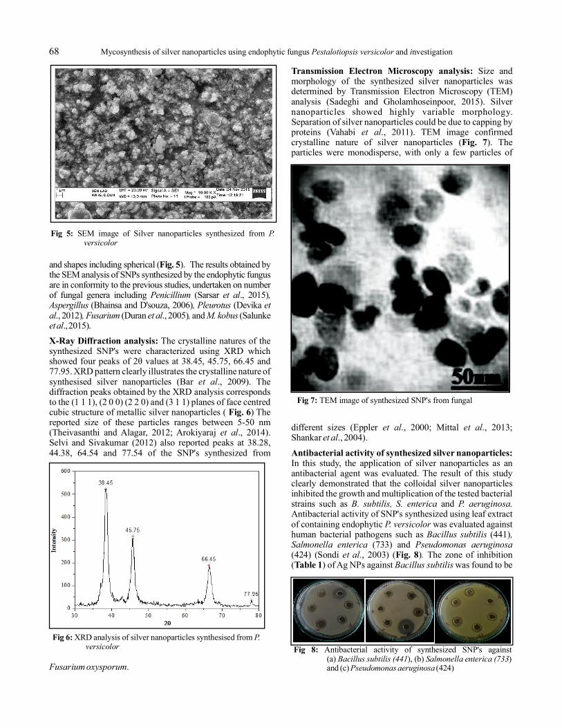
Size andmorphology of the synthesized silver nanoparticles wasdetermined by Transmission Electron Microscopy (TEM)analysis (Sadeghi and Gholamhoseinpoor, 2015). Silvernanoparticles showed highly variable morphology.Separation of silver nanoparticles could be due to capping byproteins (Vahabi ., 2011). TEM image confirmedcrystalline nature of silver nanoparticles ( ). Theparticles were monodisperse, with only a few particles of
different sizes (Eppler ., 2000; Mittal ., 2013;Shankar ., 2004).
In this study, the application of silver nanoparticles as anantibacterial agent was evaluated. The result of this studyclearly demonstrated that the colloidal silver nanoparticlesinhibited the growth and multiplication of the tested bacterialstrains such as andAntibacterial activity of SNP's synthesized using leaf extractof containing endophytic was evaluated againsthuman bacterial pathogens such as (441)
(733) and(424) (Sondi , 2003) ( ) The zone of inhibition( ) of Ag NPs against was found to be
Fig. 5
X-Ray Diffraction analysis:
Fig. 6
Transmission Electron Microscopy analysis:
Fig. 7
Antibacterial activity of synthesized silver nanoparticles:
Fig. 8Table 1
Penicillium et al ,Aspergillus , Pleurotus etal , Fusarium et al , M. kobusetal
et al
et al
Fusarium oxysporum
et al
et al et alet al
B. subtilis, S. enterica P. aeruginosa.
P. versicolorBacillus subtilis ,
Salmonella enterica Pseudomonas aeruginosaet al. .
Bacillus subtilis
2θ values
Fig 5: SEM image of Silver nanoparticles synthesized from P.versicolor
Fig 6: XRD analysis of silver nanoparticles synthesised from P.versicolor
Fig 7: TEM image of synthesized SNP's from fungal
Fig 8: Antibacterial activity of synthesized SNP's against(a) ), (b) )and (c) (424)
Bacillus subtilis (441 Salmonella enterica (733Pseudomonas aeruginosa
Mycosynthesis of silver nanoparticles using endophytic fungus and nvestigationPestalotiopsis versicolor i68

6
mm at 50 µl, 100 µl, and 150 µl and 7 mm at 200 µlconcentrations. Exactly similar pattern was observed against
. As compared zone of inhibitionobserved against was 5 mm at 50µl, 6 mm at 100 µl, 7 mm at 150 µl and 8 mm at 200 µl of thesample. The SNPs synthesized from various endophytic fungi
including (Sarsar ., 2015)(Bhainsa and D'souza, 2006) (Devika ., 2012)and (Duran ., 2005) are also reported to showantibacterial activity.
Antibacterial test of 1mM aqueous solution of silver nitratewas tested against (441)
(733) and (424). Whencompared with the silver nanoparticles synthesized from the
fungal isolates of ( ), the zone of inhibitionof AgNO against was found to be 4 mmwhile it was 1mm against and 2 mmagainst when 50 µl of the samplewas used in each case .
Decolourization of the dyes Congo red,Orange G and Rhodamine B using silver nanoparticlessynthesized from endophytic fungal extract of( ) was investigated. After 24 hour samples werewithdrawn and analysed by UV-visible spectrophotometer at498 nm for Congo red, 476 nm for Orange G and 550 nm forRhodamine B. Significant decolourization of Congo red,Orange G and Rhodamine B was observed. The
decolourization absorbance has been depicted in the table.The percentage (%) of dye decolourization was calculated byusing the formula:
% Decolourization = (O.D -O.D / O.D ) x 100
Silver nanoparticles synthesized from endophytic fungalextract of decolourized Congo red quite fast ascompared with Orange G and Rhodamine B. These resultssuggested that SNP's can be used for the treatment of textileeffluents also which is in conformity with the observations ofJalandoni-Buan (2015)
In this study, SNPs were synthesized by biological meansusing endophytic fungal extract. The SNPs synthesis processat laboratory scale is quite inexpensive and non-toxic, eco-friendly as compared to the chemical methods. This bio-process yielded stable, spherical SNPs displayingconsiderable antibacterial activity. The synthesized SNPsalso showed efficient degradation of azo dyes like Congo reddye and thus have potential in industrial application other thanpossible pharmaceutical application as antibacterial agents.
We gratefully acknowledge TEQIP-II and G. B. Pant Instituteof Engineering and Technology, Pauri, Garhwal for providingfinancial support for conducting this research.
Ahamed, M., Alsalhi, M.S. and Siddiqui, M.K. 2010. Silvernanoparticle applications and human health.
(23):1841-1848.
Arokiyaraj, S., Arasu, M.V., Vincent, S., Prakash, N.U., ChoiS.H., Oh, Y.K., Choi K.C. and Kim K.H. 2014.Rapid green synthesis of silver nanoparticles from
L and its antibacterial andcytotoxic effects: an study.
379-388.
Baker, S., Mohan Kumar, K., Santosh, P., Rakshith, D. andSatish, S. 2015. Extracellular synthesis of silvernanoparticles by novel AS41G inhabiting L. and theirbactericidal activity.
:1434-1440.
Bar, H., Bhui, D.K., Sahoo, P.G., Sarkar, P. and Sarkar, P.2009. Green synthesis of silver nanoparticles using
Salmonella entericaPseudomonas aeruginosa
Penicillium et al , Aspergillus, Pleurotus et al
Fusarium et al
Bacillus subtilis , Salmonellaenterica Pseudomonas aeruginosa
C. torulosaBacillus subtilis
Salmonella entericaPseudomonas aeruginosa
P. versicolor
P. versicolor
et al. .
Clin.Chim. Acta
Chrysanthemum indicum .in vitro Int. J. Nanomed
Pseudomonas veroniiAnnona squamosa
Spectrochimica Acta Part A:Molecular and Biomolecular Spectroscopy
Fig. 9
(Table 2)Dye decolourization by activity of synthesized silvernanoparticles:
Table 3
CONCLUSION
ACKNOWLEDGMENT
REFERENCES
411
.9:
136
3
control test control
Fig 9: Antibacterial test of AgNO against (a) )(b) ) and (c)
(424)
3 Bacillus subtilis (441Salmonella enterica (733 Pseudomonas
aeruginosa
S. No. Bacterial pathogen AgNO3 Fungi SNPs
1 Bacillus subtilis 4 6
2 Salmonella enterica 1 3
3 Pseudomonas aeruginosa 2 4
Table 2: Zone of inhibition (mm) of AgNO and silver nanoparticlessynthesized from of
3
P. versicolor C. torulosa
Time duration(in days)
% reduction of dyeCongo red
% reduction of dyeOrange G
% reduction of dyeRhodamine B
1 9.70 16.90 12.242 14.76 26.47 22.493 22.63 35.64 34.684 41.35 43.99 48.405 57.38 52.54 62.836 70.46 60.08 75.737 89.45 69.65 82.628 91.56 83.50 89.10
Table 3: % Decolourization of dyes by silver nanoparticlessynthesized from P. versicolor.0
S.No.
HumanPathogen
Antibiotic(Tetracycline)
Fungiextract
AgNP’s(50µl)
100µl 150µl 200µl
1. Bacillussubtilis
12 5 6 6 6 7
2. Salmonellaenterica
15 5 6 6 6 7
3. Pseudomonasaeruginosa
1 4 5 6 7 8
Table 1- Zone of inhibition (mm) of SNPs synthesized fromP. versicolor
Kavish Rajput, Shruti Agrawal, Jyoti Sharma, and Pavan Kr. Agrawal 69

latex of . , :134-139.
Basavaraja, S., Balaji, S.D., Lagashetty,A., Rajasab,A.H. andVenkataraman, A. 2008. Extracellular biosynthesisof silver nanoparticles using the fungus
(5):1164-1170.
Bhainsa, C.K. and D'Souza, F.S. 2006. Extracellularbiosynthesis of silver nanoparticles using the fungus
.160-164.
Bindhani, B.K. and Panigrahi, A.K. 2014. Green synthesisand characterization of gold nanoparticles using leafextracts of (Linn.)(Ashwagandha).
(6): 279-284.
Cauerhff,A. and Castro, G.R. 2013. Bionanoparticles, a greennanochemistry approach.(3): 1-10.
Chen, J.C., Lin, Z.H. and Ma, X.X. 2003. Evidence of theproduction of silver nanoparticles via pretreatmentof Phoma sp.3.2883 with silver nitrate.
. (2):105-108.
Deb, S. 2014. Synthesis and characterisation of silvernanoparticles using var(Cabbage) and (French Beans):A study on their antimicrobial activity and dyedegrading ability. (7):3909-3917.
Devika, R., Elumalai, S., Manikandan, E. andEswaramoorthy, D. 2012. Biosynthesis of silvernanoparticles using the fungusand their antibacterial activity .(1): 1-5.
Duran, N., Cuevas, R., Cordi, L., Rubilar, O. and Diez, M.C.2014. Biogenic silver nanoparticles associated withsilver chloride nanoparticles (Ag@ AgCl) producedby laccase from .
(1) 645:1-7.
Duran, N., Marcato, P.D., Alves, O.L., De Souza, G.I. andEsposito, E. 2005. Mechanistic aspects ofbiosynthesis of silver nanoparticles by several
strains.(8):1-7.
EL-Moslamy, S.H., Elkady, M.F., Rezk, A.H. and Abdel-Fattah, Y.R. 2017. Applying Taguchi design andlarge-scale strategy for mycosynthesis of nano-silver from endophyticSYAF4 and its application against phytopathogens.
(452297): 1-22
Eppler, A.S., Rupprechter, G., Anderson, E.A. and Somorjai,G.A. 2000. Thermal and chemical stability andadhesion strength of Pt nanoparticle arrayssupported on silica studied by transmission electronmicroscopy and atomic force microscopy.
(31):7286-7292.
Gade, A., Ingle, A., Bawaskar, M. and Rai, M. 2009.: a novel biological agent for
extracellular synthesis of nanoparticles.2079-2085.
Iravani, S. 2014. Bacteria in nanoparticle synthesis: currentstatus and future prospects.
.Article ID 359316, 18 pages
Ishida, K., Cipriano, T.F., Rocha, G.M., Weissmuller, G.,Gomes, F., Miranda, K. and Rozental, S. 2014.Silver nanoparticle production by the fungus
: nanoparticle characterisationand analysis of antifungal activity againstpathogenic yeasts. , Riode Janeiro, (2): 220-228.
Jalandoni-Buan, A.C., Decena-Soliven, A.L., Cao, E.P.,Barraquio, V.L. and Barraquio, W.L. 2015. CongoRed decolorizing bacteria from paper factoryeffluent. In:
. (Ed.: Shree, N.S.). SpringerInternational Publishing, 135-148pp
Mittal, A.K., Chisti, Y. and Banerjee, U.C. 2013. Synthesis ofmetallic nanoparticles using plant extracts.
(2): 346-356.
Mohanpuria, P., Rana, K.N. and Yadav, S.K. 2008.Biosynthesis of nanoparticles: technologicalconcepts and future applications. :507-517.
Mukherjee, P., Ahmad, A., Mandal, D., Senapati, S., Sainkar,S.R. and Khan, MI. ., 2001. Bioreduction ofAuCl ions by the fungus, sp andsurface trapping of the gold nanoparticles formed.
: 3585-3588.
Musarrat, J., Dwivwdi, S., Singh, B.R., Al-Khedhairy, A.,Azam, A. and Naqvi, A. 2010. Production ofantimicrobial silver nanoparticles in water extractsof the fungus strain KSU-09.
: 8772-8776.
Nabikhan,A., Kandasamy, K., Raj, A. and Alikunhi, N.M.2010 Synthesis of ant imicrobial silvernanoparticles by callus and leaf extracts fromsaltmarsh plant, L.
(2):488-493
Raja, S., Ramesh, V. and Thivaharan, V. 2017. Greenbiosynthesis of silver nanoparticles using
leaf extract, theirantibacterial activity and hydrogen peroxide sensingcapability. (2): 253-261.
Rajput, K., Bhatt, A. and Agrawal, P.K. 2016. Plant mediatedbiosynthesis, characterization and application ofsilver nanoparticles by leaves extract of
., (7): 1199-1207.
Jatropha curcas Colliod surface A
Fusariumsemitectum. Mater. Res. Bull.
Aspergil lus funigatus Col loids Surf. BBiointerfaces
Withania somniferaInternational Journal of Materials
Science andApplications
Electron J. Biotechnol.
Lett. Appl.Microbiol
Brassica oleracea .capitataPhaseolus vulgaris
Int. J. Chem. Tech. Res.
Pleurotus ostreatus. Scientific reports
Trametes versicolor Springer Plus
Fusarium oxysporum J nanobiotechnology
Trichoderma harzianum
Scientific Reports
J. Phys.
Chem. B.
Fusarium solaniJ.
Nanopart. Res
International ScholarlyResearch Notices
Fusarium oxysporum
Mem. Inst. Oswaldo Cruz.
Microbial degradation of synthetic dyesin waste waters
Biotechnol. Adv.
J. Nanopart.
et alVerticillium .
Angew. Chem. Int.
Amylomyces rouxiiBioresource Technology
Sesuvium portulacastrumColloids Surf. B Biointerfaces.
Calliandra haematocephala
Arab J. Chem.
Cupressustorulosa International Journal of AdvancedResearch
39 (3)
43
47:
3
16
37
6
12
3
3
7
104
11:
109
31
10
40
101
.
79
10
4
4−
Mycosynthesis of silver nanoparticles using endophytic fungus and nvestigationPestalotiopsis versicolor i70

Rajput, K., Raghuvanshi, S., Bhatt, A., Rai, S.K. andAgrawal, P.K. 2017. A Review on Synthesis ofSilver Nano-Particles.
(7): 1513-1528.
Ramteke, C., Chakrabarti, T., Sarangi, B.K. and Pandey,R.A. 2013. Synthesis of silver nanoparticles fromthe aqueous extract of leaves of forenhanced antibacterial activity.
ID 278925, 1-7.
Sadeghi, B. and Gholamhoseinpoor F. 2015. A study on thestability and green synthesis of silver nanoparticlesusing (Zt) extract at roomtemperature.
. : 310-315.
Salunke, B.K., Sawant, S.S., Kang, T.K., Seo, D.Y., Cha, Y.,Moon, S.A., Alkotaini, B., Sathiyamoorthi, E. andKim, B.S. 2015. Potential of biosynthesized silvernanoparticles as nanocatalyst for enhanceddegradation of cellulose by cellulase.
Article ID :1-8.
Sanghi, R. and Verma, P. 2009. Biomimetic synthesis andcharacterisation of protein capped silvernanoparticles. (1): 501-504.
Sarsar, V., Selwal, M.K. and Selwal, K.K. 2015.Biofabrication, characterization and antibacterialefficacy of extracellular silver nanoparticles usingnovel fungal strain ofKM. (6) :682-688.
Schaffer, B., Hohenester, U., Trugler, A. and Hofer, F. 2009.High-resolution surface plasmon imaging of goldnanoparticles by energy-filtered transmissionelectron microscopy. (4):1-9.
Selvi, K.V. and Sivakumar, T. 2012. Isolation andcharacterization of silver nanoparticles from
(1): 56-62.
Shankar, S.S., Rai, A., Ahmad, A. and Sastry, M. 2004. Rapidsynthesis ofAu, Ag, and bimetallic Au core-Ag shellnanoparticles using Neem ( ) leaf
broth. (2): 496-502.
Sharma, D., Pramanik,A. andAgrawal, P.K. 2016. Evaluationof bioactive secondary metabolites from endophyticfungus BAB-5510 isolatedfrom leaves of D. Don. 3
(210): 1-14.
Sondi, I., Goia, D.V. and Matijevic, E. 2003. Preparation ofhighly concentrated stable dispersions of uniformsilver nanoparticles.(1):75-81.
Sunkar, S. and Nachiyar, C.V. 2012. Biogenesis ofantibacterial silver nanoparticles using theendophytic bacterium isolated from
(12): 953-959.
Swamy, M.K, Sudipta, K.M. Jayanta , K. andBalasubramanya S. 2014. The green synthesis,characterization, and evaluation of the biologicalactivities of silver nanoparticles synthesized from
leaf extract.(1): 73-81.
Theivasanthi, T. and Alagar, M. 2012. Electrolytic synthesisand characterizations of silver nanopowder.
. (2): 58-65.
Vahabi, K., Mansoori, G.A. and Karimi, S. 2011.Biosynthesis of silver nanoparticles by fungus
. (1): 65-79.
Verma, V.C., Kharwar, R.N. and Gange, A.C. 2010.Biosynthesis of antimicrobial silver nanoparticlesby the endophytic fungus .
(1): 33-40.
Vigneshwaran, N., Ashtaputre, N.M., Varadarajan, P.V.,Nachane, R., Paralikar, K.M. and Balasubramanya,R.H. 2007. Biological synthesis of silvernanoparticles using the fungus .
: 1413-1418.
Vigneshwaran, N., Kathe, A.A., Varadarajan, P.V., Nachane,R.P. and Balasubramanya, R.H. 2006. Biomimeticsof silver nanoparticles by white rot fungus,
Int. J. Curr. Microbiol. App.Sci.
Ocimum sanctumJournal of
Chemistry
Ziziphora tenuiorSpectrochim. Acta Mol. Biomol.
Spectrosc
Journal ofNanomaterials
Bioresource Technol.
Penicillium atramentosumJournal of Saudi Chemical Society
Phys. Rev.
Fusarium oxysporum. Int. J.Curr. Microbiol. App.Sci.
Azadirachta indica
J. Colloid Interface Sci.
Pestalotiopsis neglectaCupressus torulosa Biotech.
J. Colloid Interface Sci.
Bacillus cereusGarcinia xanthochymus. Asian Pac. J. Trop.Biomed.
Leptadenia reticulata Appl. Nano sci.
NanoBiomed. Eng
Trichoderma ressei Insciences J.
Aspergillus clavatusNanomedicine
Aspergillus flavusMater. Lett.
Phaenerochaete chrysosporium. Colloids Surf. B
6
134
289410
100
19
B. 79
1
275
6
260
2
5
4
1
5
66
53
Kavish Rajput, Shruti Agrawal, Jyoti Sharma, and Pavan Kr. Agrawal 71

![Journal of Colloid and Interface Science - ntnlab.com · plex synthesis procedures. Therefore, ... [31], ferric ions [32], silver ions [33], as well as mer-cury ions [34] in water](https://img.pdfslide.us/doc/110x75/5ae38e9e7f8b9ad47c8e6e76/journal-of-colloid-and-interface-science-synthesis-procedures-therefore-.jpg)