Embed Size (px)
Citation preview

©FUNPEC-RP www.funpecrp.com.brGenetics and Molecular Research 13 (2): 2349-2358 (2014)
Diversity of endophytic bacteria inCaragana microphylla grown in the desert grassland of the Ningxia Hui Autonomous Region of China
J.X. Dai1,2, X.M. Liu1,2 and Y.J. Wang1,2
1Key Laboratory of the Ministry of Education for the Conservation and Utilization of Special Biological Resources of Western China,Ningxia University, Yinchuan, Ningxia, China2College of Life Science, Ningxia University, Yinchuan, Ningxia, China
Corresponding author: Y.J. WangE-mail: [email protected] / [email protected]
Genet. Mol. Res. 13 (2): 2349-2358 (2014)Received March 25, 2013Accepted June 10, 2013Published April 3, 2014DOI http://dx.doi.org/10.4238/2014.April.3.7
ABSTRACT. The diversity of endophytic bacteria in the sand-fixation plant Caragana microphylla was investigated by amplified rDNA restriction analysis and by sequence and phylogenetic comparisons of the 16S rRNA genes. A total of 24, 19, and 17 operational taxonomic units were identified from 16S rDNA libraries of the plant roots, stems and leaves, respectively. Homology analysis revealed a 92-100% identity of bacterial 16S rDNA sequences compared with those in the GenBank database. The bacteria identified by sequence homology fell into the following groups: α-Proteobacteria, β-Proteobacteria, γ-Proteobacteria, bacilli, and uncultured bacterium. Sequence analysis demonstrated that the roots were colonized predominantly by Bradyrhizobiaceae, while bacteria from Burkholderiaceae and Sphingomonadaceae were predominant in the stems and leaves, respectively. Additionally, the endophytic bacterial community in leaves was more diverse than those in the roots and stems.

2350
©FUNPEC-RP www.funpecrp.com.brGenetics and Molecular Research 13 (2): 2349-2358 (2014)
J.X. Dai et al.
Overall, the most abundant bacteria in all three tissues analyzed were from the Sphingomonadaceae and Burkholderiaceae families, although many bacterial populations were found in only a single tissue. These results suggest that the bacterial population of C. microphylla is diverse.
Key words: Diversity; 16S rDNA; Endophytic bacteria;Caragana microphylla
INTRODUCTION
Endophytic bacteria are defined as those bacteria that can colonize the internal tissue of a living plant for all or part of their life cycle, causing asymptomatic infections entirely within plant tissues (Wilson, 1995). It has been proposed that endophytic bacteria originated in the rhizosphere (Germida et al., 2006). Members of this microbial community have been suggested to play diverse positive roles in the development and growth of host plants, since they are capable of producing phytohormones and siderophores, increasing the hosts’ resistance to pathogens and parasites (Burd et al., 1998; Compant et al., 2005; Berg et al., 2005; Feng et al., 2006), as well as promoting biological nitrogen fixation (Han et al., 2005). Additionally, many endophytes can produce bioactive natural compounds that are a potential source of novel products for use in medicine, agriculture, and industry. These natural products are highly effective, with low toxicity and minor environmental impact, and thus are resources for discovering and developing novel antibiotics (Strobel et al., 2004), chemothera-peutic agents (Strobel and Daisy, 2003), and agrochemicals (Brooks et al., 1994; Ryan et al., 2008). To date, endophytic bacteria have been found in a variety of plants, including soybean, potato, rice, wheat, and sugarcane (Coombs and Franco, 2003; Kuklinsky-Sobral et al., 2004; Sun et al., 2008). Understanding the diversity of bacterial endophytes in plants is therefore important for increasing crop production, conserving biodiversity and sustaining agro-ecosystems, as well as for the discovery of novel drugs for the treatment of diseases in humans, plants, and animals (Ryan et al., 2008).
Caragana microphylla, a species of the genus Caragana, a subfamily of Papilionoideae in the Leguminosae, is a perennial leguminous shrub that is highly tolerant of drought, salt, and extreme cold in relatively poor or sandy, well-drained soils. In the northern regions of China, it has been widely used as a windbreak to protect soils from desertification, as well as forage for live-stock, and high-energy firewood (Yan et al., 2007). Furthermore, the roots, flowers, and seeds of C. microphylla are used in herbal medicine for their antineuralgic, antirheumatic, and antiarthritic properties. However, to date, relatively little information is available regarding the bacterial com-munity in C. microphylla. The aim of this study was to investigate the diversity of the endophytic population in the roots, stems, and leaves of C. microphylla by 16S rDNA analysis.
MATERIAL AND METHODS
Sampling of plants and DNA extraction
Plant materials were obtained from Baijitan National Nature Reserves of Ningxia Hui Autonomous Region, China. This is a typical desert-type region, located on the south rim of the Maowusu sandland (106°20'-106°37' E, 37°49'-38°20' N). In May 2011, the roots, stems, and leaves of C. microphylla were collected randomly from plants separated by at least 100 m

2351
©FUNPEC-RP www.funpecrp.com.brGenetics and Molecular Research 13 (2): 2349-2358 (2014)
Endophytic bacteria in the desert
in a plantation that was more than 10 years old. The tissues collected were washed with sterile distilled water and their surfaces disinfected by immersion in 70% ethanol and then in fresh sodium hypochlorite solution (2.5% Cl- concentration) for 3 min each, followed by extensive rinsing several times with sterile distilled water. To confirm that the sterilization process was successful, water from the final rinse was tested for bacterial growth on LB medium plates. Uncontaminated tissues were then used for DNA extraction by the CTAB procedure, as de-scribed previously (Marquez-Santacruz et al., 2010).
PCR amplification and cloning of bacterial 16S rDNA
The 16S rDNA of endophytic bacteria was amplified using the universal primers 799f: 5'-AACAGGATTAGATACCCTG-3' and 1492r: 5'-GGTTACCTTGTTACGACTT-3'. The 50-µL PCR mixture contained 50 ng plant tissue DNA, 250 pmol each primer, 5 µL 10X PCR buffer, 2.5 U Taq DNA polymerase (Takara, Dalian, China), and 100 µM dNTP mixture. The PCR temperature cycling program consisted of an initial denaturation at 94°C for 5 min; 30 cycles of 94°C for 1 min, 58°C for 45 s, and 72°C for 1 min; followed by a final extension at 72°C for 7 min. The PCR products were examined by running them on a 1% agarose gel, and the desired PCR product was purified using the PCR Purification Kit (Promega, USA) as per the manufacturer instructions. The purified PCR fragments were then cloned into the pGM-T Easy Vector (TIANGEN, Beijing, China), and positive clones with DNA insertions were identified as white colonies on LB medium containing 80 μg/mL X-Gal and 0.5 mM IPTG.
Amplified ribosomal DNA restriction analysis (ARDRA), sequencing, and phyloge-netic analyses
One hundred positive clones bearing the 16 rDNA PCR product amplified from DNA extracted from roots, stems, or leaves were randomly picked, and plasmid DNAs isolated from these clones were then digested with HaeIII or AluI restriction endonuclease (Fermentas, USA). The restriction fragments were separated on 2% agarose gels. Clones that had identical ARDRA patterns in digests with each of the two restriction endonucleases were grouped into an operational taxonomic unit (OTU). Plasmids of three representative clones from each OTU from the roots, stems, and leaves of the plants were sequenced on an ABI 3730 DNA sequenc-er at Sangon Biotech Co. Ltd. (Shanghai, China). The 16S rDNA sequences acquired were further analyzed for the presence of possible chimeric sequences by using the CHIMERA-CHECK program and then analyzed using BLAST in the GenBank database (http://www.ncbi.nlm.nih.gov/) to explore the identity by sequence alignment and the CLUSTAL X1.8 software (BiowareDB.org) was also used. Phylogenetic trees were constructed by the neighbor-joining method using the MEGA 4.0 software. The statistical significance levels of the interior nodes were determined by bootstrap analysis with 1000 replications of each sequence.
RESULTS
ARDRA analysis of bacterial communities
16S rDNA clone libraries were constructed from the endophytic bacteria of C. micro-
2352
©FUNPEC-RP www.funpecrp.com.brGenetics and Molecular Research 13 (2): 2349-2358 (2014)
J.X. Dai et al.
phylla and analyzed based on their ARDRA restriction patterns. The analysis identified 24, 19, and 17 OTUs in libraries from C. microphylla roots, stems, and leaves, respectively. Homol-ogy analysis of OTU representatives revealed that the bacterial 16S rDNA sequences were 92-100% identical to those in the GenBank database. The sequences obtained in this study can be accessed in GenBank (accession Nos. JQ795016-JQ795086).
The sequence analysis showed that all the isolated bacterial clones were from α-Proteobacteria, β-Proteobacteria, γ-Proteobacteria, bacilli, and uncultured bacterium, with a majority from the Proteobacterial phyla (Tables 1-3).
Class Family (No. of OTUs) NCBI alignment (No. of clones) Max identity %
α-Proteobacteria Bradyrhizobiaceae (6) Bradyrhizobium sp FR753090 (2) 97% Bradyrhizobium sp HQ233230 (4) 99% Bradyrhizobium sp JN377665 (5) 99% Bradyrhizobium japonicum FJ436370 (8) 96% Uncultured Bradyrhizobium sp AB512187 (6) 100% Uncultured Bradyrhizobium sp GQ129971 (4) 99% Sphingomonadaceae (1) Sphingomonas sp HQ530510 (8) 95% Erythrobacteraceae (1) Uncultured Erythrobacteraceae bacterium clone FJ516791 (4) 92% Caulobacteraceae (1) Phenylobacterium lituiforme NR_029117 (1) 99% Rhizobiaceae (1) Rhizobium sp EF638788 (6) 97% - Alpha proteobacterium FJ711201 (4) 99%β-Proteobacteria Burkholderiaceae (2) Uncultured Burkholderiales bacterium clone JF277911 (2) 98% Cupriavidus respiraculi EU221394 (6) 99%γ-Proteobacteria Xanthomonadaceae (6) Lysobacter yangpyeongensis NR_043625 (5) 96% Lysobacter sp GU385868 (2) 96% Uncultured Xanthomonadaceae bacterium clone JN082695 (3) 96% Uncultured Xanthomonadaceae bacterium clone EU305594 (1) 96% Uncultured Xanthomonadaceae bacterium clone DQ230964 (1) 99% - Uncultured gamma proteobacterium clone AY622241 (1) 97%Bacilli Paenibacillaceae (1) Paenibacillus elgii AY090110 (4) 99% Bacillaceae (2) Bacillus sp AY556409 (1) 99% Bacillus sp JN604334 (4) 99%- - Uncultured bacterium clone HQ190331 (8) 99%- - Uncultured bacterium clone FJ612147 (10) 99%
Table 1. Diversity of the community of endophytic bacteria in the roots of Caragana microphylla.
Class Family (No. of OTUs) NCBI alignment (No. of clones) Max identity %
α-Proteobacteria Sphingomonadaceae (1) Sphingomonas sp AB265150 (6) 97% Phyllobacteriaceae (1) Mesorhizobium sp AB604651 (3) 97% Bradyrhizobiaceae (1) Uncultured Bradyrhizobium sp clone AB512187 (5) 100%β-Proteobacteria Comamonadaceae (1) Delftia sp EU304256 (8) 100% Burkholderiaceae (4) Burkholderia sp FJ025138 (4) 93% Burkholderia caledonica NR_025057 (1) 94% Ralstonia sp FJ984446 (23) 97% Ralstonia solanacearum EF016361 (2) 93%γ-Proteobacteria Pseudomonadaceae (1) Pseudomonas sp AY247063 (4) 100% Pasteurellaceae (1) Haemophilus sp AM420172 (4) 95% Enterobacteriaceae (3) Serratia sp HM245061 (2) 96% Pantoea sp HM159968 (11) 95% Enterobacter sp GQ478257 (7) 97%Bacilli Streptococcaceae (2) Streptococcus sp EU071520 (12) 99% Streptococcus sp EU341245 (2) 97% Staphylococcacea (1) Staphylococcus sp FJ957767 (1) 99%- - Uncultured bacterium clone AY218754 (2) 95%- - Uncultured bacterium clone EF397376 (2) 92%- - Uncultured bacterium clone FJ983108 (1) 93%
Table 2. Diversity of the community of endophytic bacteria in the stems of Caragana microphylla.

2353
©FUNPEC-RP www.funpecrp.com.brGenetics and Molecular Research 13 (2): 2349-2358 (2014)
Endophytic bacteria in the desert
Phylogenetic analysis of the bacterial community in different plant tissues
The endophytic bacterial community of C. microphylla consisted of 20 families. Phylo-genetic analysis showed that the community composition varied in different plant tissues. The bacteria identified from the roots, stems, and leaves fell into 9, 10, and 13 families, respectively. Among them, bacteria representing Paenibacillaceae, Erythrobacteraceae, Caulobacteraceae, and Rhizobiaceae were detected only in the roots; members of Streptococcaceae, Pasteurella-ceae, and Phyllobacteriaceae were found exclusively in the stems; and bacteria from Rhodobac-teraceae, Enterococcaceae, Acetobacteraceae, Moraxellaceae, and Alcaligenaceae were identi-fied only in leaf tissues. The most common bacteria were members of the Sphingomonadaceae and Burkholderiaceae families, which were detected in all three tissues (Tables 1-3).
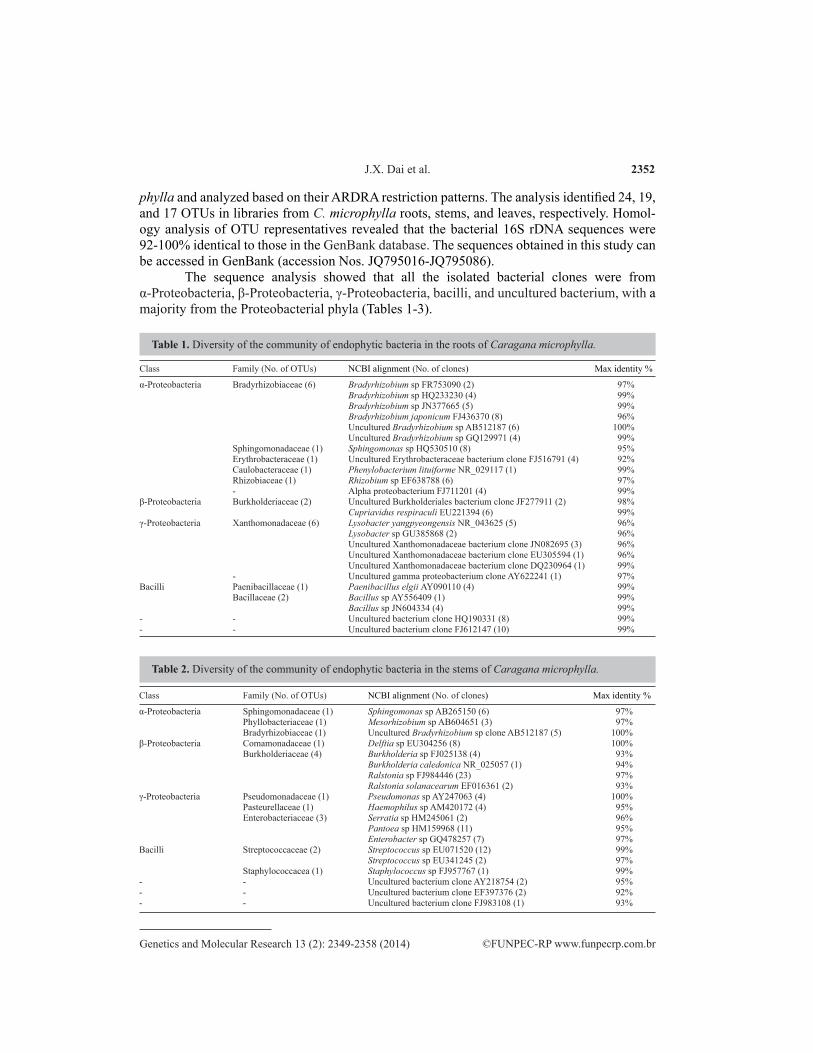
The results of phylogenetic analysis indicated that the bacterial clones from the roots of C. microphylla consisted of five groups, with a majority (52%) from the α-Proteobacteria, with Brady-rhizobium the predominant genus, making up 29% of the total (Figure 1). The γ-Proteobacteria were assigned to the family Xanthomonadaceae, which accounted for 12% of root clones and had 96-99% sequence identity with Lysobacter and uncultured bacterium. β-Proteobacteria were represented by a single genus, Cupriavidus. Bacilli consisted of Paenibacillaceae and Bacillaceae, and showed 99% 16S rDNA sequence identity with Paenibacillus and Bacillus.
Bacterial clones from the stems also fell into five groups: α-Proteobacteria (14%), β-Proteobacteria (38%), γ-Proteobacteria (28%), bacilli (15%), and uncultured bacterium (5%) (Figure 2). The predominant clones were from the Burkholderiaceae family (30%). The bacterial clones isolated from the stem were assigned to 13 genera, among which Ralstonia, Pantoea, and Streptococcus were the most common. Eleven of the 13 genera detected in the stems were not found in the root library. Only Bradyrhizobium and Sphingomonas occurred in both libraries.
The bacterial clones detected in leaf tissues fell into four classes, the most common being α-Proteobacteria (34%) and β-Proteobacteria (39%), with γ-Proteobacteria and Bacilli accounting for 18% and 9%, respectively (Figure 3). The predominant clones were from the Sphingomonadaceae family, of which Novosphingobium, Delftia, and Achromobacter were the major genera. The diversity of endophytic bacteria in the leaves of this plant was greater than that in the roots and stems, comprising 13 families. Fourteen genera could be assigned, among which eight were detected only in the leaves.
Class Family (No. of OTUs) NCBI alignment (No. of clones) Max identity %α-Proteobacteria Sphingomonadaceae (1) Novosphingobium sp FJ889321 (30) 97% Rhodobacteraceae (1) Paracoccus sp JN561152 (1) 98% Acetobacteraceae (1) Acidisphaera sp AB512155 (1) 97% - Uncultured alpha proteobacterium FJ517710 (2) 93%β-Proteobacteria Comamonadaceae (2) Pelomonas puraquae AM501441 (5) 97% Delftia sp GU560163 (12) 99% Alcaligenaceae (1) Achromobacter sp GU056301 (14) 99% Burkholderiaceae (2) Burkholderia sp EU071528 (7) 99% Burkholderia sp GQ181152 (1) 95%γ-Proteobacteria Pseudomonadaceae (2) Pseudomonas sp AM913900 (1) 97% Pseudomonas sp AY288072 (3) 97% Moraxellaceae (1) Acinetobacter sp DQ421391 (1) 99% Xanthomonadaceae (1) Stenotrophomonas sp GU179638 (1) 99% Enterobacteriaceae (1) Enterobacter sp FN433019 (12) 99%Bacilli Staphylococcaceae (1) Staphylococcus hominis JN644561 (1) 99% Bacillaceae (1) Bacillus sp JN604334 (2) 99% Enterococcaceae (1) Enterococcus sp JF346884 (6) 99%
Table 3. Diversity of the community of endophytic bacteria in the leaves of Caragana microphylla.
2354
©FUNPEC-RP www.funpecrp.com.brGenetics and Molecular Research 13 (2): 2349-2358 (2014)
J.X. Dai et al.
Figure 1. Phylogenetic tree based on the 16S rDNA sequences of endophytic bacteria clones from the roots of Caragana microphylla. The dendrogram was constructed with the neighbor-joining method. Bootstrap values greater than 50% (based on 1000 bootstrap resamplings) were labeled at the nodes. Scale bar represents 1% sequence divergence.

2355
©FUNPEC-RP www.funpecrp.com.brGenetics and Molecular Research 13 (2): 2349-2358 (2014)
Endophytic bacteria in the desert
Figure 2. Phylogenetic tree based on the 16S rDNA sequences of endophytic bacteria clones from the stems of Caragana microphylla . The dendrogram was constructed with the neighbour-joining method. Bootstrap values greater than 50% (based on 1000 bootstrap resamplings) were labeled at the nodes. Scale bar represents 1% sequence divergence.

2356
©FUNPEC-RP www.funpecrp.com.brGenetics and Molecular Research 13 (2): 2349-2358 (2014)
J.X. Dai et al.
Figure 3. Phylogenetic tree based on the 16S rDNA sequences of endophytic bacteria clones from the leaves of Caragana microphylla . The dendrogram was constructed with the neighbour-joining method. Bootstrap values greater than 50% (based on 1000 bootstrap resamplings) were labeled at the nodes. Scale bar represents 1% sequence divergence.

2357
©FUNPEC-RP www.funpecrp.com.brGenetics and Molecular Research 13 (2): 2349-2358 (2014)
Endophytic bacteria in the desert
DISCUSSION
Microbial endophytes establish communities by colonizing plant tissues. They are capable of developing interactions, not only among themselves, but also with their host plants, by which they may influence plant development and growth. Thus, understanding the diversity of endophytic bacteria is important for both ecological and biotechnological studies. Here, we show the diversity of endophytic bacteria in the root, stem, and leaf tissues of C. microphylla growing in the desert of Ningxia, China.
The results reported in this study suggest a tissue-dependent endophytic popula-tion in C. microphylla. Bradyrhizobiaceae were the predominant microbial population in the roots, while Burkholderiaceae and Sphingomonadaceae were the predominant bacteria in the stem and leaf tissues of the plant, respectively. Importantly, many clones identified in this study were found in only one of the three plant tissues. For example, members of Rhizobium, Paenibacillus, Phenylobacterium, Cupriavidus, and Lysobacter were detect-ed only in the roots; bacteria from Mesorhizobium Haemophilus, Pantoea, Serratia, and Streptococcus were found only in the stems; and bacterial members of Novosphingobium, Paracoccus, Acidisphaera, Pelomonas, Achromobacter, Acinetobacter, Stenotrophomona, and Enterococcus were identified only in leaf tissue. The endophytic bacterial community in leaves was more diverse than in the roots and stems: there was a larger number of differ-ent genera, falling into four groups, possibly a result of the selective pressure plants exert on their associated bacterial populations. Endophytic bacteria of plants colonize specific ecological niches in different tissues, which might be a reason for the diversity observed within the bacterial community in this study. This finding is consistent with those from other studies (Hallmann et al., 1997; Ulrich et al., 2008).
The most abundant bacteria detected in this study were Sphingomonadaceae and Burkholderiaceae. With their ability to fix nitrogen and their potential to promote plant de-velopment and growth, plant-associated Burkholderiaceae species have recently received increased attention. For example, endophytic Burkholderiaceae species have been detected in several crops (Sun et al., 2008). Similarly, members of the Sphingomonadaceae family are widespread in different types of soils, sediments, and pelagic aquatic environments, and are known for their ability to utilize a wide variety of carbon sources; several are in fact renowned degraders of recalcitrant (xenobiotic) molecules (Leys et al., 2004). Addi-tionally, some sphingomonads were also found to play important roles in the mycorrhizo-sphere (Boersma et al., 2009).
Other 16S rDNA sequences identified in this study also showed a high degree of identity with common genera within the Bradyrhizobiaceae, Bacillaceae, Enterobacteriaceae, and Pseudomonadacea families, such as Bradyrhizobium, Bacillus, Enterobacter, and Pseu-domonas. Members of these genera have been widely studied as endophytes because of their potential use for promoting plant growth and plant health by means of their nitrogen fixation, antifungal, and antiviral activities.
In summary, the data presented in this study add to our knowledge of the diversity of endophytic bacteria associated with C. microphylla, a major sand-fixation plant species inhab-iting desert and semi-desert areas of China. The culture-independent approach was a limitation of this study. Further studies using plants collected from different geographic location and a culture-dependent approach are required.

2358
©FUNPEC-RP www.funpecrp.com.brGenetics and Molecular Research 13 (2): 2349-2358 (2014)
J.X. Dai et al.
ACKNOWLEDGMENTS
Research financed by the National Natural Science Foundation of China (#31200103) and the Natural Science Foundation of Ningxia (#NZ12152).
REFERENCES
Berg G, Krechel A, Ditz M, Sikora RA, et al. (2005). Endophytic and ectophytic potato-associated bacterial communities differ in structure and antagonistic function against plant pathogenic fungi. FEMS Microbiol. Ecol. 51: 215-229.
Boersma FG, Warmink JA, Andreote FA and van Elsas JD (2009). Selection of Sphingomonadaceae at the base of Laccaria proxima and Russula exalbicans fruiting bodies. Appl. Environ. Microbiol. 75: 1979-1989.
Brooks D, Gonzalez C, Appel D and Filer T (1994). Evaluation of endophytic bacteria as potential biological-control agents for oak wilt. Biol. Contr. 4: 373-381.
Burd GI, Dixon DG and Glick BR (1998). A plant growth-promoting bacterium that decreases nickel toxicity in seedlings. Appl. Environ. Microbiol. 64: 3663-3668.
Compant S, Duffy B, Nowak J, Clement C, et al. (2005). Use of plant growth-promoting bacteria for biocontrol of plant diseases: principles, mechanisms of action, and future prospects. Appl. Environ. Microbiol. 71: 4951-4959.
Coombs JT and Franco CM (2003). Isolation and identification of actinobacteria from surface-sterilized wheat roots. Appl. Environ. Microbiol. 69: 5603-5608.
Feng Y, Shen D and Song W (2006). Rice endophyte Pantoea agglomerans YS19 promotes host plant growth and affects allocations of host photosynthates. J. Appl. Microbiol. 100: 938-945.
Germida JJ, Siciliano SD, Renato de Freitas J and Seib AM (2006). Diversity of root-associated bacteria associated with field-grown canola (Brassica napus L.) and wheat (Triticum aestivum L.). FEMS Microbiol. Ecol. 26: 43-50.
Hallmann J, Quadt-Hallmann A, Mahaffee W and Kloepper J (1997). Bacterial endophytes in agricultural crops. Can. J. Microbiol. 43: 895-914.
Han J, Sun L, Dong X, Cai Z, et al. (2005). Characterization of a novel plant growth-promoting bacteria strain Delftia tsuruhatensis HR4 both as a diazotroph and a potential biocontrol agent against various plant pathogens. Syst. Appl. Microbiol. 28: 66-76.
Kuklinsky-Sobral J, Araujo WL, Mendes R, Geraldi IO, et al. (2004). Isolation and characterization of soybean-associated bacteria and their potential for plant growth promotion. Environ. Microbiol. 6: 1244-1251.
Leys NM, Ryngaert A, Bastiaens L, Verstraete W, et al. (2004). Occurrence and phylogenetic diversity of Sphingomonas strains in soils contaminated with polycyclic aromatic hydrocarbons. Appl. Environ. Microbiol. 70: 1944-1955.
Marquez-Santacruz HA, Hernandez-Leon R, Orozco-Mosqueda MC, Velazquez-Sepulveda I, et al. (2010). Diversity of bacterial endophytes in roots of Mexican husk tomato plants (Physalis ixocarpa) and their detection in the rhizosphere. Genet. Mol. Res. 9: 2372-2380.
Ryan RP, Germaine K, Franks A, Ryan DJ, et al. (2008). Bacterial endophytes: recent developments and applications. FEMS Microbiol. Lett. 278: 1-9.
Strobel G and Daisy B (2003). Bioprospecting for microbial endophytes and their natural products. Microbiol. Mol. Biol. Rev. 67: 491-502.
Strobel G, Daisy B, Castillo U and Harper J (2004). Natural products from endophytic microorganisms. J. Nat. Prod. 67: 257-268.
Sun L, Qiu F, Zhang X, Dai X, et al. (2008). Endophytic bacterial diversity in rice (Oryza sativa L.) roots estimated by 16S rDNA sequence analysis. Microb. Ecol. 55: 415-424.
Ulrich K, Ulrich A and Ewald D (2008). Diversity of endophytic bacterial communities in poplar grown under field conditions. FEMS Microbiol. Ecol. 63: 169-180.
Wilson D (1995). Endophyte: The evolution of a term, and clarification of its use and definition. Oikos 73: 274-276.Yan XR, Chen WF, Fu JF, Lu YL, et al. (2007). Mesorhizobium spp. are the main microsymbionts of Caragana spp.
grown in Liaoning Province of China. FEMS Microbiol. Lett. 271: 265-273.