Embed Size (px)
Citation preview
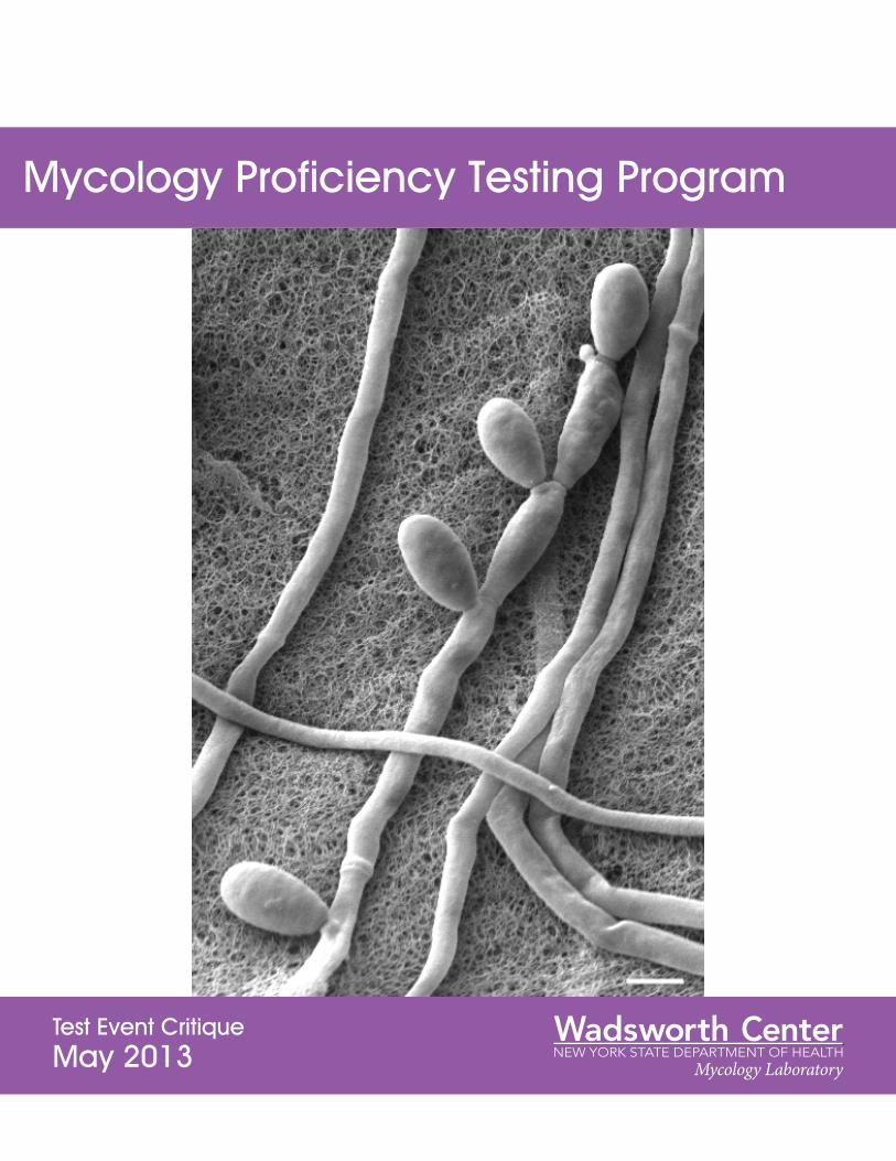
Mycology Proficiency Testing Program
Mycology Laboratory
Test Event CritiqueMay 2013

1
Table of Contents
Mycology Laboratory 2
Mycology Proficiency Testing Program 3
Test Specimens & Grading Policy 5
Test Analyte Master Lists 7
Performance Summary 9
Commercial Device Usage Statistics 10
Yeast Descriptions 11
Y-1 Candida kefyr 11
Y-2 Candida tropicalis 14
Y-3 Candida guilliermondii 17
Y-4 Candida krusei 20
Y-5 Candida lusitaniae 23
Antifungal Susceptibility Testing - Yeast 26
Antifungal Susceptibility Testing - Mold (Educational) 28
2
Mycology Laboratory
Mycology Laboratory at the Wadsworth Center, New York State Department of Health
(NYSDOH) is a reference diagnostic laboratory for the fungal diseases. The laboratory services
include testing for the dimorphic pathogenic fungi, unusual molds and yeasts pathogens,
antifungal susceptibility testing including tests with research protocols, molecular tests including
rapid identification and strain typing, outbreak and pseudo-outbreak investigations, laboratory
contamination and accident investigations and related environmental surveys. The Fungal
Culture Collection of the Mycology Laboratory is an important resource for high quality cultures
used in the proficiency-testing program and for the in-house development and standardization of
new diagnostic tests.
Mycology Proficiency Testing Program provides technical expertise to NYSDOH
Clinical Laboratory Evaluation Program (CLEP). The program is responsible for conducting the
Clinical Laboratory Improvement Amendments (CLIA)-compliant Proficiency Testing
(Mycology) for clinical laboratories in New York State. All analytes for these test events are
prepared and standardized internally. The program also provides continuing educational
activities in the form of detailed critiques of test events, workshops and occasional one-on-one
training of laboratory professionals.
Mycology Laboratory Staff and Contact Details
Name Responsibility Phone Email
Dr. Vishnu Chaturvedi
Director
(on leave of absence)
518-474-4177 [email protected]
Dr. Sudha Chaturvedi Deputy Director 518-474-4177 [email protected]
Dr. Ping Ren PT Program
Coordinator 518-474-4177
or
Ms. Xiaojiang Li Research Scientist
(Diagnostic Section) 518-486-3820
Ms. Tanya Victor Research Scientist
(Molecular Section) 518-474-4177
3
Mycology Proficiency Testing Program (PTP)
CATEGORY DESCRIPTION
COMPREHENSIVE: This category is for the laboratories that examine specimens for the
pathogenic molds and yeasts encountered in a clinical microbiology laboratory. These
laboratories are expected to identify fungal pathogens to the genus and species level (for detail,
please see mold and yeast master lists). Laboratories holding this category may also perform
antifungal susceptibility testing, antigen detection, molecular identification or other tests
described under any of the categories listed below.
RESTRICTED: This category is for the laboratories that restrict their testing to one or more of
the following:
Identification yeast only: This category is for laboratories that isolate and identify
pathogenic yeasts or yeast-like fungi to genus and species level (for detail, please see yeast
master list). Laboratories holding this category may also perform susceptibility testing on
yeasts. These laboratories are expected to refer mold specimens to another laboratory
holding Mycology – Comprehensive permit.
Antigen detection: This category is for laboratories that perform direct antigen detection
methods.
Molecular methods: This category is for laboratories that use FDA-approved or lab-
developed molecular methods for detection, identification, typing, characterization or
determination of drug resistance against fungal pathogens. Laboratories using molecular
methods under another Restricted permit category (e.g. Restricted: Antigen detection) or
those holding a Comprehensive category permit are exempt from this category.
OTHER: This category is for laboratories that perform only specialized tests such as KOH
mounts, wet mounts, PNA-FISH or any other mycology test not covered in the categories above
or when no New York State Proficiency Test is available.
4
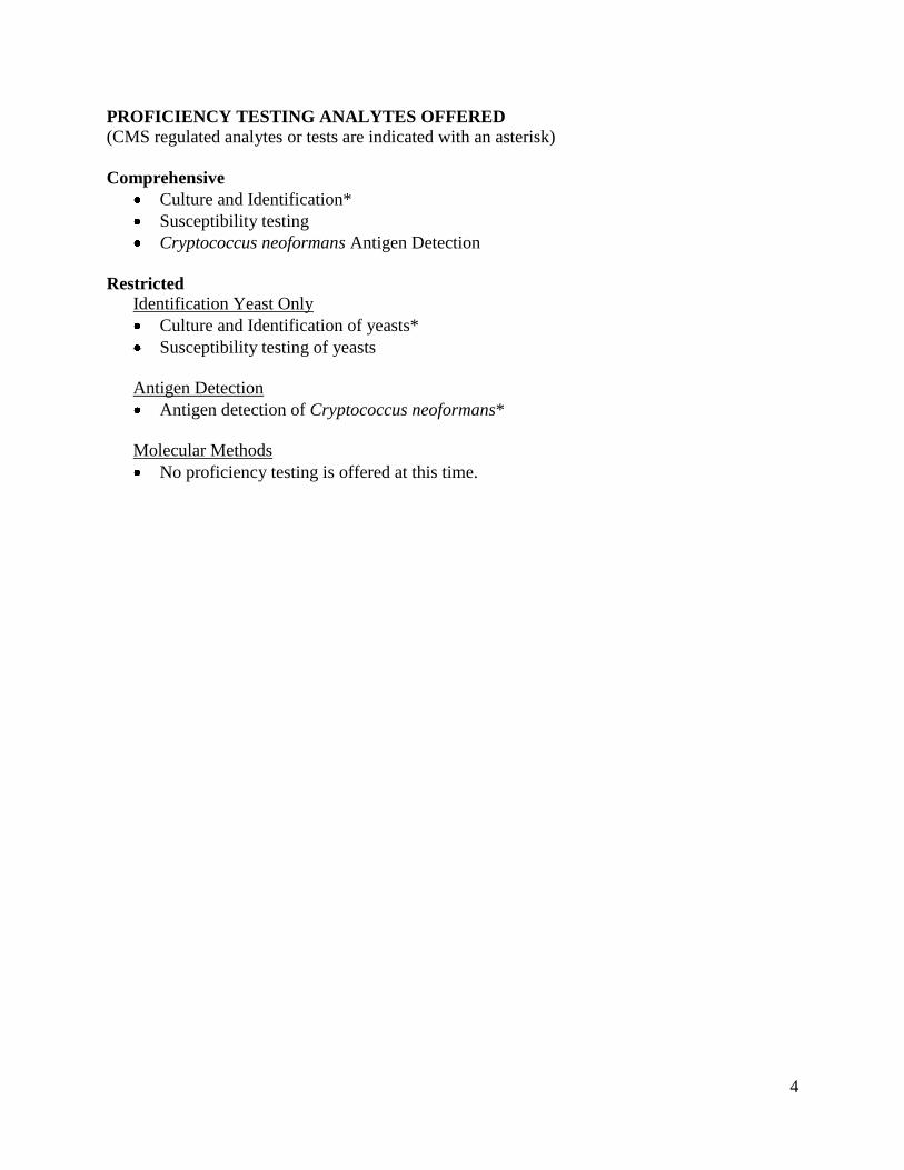
PROFICIENCY TESTING ANALYTES OFFERED
(CMS regulated analytes or tests are indicated with an asterisk)
Comprehensive
Culture and Identification*
Susceptibility testing
Cryptococcus neoformans Antigen Detection
Restricted
Identification Yeast Only
Culture and Identification of yeasts*
Susceptibility testing of yeasts
Antigen Detection
Antigen detection of Cryptococcus neoformans*
Molecular Methods
No proficiency testing is offered at this time.

5
TEST SPECIMENS & GRADING POLICY
Test Specimens
At least two strains of each mold or yeast species are examined for inclusion in the
proficiency test event. The colony morphology of molds is studied on Sabouraud dextrose agar.
The microscopic morphologic features are examined by potato dextrose agar slide cultures. The
physiological characteristics such as cycloheximide sensitivity and growth at higher temperatures
are investigated with appropriate test media. The strain that best demonstrates the morphologic
and physiologic characteristics typical of the species is included as a test analyte. Similarly, two
or more strains of yeast species are examined for inclusion in the proficiency test. The colony
morphology of all yeast strains is studied on corn meal agar with Tween 80 plates inoculated by
Dalmau or streak-cut method. Carbohydrate assimilation is studied with the API 20C AUX
identification kit (The use of brand and/or trade names in this report does not constitute an
endorsement of the products on the part of the Wadsworth Center or the New York State
Department of Health). The fermentations of carbohydrates, i.e., glucose, maltose, sucrose,
lactose, trehalose, and cellobiose, are also documented using classical approaches. Additional
physiologic characteristics such as nitrate assimilation, urease activity, and cycloheximide
sensitivity are investigated with the appropriate test media. The strain that best demonstrates the
morphologic and physiologic characteristics of the proposed test analyte is included as test
analyte. The morphologic features are matched with molecular identification using PCR and
nucleotide sequencing of ribosomal ITS1 – ITS2 regions.
Grading Policy
A laboratory’s response for each sample is compared with the responses that reflect 80%
agreement of 10 referee laboratories and/or 80% of all participating laboratories. The referee
laboratories are selected at random from among hospital laboratories participating in the
program. They represent all geographical areas of New York State and must have a record of
excellent performance during the preceding three years. The score in each event is established by
total number of correct responses submitted by the laboratory divided by the number of
organisms present plus the number of incorrect organisms reported by the laboratory multiplied
by 100 as per the formula shown on the next page.
# of acceptable responses 100
# of fungi present + # incorrect responses
For molds and yeast specimens, a facility can elect to process only those analytes that
match the type of clinical materials included within the scope of the facility’s standard operating
procedures (SOP). Similarly, the participating laboratory can elect to provide only genus level
identification if it reflects the SOP for patient testing in the concerned facility. In all such
instances, a maximum score of 100 will be equally distributed among the number of test analytes
selected by the laboratory. The rest of the score algorithm will be similar to the aforementioned
formula.
6
Acceptable results for antifungal susceptibility testing are based on the
consensus/reference laboratories’ MIC values within +/- 2 dilutions and the interpretation per
CLSI (NCCLS) guidelines or related, peer-reviewed publications. One yeast species is to be
tested against following drugs: amphotericin B, anidulafungin, caspofungin, flucytosine,
fluconazole, itraconazole, ketoconazole, micafungin, posaconazole, and voriconazole. The
participating laboratories are free to select any number of antifungal drugs from the test panel
based upon test practices in their facilities. A maximum score of 100 is equally distributed to
account for the drugs selected by an individual laboratory. If the result for any drug is incorrect
then laboratory gets a score of zero for that particular test component or set.
For Cryptococcus neoformans antigen test, laboratories are evaluated on the basis of their
responses and on overall performance for all the analytes tested in the Direct Detection category.
The maximum score for this event is 100. Appropriate responses are determined by 80%
agreement among participant responses. Target values and acceptable ranges are mean value +/-
2 dilutions; positive or negative answers will be acceptable from laboratories that do not report
antigen titers. When both qualitative and quantitative results are reported for an analyte, ten
points are deducted for each incorrect result. When only qualitative OR quantitative results are
reported, twenty points are deducted from each incorrect result.
A failure to attain an overall score of 80% is considered unsatisfactory performance.
Laboratories receiving unsatisfactory scores in two out of three consecutive proficiency test
events may be subject to ‘cease testing’.
7
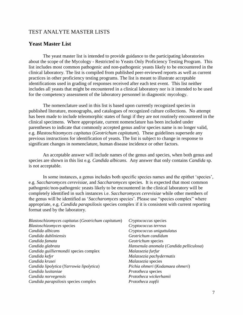
TEST ANALYTE MASTER LISTS
Yeast Master List
The yeast master list is intended to provide guidance to the participating laboratories
about the scope of the Mycology - Restricted to Yeasts Only Proficiency Testing Program. This
list includes most common pathogenic and non-pathogenic yeasts likely to be encountered in the
clinical laboratory. The list is compiled from published peer-reviewed reports as well as current
practices in other proficiency testing programs. The list is meant to illustrate acceptable
identifications used in grading of responses received after each test event. This list neither
includes all yeasts that might be encountered in a clinical laboratory nor is it intended to be used
for the competency assessment of the laboratory personnel in diagnostic mycology.
The nomenclature used in this list is based upon currently recognized species in
published literature, monographs, and catalogues of recognized culture collections. No attempt
has been made to include teleomorphic states of fungi if they are not routinely encountered in the
clinical specimens. Where appropriate, current nomenclature has been included under
parentheses to indicate that commonly accepted genus and/or species name is no longer valid,
e.g. Blastoschizomyces capitatus (Geotrichum capitatum). These guidelines supersede any
previous instructions for identification of yeasts. The list is subject to change in response to
significant changes in nomenclature, human disease incidence or other factors.
An acceptable answer will include names of the genus and species, when both genus and
species are shown in this list e.g. Candida albicans. Any answer that only contains Candida sp.
is not acceptable.
In some instances, a genus includes both specific species names and the epithet ‘species’,
e.g. Saccharomyces cerevisiae, and Saccharomyces species. It is expected that most common
pathogenic/non-pathogenic yeasts likely to be encountered in the clinical laboratory will be
completely identified in such instances i.e. Saccharomyces cerevisiae while other members of
the genus will be identified as ‘Saccharomyces species’. Please use “species complex” where
appropriate, e.g. Candida parapsilosis species complex if it is consistent with current reporting
format used by the laboratory.
Blastoschizomyces capitatus (Geotrichum capitatum) Cryptococcus species Blastoschizomyces species Cryptococcus terreus Candida albicans Cryptococcus uniguttulatus Candida dubliniensis Geotrichum candidum Candida famata Geotrichum species Candida glabrata Hansenula anomala (Candida pelliculosa) Candida guilliermondii species complex Malassezia furfur Candida kefyr Malassezia pachydermatis Candida krusei Malassezia species Candida lipolytica (Yarrowia lipolytica) Pichia ohmeri (Kodamaea ohmeri) Candida lusitaniae Prototheca species Candida norvegensis Prototheca wickerhamii Candida parapsilosis species complex Prototheca zopfii
8
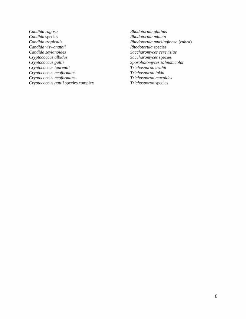
Candida rugosa Rhodotorula glutinis Candida species Rhodotorula minuta Candida tropicalis Rhodotorula mucilaginosa (rubra) Candida viswanathii Rhodotorula species Candida zeylanoides Saccharomyces cerevisiae Cryptococcus albidus Saccharomyces species Cryptococcus gattii Sporobolomyces salmonicolor Cryptococcus laurentii Trichosporon asahii Cryptococcus neoformans Trichosporon inkin Cryptococcus neoformans- Trichosporon mucoides Cryptococcus gattii species complex Trichosporon species
9
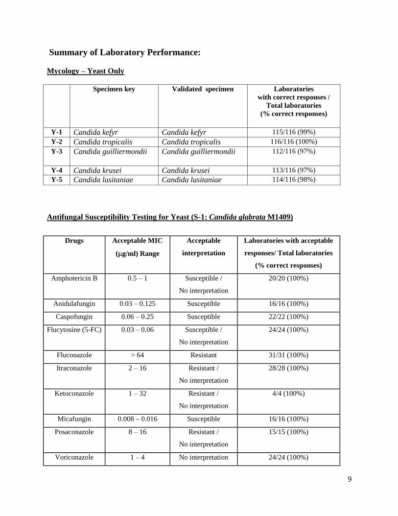
Summary of Laboratory Performance:
Mycology – Yeast Only
Antifungal Susceptibility Testing for Yeast (S-1: Candida glabrata M1409)
Specimen key Validated specimen Laboratories with correct responses /
Total laboratories (% correct responses)
Y-1 Candida kefyr Candida kefyr 115/116 (99%)
Y-2 Candida tropicalis Candida tropicalis 116/116 (100%)
Y-3 Candida guilliermondii Candida guilliermondii 112/116 (97%)
Y-4 Candida krusei Candida krusei 113/116 (97%)
Y-5 Candida lusitaniae Candida lusitaniae 114/116 (98%)
Drugs Acceptable MIC
( g/ml) Range
Acceptable
interpretation
Laboratories with acceptable
responses/ Total laboratories
(% correct responses)
Amphotericin B 0.5 – 1 Susceptible /
No interpretation
20/20 (100%)
Anidulafungin 0.03 – 0.125 Susceptible 16/16 (100%)
Caspofungin 0.06 – 0.25 Susceptible 22/22 (100%)
Flucytosine (5-FC) 0.03 – 0.06 Susceptible /
No interpretation
24/24 (100%)
Fluconazole > 64 Resistant 31/31 (100%)
Itraconazole 2 – 16 Resistant /
No interpretation
28/28 (100%)
Ketoconazole 1 – 32 Resistant /
No interpretation
4/4 (100%)
Micafungin 0.008 – 0.016 Susceptible 16/16 (100%)
Posaconazole 8 – 16 Resistant /
No interpretation
15/15 (100%)
Voriconazole 1 – 4 No interpretation 24/24 (100%)
10
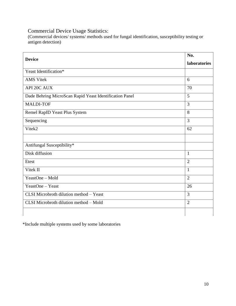
Commercial Device Usage Statistics: (Commercial devices/ systems/ methods used for fungal identification, susceptibility testing or
antigen detection)
*Include multiple systems used by some laboratories
Device No.
laboratories
Yeast Identification*
AMS Vitek 6
API 20C AUX 70
Dade Behring MicroScan Rapid Yeast Identification Panel 5
MALDI-TOF 3
Remel RapID Yeast Plus System 8
Sequencing 3
Vitek2 62
Antifungal Susceptibility*
Disk diffusion 1
Etest 2
Vitek II 1
YeastOne – Mold 2
YeastOne – Yeast 26
CLSI Microbroth dilution method – Yeast 3
CLSI Microbroth dilution method – Mold 2
11
YEAST DESCRIPTIONS
Y-1 Candida kefyr
Source: Urine / Vaginal / Wound
Clinical significance: Candida kefyr is rarely isolated in the clinical laboratory. Candida kefyr
infections are reported from the reproductive and digestive tracts and the mucous linings.
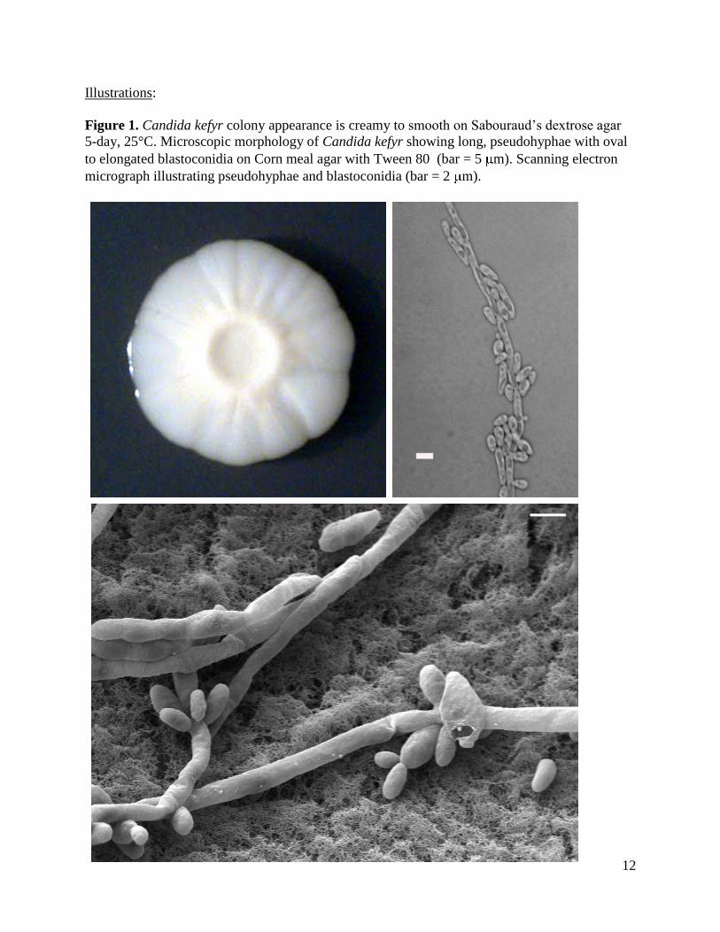
Colony: Candida kefyr colonies appear smooth, creamy, and soft on Sabouraud’s dextrose agar
after 3 to 5 days, 25°C (Figure 1).
Microscopy: Candida kefyr shows abundant long pseudohyphae, and oval to elongated
blastoconidia on cornmeal agar with Tween 80 (Figure 1). Ascospores within asci are observed
in cultures on V-8 or malt extract agar (details not shown). The sexual or teleomorphic state is
termed Kluyveromyces marxianus.
Differentiation: C. kefyr grows at 45 C and on the media containing cycloheximide. C. kefyr
ferments glucose, sucrose, lactose, galactose, but not maltose, trehalose, and cellobiose, which
differentiates it from other medically important Candida species.
Molecular test: Randomly amplified polymorphic DNA-polymorase chain reaction (RADP-PCR)
was applied for the identification of C. kefyr. The ribosomal ITS1 and ITS2 regions of the test
isolate showed 100 % nucleotide identity with a reference strain of Candida kefyr UWFP-208
(GenBank accession no. AF336841)
Antifungal susceptibility: C. kefyr is susceptible to amphotericin B, caspofungin, different
azoles, and 5-fluorocytosine.
Participant performance:
Referee Laboratories with correct ID: 10
Laboratories with correct ID: 115
Laboratories with incorrect ID: 01
(Candida species) (1)
12
Illustrations:
Figure 1. Candida kefyr colony appearance is creamy to smooth on Sabouraud’s dextrose agar
5-day, 25°C. Microscopic morphology of Candida kefyr showing long, pseudohyphae with oval
to elongated blastoconidia on Corn meal agar with Tween 80 (bar = 5 m). Scanning electron
micrograph illustrating pseudohyphae and blastoconidia (bar = 2 m).
13
Further reading:
Garcia-Martos P, Mira J, Galan F, Hernandez JM. Sexual forms of yeasts in clinical samples. 1996.
Mycopathologia. 136: 67-70.
Garcia-Martos P, Dominguez I, Marin P, Garcia-Agudo R, Aoufi S, Mira J. 2001. Antifungal susceptibility of
emerging yeast pathogens. Enferm. Infecc. Microbiol. Clin. 19: 249-256.
Corpus K, Hegeman-Dingle R, Bajjoka I. 2004. Candida kefyr, an uncommon but emerging fungal pathogen: report
of two cases. Pharmacotherapy. 24: 1084-1088.
Gil-Lamaignere C, Muller FM. 2004. Differential effects of the combination of caspofungin and terbinafine against
Candida albicans, Candida dubliniensis and Candida kefyr. Int J Antimicrob Agents. 23: 520-523.
Reuter CW, Morgan MA, Bange FC, Gunzer F, Eder M, Hertenstein B, Ganser A. 2005. Candida kefyr as an
emerging pathogen causing nosocomial bloodstream infections in neutropenic leukemia patients. Clin Infect Dis. 41:
1365-1366.
Sendid B, Lacroix C, Bougnoux ME. 2006. Is Candida kefyr an emerging pathogen in patients with
oncohematological diseases? Clin Infect Dis. 43: 666-667.
Chopra T, Bhargava A, Kumar S, Chopra A, Dhar S, Afonso L, Sobel JD. 2010. Candida kefyr endocarditis in a
patient with hypertrophic obstructive cardiomyopathy. Am J Med Sci. 339: 188-189.
Gomez-Lopez A, Pan D, Cuesta I, Alastruey-Izquierdo A, Rodriguez-Tudela JL, Cuenca-Estrella M. 2010.
Molecular identification and susceptibility profile in vitro of the emerging pathogen Candida kefyr. Diagn Microbiol
Infect Dis. 66: 116-119.
14
Y-2 Candida tropicalis
Source: Body fluid / Urine / Stool
Clinical significance: Candida tropicalis causes sepsis, wound infections, and disseminated
infections in immunocompromised patients.
Colony: C. tropicalis colony is smooth to wrinkled, cream-colored and rapid-growing on
Sabouraud’s dextrose agar after 7 days of incubation at 25°C, (Figure 2).
Microscopy: C. tropicalis shows long true hyphae and pseudohyphae, with either single or small
clusters of blastoconidia on Corn meal agar with Tween 80 (Figure 2).
Differentiation: C. tropicalis is differentiated from C. albicans and C. dubliniensis by variable
growth on media containing cycloheximide, and by its fermentation of glucose, maltose, sucrose,
and trehalose. Occasionally, C. tropicalis produces chlamydospores on corn meal agar.
Molecular test: Reverse-hybridization line probe assay combined with PCR amplification of
internal transcribed-spacer (ITS) regions are used for rapid identification of clinically significant
fungal pathogens including C. tropicalis. The combination of pan-fungal PCR and multiplex
liquid hybridization of ITS regions are developed for detection and identification of fungi in
tissue specimens. The ribosomal ITS1 and ITS2 regions of the test isolate showed 100 %
nucleotide identity with C. tropicalis CBL Cd-3 (Genebank accession no. EU924133)
Antifungal susceptibility: C. tropicalis is generally susceptible to azoles and echinocandins, but
variably susceptible to flucytosine. Few strains of C. tropicalis have been reported with high
amphotericin B MIC.
Participant performance:
Referee Laboratories with correct ID: 10
Laboratories with correct ID: 116
Laboratories with incorrect ID: 0
15
Illustrations:
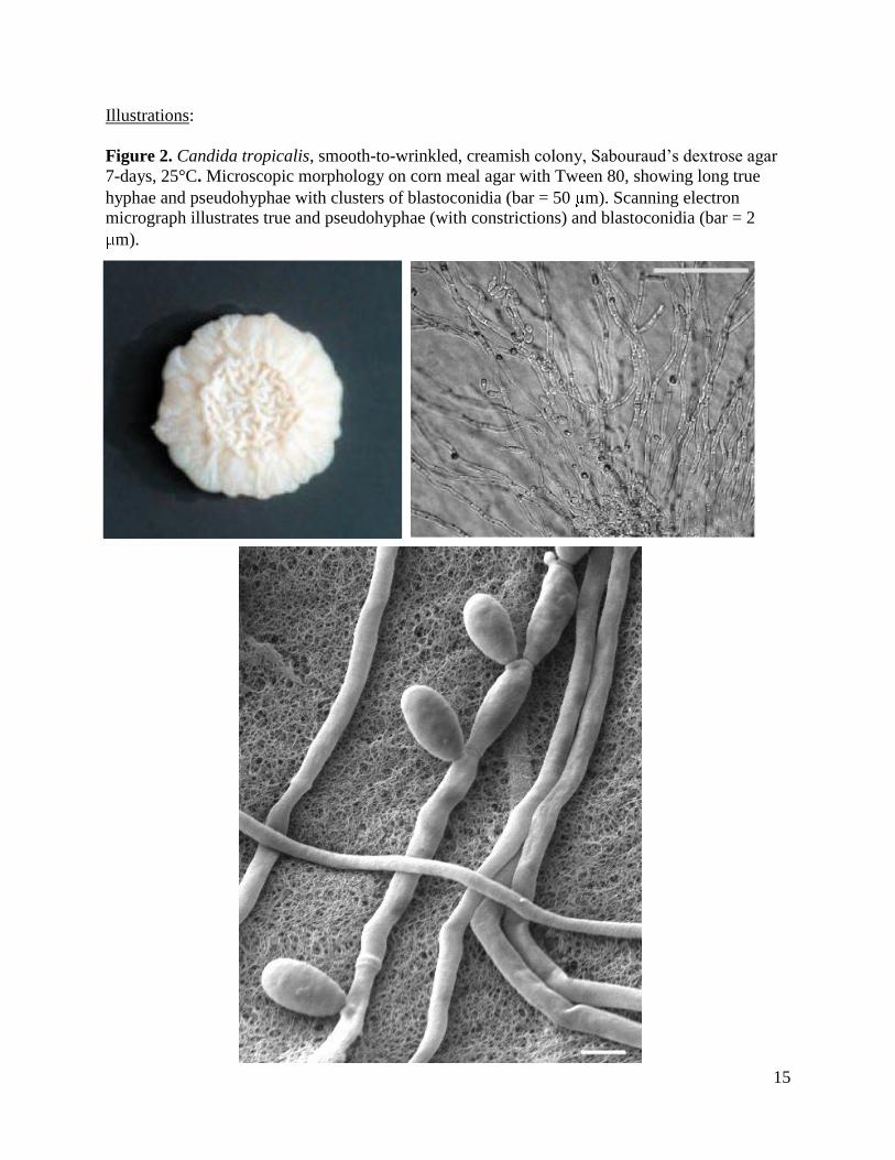
Figure 2. Candida tropicalis, smooth-to-wrinkled, creamish colony, Sabouraud’s dextrose agar
7-days, 25°C. Microscopic morphology on corn meal agar with Tween 80, showing long true
hyphae and pseudohyphae with clusters of blastoconidia (bar = 50 m). Scanning electron
micrograph illustrates true and pseudohyphae (with constrictions) and blastoconidia (bar = 2
m).

16
Further reading:
Hilmioglu S, Ilkit M, Badak Z. 2007. Comparison of 12 liquid media for germ tube production of Candida albicans
and C. tropicalis. Mycoses. 50: 282-285.
Nucci M, Colombo AL. 2007. Candidemia due to Candida tropicalis: clinical, epidemiologic, and microbiologic
characteristics of 188 episodes occurring in tertiary care hospitals. Diagn Microbiol Infect Dis. 58: 77-82.
Pfaller MA, Castanheira M, Messer SA, Moet GJ, Jones RN. 2010. Variation in Candida spp. distribution and
antifungal resistance rates among bloodstream infection isolates by patient age: report from the SENTRY
Antimicrobial Surveillance Program (2008-2009). Diagn Microbiol Infect Dis. 68: 278-283.
Chai LY, Denning DW, Warn P. 2010. Candida tropicalis in human disease. Crit Rev Microbiol. 36: 282-98.
de Carvalho Parahym AM, da Silva CM, Leão MP, Macario MC, Filho GA, de Oliveira NT, Neves RP. 2011.
Invasive infection in an acute myeloblastic leukemia patient due to triazole-resistant Candida tropicalis. Diagn
Microbiol Infect Dis. 71: 291-293.
Muñoz P, Giannella M, Fanciulli C, Guinea J, Valerio M, Rojas L, Rodríguez-Créixems M, Bouza E. 2011. Candida
tropicalis fungemia: incidence, risk factors, and mortality in a general hospital. Clin Microbiol Infect. 17: 1538-
1545.
de Carvalho Parahym AM, da Silva CM, Leao MP, Macario MC, Filho GA, de Oliveira NT, Neves RP. 2011.
Invasive infection in an acute myeloblastic leukemia patient due to triazole-resistant Candida tropicalis. Diagn
Microbiol Infect Dis. 71: 291-293.
Negri M, Silva S, Henriques M, Oliveira R. 2011. Insights into Candida tropicalis nosocomial infections and
virulence factors. Eur J Clin Microbiol Infect Dis. DOI. 10.1007/s10096-011-1455-z.
17
Y-3 Candida guilliermondii
Source: Urine / Blood / Nail
Clinical significance: Candida guilliermondii is a frequent cause of nosocomial fungemia in
immunosuppressed patients. It rarely causes infection of urinary tract, brain and eye.
Colony: C. guilliermondii colony is flat, smooth, cream-yellow on Sabouraud’s dextrose agar
after 7 days of incubation at 25 C (Figure 3).
Microscopy: C. guilliermondii shows few short pseudohyphae with clusters of blastoconidia on
Corn meal agar with Tween 80 (Figure 3). Please check corn meal how it is written in book and
change accordingly?
Differentiation: C. guilliermondii is the anamorph (asexual form) of Pichia guilliermondii/
Kodamaea ohmeri. It ferments glucose, sucrose, and trehalose, grows at 37 C, and on media
containing cycloheximide. It does not form pink pigment thereby differentiating it from
Rhodotorula species. It does not produce true hyphae, which differentiates it from Candida
ciferrii and Trichosporon beigelii. Unlike Candida lusitaniae, it is unable to grow at 45 C.
Molecular test: Primers for large ribosomal subunit DNA sequences are used in PCR to
differentiate C. guilliermondii from C. famata/ Debaryomyces hansenii complex. Isolates of C.
guilliermondii are identified using PCR to amplify ribosomal DNA, followed by restriction
digestion of the PCR product.
The ribosomal ITS1 and ITS2 regions of the test isolate showed 100 % nucleotide identity with
Candida guilliermondii (Pichia guilliermondii) isolate SMB (GenBank accession no.
GU385845.1).
Antifungal susceptibility: Most clinical isolates are susceptible to amphotericin B, 5-flucytosine,
echinocandins and azoles such as fluconazole, ketocoanzole, itraconazole. A few isolates are
reported to have high MIC to azoles.
Participant performance:
Referee Laboratories with correct ID: 10
Laboratories with correct ID: 112
Laboratories with incorrect ID: 06
(Candida famata) (1)
(Candida sp.) (2)
(Candida viswanathii) (1)
18
Illustrations:
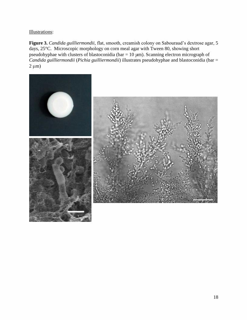
Figure 3. Candida guilliermondii, flat, smooth, creamish colony on Sabouraud’s dextrose agar, 5
days, 25°C. Microscopic morphology on corn meal agar with Tween 80, showing short
pseudohyphae with clusters of blastoconidia (bar = 10 m). Scanning electron micrograph of
Candida guilliermondii (Pichia guilliermondii) illustrates pseudohyphae and blastoconidia (bar =
2 m)
19
Further reading:
Kabbara N, Lacroix C, de Latour RP, Socié G, Ghannoum M, Ribaud P. 2008. Breakthrough C. parapsilosis and C.
guilliermondii blood stream infections in allogeneic hematopoietic stem cell transplant recipients receiving long-
term caspofungin therapy. Haematologica. 93: 639-640.
Lee GW, Kim TH, Son JH. 2012. Primary Candida guilliermondii infection of the knee in a patient without
predisposing factors. Case Report Med. 2012:375682. Epub 2012 Feb 28.
Macêdo DP, Oliveira NT, Farias AM, Silva VK, Wilheim AB, Couto FM, Neves RP. 2010. Esophagitis caused by
Candida guilliermondii in diabetes mellitus: first reported case. Med Mycol. 48: 862-865.
Mardani M, Hanna, HA, Girgawy, E, Raad, I. 2000. Nosocomial Candida guilliermondii fungemia in cancer
patients. Infect Control Hosp. Epidemiol. 21: 336-337.
Pemán J, Bosch M, Cantón E, Viudes A, Jarque I, Gómez-García M, García-Martínez JM., Gobernado M. 2008.
Fungemia due to Candida guilliermondii in a pediatric and adult population during a 12-year period. Diagn
Microbiol Infect Dis. 60: 109-112.
Pfaller MA, Boyken L, Hollis RJ, Messer SA, Tendolkar S, Diekema DJ. 2006. In Vitro Susceptibilities of Candida
spp. to Caspofungin: Four Years of Global Surveillance. J. Clin. Microbiol. 44: 760-763.
Savini V, Catavitello C, Onofrillo D, Masciarelli G, Astolfi D, Balbinot A, Febbo F, D'Amario C, D'Antonio D.
2011. What do we know about Candida guilliermondii? A voyage throughout past and current literature about this
emerging yeast. Mycoses. 54:434-41.
20
Y-4 Candida krusei
Source: Tissue / Sputum / Urine / Stool
Clinical significance: Candida krusei causes nosocomial fungemia in immunosuppressed
patients. It also causes disseminated disease including endocarditis, peritonitis, vaginitis, urinary
tract infections, and sinusitis.
Colony: C. krusei colony is soft, cream to buff, glassy and wrinkled on Sabouraud’s dextrose
agar, after7 days of incubation at 25°C (Figure 4).
Microscopy: C. krusei shows branched pseudohyphae with elongated blastoconidia on Corn meal
agar with Tween 80 (Figure 4).
Differentiation: C. krusei ferments glucose, but not sucrose or cellobiose, and does not grow on
the media containing cycloheximide. C. krusei does not assimilate sucrose, which differentiates
it from C. parapsilosis and C. lusitaniae. C. krusei grows well at 42°C, differentiating it from C.
lambica. C. krusei does not produce arthroconidia, thus differentiating it from Blastoschizomyces
capitatus.
Molecular test: DNA probes from the ITS regions are incorporated in a reverse hybridization line
probe assay for the detection of ITS PCR products for identification of fungal pathogens.
Panfungal PCR and multiplex liquid hybridization are developed for the detection of clinically
important yeasts in tissue specimens. PFGE, RFLP, and RAPD procedures are used for DNA
fingerprinting and electrophoretic karyotyping of oral C. krusei isolates. The ribosomal ITS1 and
ITS2 regions of the test isolate showed 100% nucleotide identity with a reference strain of C.
krusei (Pichia kudriavzevii) GenBank accession no. AF411417.
Antifungal susceptibility: C. krusei is susceptible to amphotericin B and flucytosine. C. krusei is
innately resistant to fluconazole and variably resistant to other azoles such as itraconazole and
ketoconazole, but not voriconazole. C. krusei is also susceptible to echinocandins.
Participant performance:
Referee Laboratories with correct ID: 10
Laboratories with correct ID: 113
Laboratories with incorrect ID: 05
(Candida norvegensis) (2)
(Blastoschizomyces capitatus) (1)
21
Illustrations:
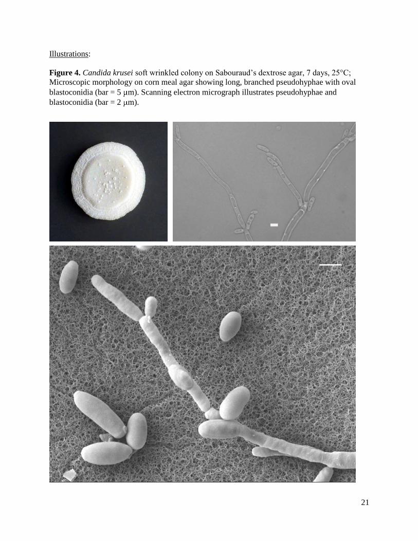
Figure 4. Candida krusei soft wrinkled colony on Sabouraud’s dextrose agar, 7 days, 25°C;
Microscopic morphology on corn meal agar showing long, branched pseudohyphae with oval
blastoconidia (bar = 5 m). Scanning electron micrograph illustrates pseudohyphae and
blastoconidia (bar = 2 m).
22
Further reading:
Sili U, Yilmaz M, Ferhanoglu B, Mert A. 2007. Candida krusei arthritis in a patient with hematologic malignancy:
successful treatment with voriconazole. Clin Infect Dis. 45: 897-898.
Jacobsen MD, Gow NA, Maiden MC, Shaw DJ, Odds FC. 2007. Strain typing and determination of population
structure of Candida krusei by multilocus sequence typing. J Clin Microbiol. 45: 317-323.
Pfaller MA, Diekema DJ, Gibbs DL, Newell VA, Nagy E, Dobiasova S, Rinaldi M, Barton R, Veselov A; the
Global Antifungal Surveillance Group. 2008. Candida krusei, a Multidrug-Resistant Opportunistic Fungal Pathogen:
Geographic and Temporal Trends from the ARTEMIS DISK Antifungal Surveillance Program, 2001-2005. J Clin
Microbiol. 46: 515-521.
Natale F, Castronovo A, Regoli D, De Curtis M, Manzoni P. 2009. Successful treatment with caspofungin of
refractory Candida krusei candidemia in a very low birth weight preterm infant. Pediatr Infect Dis J. 28: 452.
Cascio A, Barone M, Micali V, Iaria C, Delfino D, David A, Monaco M, Monaco F. 2010. On a fatal case of
Candida krusei pleural empyema in a pregnant woman with spontaneous esophagus perforation. Mycopathologia.
169: 451-455.
Hager JL, Mir MR, Hsu S. 2010. Candida krusei fungemia in an immunocompromised patient. Dermatol Online J.
16: 5.
Schilling A, Seibold M, Mansmann V, Gleissner B. 2007. Successfully treated Candida krusei infection of the
lumbar spine with combined caspofungin/posaconazole therapy. Med Mycol. 46: 79-83.
Shorr AF, Wu C, Kothari S. 2011. Outcomes with micafungin in patients with candidaemia or invasive candidiasis
due to Candida glabrata and Candida krusei. J Antimicrob Chemother. 66: 375-380.
23
Y-5 Candida lusitaniae
Source: Stool / Blood / Eye / Urine
Clinical significance: Candida lusitaniae causes fungemia and sepsis in immunocompromised
and debilitated patients with cancer, diabetes, or asthma, and also neonates in intensive care
units. The common clinical samples are blood, urine, and respiratory tract secretions.
Colony: C. lusitaniae colony is white to creamish, shiny, and slightly raised in the center on
Sabouraud’s dextrose agar, after 7 days of incubation at 25°C (Figure 5).
Microscopy: C. lusitaniae produced many short, branched (“bushy”) pseudohyphae. Along the
length of the pseudohyphae, elongated blastoconidia formed in short chains on Corn Meal Agar
with Tween 80 (Figure 5).
Differentiation: C. lusitaniae is able to ferment and assimilate cellobiose, which differentiates it
from C. parapsilosis.
Molecular test: Specific nucleic acid probes targeting the large subunit rRNA genes have been
developed for identification of C. lusitaniae. Three pulsed-field electrophoretic methods and a
random amplified polymorphic DNA (RAPD) method were also reported to delineate strains of
C. lusitaniae.
The ribosomal ITS1 and ITS2 regions of the test isolate showed 100 % nucleotide identity with
Candida lusitaniae (Clavispora lusitaniae) isolate F47819-04 (GenBank accession no.
HQ693785.1).
Antifungal susceptibility: Some C. lusitaniae strains are reported to be inherently resistant to
amphotericin B. Amphotericin B susceptible strains are also known to develop resistance during
the course of treatment with this drug. C. lusitaniae is reported as more susceptible to
voriconazole than fluconazole.
Participant performance:
Referee Laboratories with correct ID: 10
Laboratories with correct ID: 57
Laboratories with incorrect ID: 01
(Candida famata) (1)
(Candida species) (1)
24
Illustrations:
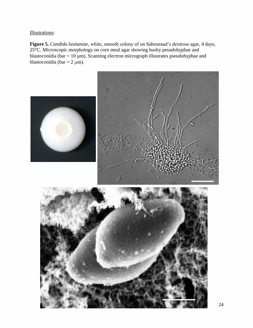
Figure 5. Candida lusitaniae, white, smooth colony of on Sabouraud’s dextrose agar, 4 days,
25°C. Microscopic morphology on corn meal agar showing bushy pesudohyphae and
blastoconidia (bar = 10 m). Scanning electron micrograph illustrates pseudohyphae and
blastoconidia (bar = 2 m).

25
Further reading:
Alberth M, Majoros L, Kovalecz G, Borbas E, Szegedi I, J Marton I, Kiss C. 2006. Significance of oral Candida
infections in children with cancer. Pathol Oncol Res. 12: 237-241.
Atkinson BJ, Lewis RE, Kontoyiannis DP. 2008. Candida lusitaniae fungemia in cancer patients: risk factors for
amphotericin B failure and outcome. Med Mycol. 46: 541-546.
Bariola JR, Saccente M. 2008. Candida lusitaniae septic arthritis: case report and review of the literature. Diagn
Microbiol Infect Dis. 61: 61-63.
De Carolis E, Sanguinetti M, Florio AR, La Sorda M, D'Inzeo T, Morandotti GA, Fadda G, Posteraro B. 2010. In
vitro susceptibility to seven antifungal agents of Candida lusitaniae isolates from an italian university hospital.
J Chemother. 22: 68-70.
Estrada B, Mancao MY, Polski JM, Figarola MS. 2006. Candida lusitaniae and chronic granulomatous disease.
Pediatr Infect Dis J. 25: 758-759.
McClenny NB, Fei H, Baron EJ, Gales AC, Houston A, Hollis RJ, Pfaller MA. 2002. Change in colony morphology
of Candida lusitaniae in association with development of amphotericin B resistance. Antimicrob Agnets Chemother.
46: 1325-1328.
Michel RG, Kinasewitz GT, Drevets DA, Levin JH, Warden DW. 2009. Prosthetic valve endocarditis caused by
Candida lusitaniae, an uncommon pathogen: a case report. J Med Case Reports. 3: 7611.
Parentin F, Liberali T, Perissutti P. 2006. Polymicrobial keratomycosis in a three-year-old child. Ocul Immunol
Inflamm. 14: 129-131.
Pfaller MA, Woosley LN, Messer SA, Jones RN, Castanheira M. 2012. Significance of molecular identification and
antifungal susceptibility of clinically significant yeasts and moulds in a Global Antifungal Surveillance Programme.
Mycopathologia. DOI 10.1007/s11046-012-9551-x
Prigitano A, Biraghi E, Pozzi C, Viviani MA, Tortorano AM. 2010. In vitro activity of amphotericin B against
Candida lusitaniae clinical isolates. J Chemother. 22: 71-72.
Werner BC, Hogan MV, Shen FH. 2011. Candida lusitaniae discitis after discogram in an immunocompetent
patient. Spine J. 11: e1-6.
26
ANTIFUNGAL SUSCEPTIBILITY TESTING FOR YEASTS
Introduction: Clinical laboratories perform susceptibility testing of pathogenic yeasts to
determine their in vitro resistance to antifungal drugs. This test is also useful in conducting
surveillance for evolving patterns of antifungal drug resistance in a healthcare facility. The
results are likely to facilitate the selection of appropriate drugs for treatment. Clinical
Laboratory Standards Institute (CLSI) documents of M27-A3, M27-S3, M27-S4, and M44-A,
describe the current standard methods for antifungal susceptibility testing of pathogenic yeasts.
Another resource for standardized method is the EUCAST Definitive Document EDef 7.1:
method for the determination of broth dilution MICs of antifungal agents for fermentative yeasts.
The FDA approved devices for antifungal susceptibility testing of yeasts include Sensititre
YeastOne Colorimetric Panel (Trek Diagnostic Systems Inc. Cleveland, OH) and Etest
(bioMérieux, Inc., Durham, NC). The following ten drugs are included in the Mycology
Proficiency Test Program - amphotericin B, anidulafungin, caspofungin, flucytosine (5-FC),
fluconazole, itraconazole, ketoconazole, micafungin, posaconazole, and voriconazole. The
participating laboratories are allowed to select any number of antifungal drug(s) from this test
panel based upon practices in their facilities.
Materials: Candida glabrata (S-1) was the analyte in the May 29, 2013 antifungal proficiency
testing event. The interpretation of MIC values for antifungal susceptibility testing of yeasts and
molds is in a state of constant change. These changes are necessitated by new information
emerging from clinical trials and laboratory susceptibility testing. NYSDOH Mycology
Laboratory uses latest CLSI and EUCAST documents to score proficiency testing results.
However, the participating laboratories are advised to regularly consult these organizations for
the latest version of their standard documents.
Comments: Acceptable results were MICs +/-2 dilutions of the reference laboratory results for
any single drug. Only 2 of the 31 laboratories participating in this test event tested all 10
antifungal drugs. The reported results were as follows: itraconazole (28 laboratories), flucytosine
(24 laboratories), voriconasole (24 laboratories), caspofungin (22 laboratories), amphotericin B
(20 laboratories), anidulafungin (16 laboratories), micafungin (16 laboratories), posacoanazole
(15 laboratories), and ketocoanzole (4 laboratories). Fluconazole was the only drug tested by all
31 laboratories.
27
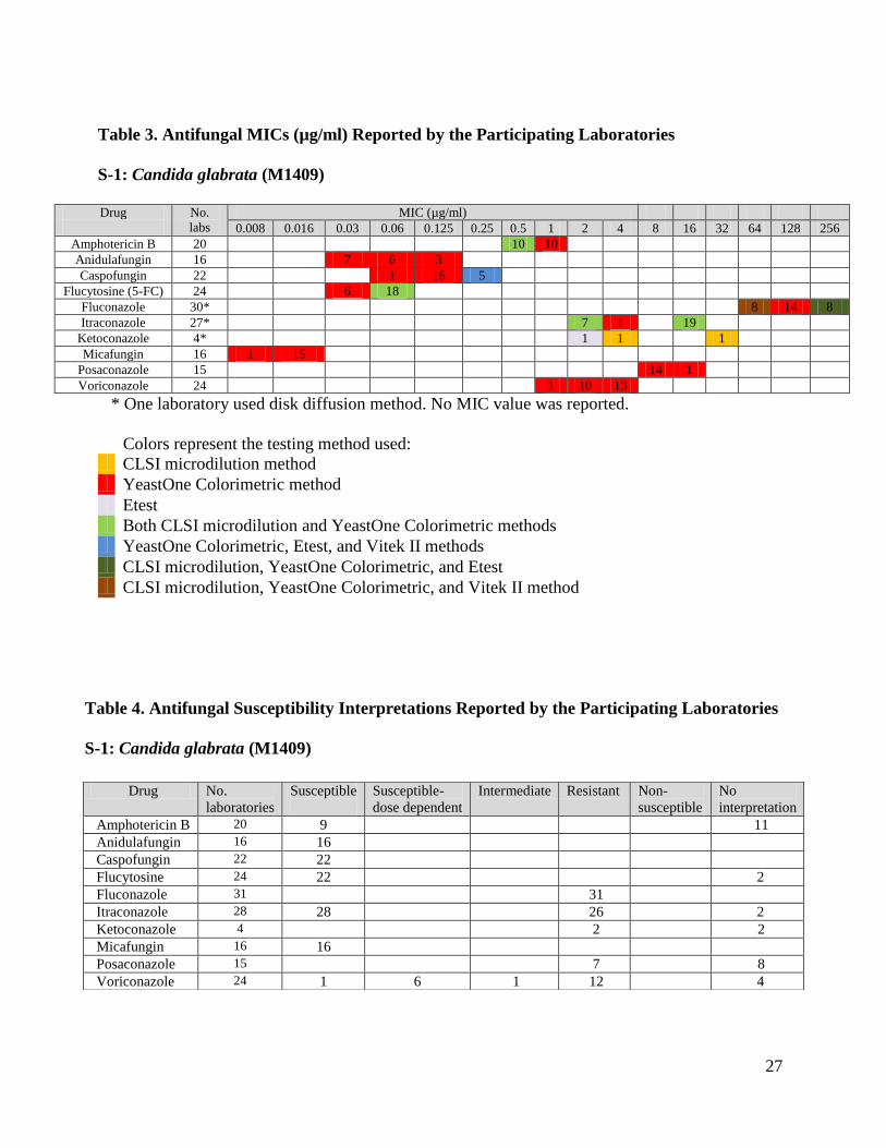
Table 3. Antifungal MICs (µg/ml) Reported by the Participating Laboratories
S-1: Candida glabrata (M1409)
Drug
No.
labs
MIC (µg/ml)
0.008 0.016 0.03 0.06 0.125 0.25 0.5 1 2 4 8 16 32 64 128 256
Amphotericin B 20 10 10
Anidulafungin 16 7 6 3
Caspofungin 22 1 16 5
Flucytosine (5-FC) 24 6 18
Fluconazole 30* 8 14 8
Itraconazole 27* 7 1 19
Ketoconazole 4* 1 1 1
Micafungin 16 1 15
Posaconazole 15 14 1
Voriconazole 24 1 10 13
* One laboratory used disk diffusion method. No MIC value was reported.
Colors represent the testing method used: CLSI microdilution method YeastOne Colorimetric method Etest Both CLSI microdilution and YeastOne Colorimetric methods YeastOne Colorimetric, Etest, and Vitek II methods CLSI microdilution, YeastOne Colorimetric, and Etest CLSI microdilution, YeastOne Colorimetric, and Vitek II method
Table 4. Antifungal Susceptibility Interpretations Reported by the Participating Laboratories
S-1: Candida glabrata (M1409)
Drug No.
laboratories
Susceptible
Susceptible-
dose dependent
Intermediate Resistant Non-
susceptible
No
interpretation
Amphotericin B 20 9 11
Anidulafungin 16 16
Caspofungin 22 22
Flucytosine 24 22 2
Fluconazole 31 31
Itraconazole 28 28 26 2
Ketoconazole 4 2 2
Micafungin 16 16
Posaconazole 15 7 8
Voriconazole 24 1 6 1 12 4
28
ANTIFUNGAL SUSCEPTIBILITY TESTING FOR MOLDS
(EDUCATIONAL)
Introduction: Clinical laboratories perform susceptibility testing of pathogenic molds to
determine their in vitro resistance to antifungal drugs. This test is also useful in conducting
surveillance for evolving patterns of antifungal drug resistance in a healthcare facility. It is not
clear at this juncture if the results of mold susceptibility testing have direct relevance in the
selection of appropriate drugs for treatment. Clinical Laboratory Standards Institute (CLSI)
document of M38-A2 describes the current standard methods for antifungal susceptibility testing
of pathogenic molds. Another resource for standardized method is the EUCAST Technical Note
on the method for the determination of broth dilution minimum inhibitory concentrations of
antifungal agents for conidia-forming moulds. The following nine drugs are included in the
antifungal susceptibility panel - amphotericin B, anidulafungin, caspofungin, fluconazole,
itraconazole, ketoconazole, micafungin, posaconazole, and voriconazole.
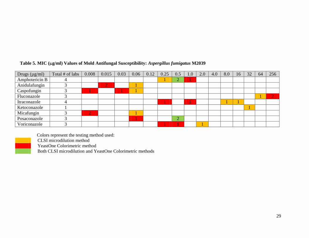
Materials: Aspergillus fumigatus M2039 was used as a test analyte; it was obtained from a
reference laboratory. Participating laboratories volunteered to perform the test and they were free
to choose any number of drugs and a test method. Two laboratories used CLSI broth
microdilution method while the remaining two used TREK YeastOne Colorimetric method.
Comments: Four out of thirty-one laboratories, which hold antifungal susceptibility testing for
yeasts permit, voluntarily participated in this test event for molds. Please refer to Table 5 for
summary of performances. Since too few laboratories have participated in this test, no consensus
data could be generated.
29
Table 5. MIC ( g/ml) Values of Mold Antifungal Susceptibility: Aspergillus fumigatus M2039
Drugs (µg/ml) Total # of labs 0.008 0.015 0.03 0.06 0.12 0.25 0.5 1.0 2.0 4.0 8.0 16 32 64 256
Amphotericin B 4 1 2 1
Anidulafungin 3 2 1
Caspofungin 3 1 1 1
Fluconazole 3 1 2
Itraconazole 4 1 1 1 1
Ketoconazole 1 1
Micafungin 3 2 1
Posaconazole 3 1 2
Voriconazole 3 1 1 1
Colors represent the testing method used:
CLSI microdilution method
YeastOne Colorimetric method
Both CLSI microdilution and YeastOne Colorimetric methods
30
Further Reading: Canton E, Peman J, Gobernado M, Alvarez E, Baquero F, Cisterna R, Gil J, Martin-Mazuelos E, Rubio C, Sanchez-
Sousa A, Settano C. 2005. Sensititre YeastOne caspofungin susceptibility testing of Candida clinical isolates:
correlation with results of NCCLS M27-A2 multicenter study. Antimicrobiol Agents Chemother. 49: 1604-1607.
Clinical and Laboratory Standards Institute. 2008. Reference Method for Broth Dilution Antifungal Susceptibility
Testing of Yeasts; Approved Standard - Third Edition. CLSI document M27-A3 (ISBN 1-56238-666-2).
Clinical and Laboratory Standards Institute. 2008. Quality Control Minimal Inhibitory Concentration (MIC) Limits
for Broth Microdilution and MIC Interpretive Breakpoints; Informational Supplement - Third Edition. CLSI
document M27-S3 (ISBN 1-56238-667-0).
Clinical and Laboratory Standards Institute. 2008. Reference Method for Broth Dilution Antifungal Susceptibility
Testing of Filamentous Fungi; Approved Standard – Second Edition. CLSI document M38-A2 (1-56238-668-9).
Clinical and Laboratory Standards Institute. 2009. Method for Antifungal Disk Diffusion Susceptibility Testing of
Yeasts; Approved Guideline – Second Edition. CLSI document M44-A2 (ISBN 1-56238-703-0).
Clinical and Laboratory Standards Institute. 2009. Zone Diameter Interpretive Standards, Corresponding Minimal
Inhibitory Concentration (MIC) Interpretive Breakpoints, and Quality Control Limits for Antifungal Disk Diffusion
Susceptibility Testing of Yeasts; Informational Supplement. CLSI document M44-S3.
Clinical and Laboratory Standards Institute. 2010. Method for Antifungal Disk Diffusion Susceptibility Testing of
Nondermatophyte Filamentous Fungi; Approved Guideline. CLSI document M51-A (ISBN 1-56238-725-1).
Clinical and Laboratory Standards Institute. 2010. Performance Standards for Antifungal Disk Diffusion
Susceptibility Testing of Filamentous Fungi; Informational Supplement. CLSI document M51-S1 (ISBN 1-56238-
725-1).
Clinical and Laboratory Standards Institute. 2012. Reference Method for Broth Dilution Antifungal Susceptibility
Testing of Yeasts; Fourth Informational Supplement. CLSI document M27-S4 (ISBN 1-56238-863-0).
Subcommittee on Antifungal Susceptibility Testing (AFST) of the ESCMID European Committee for Antimicrobial
Susceptibility Testing (EUCAST). 2008. EUCAST technical note on fluconazole. Clin Microbiol Infect. 14: 193-
195.
Subcommittee on Antifungal Susceptibility Testing (AFST) of the ESCMID European Committee for Antimicrobial
Susceptibility Testing (EUCAST). 2008. EUCAST definitive document Edef 7.1: method for the determination of
broth dilution MICs of antifungal agents for fermentative yeasts. Clin Microbiol Infect. 14: 398-405.
Subcommittee on Antifungal Susceptibility Testing (AFST) of the ESCMID European Committee for Antimicrobial
Susceptibility Testing (EUCAST). 2008. EUCAST technical note on the method for the determination of broth
dilution minimum inhibitory concentrations of antifungal agents for conidia–forming moulds. Clin Microbiol Infect.
14: 982-984.
Subcommittee on Antifungal Susceptibility Testing (AFST) of the ESCMID European Committee for Antimicrobial
Susceptibility Testing (EUCAST). 2008. EUCAST technical note on voriconazole. Clin Microbiol Infect. 14: 985-
987.
Copyright 2013 Wadsworth Center
New York State Department of Health