Embed Size (px)
Citation preview
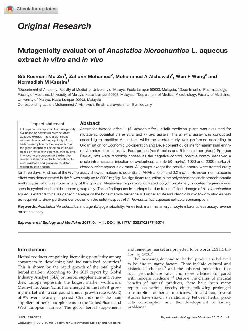
Original Research
Mutagenicity evaluation of Anastatica hierochuntica L. aqueous
extract in vitro and in vivo
Siti Rosmani Md Zin1, Zahurin Mohamed2, Mohammed A Alshawsh2, Won F Wong3 andNormadiah M Kassim1
1Department of Anatomy, Faculty of Medicine, University of Malaya, Kuala Lumpur 50603, Malaysia; 2Department of Pharmacology,
Faculty of Medicine, University of Malaya, Kuala Lumpur 50603, Malaysia; 3Department of Medical Microbiology, Faculty of Medicine,
University of Malaya, Kuala Lumpur 50603, Malaysia
Corresponding author: Mohammed A Alshawsh. Email: [email protected]
AbstractAnastatica hierochuntica L. (A. hierochuntica), a folk medicinal plant, was evaluated for
mutagenic potential via in vitro and in vivo assays. The in vitro assay was conducted
according to modified Ames test, while the in vivo study was performed according to
Organisation for Economic Co-operation and Development guideline for mammalian eryth-
rocyte micronucleus assay. Four groups (n¼ 5 males and 5 females per group) Sprague
Dawley rats were randomly chosen as the negative control, positive control (received a
single intramuscular injection of cyclophosphamide 50 mg/kg), 1000 and, 2000 mg/kg A.
hierochuntica aqueous extracts. All groups except the positive control were treated orally
for three days. Findings of the in vitro assay showed mutagenic potential of AHAE at 0.04 and 0.2 mg/ml. However, no mutagenic
effect was demonstrated in the in vivo study up to 2000 mg/kg. No significant reduction in the polychromatic and normochromatic
erythrocytes ratio was noted in any of the groups. Meanwhile, high micronucleated polychromatic erythrocytes frequency was
seen in cyclophosphamide-treated group only. These findings could perhaps be due to insufficient dosage of A. hierochuntica
aqueous extracts to cause genetic damage on the bone marrow target cells. Further acute and chronic in vivo toxicity studies may
be required to draw pertinent conclusion on the safety aspect of A. hierochuntica aqueous extracts consumption.
Keywords: Anastatica hierochuntica, mutagenicity, genotoxicity, Ames test, mammalian erythrocyte micronucleus assay, reverse
mutation assay
Experimental Biology and Medicine 2017; 0: 1–11. DOI: 10.1177/1535370217748574
Introduction
Herbal products are gaining increasing popularity amongconsumers in developing and industrialized countries.1
This is shown by the rapid growth of the total globalherbal market. According to the 2015 report by GlobalIndustry Analyst (GIA) on herbal supplements and reme-dies, Europe represents the largest market worldwide.Meanwhile, Asia-Pacific has emerged as the fastest grow-ing market with a compound annual growth rate (CAGR)of 9% over the analysis period. China is one of the mainsuppliers of herbal supplements to the United States andWest European markets. The global herbal supplements
and remedies market are projected to be worth US$115 bil-lion by 2020.2
The increasing demand for herbal products is believedto be due to many factors. These include cultural andhistorical influences3 and the inherent perception thatsuch products are safer and more efficient comparedwith modern medicine.4,5 Despite the claims of medicalbenefits of natural products, there have been manyreports on various toxicity effects following prolongedconsumption of herbal medicines.6 In addition, severalstudies have shown a relationship between herbal prod-ucts consumption and the development of kidneyproblems.7
Impact statementIn this paper, we report on the mutagenicity
evaluation of Anastatica hierochuntica
aqueous extract. This is a significant
research in view of the popularity of this
herb consumption by the people across
the globe despite of limited scientific evi-
dence on its toxicity potential. This study is
intended to encourage more extensive
related research in order to provide suffi-
cient evidence and guidance for deter-
mining its safe dosage.
ISSN 1535-3702 Experimental Biology and Medicine 2017; 0: 1–11
Copyright ! 2017 by the Society for Experimental Biology and Medicine
The World Health Organization (WHO) has expressedgreat concern regarding the safety of herbal products. Thisis shown by the efforts made by theWHO in developing theguidelines on the collection, preparation, and manufactureof herbal products. The WHO also supports a global regu-latory network of the International Regulatory Cooperationon Herbal Medicines (IRCH) aimed at coordinating regu-lations on herbal medicines.3 The safety of herbal medi-cines is a major concern both to national healthauthorities and to the public.8 Therefore, in-depth studieson the safety and potential toxicity effects of herbal prod-ucts would be helpful in this regard.
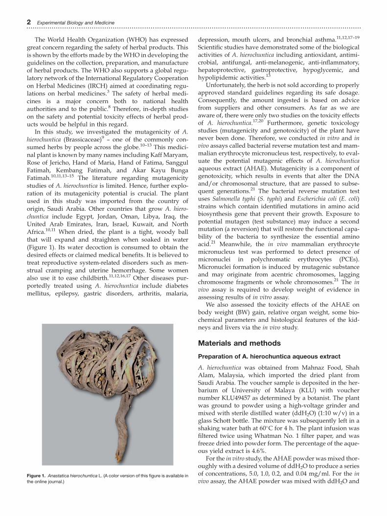
In this study, we investigated the mutagenicity of A.hierochuntica (Brassicaceae)9 – one of the commonly con-sumed herbs by people across the globe.10–13 This medici-nal plant is known bymany names including KaffMaryam,Rose of Jericho, Hand of Maria, Hand of Fatima, SanggulFatimah, Kembang Fatimah, and Akar Kayu BungaFatimah.10,11,13–15 The literature regarding mutagenicitystudies of A. hierochuntica is limited. Hence, further explo-ration of its mutagenicity potential is crucial. The plantused in this study was imported from the country oforigin, Saudi Arabia. Other countries that grow A. hiero-chuntica include Egypt, Jordan, Oman, Libya, Iraq, theUnited Arab Emirates, Iran, Israel, Kuwait, and NorthAfrica.10,11 When dried, the plant is a tight, woody ballthat will expand and straighten when soaked in water(Figure 1). Its water decoction is consumed to obtain thedesired effects or claimed medical benefits. It is believed totreat reproductive system-related disorders such as men-strual cramping and uterine hemorrhage. Some womenalso use it to ease childbirth.11,12,16,17 Other diseases pur-portedly treated using A. hierochuntica include diabetesmellitus, epilepsy, gastric disorders, arthritis, malaria,
depression, mouth ulcers, and bronchial asthma.11,12,17–19
Scientific studies have demonstrated some of the biologicalactivities of A. hierochuntica including antioxidant, antimi-crobial, antifungal, anti-melanogenic, anti-inflammatory,hepatoprotective, gastroprotective, hypoglycemic, andhypolipidemic activities.13
Unfortunately, the herb is not sold according to properlyapproved standard guidelines regarding its safe dosage.Consequently, the amount ingested is based on advicefrom suppliers and other consumers. As far as we areaware of, there were only two studies on the toxicity effectsof A. hierochuntica.17,20 Furthermore, genetic toxicologystudies (mutagenicity and genotoxicity) of the plant havenever been done. Therefore, we conducted in vitro and invivo assays called bacterial reverse mutation test and mam-malian erythrocyte micronucleus test, respectively, to eval-uate the potential mutagenic effects of A. hierochunticaaqueous extract (AHAE). Mutagenicity is a component ofgenotoxicity, which results in events that alter the DNAand/or chromosomal structure, that are passed to subse-quent generations.21 The bacterial reverse mutation testuses Salmonella typhi (S. typhi) and Escherichia coli (E. coli)strains which contain identified mutations in amino acidbiosynthesis gene that prevent their growth. Exposure topotential mutagen (test substance) may induce a secondmutation (a reversion) that will restore the functional capa-bility of the bacteria to synthesize the essential aminoacid.21 Meanwhile, the in vivo mammalian erythrocytemicronucleus test was performed to detect presence ofmicronuclei in polychromatic erythrocytes (PCEs).Micronuclei formation is induced by mutagenic substanceand may originate from acentric chromosomes, laggingchromosome fragments or whole chromosomes.21 The invivo assay is required to develop weight of evidence inassessing results of in vitro assay.
We also assessed the toxicity effects of the AHAE onbody weight (BW) gain, relative organ weight, some bio-chemical parameters and histological features of the kid-neys and livers via the in vivo study.
Materials and methods
Preparation of A. hierochuntica aqueous extract
A. hierochuntica was obtained from Mahnaz Food, ShahAlam, Malaysia, which imported the dried plant fromSaudi Arabia. The voucher sample is deposited in the her-barium of University of Malaya (KLU) with vouchernumber KLU49457 as determined by a botanist. The plantwas ground to powder using a high-voltage grinder andmixed with sterile distilled water (ddH2O) (1:10 w/v) in aglass Schott bottle. The mixture was subsequently left in ashaking water bath at 60�C for 4 h. The plant infusion wasfiltered twice using Whatman No. 1 filter paper, and wasfreeze dried into powder form. The percentage of the aque-ous yield extract is 4.6%.
For the in vitro study, the AHAE powder wasmixed thor-oughly with a desired volume of ddH2O to produce a seriesof concentrations, 5.0, 1.0, 0.2, and 0.04 mg/ml. For the invivo assay, the AHAE powder was mixed with ddH2O and
Figure 1. Anastatica hierochuntica L. (A color version of this figure is available in
the online journal.)
2 Experimental Biology and Medicine...............................................................................................................................................................
doses of 1000 and 2000mg/kgwere used for treatment. Theconcentrations for in vitro and doses for in vivo assays weredetermined by the results of preliminary studies.
In vitro Ames test
Bacteria strains and mutagenicity assay
The in vitro Ames test was conducted according toOrganization for Economic Co-operation andDevelopment (OECD) guidelines no. 471.22 The study pro-posal was approved by the Institutional Biosafety andBiosecurity Committee (IBBC), University of Malaya. Itinvolved four different strains of S. typhi, TA 100, TA 98,TA 97a and TA, 1535 and one strain of E. coli calledWP2. Allbacterial strains were purchased from Environmental Bio-detection Product Inc. (EBPI), Canada. One day prior to theday of assay, one vial of growth media was thoroughlymixed with one vial of bacterial strain followed by incuba-tion in 37�C incubator for 16 to 18 h.
Chemicals and reagents
The in vitroAmes test or so-called bacterial reverse mutationassay was performed using Muta-Chromo plate assay, pur-chased from Environmental Bio-Detection Products (EBPI,Canada) according to the manufacturer’s protocol. Theplate assay consists of five strains of bacteria, nutrientbroth (growth media), S-9 components, and standard muta-gens. Other reagents are components of ‘reaction mixture’that consist of Davis Mingioli salts (A), D-glucose (B), bro-mocresol purple (C), D-biotin (D), and L-histidine (for S.typhi) or L-tryptophan (for E. coli) (E). Theywere mixed thor-oughly with proper volume [A (43.24ml)þB (9.50ml)þC(4.76ml)þD (2.38ml)þE (0.12ml)] according to the suppli-er guideline in order to produce 60ml of reaction mixture.The S-9 components are magnesium chloride (MgCl2)þpotassium chloride (KCl), glucose-6-phosphate (S9B), nico-tine amide di-nucleotide phosphate (NADP), phosphatebuffer (pH 7.4) (S9D), ddH2O (S9E), and rat-liver extract(S9F). Components S9A to S9F were mixed thoroughly[S9A (0.40ml)þ S9B (0.09ml)þ S9C (0.81ml)þ S9D(9.98ml)þ S9E (8.47ml)þ S9F (0.25ml)] according to thesupplier guideline to produce 20ml of S-9 mixture.
The standard mutagen used for all bacteria strains in thein vitro assays with metabolic activity was 2-AminoAnthracene (2 AA). For the assays without metabolic activ-ity, sodium azide (SA) was the standard mutagen used forTA, 1535, and TA 100 bacteria strains. Meanwhile,Nitrofluorene (NF), 9- Amino Acridine (9 AA) and 4- nitro-quinolone (4 NQ) were the standard mutagens for TA 98,TA 97a, and WP2 bacteria strains, respectively.
Reverse mutation assay (muta-chromo plateassay)
Muta-chromo plate assay without S-9 (withoutmetabolic activation)
In the assay without metabolic activation, 2.5ml of the“reaction mixture” were dispensed into each sterile tube.
Then, a proportion of AHAE with concentration of (5.0, 1.0,0.2, and 0.04 mg/ml), standard mutagen and sterile waterwere added to a total volume of 17.5ml into each respectivetube. No S-9 mixture is required for this assay.
Muta-chromo plate assay with S-9 (with metabolicactivation)
For the assay with S-9 activation, 2.5ml of the “reactionmixture” were dispensed into each sterile tube followedby 2.0ml of S-9 mixture. After that, AHAE with concentra-tion of (5.0, 1.0, 0.2, and 0.04 mg/ml), standard mutagenand sterile water were added to a total volume of 15.5mlinto each respective tube. The total volume of the final solu-tion then became 17.5 ml. The mixture was mixed thor-oughly. Then, 5 ml of grown bacterial strains were addedand mixed with the prepared solution in each sterile tubeexcept blank tubes of both assays, with and without S-9.Subsequently, 200 ml final solutions were added into eachwell of a 96-well microtiter plate using a multichannelpipette. All procedures were conducted under a biosafetycabinet to assure aseptic conditions. Finally, the plates wereincubated in a 37�C incubator for five days.
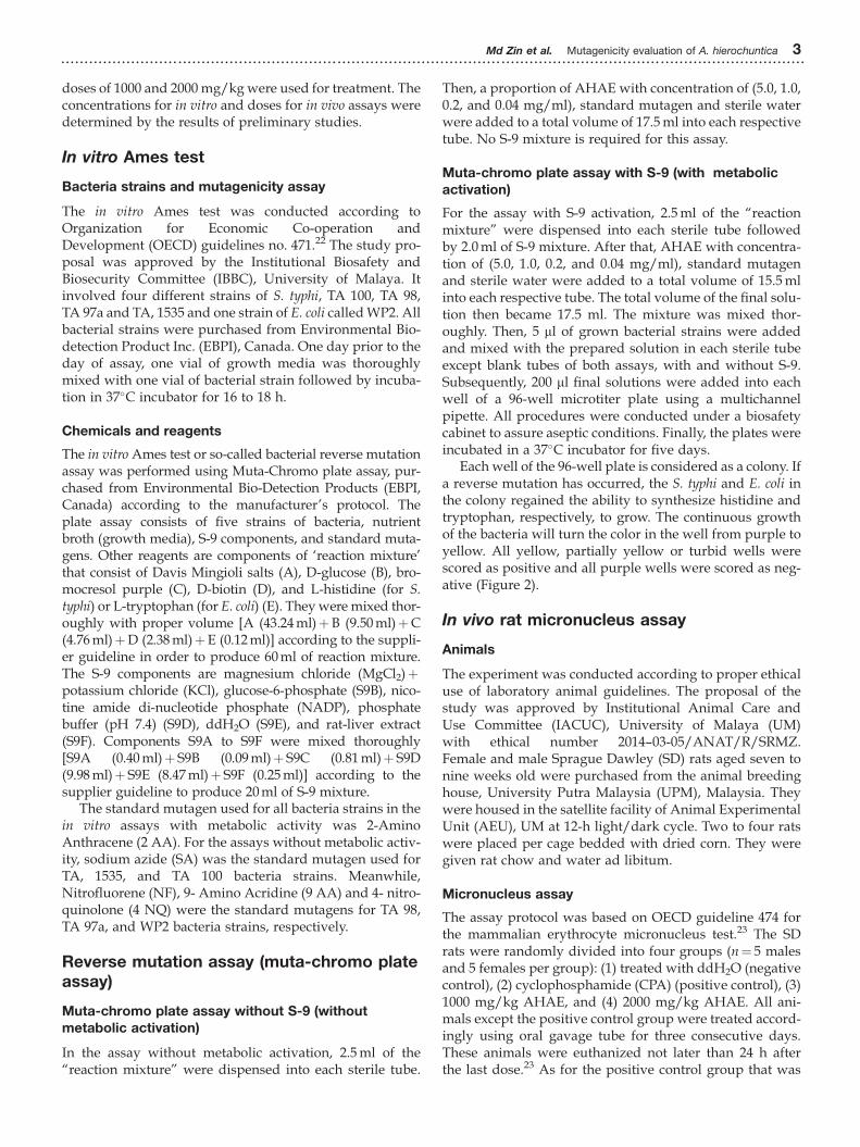
Each well of the 96-well plate is considered as a colony. Ifa reverse mutation has occurred, the S. typhi and E. coli inthe colony regained the ability to synthesize histidine andtryptophan, respectively, to grow. The continuous growthof the bacteria will turn the color in the well from purple toyellow. All yellow, partially yellow or turbid wells werescored as positive and all purple wells were scored as neg-ative (Figure 2).
In vivo rat micronucleus assay
Animals
The experiment was conducted according to proper ethicaluse of laboratory animal guidelines. The proposal of thestudy was approved by Institutional Animal Care andUse Committee (IACUC), University of Malaya (UM)with ethical number 2014–03-05/ANAT/R/SRMZ.Female and male Sprague Dawley (SD) rats aged seven tonine weeks old were purchased from the animal breedinghouse, University Putra Malaysia (UPM), Malaysia. Theywere housed in the satellite facility of Animal ExperimentalUnit (AEU), UM at 12-h light/dark cycle. Two to four ratswere placed per cage bedded with dried corn. They weregiven rat chow and water ad libitum.
Micronucleus assay
The assay protocol was based on OECD guideline 474 forthe mammalian erythrocyte micronucleus test.23 The SDrats were randomly divided into four groups (n¼ 5 malesand 5 females per group): (1) treated with ddH2O (negativecontrol), (2) cyclophosphamide (CPA) (positive control), (3)1000 mg/kg AHAE, and (4) 2000 mg/kg AHAE. All ani-mals except the positive control group were treated accord-ingly using oral gavage tube for three consecutive days.These animals were euthanized not later than 24 h afterthe last dose.23 As for the positive control group that was
Md Zin et al. Mutagenicity evaluation of A. hierochuntica 3...............................................................................................................................................................
treated with intraperitoneal (IP) injection of 50 mg/kg CPA(CAS 6055–19-2, Merck Millipore, US), these animals wereeuthanized 24 h after the injection,23 using carbon dioxide(CO2) chamber.
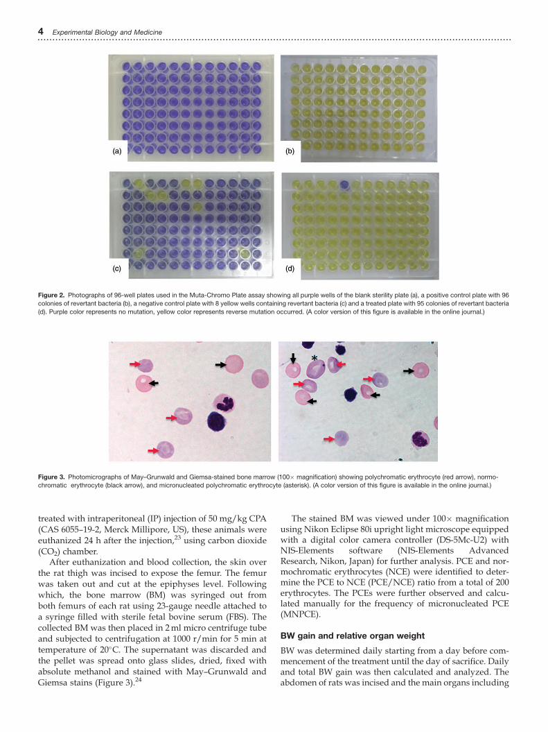
After euthanization and blood collection, the skin overthe rat thigh was incised to expose the femur. The femurwas taken out and cut at the epiphyses level. Followingwhich, the bone marrow (BM) was syringed out fromboth femurs of each rat using 23-gauge needle attached toa syringe filled with sterile fetal bovine serum (FBS). Thecollected BM was then placed in 2ml micro centrifuge tubeand subjected to centrifugation at 1000 r/min for 5 min attemperature of 20�C. The supernatant was discarded andthe pellet was spread onto glass slides, dried, fixed withabsolute methanol and stained with May–Grunwald andGiemsa stains (Figure 3).24
The stained BM was viewed under 100� magnificationusing Nikon Eclipse 80i upright light microscope equippedwith a digital color camera controller (DS-5Mc-U2) withNIS-Elements software (NIS-Elements AdvancedResearch, Nikon, Japan) for further analysis. PCE and nor-mochromatic erythrocytes (NCE) were identified to deter-mine the PCE to NCE (PCE/NCE) ratio from a total of 200erythrocytes. The PCEs were further observed and calcu-lated manually for the frequency of micronucleated PCE(MNPCE).
BW gain and relative organ weight
BW was determined daily starting from a day before com-mencement of the treatment until the day of sacrifice. Dailyand total BW gain was then calculated and analyzed. Theabdomen of rats was incised and the main organs including
Figure 2. Photographs of 96-well plates used in the Muta-Chromo Plate assay showing all purple wells of the blank sterility plate (a), a positive control plate with 96
colonies of revertant bacteria (b), a negative control plate with 8 yellow wells containing revertant bacteria (c) and a treated plate with 95 colonies of revertant bacteria
(d). Purple color represents no mutation, yellow color represents reverse mutation occurred. (A color version of this figure is available in the online journal.)
Figure 3. Photomicrographs of May–Grunwald and Giemsa-stained bone marrow (100� magnification) showing polychromatic erythrocyte (red arrow), normo-
chromatic erythrocyte (black arrow), and micronucleated polychromatic erythrocyte (asterisk). (A color version of this figure is available in the online journal.)
4 Experimental Biology and Medicine...............................................................................................................................................................
the liver, kidney, spleen, heart, and lung were harvestedand weighed. The organ weight data were expressed rela-tive to the animal’s BW.
Serum biochemical analysis
After euthanization, blood sample was collected and sent toGribble Pathology laboratory, Petaling Jaya, Malaysia, forserum renal profile (RP) and protein level.
Histological analysis
Liver and kidney of rats were fixed with 10% formalin.After that, the tissue samples were processed with seriesof alcohol (Thermo Fisher Scientific, USA) and xylene(BDH, England) and then embedded in paraffin wax(Paraplast, USA). Paraffin blocks were sectioned at 5 mmthickness placed on glass slides, incubated and stainedwith hematoxylin and eosin (H&E). The slides wereviewed under 100� magnification using Nikon Eclipse80i upright light microscope (Nikon, Japan), equippedwith a digital color camera controller (DS-5Mc-U2) andNIS-Elements software for histological analysis.
Statistical analysis
The muta-chromo plate kit was used, and it utilized thesame principle of an Ames test which compares the rateof spontaneous reverse mutation in the backgroundgroup to the rate of reverse mutation within a sampleassay. The statistical significance of the data was deter-mined using the table for analysis of fluctuation tests(Table S1) provided by the manufacturer (EBPI, Canada).25
The BW gain, serum biochemical parameters, percent-age of MNPCE frequency, and statistical comparisons ofPCE/NCE ratio were analyzed using univariate analysisof variance (SPSS, version 22.0). The results were consid-ered as statistically significant when the difference has Pvalue of less than 0.05 (P <0.05).
Results
In vitro reverse mutation assay
A significant increase in the number of revertants wasobserved in the presence of two AA as the positive refer-ence (standard mutagen) in all bacteria strains except forTA 97a with S-9 (with metabolic activation). There was alsoa significant increase in the number of revertants in all theS. typhi strains tested with 0.2 mg/ml AHAE with metabol-ic activation. As for the WP2 E. coli tested with the sameAHAE concentration, the number of revertants was notedto be increased in well plates with and without S-9.Exposure to the lowest concentration, 0.04 mg/ml resultedin increased revertants in WP2 E. coli and only two S. typhistrains namely TA, 1535, and TA 97a with presence of met-abolic activation. Absence of revertant bacteria wasobserved in most of the well plates containing higher con-centrations (1.0 and 5.0 mg/ml) of AHAE (Table 1).
In vivo micronucleus assay
Polychromatic and normochromatic erythrocytes ratio
The PCE/NCE ratio of female and male SD rats treatedwith 2000 mg/kg AHAE showed significant increase(P< 0.05) compared with the control group. In contrast,decreased PCE/NCE ratio was observed in positive controlgroup in both genders. Although the result was not statis-tically significant (Tables 2 and 3), the trend suggests CPAas an effective positive control as shown by lowest PCE/NCE ratio in CPA-treated group compared with the othergroups.
Percentage of MNPCE frequency in 2000 PCE
In the male rats, the percentage of MNPCE frequency in2000 PCE in the positive control group was significantlyincreased compared with the negative control group andother groups. The same trend was also observed in theCPA-treated female rats but the results were not
Table 1. Reverse mutation assay results for A. hierochuntica aqueous extract in four strains of S. typhi and one strain of E. coli.
Groups
Colony of revertants per plate
S. typhi
TA 100
S. typhi
TA 1535
S. typhi
TA 98
S. typhi
TA 97a
E. coli
WP2
2S9 1S9 2S9 1S9 2S9 1S9 2S9 1S9 2S9 1S9
Blanka 0 0 0 0 0 0 0 0 0 0
�ve controlb 15 75 2 8 6 22 7 80 59 52
AHAEc 5.0 mg/ml 0 1 0 0 0 0 0 0 0 0
AHAEc 1.0 mg/ml 12 0 0 0 0 0 2 1 0 0
AHAEc 0.2 mg/ml 18 95** 0 41** 10 96** 0 96** 74* 70**
AHAEc 0.04 mg/ml 5 81 1 75** 5 27 1 88* 66 66*
þve controlsd 37** 90** 94** 45** 91** 96** 96** 84 95** 82**
AHAE: A. hierochuntica aqueous extract.aBlank: Sterility check.bNegative control: Sterile distilled water.cAnastatica hierochuntica aqueous extract (mg/ml).
�S9: without metabolic activator rat liver extract. þS9: with metabolic activator rat liver extract.dPositive controls: 2- Amino anthracene (all bacterial strains; þS9); Sodium azide (TA 100 and TA 1535; �S9); Nitrofluorene (TA 98; �S9); 9- Amino acridine (TA 97a;
�S9) and 4-Nitroquinolone (WP2; �S9). **P <0.001,*P<0.05.
Md Zin et al. Mutagenicity evaluation of A. hierochuntica 5...............................................................................................................................................................
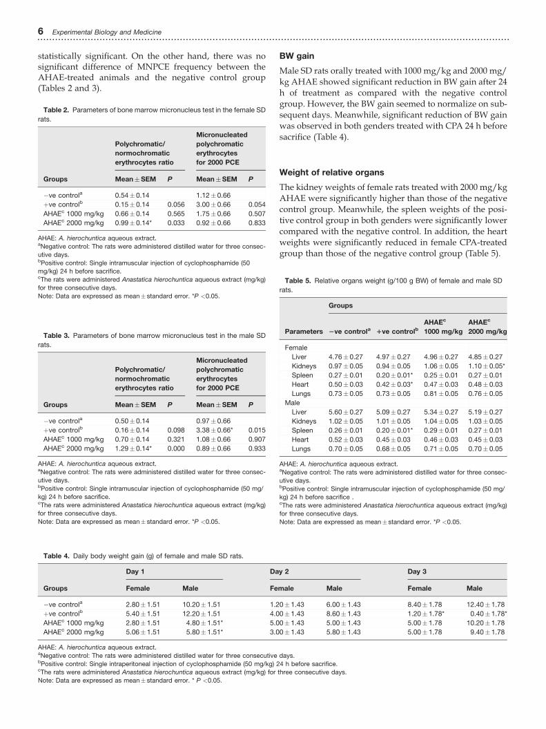
statistically significant. On the other hand, there was nosignificant difference of MNPCE frequency between theAHAE-treated animals and the negative control group(Tables 2 and 3).
BW gain
Male SD rats orally treated with 1000 mg/kg and 2000 mg/kg AHAE showed significant reduction in BW gain after 24h of treatment as compared with the negative controlgroup. However, the BW gain seemed to normalize on sub-sequent days. Meanwhile, significant reduction of BW gainwas observed in both genders treated with CPA 24 h beforesacrifice (Table 4).
Weight of relative organs
The kidney weights of female rats treated with 2000 mg/kgAHAE were significantly higher than those of the negativecontrol group. Meanwhile, the spleen weights of the posi-tive control group in both genders were significantly lowercompared with the negative control. In addition, the heartweights were significantly reduced in female CPA-treatedgroup than those of the negative control group (Table 5).
Table 2. Parameters of bone marrow micronucleus test in the female SD
rats.
Groups
Polychromatic/
normochromatic
erythrocytes ratio
Micronucleated
polychromatic
erythrocytes
for 2000 PCE
Mean�SEM P Mean�SEM P
�ve controla 0.54� 0.14 1.12� 0.66
þve controlb 0.15� 0.14 0.056 3.00� 0.66 0.054
AHAEc 1000 mg/kg 0.66� 0.14 0.565 1.75� 0.66 0.507
AHAEc 2000 mg/kg 0.99� 0.14* 0.033 0.92� 0.66 0.833
AHAE: A. hierochuntica aqueous extract.aNegative control: The rats were administered distilled water for three consec-
utive days.bPositive control: Single intramuscular injection of cyclophosphamide (50
mg/kg) 24 h before sacrifice.cThe rats were administered Anastatica hierochuntica aqueous extract (mg/kg)
for three consecutive days.
Note: Data are expressed as mean� standard error. *P <0.05.
Table 3. Parameters of bone marrow micronucleus test in the male SD
rats.
Groups
Polychromatic/
normochromatic
erythrocytes ratio
Micronucleated
polychromatic
erythrocytes
for 2000 PCE
Mean�SEM P Mean�SEM P
�ve controla 0.50� 0.14 0.97� 0.66
þve controlb 0.16� 0.14 0.098 3.38� 0.66* 0.015
AHAEc 1000 mg/kg 0.70� 0.14 0.321 1.08� 0.66 0.907
AHAEc 2000 mg/kg 1.29� 0.14* 0.000 0.89� 0.66 0.933
AHAE: A. hierochuntica aqueous extract.aNegative control: The rats were administered distilled water for three consec-
utive days.bPositive control: Single intramuscular injection of cyclophosphamide (50 mg/
kg) 24 h before sacrifice.cThe rats were administered Anastatica hierochuntica aqueous extract (mg/kg)
for three consecutive days.
Note: Data are expressed as mean� standard error. *P <0.05.
Table 4. Daily body weight gain (g) of female and male SD rats.
Groups
Day 1 Day 2 Day 3
Female Male Female Male Female Male
�ve controla 2.80� 1.51 10.20� 1.51 1.20� 1.43 6.00� 1.43 8.40� 1.78 12.40� 1.78
þve controlb 5.40� 1.51 12.20� 1.51 4.00� 1.43 8.60� 1.43 1.20� 1.78* 0.40� 1.78*
AHAEc 1000 mg/kg 2.80� 1.51 4.80� 1.51* 5.00� 1.43 5.00� 1.43 5.00� 1.78 10.20� 1.78
AHAEc 2000 mg/kg 5.06� 1.51 5.80� 1.51* 3.00� 1.43 5.80� 1.43 5.00� 1.78 9.40� 1.78
AHAE: A. hierochuntica aqueous extract.aNegative control: The rats were administered distilled water for three consecutive days.bPositive control: Single intraperitoneal injection of cyclophosphamide (50 mg/kg) 24 h before sacrifice.cThe rats were administered Anastatica hierochuntica aqueous extract (mg/kg) for three consecutive days.
Note: Data are expressed as mean� standard error. * P <0.05.
Table 5. Relative organs weight (g/100 g BW) of female and male SD
rats.
Parameters
Groups
2ve controla 1ve controlbAHAEc
1000 mg/kg
AHAEc
2000 mg/kg
Female
Liver 4.76� 0.27 4.97� 0.27 4.96� 0.27 4.85� 0.27
Kidneys 0.97� 0.05 0.94� 0.05 1.06� 0.05 1.10� 0.05*
Spleen 0.27� 0.01 0.20� 0.01* 0.25� 0.01 0.27� 0.01
Heart 0.50� 0.03 0.42� 0.03* 0.47� 0.03 0.48� 0.03
Lungs 0.73� 0.05 0.73� 0.05 0.81� 0.05 0.76� 0.05
Male
Liver 5.60� 0.27 5.09� 0.27 5.34� 0.27 5.19� 0.27
Kidneys 1.02� 0.05 1.01� 0.05 1.04� 0.05 1.03� 0.05
Spleen 0.26� 0.01 0.20� 0.01* 0.29� 0.01 0.27� 0.01
Heart 0.52� 0.03 0.45� 0.03 0.46� 0.03 0.45� 0.03
Lungs 0.70� 0.05 0.68� 0.05 0.71� 0.05 0.70� 0.05
AHAE: A. hierochuntica aqueous extract.aNegative control: The rats were administered distilled water for three consec-
utive days.bPositive control: Single intramuscular injection of cyclophosphamide (50 mg/
kg) 24 h before sacrifice .cThe rats were administered Anastatica hierochuntica aqueous extract (mg/kg)
for three consecutive days.
Note: Data are expressed as mean� standard error. *P <0.05.
6 Experimental Biology and Medicine...............................................................................................................................................................
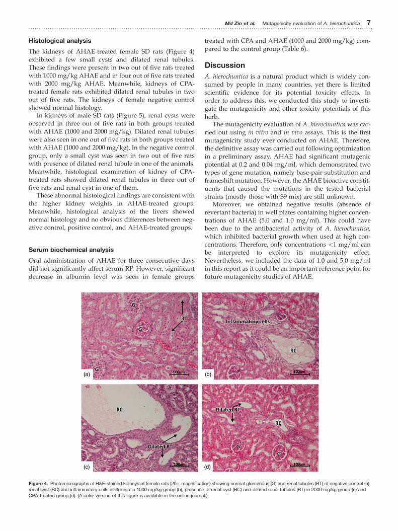
Histological analysis
The kidneys of AHAE-treated female SD rats (Figure 4)exhibited a few small cysts and dilated renal tubules.These findings were present in two out of five rats treatedwith 1000 mg/kg AHAE and in four out of five rats treatedwith 2000 mg/kg AHAE. Meanwhile, kidneys of CPA-treated female rats exhibited dilated renal tubules in twoout of five rats. The kidneys of female negative controlshowed normal histology.
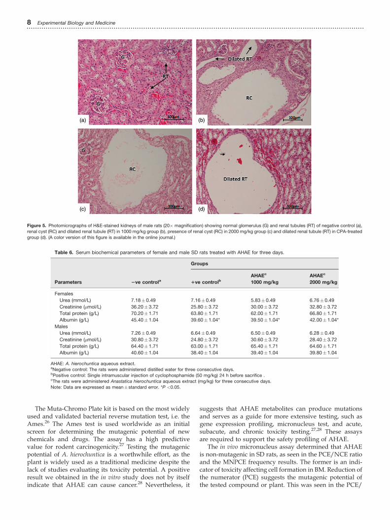
In kidneys of male SD rats (Figure 5), renal cysts wereobserved in three out of five rats in both groups treatedwith AHAE (1000 and 2000 mg/kg). Dilated renal tubuleswere also seen in one out of five rats in both groups treatedwith AHAE (1000 and 2000 mg/kg). In the negative controlgroup, only a small cyst was seen in two out of five ratswith presence of dilated renal tubule in one of the animals.Meanwhile, histological examination of kidney of CPA-treated rats showed dilated renal tubules in three out offive rats and renal cyst in one of them.
These abnormal histological findings are consistent withthe higher kidney weights in AHAE-treated groups.Meanwhile, histological analysis of the livers showednormal histology and no obvious differences between neg-ative control, positive control, and AHAE-treated groups.
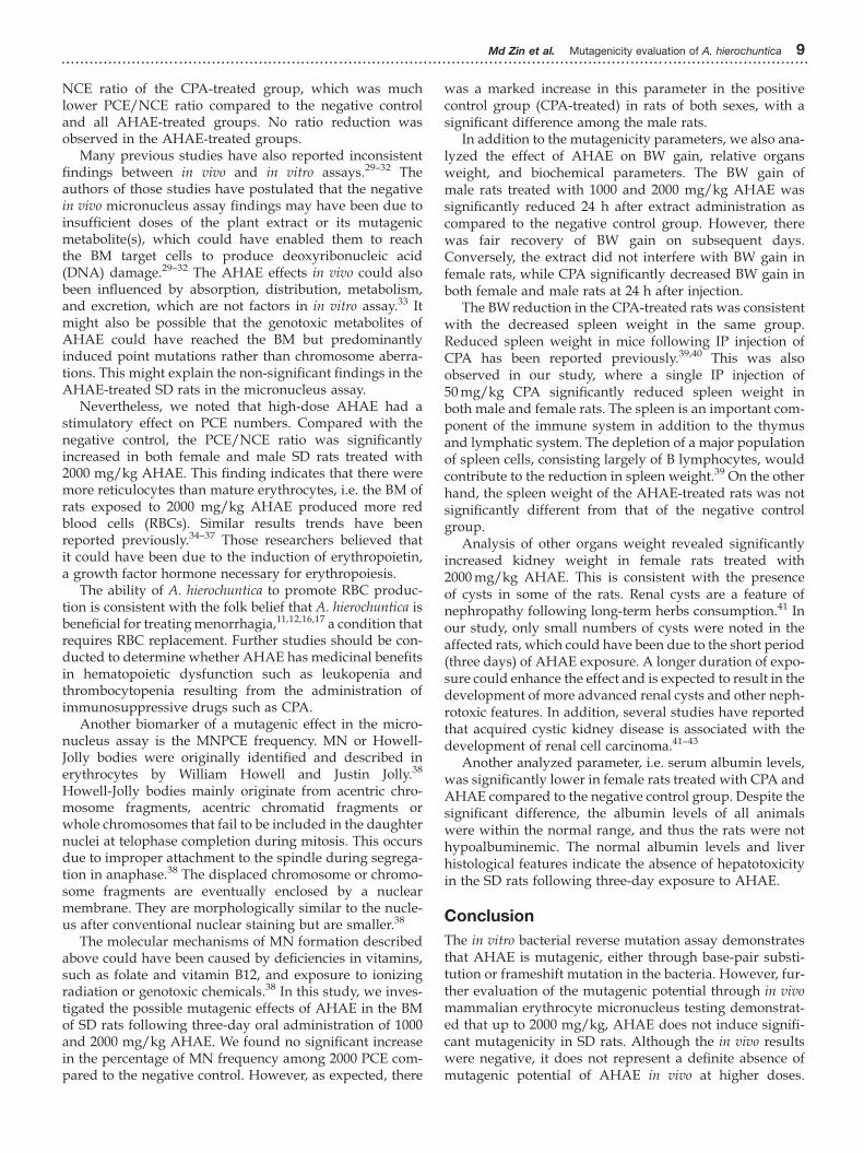
Serum biochemical analysis
Oral administration of AHAE for three consecutive daysdid not significantly affect serum RP. However, significantdecrease in albumin level was seen in female groups
treated with CPA and AHAE (1000 and 2000 mg/kg) com-pared to the control group (Table 6).
Discussion
A. hierochuntica is a natural product which is widely con-sumed by people in many countries, yet there is limitedscientific evidence for its potential toxicity effects. Inorder to address this, we conducted this study to investi-gate the mutagenicity and other toxicity potentials of thisherb.
The mutagenicity evaluation of A. hierochuntica was car-ried out using in vitro and in vivo assays. This is the firstmutagenicity study ever conducted on AHAE. Therefore,the definitive assay was carried out following optimizationin a preliminary assay. AHAE had significant mutagenicpotential at 0.2 and 0.04 mg/ml, which demonstrated twotypes of gene mutation, namely base-pair substitution andframeshift mutation. However, the AHAE bioactive constit-uents that caused the mutations in the tested bacterialstrains (mostly those with S9 mix) are still unknown.
Moreover, we obtained negative results (absence ofrevertant bacteria) in well plates containing higher concen-trations of AHAE (5.0 and 1.0 mg/ml). This could havebeen due to the antibacterial activity of A. hierochuntica,which inhibited bacterial growth when used at high con-centrations. Therefore, only concentrations <1 mg/ml canbe interpreted to explore its mutagenicity effect.Nevertheless, we included the data of 1.0 and 5.0 mg/mlin this report as it could be an important reference point forfuture mutagenicity studies of AHAE.
Figure 4. Photomicrographs of H&E-stained kidneys of female rats (20� magnification) showing normal glomerulus (G) and renal tubules (RT) of negative control (a),
renal cyst (RC) and inflammatory cells infiltration in 1000 mg/kg group (b), presence of renal cyst (RC) and dilated renal tubules (RT) in 2000 mg/kg group (c) and
CPA-treated group (d). (A color version of this figure is available in the online journal.)
Md Zin et al. Mutagenicity evaluation of A. hierochuntica 7...............................................................................................................................................................
The Muta-Chromo Plate kit is based on the most widelyused and validated bacterial reverse mutation test, i.e. theAmes.26 The Ames test is used worldwide as an initialscreen for determining the mutagenic potential of newchemicals and drugs. The assay has a high predictivevalue for rodent carcinogenicity.27 Testing the mutagenicpotential of A. hierochuntica is a worthwhile effort, as theplant is widely used as a traditional medicine despite thelack of studies evaluating its toxicity potential. A positiveresult we obtained in the in vitro study does not by itselfindicate that AHAE can cause cancer.28 Nevertheless, it
suggests that AHAE metabolites can produce mutationsand serves as a guide for more extensive testing, such asgene expression profiling, micronucleus test, and acute,subacute, and chronic toxicity testing.27,28 These assaysare required to support the safety profiling of AHAE.
The in vivo micronucleus assay determined that AHAEis non-mutagenic in SD rats, as seen in the PCE/NCE ratioand the MNPCE frequency results. The former is an indi-cator of toxicity affecting cell formation in BM. Reduction ofthe numerator (PCE) suggests the mutagenic potential ofthe tested compound or plant. This was seen in the PCE/
Figure 5. Photomicrographs of H&E-stained kidneys of male rats (20� magnification) showing normal glomerulus (G) and renal tubules (RT) of negative control (a),
renal cyst (RC) and dilated renal tubule (RT) in 1000 mg/kg group (b), presence of renal cyst (RC) in 2000 mg/kg group (c) and dilated renal tubule (RT) in CPA-treated
group (d). (A color version of this figure is available in the online journal.)
Table 6. Serum biochemical parameters of female and male SD rats treated with AHAE for three days.
Parameters
Groups
2ve controla 1ve controlbAHAEc
1000 mg/kg
AHAEc
2000 mg/kg
Females
Urea (mmol/L) 7.18� 0.49 7.16� 0.49 5.83� 0.49 6.76� 0.49
Creatinine (lmol/L) 36.20� 3.72 25.80� 3.72 30.00� 3.72 32.80� 3.72
Total protein (g/L) 70.20� 1.71 63.80� 1.71 62.00� 1.71 66.80� 1.71
Albumin (g/L) 45.40� 1.04 39.60� 1.04* 39.50� 1.04* 42.00� 1.04*
Males
Urea (mmol/L) 7.26� 0.49 6.64� 0.49 6.50� 0.49 6.28� 0.49
Creatinine (lmol/L) 30.80� 3.72 24.80� 3.72 30.60� 3.72 28.40� 3.72
Total protein (g/L) 64.40� 1.71 63.00� 1.71 65.40� 1.71 64.60� 1.71
Albumin (g/L) 40.60� 1.04 38.40� 1.04 39.40� 1.04 39.80� 1.04
AHAE: A. hierochuntica aqueous extract.aNegative control: The rats were administered distilled water for three consecutive days.bPositive control: Single intramuscular injection of cyclophosphamide (50 mg/kg) 24 h before sacrifice .cThe rats were administered Anastatica hierochuntica aqueous extract (mg/kg) for three consecutive days.
Note: Data are expressed as mean� standard error. *P <0.05.
8 Experimental Biology and Medicine...............................................................................................................................................................
NCE ratio of the CPA-treated group, which was muchlower PCE/NCE ratio compared to the negative controland all AHAE-treated groups. No ratio reduction wasobserved in the AHAE-treated groups.
Many previous studies have also reported inconsistentfindings between in vivo and in vitro assays.29–32 Theauthors of those studies have postulated that the negativein vivo micronucleus assay findings may have been due toinsufficient doses of the plant extract or its mutagenicmetabolite(s), which could have enabled them to reachthe BM target cells to produce deoxyribonucleic acid(DNA) damage.29–32 The AHAE effects in vivo could alsobeen influenced by absorption, distribution, metabolism,and excretion, which are not factors in in vitro assay.33 Itmight also be possible that the genotoxic metabolites ofAHAE could have reached the BM but predominantlyinduced point mutations rather than chromosome aberra-tions. This might explain the non-significant findings in theAHAE-treated SD rats in the micronucleus assay.
Nevertheless, we noted that high-dose AHAE had astimulatory effect on PCE numbers. Compared with thenegative control, the PCE/NCE ratio was significantlyincreased in both female and male SD rats treated with2000 mg/kg AHAE. This finding indicates that there weremore reticulocytes than mature erythrocytes, i.e. the BM ofrats exposed to 2000 mg/kg AHAE produced more redblood cells (RBCs). Similar results trends have beenreported previously.34–37 Those researchers believed thatit could have been due to the induction of erythropoietin,a growth factor hormone necessary for erythropoiesis.
The ability of A. hierochuntica to promote RBC produc-tion is consistent with the folk belief that A. hierochuntica isbeneficial for treatingmenorrhagia,11,12,16,17 a condition thatrequires RBC replacement. Further studies should be con-ducted to determine whether AHAE has medicinal benefitsin hematopoietic dysfunction such as leukopenia andthrombocytopenia resulting from the administration ofimmunosuppressive drugs such as CPA.
Another biomarker of a mutagenic effect in the micro-nucleus assay is the MNPCE frequency. MN or Howell-Jolly bodies were originally identified and described inerythrocytes by William Howell and Justin Jolly.38
Howell-Jolly bodies mainly originate from acentric chro-mosome fragments, acentric chromatid fragments orwhole chromosomes that fail to be included in the daughternuclei at telophase completion during mitosis. This occursdue to improper attachment to the spindle during segrega-tion in anaphase.38 The displaced chromosome or chromo-some fragments are eventually enclosed by a nuclearmembrane. They are morphologically similar to the nucle-us after conventional nuclear staining but are smaller.38
The molecular mechanisms of MN formation describedabove could have been caused by deficiencies in vitamins,such as folate and vitamin B12, and exposure to ionizingradiation or genotoxic chemicals.38 In this study, we inves-tigated the possible mutagenic effects of AHAE in the BMof SD rats following three-day oral administration of 1000and 2000 mg/kg AHAE. We found no significant increasein the percentage of MN frequency among 2000 PCE com-pared to the negative control. However, as expected, there
was a marked increase in this parameter in the positivecontrol group (CPA-treated) in rats of both sexes, with asignificant difference among the male rats.
In addition to the mutagenicity parameters, we also ana-lyzed the effect of AHAE on BW gain, relative organsweight, and biochemical parameters. The BW gain ofmale rats treated with 1000 and 2000 mg/kg AHAE wassignificantly reduced 24 h after extract administration ascompared to the negative control group. However, therewas fair recovery of BW gain on subsequent days.Conversely, the extract did not interfere with BW gain infemale rats, while CPA significantly decreased BW gain inboth female and male rats at 24 h after injection.
The BWreduction in the CPA-treated rats was consistentwith the decreased spleen weight in the same group.Reduced spleen weight in mice following IP injection ofCPA has been reported previously.39,40 This was alsoobserved in our study, where a single IP injection of50mg/kg CPA significantly reduced spleen weight inboth male and female rats. The spleen is an important com-ponent of the immune system in addition to the thymusand lymphatic system. The depletion of a major populationof spleen cells, consisting largely of B lymphocytes, wouldcontribute to the reduction in spleen weight.39 On the otherhand, the spleen weight of the AHAE-treated rats was notsignificantly different from that of the negative controlgroup.
Analysis of other organs weight revealed significantlyincreased kidney weight in female rats treated with2000mg/kg AHAE. This is consistent with the presenceof cysts in some of the rats. Renal cysts are a feature ofnephropathy following long-term herbs consumption.41 Inour study, only small numbers of cysts were noted in theaffected rats, which could have been due to the short period(three days) of AHAE exposure. A longer duration of expo-sure could enhance the effect and is expected to result in thedevelopment of more advanced renal cysts and other neph-rotoxic features. In addition, several studies have reportedthat acquired cystic kidney disease is associated with thedevelopment of renal cell carcinoma.41–43
Another analyzed parameter, i.e. serum albumin levels,was significantly lower in female rats treated with CPA andAHAE compared to the negative control group. Despite thesignificant difference, the albumin levels of all animalswere within the normal range, and thus the rats were nothypoalbuminemic. The normal albumin levels and liverhistological features indicate the absence of hepatotoxicityin the SD rats following three-day exposure to AHAE.
Conclusion
The in vitro bacterial reverse mutation assay demonstratesthat AHAE is mutagenic, either through base-pair substi-tution or frameshift mutation in the bacteria. However, fur-ther evaluation of the mutagenic potential through in vivomammalian erythrocyte micronucleus testing demonstrat-ed that up to 2000 mg/kg, AHAE does not induce signifi-cant mutagenicity in SD rats. Although the in vivo resultswere negative, it does not represent a definite absence ofmutagenic potential of AHAE in vivo at higher doses.
Md Zin et al. Mutagenicity evaluation of A. hierochuntica 9...............................................................................................................................................................
Further work is necessary to identify the genotoxic risk ofAHAE in humans. It may also be worthwhile to performacute and chronic toxicity studies to draw firm conclusionsregarding the safety of AHAE consumption.
Authors’ contributions:MAA, ZM and SRMZ designed the study.SRMZ conducted the experiments, analyzed data and draftedthe manuscript. MAA analyzed data and wrote parts of themanuscript. ZM, WFW and NMK wrote parts of the manu-script. The manuscript was reviewed by all authors.
FUNDING
The authors would like to thank the Bantuan Kecil Peruntukan(BK040–2013) grant of University of Malaya for funding thisresearch.
DECLARATION OF CONFLICTING INTERESTS
The authors declared no conflicts of interest with respect to theresearch, authorship, and/or publication of this article.
REFERENCES
1. Pan SY, Litscher G, Gao SH, Zhou SF, Yu ZL, Chen HQ, Zhang SF, Tang
MK, Sun JN, Ko KM. Historical perspective of traditional indigenous
medical practices: the current renaissance and conservation of herbal
resources. Evid-Based Compl Alt 2014;2014:1–202. GIA. Herbal Supplements and remedies – a global strategic business
report. San Jose, CA: Global Industry Analyst Inc., 2015.
3. WHO. WHO traditional medicine strategy:2014-2023. Geneva: World
Health Organization, 2013.
4. Ernst E, White A. The BBC survey of complementary medicine use in
the UK. Complement Ther Med 2000;8:32–6
5. Kamboj V. Herbal medicine. Curr Sci Bangalore 2000;78:35–86. Jantan I. The scientific values of Malaysian herbal products. J Sains
Kesihatan Malays 2006;4:59–707. Jha V. Herbal medicines and chronic kidney disease. Nephrology
2010;15:10–7
8. WHO. WHO guidelines on safety monitoring of herbal medicines in pharma-
covigilance systems. Geneva: World Health Organization, 2004.
9. Law KS, Soon LK, Mohsin SSS, Farid CG. Ultrastructural findings of
Anastatica hierochuntica L.,(Sanggul Fatimah) towards explaining its
medicinal properties. Ann Microsc 2009;9:50–610. Abdulfattah SY. Study of immunological effect of Anastatica hierochun-
tica (Kaff Maryam) plant methanolic extract on albino male mice. J
Biotechnol Res Cent 2013;7:3–1011. Daur I. Chemical properties of the medicinal herb Kaff Maryam
(Anastatica hierochuntica L.) and its relation to folk medicine use. Afr J
Microbiol Res 2012;6:5048–5112. Sooi LK, Keng SL. Herbal medicines: Malaysian women’s knowledge
and practice. Evid Based Compl Alt 2013;2013:1–1013. Zin SRM, Kassim NM, Alshawsh MA, Hashim NE, Mohamed Z.
Biological activities of Anastatica hierochuntica L.: a systematic review.
Biomed Pharmacother 2017;91:611–2014. El-Sayed M, El-Sherif F, Elhassaneen Y, El-Rahman AA. Potential ther-
apeutic effects of some Egyptian plant parts on hepatic toxicity induced
by carbon tetrachloride in rats. Life Sci J 2012;9:3747–5515. Saleh J, Machado L. Rose of Jericho: a word of caution. Oman Med J
2012;27:338
16. El-Ghazali GE, Al-Khalifa KS, Saleem GA, Abdallah EM. Traditional
medicinal plants indigenous to Al-Rass province, Saudi Arabia. J Med
Plants Res 2010;4:2680–317. Shah AH, Bhandari MP, Al-Harbi NO, Al-Ashban RM. Kaff-E-Maryam
(Anastatica hierochuntica L.): evaluation of gastro-protective activity and
toxicity in different experimental models. Biol Med 2014;6:1–10
18. Jaradat N. Ethnopharmacological survey of natural products in pales-
tine. An-Najah Univ J Res 2005a;19:13–6719. Jaradat NA. Medical plants utilized in Palestinian folk medicine for
treatment of diabetes mellitus and cardiac diseases. J Al-Aqsa Univ
2005b;9:1–28.
20. Rasheed RA, Bashir AK, Ali BH (eds). Fetal toxicity of Anastatica hier-
ochuntica L. in mice. Fed Am Soc Exp Biol 1997;11:A417
21. Genetic toxicology guidance document: second commenting round,
30 November 2015, http://www.oecd.org/env/ehs/testing/Draft%
20Guidance%20Document%20on%20OECD%20Genetic%
20Toxicology%20Test%20Guidelines.pdf (accessed 27 July 2017).
22. OECD. 471, bacterial reverse mutation test. OECD Guidelines for test-
ing of chemicals, OECD, Paris, France OECD (Organization for
Economic Cooperation and Development), 1997a
23. OECD. 474: mammalian erythrocyte micronucleus test. OECD
Guidelines for Testing of Chemicals, 1997b
24. Shahrim Z, Baharuddin P, Yahya NA, Muhammad H, Bakar RA, Ismail
Z. The in vivo rodent micronucleus assay of Kacip Fatimah (Labisia
pumila) extract. Trop Biomed 2006;23:214–9
25. Gilbert R. The analysis of fluctuation tests. Mutat Res 1980;74:283–926. Ames BN, McCann J, Yamasaki E. Methods for detecting carcinogens
and mutagens with the Salmonella/mammalian-microsome mutage-
nicity test. Mutat Res 1975;31:347–6327. Chang JB, Lu HF, Liao NC, Lee CS, Yeh MY, Liu CM, Chung MT, Kuan
AM, Lin JJ, Wu MF, Chung JG. Evaluation of genotoxicity and subclin-
ical toxicity of Agaricus blazei Murrill in the Ames test and in histo-
pathological and biochemical analysis. In Vivo 2012;26:437–45
28. Wu MF, Peng FC, Chen YL, Lee CS, Yang YY, Yeh MY, Liu CM, Chang
JB, Wu RSC, Yu CC, Lu HF, Chung JG. Evaluation of genotoxicity of
Antrodia cinnamomea in the Ames test and the in vitro chromosomal
aberration test. In Vivo 2011;25:419–23
29. Dearfield KL, Cimino MC, McCarroll NE, Mauer I, Valcovic LR.
Genotoxicity risk assessment: a proposed classification strategy.
Mutat Res 2002;521:121–3530. Herbold BA, Brendler-Schwaab SY, Ahr HJ. Ciprofloxacin: in vivo gen-
otoxicity studies. Mutat Res 2001;498:193–20531. Kirkland D, Zeiger E, Madia F, Gooderham N, Kasper P, Lynch A,
Morita T, Ouedraogo G, Parra Morte JM, Pfuhler S, Rogiers V, Schulz
M, Thybaud V, van Benthem J, Vanparys P, Worth A, Corvi R. Can in
vitro mammalian cell genotoxicity test results be used to complement
positive results in the Ames test and help predict carcinogenic or in vivo
genotoxic activity? I. Reports of individual databases presented at an
EURL ECVAM Workshop. Mutat Res 2014;775:55–6832. Mayer C, Klein RG, Wesch H, Schmezer P. Nickel subsulfide is geno-
toxic in vitro but shows no mutagenic potential in respiratory tract
tissues of BigBlueTM rats and MutaTM Mouse mice in vivo after inha-
lation. Mutat Res 1998;420:85–9833. HHS US, FDA, CDER, CBER. Guidance for Industry S2(R1)
Genotoxicity testing and data interpretation for pharmaceuticals
intended for human use. June 2012;2012:1–3134. Ezzi L, Salah IB, Haouas Z, Sakly A, Grissa I, Chakroun S, Kerkeni E,
Hassine M, Mehdi M, Cheikh BH. Histopathological and genotoxic
effects of chlorpyrifos in rats. Environ Sci Pollut Res 2016;23:4859–6735. Gandhi G, Chowdhury J, Sareen P, Dhillon V. Genotoxic effects of del-
tamethrin in the mouse bone marrow micronucleus assay. Mutat Res
Lett 1995;346:203–636. Suzuki Y, Nagae Y, Li J, Sakaba H, Mozawa K, Takahashi A, Shimizu H.
The micronucleus test and erythropoiesis. Effects of erythropoietin and
a mutagen on the ratio of polychromatic to normochromatic erythro-
cytes (P/N ratio). Mutagenesis 1989;4:420–437. Suzuki Y, Shimizu H, Nagae Y, Fukumoto M, Okonogi H, Kadokura M.
Micronucleus test and erythropoiesis: effect of cobalt on the induction
of micronuclei by mutagens. Environ Mol Mutagen 1993;22:101–6
38. Fenech M, Kirsch-Volders M, Natarajan A, Surralles J, Crott J, Parry J,
Norppa H, Eastmond DA, Tucker JD, Thomas P. Molecular
mechanisms of micronucleus, nucleoplasmic bridge and nuclear
bud formation in mammalian and human cells. Mutagenesis
2011;26:125–32
10 Experimental Biology and Medicine...............................................................................................................................................................
39. Miyauchi A, Kiramine C, Tanaka S, Hojo K. Differential effects of a
single dose of cyclophosphamide on T cell subsets of the thymus and
spleen in mice: flow cytofluorometry analysis. Tohoku J Exp Med1990;162:147–67
40. P KJ, Lee BC, Lee JS, Cho MH. Angelica gigas Nakai extract ameliorates
the effects of cyclophosphamide on immunological and hematopoietic
dysfunction in mice. J Med Plants Res 2014;8:657–6341. Smith A, Feddersen R, Gardner K, Jr, Davis C.Jr Cystic renal cell carci-
noma and acquired renal cystic disease associated with consumption of
chaparral tea: a case report. J Urol 1994;152:2089–91
42. Cossu-Rocca P, Eble JN, Zhang S, Martignoni G, Brunelli M, Cheng L.
Acquired cystic disease-associated renal tumors: an immunohisto-
chemical and fluorescence in situ hybridization study. Mod Pathol2006;19:780–7
43. Rivera M, Tickoo SK, Saqi A, Lin O. Cytologic findings of acquired
cystic disease-associated renal cell carcinoma: a report of two cases.
Diagn Cytopathol 2008;36:344–7
(Received July 28, 2017, Accepted November 22, 2017)
Md Zin et al. Mutagenicity evaluation of A. hierochuntica 11...............................................................................................................................................................