Embed Size (px)
Citation preview
Muscular System 3 Types of Muscle Tissue – Cells are called muscle fibers.
SKELETAL
CARDIAC
SMOOTH
Location Attached to bones or skin Forms walls of heart In walls of hollow visceral organs – stomach, bladder, respiratory passages, etc
Cell shape & appearance
Single, long cylindrical cells arranged in bundles
Multinucleated cells
Striated due to arrangement of protein filaments
Branching chains of cylindrical cells
Uninucleated cells
Striated
Fibers arranged in spiral bundles joined by intercalated discs
Spindle shaped cells
muscle arranged in longitudinal and circular layers
Uninucleated cells
No striations
Regulation of contraction
Voluntary – subject to conscious control via somatic nervous system
Involuntary – controlled by intrinsic nodal system (pacemaker), autonomic nervous system and endocrine system
Involuntary – controlled by autonomic nervous system and endocrine system
Speed of contraction
Slow to fast -- Contracts rapidly, tires easily, powerful
Slow – rhythmic contractions
Very slow – rhythmic contractions
Function(s) Produces movement
Maintains posture and provides support
Stabilizes joints
Body heat production
Changes size & shape of organ space to TRANSPORT MATERIALS through the body – pushing blood through vessels
Changes size & shape of organ space to TRANSPORT MATERIALS through the body
SPECIAL CHARACTERISTICS of muscular tissue:
Excitability – ability to receive and respond to electrical stimuli
Contractility – ability to shorten forcibly when stimulated
Extensibility – ability to be stretched
Elasticity – ability to recoil to resting length
Gross Anatomy of Skeletal Muscle
Each muscle served by one artery, one nerve, and one or more veins that enter or exit near the central part of the muscle and branch through the connective tissue sheaths
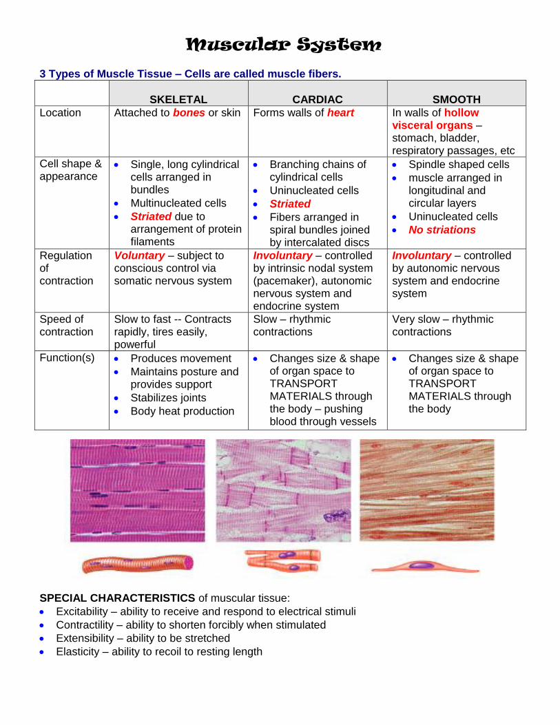
Every skeletal muscle fiber supplied by nerve ending that controls its activity. Contracting muscle fibers use huge amount of energy – needs lots of oxygen and nutrients. Muscles generate lots of metabolic waste that is removed through veins. Individual muscle fibers are wrapped and help together by several different connective tissue
sheaths that support each cell and reinforce the entire muscle. o Epimysium – dense irregular connective tissue surrounding entire muscle; may blend with
fascia o Perimysium – fibrous connective tissue surrounding fascicles (groups of muscle fibers) o Endomysium – fine areolar connective tissue surrounding each muscle fiber
All the connective tissue sheaths are continuous with each other and with the tendons that join
muscle to bone. When muscle fibers contract, they pull on these sheaths which transmit the pulling force to the bone to be moved. These sheaths contribute to the natural elasticity of muscle tissue and provide routes for blood vessels and nerves that serve the muscle.
Most skeletal muscles span joints and are attached to bones at least two places. When a muscle contracts, the moveable bone, the muscle insertion, moves toward the immovable or less movable bone, the muscles origin.
Muscle attachment may be direct or indirect. In direct attachment – epimysium is fused to the periosteum of bone or perichondrium of cartilage. In indirect attachment – the epimysium extends beyond the muscle either as a tendon or an aponeurosis which anchor the muscle to the bone, cartilage or to fascia of other muscles. Indirect attachments are more common because of durability and small size.
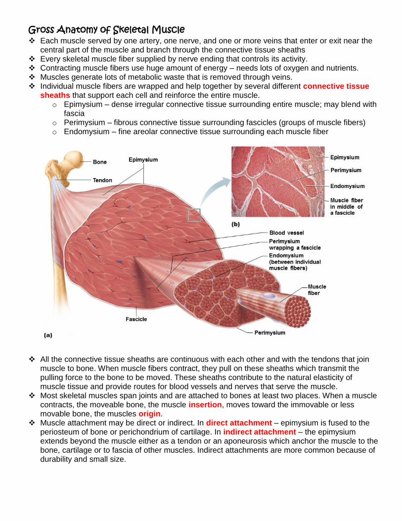
Microscopic Anatomy of Skeletal Muscle Fiber Muscle fibers are long, cylindrical cells (10 to 100 µm in diameter; up to 30 cm long) --- have multiple peripheral nuclei. Fiber components: o Sarcolemma – cell membrane o Sarcoplasm – cytoplasm; contains
glycosomes for glycogen storage, myoglobin for O2 storage
o Sarcoplasmic reticulum – contains interconnecting tubules (terminal cisternae) surrounding each myofibril; regulates intracellular levels of calcium ions
o Myofibrils – densely packed rodlike organelle that contains contracting units called sarcomeres
Sarcomeres are made up of an A band and I band. Striations are caused by repeating series of dark A bands and light I bands. Sarcomeres are regions on the myofibrils between two Z discs. o Z disc (line) – proteins on midline of I-band that anchors thin filament and connects myofibrils to
each other o H zone – lighter region in midsection of dark A-band where filaments do not overlap. Each H
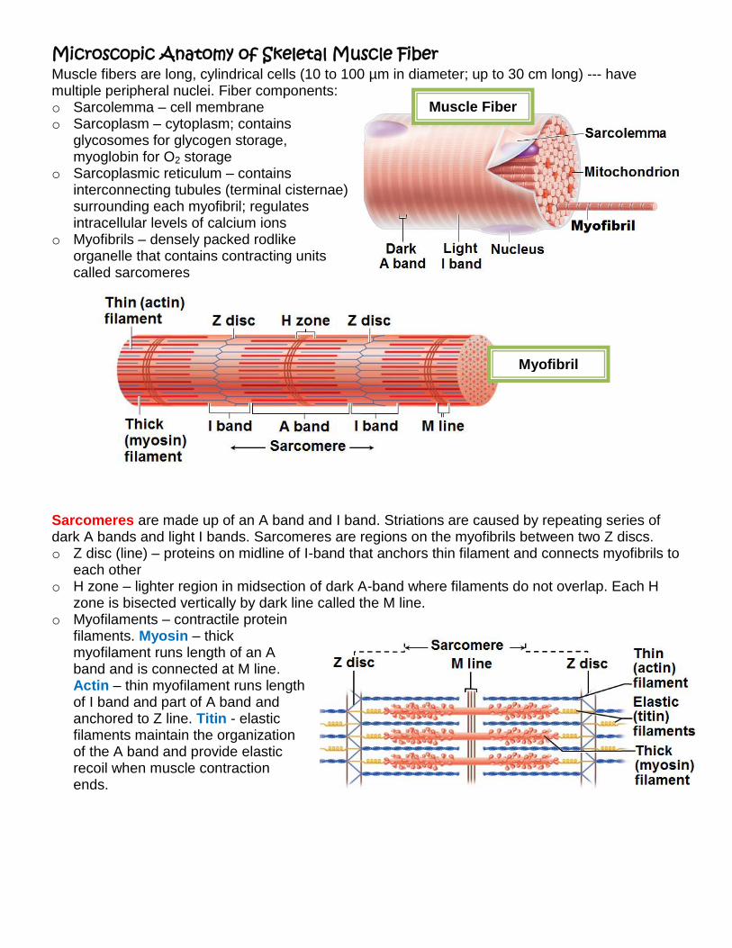
zone is bisected vertically by dark line called the M line. o Myofilaments – contractile protein
filaments. Myosin – thick myofilament runs length of an A band and is connected at M line. Actin – thin myofilament runs length of I band and part of A band and anchored to Z line. Titin - elastic filaments maintain the organization of the A band and provide elastic recoil when muscle contraction ends.
Muscle Fiber
Myofibril
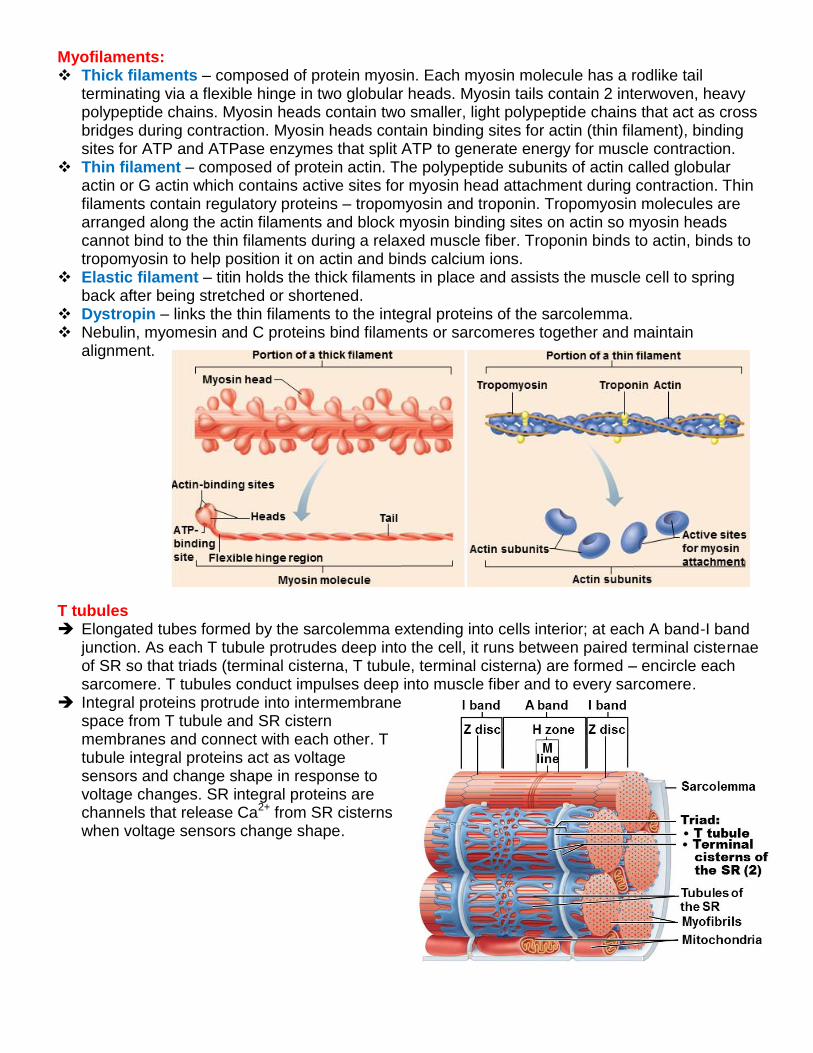
Myofilaments: Thick filaments – composed of protein myosin. Each myosin molecule has a rodlike tail
terminating via a flexible hinge in two globular heads. Myosin tails contain 2 interwoven, heavy polypeptide chains. Myosin heads contain two smaller, light polypeptide chains that act as cross bridges during contraction. Myosin heads contain binding sites for actin (thin filament), binding sites for ATP and ATPase enzymes that split ATP to generate energy for muscle contraction.
Thin filament – composed of protein actin. The polypeptide subunits of actin called globular actin or G actin which contains active sites for myosin head attachment during contraction. Thin filaments contain regulatory proteins – tropomyosin and troponin. Tropomyosin molecules are arranged along the actin filaments and block myosin binding sites on actin so myosin heads cannot bind to the thin filaments during a relaxed muscle fiber. Troponin binds to actin, binds to tropomyosin to help position it on actin and binds calcium ions.
Elastic filament – titin holds the thick filaments in place and assists the muscle cell to spring back after being stretched or shortened.
Dystropin – links the thin filaments to the integral proteins of the sarcolemma. Nebulin, myomesin and C proteins bind filaments or sarcomeres together and maintain
alignment.
T tubules Elongated tubes formed by the sarcolemma extending into cells interior; at each A band-I band
junction. As each T tubule protrudes deep into the cell, it runs between paired terminal cisternae of SR so that triads (terminal cisterna, T tubule, terminal cisterna) are formed – encircle each sarcomere. T tubules conduct impulses deep into muscle fiber and to every sarcomere.
Integral proteins protrude into intermembrane space from T tubule and SR cistern membranes and connect with each other. T tubule integral proteins act as voltage sensors and change shape in response to voltage changes. SR integral proteins are channels that release Ca2+ from SR cisterns when voltage sensors change shape.
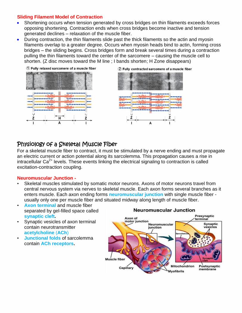
Sliding Filament Model of Contraction
Shortening occurs when tension generated by cross bridges on thin filaments exceeds forces opposing shortening. Contraction ends when cross bridges become inactive and tension generated declines – relaxation of the muscle fiber.
During contraction, the thin filaments slide past the thick filaments so the actin and myosin filaments overlap to a greater degree. Occurs when myosin heads bind to actin, forming cross bridges – the sliding begins. Cross bridges form and break several times during a contraction pulling the thin filaments toward the center of the sarcomere – causing the muscle cell to shorten. (Z disc moves toward the M line ; I bands shorten; H Zone disappears)
Physiology of a Skeletal Muscle Fiber For a skeletal muscle fiber to contract, it must be stimulated by a nerve ending and must propagate an electric current or action potential along its sarcolemma. This propagation causes a rise in intracellular Ca2+ levels. These events linking the electrical signaling to contraction is called excitation-contraction coupling. Neuromuscular Junction - • Skeletal muscles stimulated by somatic motor neurons. Axons of motor neurons travel from
central nervous system via nerves to skeletal muscle. Each axon forms several branches as it enters muscle. Each axon ending forms neuromuscular junction with single muscle fiber - usually only one per muscle fiber and situated midway along length of muscle fiber.
• Axon terminal and muscle fiber separated by gel-filled space called synaptic cleft.
• Synaptic vesicles of axon terminal contain neurotransmitter acetylcholine (ACh)
• Junctional folds of sarcolemma contain ACh receptors.

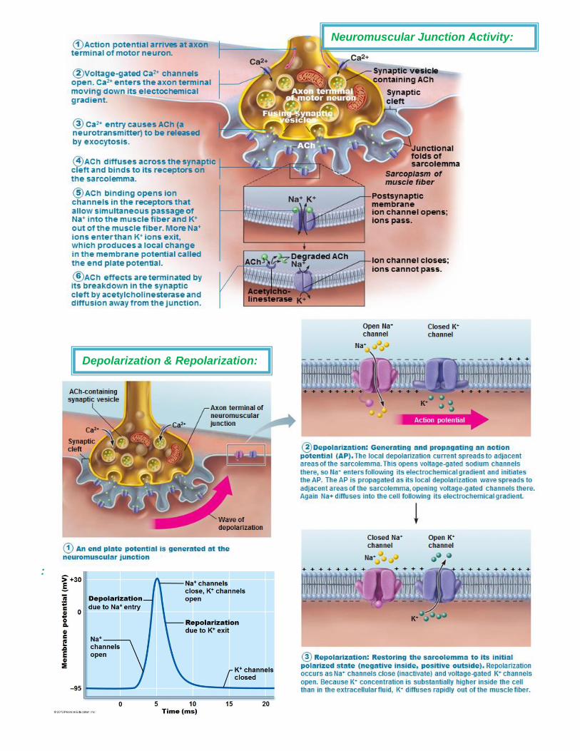
Events at the neuromuscular junction: 1. The nerve impulse arrives at axon terminal ACh released into synaptic cleft 2. ACh diffuses across cleft and binds with receptors on sarcolemma
ACh effects quickly terminated by enzyme acetylcholinesterase in synaptic cleft. Enzyme breaks down ACh to acetic acid and choline to prevent continued muscle fiber contraction in absence of additional stimulation.
3. ACh binding opens chemically gated ion channels -- simultaneous diffusion of Na+ (inward) and K+ (outward). More Na+ diffuses in, so interior of sarcolemma becomes less negative. (Resting membrane is polarized – more negative on the inside.)
4. Depolarization (end plate potential) – generation and propagation of an action potential (AP). End plate potentials spread to adjacent membrane areas – opening voltage gated Na+ channels. Na+ influx decreases membrane voltage toward critical voltage called threshold. If threshold is reached, AP initiated. Once initiated, AP is unstoppable muscle fiber contraction
5. Action Potential spread across sarcolemma voltage gated Na+ channels open in adjacent patch, causing it to depolarize to threshold.
6. Repolarization – restoring electrical conditions resting membrane potential. Na+ channels close and voltage-gated K+ channels open. K+ efflux rapidly restores resting polarity. Fibers cannot be stimulated – in refractory period until repolarization is complete. Ionic conditions of resting state restored by Na+-K+ pump
7. Action potential propagated along sarcomere to T tubules causing voltage-sensitive proteins to stimulate Ca2+ release from SR. Ca2+ binds to troponin – troponin moves tropomyosin away from myosin-binding sites. Myosin heads bind to actin, causing sarcomere shortening and muscle contraction. Cross-bridge cycling continues as Ca2+ signal and adequate ATP is present.
• Cross bridge formation – high energy myosin head attaches to thin filament. Power stroke – myosin head pivots and pulls thin filament toward the M line. Cross bridge detachment – ATP attaches to myosin head and cross bridge detaches. “Cocking” of myosin head – energy from hydrolysis of ATP cocks myosin head into high-energy state.
• When nervous stimulation ceases, Ca2+ pumped back into SR and contraction ends • At low intracellular Ca2+ concentration – tropomyosin blocks active sites on actin, myosin
heads cannot attach to actin and muscle fiber is relaxed.
Although the action potential is very brief (1-2
milliseconds), the contraction phase of the muscle fiber may persist for 100 ms and outlast the electrical event that triggers. The action potential ends before any signs of contraction are obvious. The events of excitation-coupling occur during the latent phase – between action potential initiation and beginning of mechanical activity (shortening). The electrical signal does not act directly on the myofilaments -- it causes the rise of intracellular calcium ions concentration that allows the filaments to slide.
:
Depolarization & Repolarization:
Neuromuscular Junction Activity:
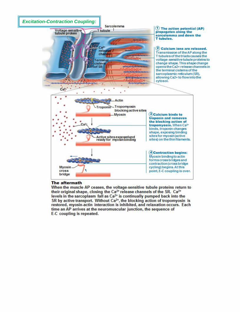
Excitation-Contraction Coupling:
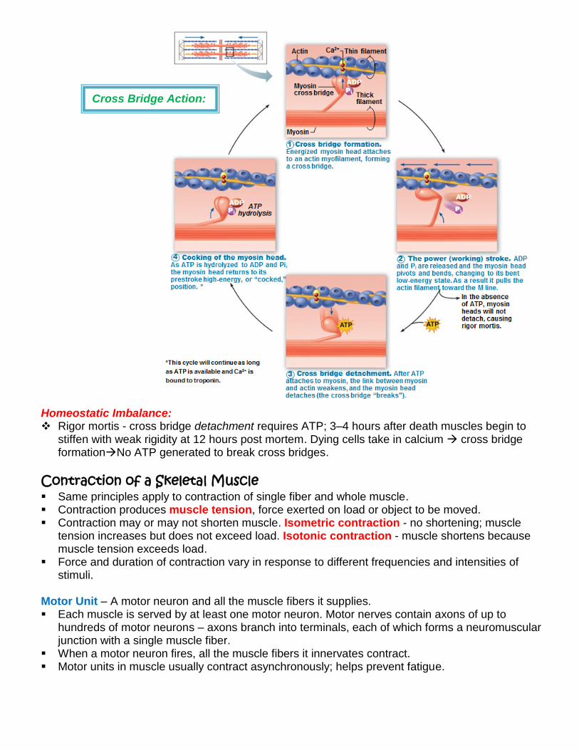
Homeostatic Imbalance: Rigor mortis - cross bridge detachment requires ATP; 3–4 hours after death muscles begin to
stiffen with weak rigidity at 12 hours post mortem. Dying cells take in calcium cross bridge formationNo ATP generated to break cross bridges.
Contraction of a Skeletal Muscle Same principles apply to contraction of single fiber and whole muscle. Contraction produces muscle tension, force exerted on load or object to be moved. Contraction may or may not shorten muscle. Isometric contraction - no shortening; muscle
tension increases but does not exceed load. Isotonic contraction - muscle shortens because muscle tension exceeds load.
Force and duration of contraction vary in response to different frequencies and intensities of stimuli.
Motor Unit – A motor neuron and all the muscle fibers it supplies. Each muscle is served by at least one motor neuron. Motor nerves contain axons of up to
hundreds of motor neurons – axons branch into terminals, each of which forms a neuromuscular junction with a single muscle fiber.
When a motor neuron fires, all the muscle fibers it innervates contract. Motor units in muscle usually contract asynchronously; helps prevent fatigue.
Cross Bridge Action:
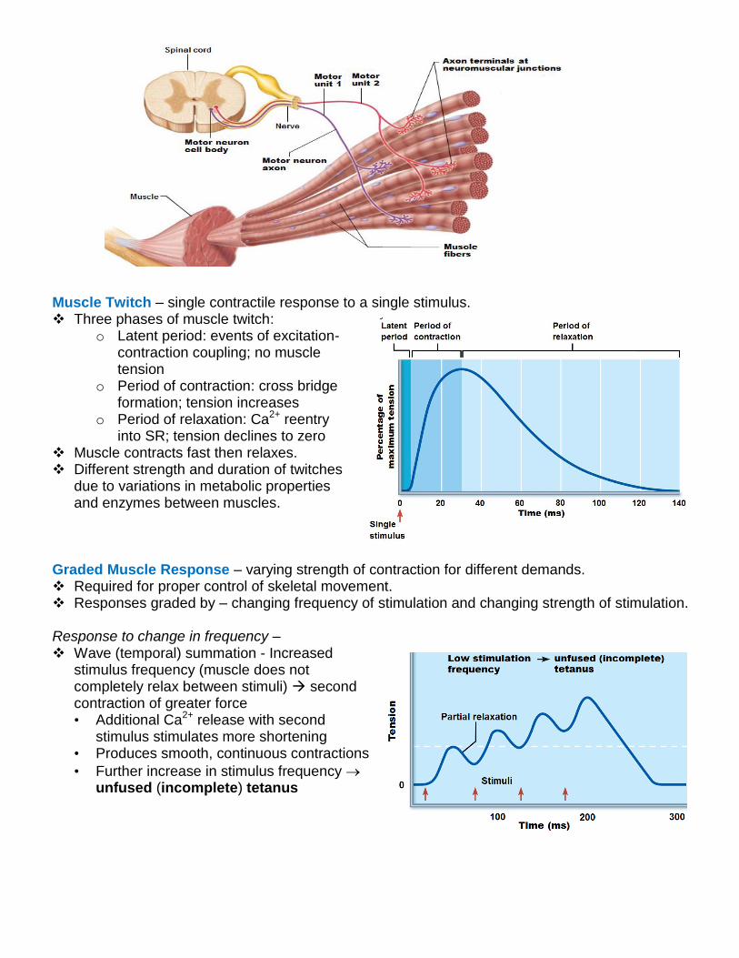
Muscle Twitch – single contractile response to a single stimulus. Three phases of muscle twitch:
o Latent period: events of excitation-contraction coupling; no muscle tension
o Period of contraction: cross bridge formation; tension increases
o Period of relaxation: Ca2+ reentry into SR; tension declines to zero
Muscle contracts fast then relaxes. Different strength and duration of twitches
due to variations in metabolic properties and enzymes between muscles.
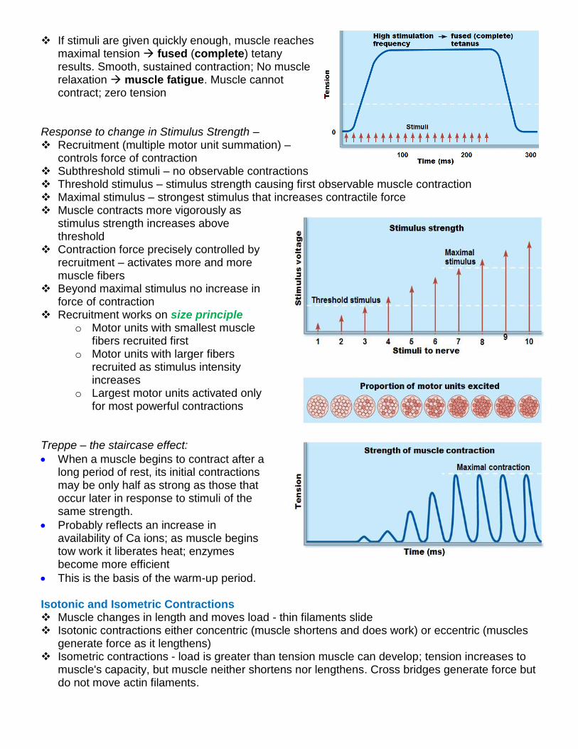
Graded Muscle Response – varying strength of contraction for different demands. Required for proper control of skeletal movement. Responses graded by – changing frequency of stimulation and changing strength of stimulation. Response to change in frequency – Wave (temporal) summation - Increased
stimulus frequency (muscle does not completely relax between stimuli) second contraction of greater force • Additional Ca2+ release with second
stimulus stimulates more shortening • Produces smooth, continuous contractions
• Further increase in stimulus frequency unfused (incomplete) tetanus
If stimuli are given quickly enough, muscle reaches maximal tension fused (complete) tetany results. Smooth, sustained contraction; No muscle relaxation muscle fatigue. Muscle cannot contract; zero tension
Response to change in Stimulus Strength – Recruitment (multiple motor unit summation) –
controls force of contraction Subthreshold stimuli – no observable contractions Threshold stimulus – stimulus strength causing first observable muscle contraction Maximal stimulus – strongest stimulus that increases contractile force Muscle contracts more vigorously as
stimulus strength increases above threshold
Contraction force precisely controlled by recruitment – activates more and more muscle fibers
Beyond maximal stimulus no increase in force of contraction
Recruitment works on size principle o Motor units with smallest muscle
fibers recruited first o Motor units with larger fibers
recruited as stimulus intensity increases
o Largest motor units activated only for most powerful contractions
Treppe – the staircase effect:
When a muscle begins to contract after a long period of rest, its initial contractions may be only half as strong as those that occur later in response to stimuli of the same strength.
Probably reflects an increase in availability of Ca ions; as muscle begins tow work it liberates heat; enzymes become more efficient
This is the basis of the warm-up period. Isotonic and Isometric Contractions Muscle changes in length and moves load - thin filaments slide Isotonic contractions either concentric (muscle shortens and does work) or eccentric (muscles
generate force as it lengthens) Isometric contractions - load is greater than tension muscle can develop; tension increases to
muscle's capacity, but muscle neither shortens nor lengthens. Cross bridges generate force but do not move actin filaments.
Muscle Tone Constant, slightly contracted state of all muscles Due to spinal reflexes -- groups of motor units alternately activated in response to input from
stretch receptors in muscles Keeps muscles firm, healthy, and ready to respond
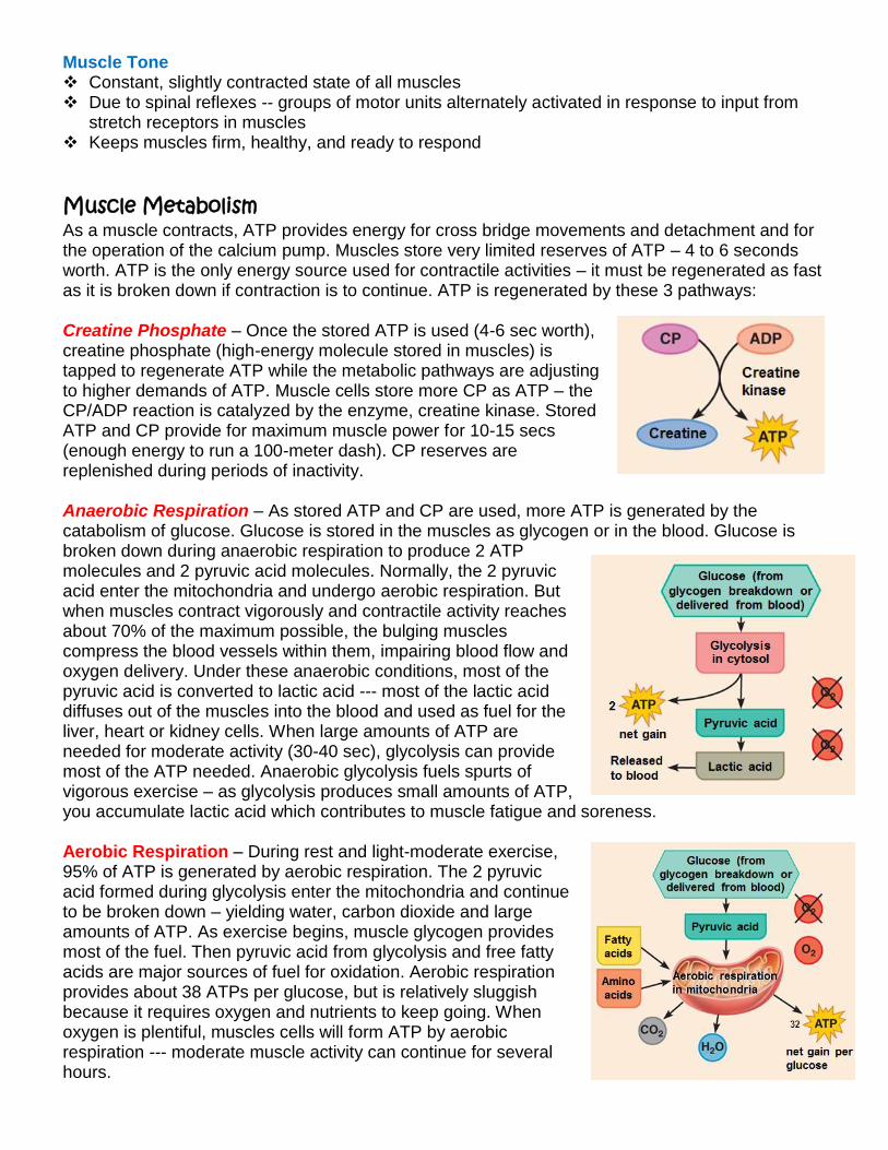
Muscle Metabolism As a muscle contracts, ATP provides energy for cross bridge movements and detachment and for the operation of the calcium pump. Muscles store very limited reserves of ATP – 4 to 6 seconds worth. ATP is the only energy source used for contractile activities – it must be regenerated as fast as it is broken down if contraction is to continue. ATP is regenerated by these 3 pathways: Creatine Phosphate – Once the stored ATP is used (4-6 sec worth), creatine phosphate (high-energy molecule stored in muscles) is tapped to regenerate ATP while the metabolic pathways are adjusting to higher demands of ATP. Muscle cells store more CP as ATP – the CP/ADP reaction is catalyzed by the enzyme, creatine kinase. Stored ATP and CP provide for maximum muscle power for 10-15 secs (enough energy to run a 100-meter dash). CP reserves are replenished during periods of inactivity. Anaerobic Respiration – As stored ATP and CP are used, more ATP is generated by the catabolism of glucose. Glucose is stored in the muscles as glycogen or in the blood. Glucose is broken down during anaerobic respiration to produce 2 ATP molecules and 2 pyruvic acid molecules. Normally, the 2 pyruvic acid enter the mitochondria and undergo aerobic respiration. But when muscles contract vigorously and contractile activity reaches about 70% of the maximum possible, the bulging muscles compress the blood vessels within them, impairing blood flow and oxygen delivery. Under these anaerobic conditions, most of the pyruvic acid is converted to lactic acid --- most of the lactic acid diffuses out of the muscles into the blood and used as fuel for the liver, heart or kidney cells. When large amounts of ATP are needed for moderate activity (30-40 sec), glycolysis can provide most of the ATP needed. Anaerobic glycolysis fuels spurts of vigorous exercise – as glycolysis produces small amounts of ATP, you accumulate lactic acid which contributes to muscle fatigue and soreness. Aerobic Respiration – During rest and light-moderate exercise, 95% of ATP is generated by aerobic respiration. The 2 pyruvic acid formed during glycolysis enter the mitochondria and continue to be broken down – yielding water, carbon dioxide and large amounts of ATP. As exercise begins, muscle glycogen provides most of the fuel. Then pyruvic acid from glycolysis and free fatty acids are major sources of fuel for oxidation. Aerobic respiration provides about 38 ATPs per glucose, but is relatively sluggish because it requires oxygen and nutrients to keep going. When oxygen is plentiful, muscles cells will form ATP by aerobic respiration --- moderate muscle activity can continue for several hours.
When exercise demands begin to exceed the ability of muscle cells to carry out aerobic respiration, glycolysis beings to contribute more of the ATP generated. The length of time a muscle can continue to contract using aerobic pathways is aerobic endurance and the points at which muscle metabolism converts to anaerobic glycolysis is anaerobic threshold.
Muscle Fatigue The state of physiological inability to contract even though the muscle may be receiving stimuli.
Occurs when ionic imbalances (K+, Ca2+, Pi) interfere with Excitation-Contraction coupling or prolonged exercise may damage SR and interferes with Ca2+ regulation and release. ATP availability declines during contraction and total lack of ATP results in contractures – continuous contraction because the cross brides are unable to detach. (Cramps are temporary contractures.)
For muscles to return to a resting state, oxygen reserves must be replenished, the accumulated lactic acid must be converted to pyruvic acid, glycogen stores must be replaced and ATP/CP reserves must be resynthesized. Oxygen debt is the extra amounts of oxygen that the body must take in for these restoration processes.
About 40% of energy released during muscle contraction is useful as work and the remaining energy (60%) is given off as heat. Heat buildup is prevented from reaching dangerous levels by several homeostatic mechanisms – sweat and radiation of heat from skin surfaces. Shivering produces more heat by contracting muscles when cold.
Force of Muscle Contraction The force of muscle contraction depends on number of cross bridges attached, which is affected by: 1. As more muscle fibers are recruited (as more are stimulated) more force 2. Relative size of fibers – bulkier muscles & hypertrophy of cells more force
3. Frequency of stimulation - frequency contractions are summed up, becoming stronger, more vigorous and ultimately producing tetanus – more time for transfer of tension to noncontractile components more force
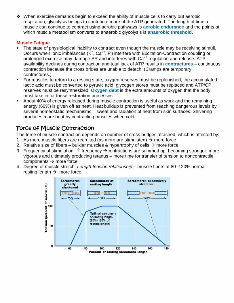
4. Degree of muscle stretch: Length-tension relationship – muscle fibers at 80–120% normal resting length more force
Velocity and Duration of Contraction
Muscles vary in how fast they can contract and in how long they can continue to contract before they fatigue. These characteristics are influenced by muscle fiber type, load and recruitment. Muscle fiber types are classified according to two characteristics: 1. Speed of contraction: slow or fast fibers according to speed at which myosin ATPases split
ATP and pattern of electrical activity of motor neurons. 2. Metabolic pathways for ATP synthesis: Oxidative fibers—use aerobic pathways; Glycolytic
fibers—use anaerobic glycolysis Three types of muscle fibers – Slow oxidative fibers; Fast oxidative fibers; Fast glycolytic fibers Most muscles contain mixture of fiber types this provides a range of contractile speed and
fatigue resistance. All muscle fibers in a particular motor unit are the same type. Some people have relatively more of one variety of fibers – initially due to genetics, but are
modified by exercise and determine endurance vs. strength.
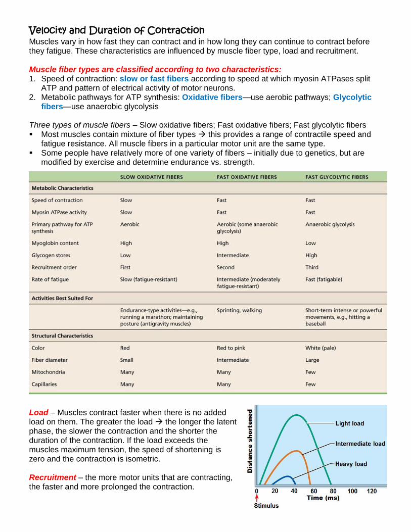
Load – Muscles contract faster when there is no added load on them. The greater the load the longer the latent phase, the slower the contraction and the shorter the duration of the contraction. If the load exceeds the muscles maximum tension, the speed of shortening is zero and the contraction is isometric. Recruitment – the more motor units that are contracting, the faster and more prolonged the contraction.
Effects of Exercise on Muscles Aerobic (endurance) exercise – leads to increased muscle capillaries, number of mitochondria,
myoglobin synthesis; results in greater endurance, strength and resistance to fatigue; may convert fast glycolytic fibers into fast oxidative fibers.
Resistance exercise (typically anaerobic) – results in muscle hypertrophy due to increase in fiber size; increased mitochondria, myofilaments, glycogen stores and connective tissue Increased muscle strength and size
Balanced exercise program – Exercise gains adhere to the overload principle. Forcing muscle to work hard promotes increased muscle strength and endurance Muscles adapt to increased demands and must be overloaded to produce further gains. Overuse injuries may result from lack of rest. Best programs alternate aerobic and anaerobic activities. Homeostatic Imbalances Disuse atrophy – results from immobilization; muscle strength declines 5% per day. Without
neural stimulation muscles atrophy to ¼ initial size. Fibrous connective tissue replaces lost muscle tissue rehabilitation impossible.
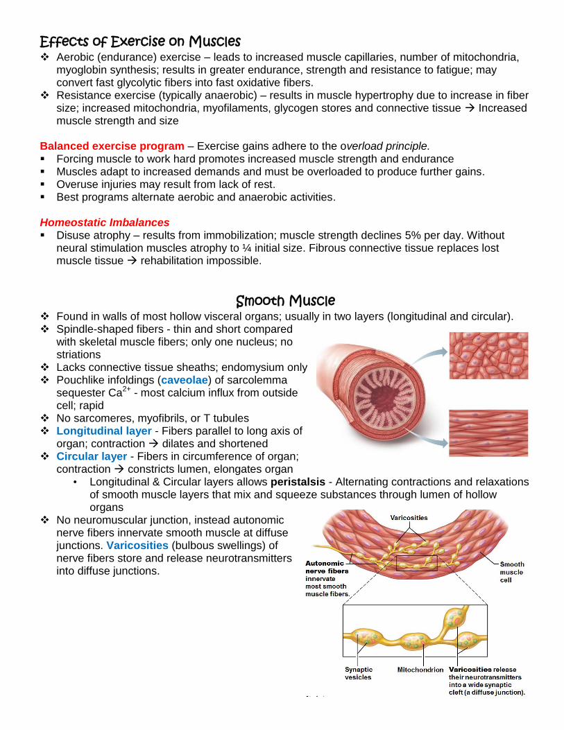
Smooth Muscle Found in walls of most hollow visceral organs; usually in two layers (longitudinal and circular). Spindle-shaped fibers - thin and short compared
with skeletal muscle fibers; only one nucleus; no striations
Lacks connective tissue sheaths; endomysium only Pouchlike infoldings (caveolae) of sarcolemma
sequester Ca2+ - most calcium influx from outside cell; rapid
No sarcomeres, myofibrils, or T tubules Longitudinal layer - Fibers parallel to long axis of
organ; contraction dilates and shortened Circular layer - Fibers in circumference of organ;
contraction constricts lumen, elongates organ • Longitudinal & Circular layers allows peristalsis - Alternating contractions and relaxations
of smooth muscle layers that mix and squeeze substances through lumen of hollow organs
No neuromuscular junction, instead autonomic nerve fibers innervate smooth muscle at diffuse junctions. Varicosities (bulbous swellings) of nerve fibers store and release neurotransmitters into diffuse junctions.
Myofilaments in Smooth Muscle • Ratio of thick to thin filaments (1:13) is much lower than in skeletal muscle (1:2) • Thick filaments have heads along entire length. • No troponin complex; protein
calmodulin binds Ca2+ • Myofilaments are spirally arranged,
causing smooth muscle to contract in corkscrew manner
• Dense bodies - Proteins that anchor noncontractile intermediate filaments to sarcolemma at regular intervals; correspond to Z discs of skeletal muscle
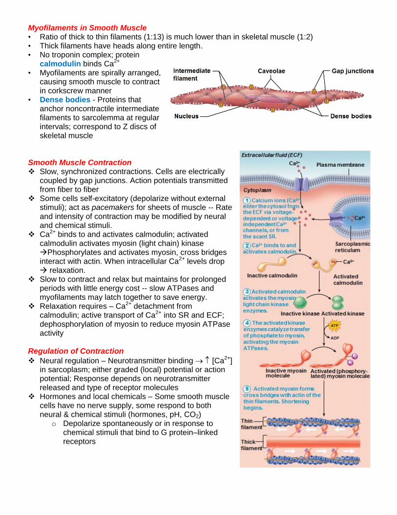
Smooth Muscle Contraction Slow, synchronized contractions. Cells are electrically
coupled by gap junctions. Action potentials transmitted from fiber to fiber
Some cells self-excitatory (depolarize without external stimuli); act as pacemakers for sheets of muscle -- Rate and intensity of contraction may be modified by neural and chemical stimuli.
Ca2+ binds to and activates calmodulin; activated calmodulin activates myosin (light chain) kinase Phosphorylates and activates myosin, cross bridges interact with actin. When intracellular Ca2+ levels drop relaxation.
Slow to contract and relax but maintains for prolonged periods with little energy cost -- slow ATPases and myofilaments may latch together to save energy.
Relaxation requires – Ca2+ detachment from calmodulin; active transport of Ca2+ into SR and ECF; dephosphorylation of myosin to reduce myosin ATPase activity
Regulation of Contraction
Neural regulation – Neurotransmitter binding [Ca2+] in sarcoplasm; either graded (local) potential or action potential; Response depends on neurotransmitter released and type of receptor molecules
Hormones and local chemicals – Some smooth muscle cells have no nerve supply, some respond to both neural & chemical stimuli (hormones, pH, CO2)
o Depolarize spontaneously or in response to chemical stimuli that bind to G protein–linked receptors
Special Features of Smooth Muscle Contraction • Stress-relaxation response – Responds to stretch only briefly, then adapts to new length; retains
ability to contract on demand; enables organs such as stomach and bladder to temporarily store contents.
• Length and tension changes – can contract when between half and twice its resting length. • Hyperplasia - Smooth muscle cells can divide and increase numbers. Types of Smooth Muscle Smooth muscle varies in different organs – fiber arrangement/organization, innervation,
responsiveness to various stimuli. Categorized as unitary and multi-unit. Unitary (visceral) smooth muscle – in all hollow organs except heart; arranged in opposing
sheets; innervated by varicosities; often exhibit spontaneous action potentials; electrically coupled by gap junctions; respond to various chemical stimuli
Multiunit smooth muscle – located in large airways, large arteries, arrector pili muscles, and iris of eye; gap junctions; spontaneous depolarization rare; independent muscle fibers; innervated by autonomic NS; graded contractions occur in response to neural stimuli; has motor units; responds to hormones
Developmental Aspects of Muscle • All muscle tissues develop from embryonic myoblasts • Multinucleated skeletal muscle cells form by fusion • Growth factor agrin stimulates clustering of ACh receptors at neuromuscular junctions • Cardiac and smooth muscle myoblasts develop gap junctions • Cardiac and skeletal muscle become amitotic, but can lengthen and thicken in growing child • Myoblast-like skeletal muscle satellite cells have limited regenerative ability • Cardiomyocytes can divide at modest rate, but injured heart muscle mostly replaced by
connective tissue • Smooth muscle regenerates throughout life • Muscular development reflects neuromuscular coordination • Peak natural neural control occurs by mid-adolescence; athletics can improve neuromuscular
control • Female skeletal muscle makes up 36% of body mass • Male skeletal muscle makes up 42% of body mass, primarily due to testosterone • With age, connective tissue increases and muscle fibers decrease; by age 30, loss of muscle
mass (sarcopenia) begins --- regular exercise reverses sarcopenia Muscular Dystrophy Group of inherited muscle-destroying diseases; generally appear in childhood Muscles enlarge due to fat and connective tissue deposits. Muscle fibers atrophy and
degenerate. Duchenne muscular dystrophy (DMD): most common and severe type; inherited, sex-linked,
carried by females and expressed in males (1/3500) as lack of dystrophin (cytoplasmic protein that stabilizes sarcolemma)Fragile sarcolemma tears Ca2+ entry damaged contractile fibers inflammatory cells muscle mass drops
Victims become clumsy and fall frequently; usually die of respiratory failure in 20s; no cure; Prednisone improves muscle strength and function