Embed Size (px)
Citation preview

Physical Education IES CONDESTABLE ÁLVARO DE LUNA European Section.
1 Victor Gonzalez
CREATED (2016) – 1ST REVISION (2017)
MUSCLE WORKING IN PAIRS: Muscles usually work in pairs or groups (e.g. the
biceps flexes the elbow and the triceps extends it). This is called antagonistic muscle
action.
The working muscle is called the prime mover or agonist. The relaxing muscle is
the antagonist. The other main pair of muscle that work together are the quadriceps
and hamstrings.
The prime mover is helped by other muscles called synergists. These contract at
the same time as the prime mover. They hold the body in position so that the prime
mover can work smoothly.
When muscles cause a limb to move through the joint's range of motion, they usually
act in the following cooperating groups:
Agonists. These muscles cause the movement to occur. They create the normal range
of movement in a joint by contracting.
Agonists are also referred to as prime movers since they are the muscles that are
primarily responsible for generating the movement.
Antagonists. These muscles act in opposition to the movement generated by the
agonists and are responsible for returning a limb to its initial position.
Synergists. These muscles perform, or assist in performing, the same set of joint
motion as the agonists. Synergists are sometimes referred to as neutralizers because
they help cancel out, or neutralize, extra motion from the agonists to make sure that
the force generated works within the desired plane of motion.
Fixators. These muscles provide the necessary support to assist in holding the rest of
the body in place while the movement occurs. Fixators are also sometimes called
stabilizers.
The contraction of a muscle does not necessarily imply that the muscle shortens; it
only means that tension has been generated. Muscles can contract in the following
ways:
Isometric contraction: This is a contraction in which no movement takes place,
because the load on the muscle exceeds the tension generated by the contracting
muscle. This occurs when a muscle attempts to push or pull an immovable object.
Isotonic contraction: This is a contraction in which movement does take place,
because the tension generated by the contracting muscle exceeds the load on the
muscle. This occurs when you use your muscles to successfully push or pull an object.
Isotonic contractions are further divided into two types:
MUSCLES & MOVEMENTS
1. Introduction
2. Types of contraction.
Physical Education IES CONDESTABLE ÁLVARO DE LUNA European Section.
2 Victor Gonzalez
CREATED (2016) – 1ST REVISION (2017)
Concentric contraction
This is a contraction in which the muscle decreases in length (shortens) against an
opposing load, such as lifting a weight up.
Eccentric contraction
This is a contraction in which the muscle increases in length (lengthens) as it resists a
load, such as lowering a weight down in a slow, controlled fashion.
During a concentric contraction, the muscles that are shortening serve as the agonists
and hence do all of the work. During an eccentric contraction the muscles that are
lengthening serve as the agonists (and do all of the work)
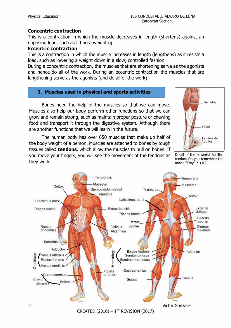
Bones need the help of the muscles so that we can move.
Muscles also help our body perform other functions so that we can
grow and remain strong, such as maintain proper posture or chewing
food and transport it through the digestive system. Although there
are another functions that we will learn in the future.
The human body has over 650 muscles that make up half of
the body weight of a person. Muscles are attached to bones by tough
tissues called tendons, which allow the muscles to pull on bones. If
you move your fingers, you will see the movement of the tendons as
they work.
3. Muscles used in physical and sports activities
Detail of the powerful Achilles tendon. Do you remember the movie "Troy" ?. (10)

Physical Education IES CONDESTABLE ÁLVARO DE LUNA European Section.
3 Victor Gonzalez
CREATED (2016) – 1ST REVISION (2017)
1. A good trick to store and organize what we have learned, is to make an outline.
Organizes the muscles of the body that we have learned in groups: muscles of the
trunk, muscles of the arm and muscles of the legs.Put each of them in the correct
box. To avoid mistakes, fill it first to other paper, and when you are reliable,
complete the diagram. If in doubt, you can help Internet or any book you have on
hand.
3. Activities.

Physical Education IES CONDESTABLE ÁLVARO DE LUNA European Section.
4 Victor Gonzalez
CREATED (2016) – 1ST REVISION (2017)
2. Up to now, we have study muscles and bones names, but now we are going to localize in the body. First study the previous
outline, and then study the pictures. Can you fill the blank pictures with muscles and bones names?

Physical Education IES CONDESTABLE ÁLVARO DE LUNA European Section.
5 Victor Gonzalez
CREATED (2016) – 1ST REVISION (2017)
4.1 Plains of movements: Movements of the human body are often described in terms of the ‘plane’ in which they pass
through. There are three planes of the human body, these planes are shown in the following
table.
4.2 Plains of movements: Knowing how the body moves and the actions that various joints allow is crucial for safe and
effective exercise instruction. Some of the key joint actions that you should know are detailed in
the following tables.
Flexion:
Refers to movement where the angle between two bones
decreases. Flexion is commonly known as bending.
Extension:
Refers to movement where the angle between two bones
increases. Extension is otherwise known as straightening.
Frontal Transverse Sagittal
Description
The frontal plane passes through
the body from left to right,
dividing the body into anterior
and posterior portions.
Description
The transverse plane passes through
the body in a line parallel to the floor,
dividing the body into top and bottom
portions.
Description
The sagittal plane passes
through the body from front to
back, dividing the body into left
and right portions.
Example
Side to side movements occur in
the frontal plane, such as raising
your arms or legs out to the side
like in a star jump.
Example
Twisting or rotational movements
occur in the transverse plane, such as
twisting your head from side to side.
Example
Front to back movements
occur in the sagittal plane,
such as walking, pushing,
pulling and squatting.
4. BODY MOVEMENTS.

Physical Education IES CONDESTABLE ÁLVARO DE LUNA European Section.
6 Victor Gonzalez
CREATED (2016) – 1ST REVISION (2017)
Horizontal flexion:
Refers to movement where the angle between two bones decreases and
on the horizontal plane.
Horizontal extension:
Refers to movement where the angle between two bones increases and
occurs on the horizontal plane.
Lateral Flexion:
Refers to movement of the spine laterally away from the midline of the
body. This can be seen when we bend to one side.
Abduction:
Is movement of a body segment away from the midline of the body.
Adduction:
Is movement of a body segment toward the midline of the body.
Circumduction:
This is a movement where the joint is the pivot and the body segment
moves in a combination of flexion, extension, adduction and
abduction.
Elevation:
Refers to the raising of the scapula to a superior level (shrugging
the shoulders).
Depression:
Refers to the scapula moving to a inferior position as they are
pulled downwards.

Physical Education IES CONDESTABLE ÁLVARO DE LUNA European Section.
7 Victor Gonzalez
CREATED (2016) – 1ST REVISION (2017)
Supination:
Hand – movement so the palm of the hand faces
upward or forward (anteriorly).
Foot – combination of inversion, plantar flexion and
adduction of the foot occurring at the same time.
Pronation:
Hand – movement so the palm of the hand faces
downward or backward (posteriorly).
Foot – combination of eversion, dorsiflexion and
abduction of the foot occurring at the same time.
Medial rotation: The movement of a body segment where the
front (anterior) of the segment rotates medially (inwards) towards
the midline of the body.
Lateral rotation: The movement of a body segment where the
front (anterior) of the segment rotates laterally (outwards) away
from the midline of the body.
4.3 Muscles actions:
Major muscles of the back, shoulder and chest:
Rotation:
Refers to a pivoting or ‘twisting’ movement. Rotation is broken
down further into medial and lateral rotation.
Plantar flexion (right):
Is moving the top of the foot away from the shin or ‘pointing’ the
toes.
Dorsiflexion (left):
Is moving the top of the foot toward the shin or ‘raising’ the
toes.

Physical Education IES CONDESTABLE ÁLVARO DE LUNA European Section.
8 Victor Gonzalez
CREATED (2016) – 1ST REVISION (2017)
Muscle Origin Insertion Movement/Action
Erector spinae
Lower four thoracic
vertebrae
Upper thoracic
vertebrae and the
cervical vertebrae
Extension of the
vertebral column
Latissimus dorsi
Thoracic, lumbar
vertebrae, sacrum
and top of pelvis
Upper part of
humerus
Adduction, and
extension of the arm.
Trapezius
Cervical and
thoracic vertebrae,
base of skull
Clavicle and
scapula
Elevation and depression
of the scapula
Deltoid
Clavicle and spine
of scapula
Upper part of
humerus
Abduction of arm.

Physical Education IES CONDESTABLE ÁLVARO DE LUNA European Section.
9 Victor Gonzalez
CREATED (2016) – 1ST REVISION (2017)
Pectoralis major
Sternum, clavicle
and 1st-6th ribs
Upper front area
of humerus
Adduction and flexion
of arm
Major muscles of the upper arm:
Muscle Origin Insertion Movement/Action
Biceps brachii
Scapula Radius Flexion of elbow
Triceps brachii
Scapula and upper
part of humerus
Ulna Extension of elbow
Major muscles of the Abdomen:
Muscle Origin Insertion Movement/Action

Physical Education IES CONDESTABLE ÁLVARO DE LUNA European Section.
10 Victor Gonzalez
CREATED (2016) – 1ST REVISION (2017)
Rectus
abdominus
Front lower
part of
pelvis
5th, 6th and 7th ribs
and lowest part of
sternum
Flexion of vertebral column
External
oblique’s
Lower ribs Front upper part of
pelvis
Rotation of vertebral column and
flexion of vertebral column (both sides
contraction)
Internal
oblique’s
Top of
pelvis
Lower three ribs Rotation of vertebral column and
flexion of vertebral column (both sides
contraction)
Major muscles of the hip region:
Muscle Origin Insertion Movement/Action

Physical Education IES CONDESTABLE ÁLVARO DE LUNA European Section.
11 Victor Gonzalez
CREATED (2016) – 1ST REVISION (2017)
Gluteus maximus
Rear part of pelvis,
sacrum and coccyx
Top of femur Extension and lateral rotation
of leg
Gluteus medius
Upper part of pelvis Outside of upper
part of femur
Abduction and medial
rotation of leg
Psoas major
Lumbar vertebrae
and top of pelvis
Upper part of
femur
Flexion of femur and vertebral
column.
Muscles of thigh:
Muscle Origin Insertion Movement/Action
Quadriceps
Front lower
part of pelvis
and upper part
of femur
Top front part of
tibia
Flexion of the hip
and extension of
the knee.

Physical Education IES CONDESTABLE ÁLVARO DE LUNA European Section.
12 Victor Gonzalez
CREATED (2016) – 1ST REVISION (2017)
(Rectus femoris, Vastus
lateralis, Vastus intermedius,
Vastus medialis)
Hamstrings
(Semimembranosus,
Semitendinosus, Biceps
femoris)
Lower back
part of pelvis
and femur
Upper part of tibia
and fibula
Extension of hip
and flexion of
knee.
Adductor
Pubic Bone Posterior and
upper surface of
the femur (Linea
aspera)
Adduction of leg.
Muscles of the leg:
Muscle Origin Insertion Movement/
Action

Physical Education IES CONDESTABLE ÁLVARO DE LUNA European Section.
13 Victor Gonzalez
CREATED (2016) – 1ST REVISION (2017)
Gastrocnemius
Lower rear
part of femur
Heel bone Plantar
flexion of
ankle and
flexion of
knee
Soleus
Upper rear
part of tibia
and fibula
Heel bone Plantar
flexion of
ankle
Tibialis anterior
Upper two-
thirds of the
lateral
(outside)
surface of the
tibia
The medial
cuneiform and
first metatarsal
bones of the foot
Dorsi
flexion of
ankle.
5. Information Resources.
Physical Education IES CONDESTABLE ÁLVARO DE LUNA European Section.
14 Victor Gonzalez
CREATED (2016) – 1ST REVISION (2017)
Bibliography:
http://www.medioscan.com/ies/musculosyhuesos
https://www.dartmouth.edu/~humananatomy/part_1/chapter_2.html
https://www.boundless.com/physiology/textbooks/boundless-anatomy-and-physiology-
textbook/skeletal-system-parts-of-the-skeleton-7/the-skull-79/general-features-and-functions-of-the-
skull-458-5063/
http://www.teachpe.com/anatomy/joints.php
http://www2.highlands.edu/academics/divisions/scipe/biology/faculty/harnden/2121/notes/art.htm
http://www.ptdirect.com/training-design/anatomy-and-physiology/joints-joint-actions-planes-of-
movement
http://www.ptdirect.com/training-design/anatomy-and-physiology/key-muscle-locations-and-actions
Pictures:
1) https://pekewiki-loja.wikispaces.com/Your+body+moves
2) https://askabiologist.asu.edu/bone-anatomy
3) http://www.surfertoday.com/surfing/8121-how-to-prevent-cramps-in-surfing
4) http://www.crossfitsouthbay.com/muscle-spotlight-diaphragm/
5) https://upload.wikimedia.org/wikipedia/commons/thumb/3/39/Cranial_bones_en_v2.svg/2000
px-Cranial_bones_en_v2.svg.png
6) https://upload.wikimedia.org/wikipedia/commons/thumb/2/25/Facial_skeleton_-
_en.svg/250px-Facial_skeleton_-_en.svg.png
7) http://classconnection.s3.amazonaws.com/592/flashcards/33592/png/symph1352425780294.
png
8) http://www.healthhype.com/wrist-joint-hand-and-finger-joints-and-bones-pictures.html
9) http://teachmeanatomy.info/lower-limb/bones/bones-of-the-foot-tarsals-metatarsals-and-
phalanges/