Embed Size (px)
Citation preview

MUSCLES

Muscular System

Muscle Tissue: histology
• 3 muscle types: skeletal muscle, cardiac muscle, and smooth muscle
• All muscles show similarities and differences
• All muscles composed of elongated cells called fibers
• Muscle cytoplasm is sarcoplasm, and muscle cell membrane is sarcolemma
• Muscle fibers contain myofibrils made of contractile proteins actin and myosin

Skeletal Muscle Fibers are multinucleated cells with peripheral nuclei: multiple nuclei due to
fusion of mesenchyme myoblasts during embryonic development Each muscle fiber is composed of myofibrils and myofilaments

Skeletal Muscle Actin and myosin filaments form distinct cross-striation patterns Light I bands contain thin actin, and dark A bands contain thick myosin
filaments Dense Z line bisects I bands; between Z lines is the contractile unit, the
sarcomere

Skeletal Muscle Accessory proteins align and stabilize actin and myosin filaments Titin protein anchors myosin filaments, and a-actinin binds actin filaments to Z
lines Titin centers, positions, and acts like a spring between myosin and Z lines

Skeletal Muscle Muscle is surrounded by connective tissue epimysium Muscle fascicles are surrounded by connective tissue perimysium Each muscle fiber is surrounded by connective tissue endomysium

Skeletal Muscle Voluntary muscles are under conscious control Neuromuscular spindles are specialized stretch receptors in almost all skeletal
muscles Intrafusal fibers and nerve endings are found in spindle capsules Stretching of muscle produces a stretch reflex and movement to shorten
muscle

Skeletal Muscle

Skeletal Muscle

Skeletal Muscle

Skeletal Muscle

Skeletal Muscle:Transmission Electron Microscopy
◊ Light bands = I bands, formed by thin actin filaments: are crossed by dense Z lines
◊ Between Z lines = smallest contractile unit = sarcomere ◊ Dark bands = A bands, located in the middle of sarcomere,
formed by overlapping actin and myosin filaments ◊ M bands = in the middle of A bands represent linkage of myosin
filaments ◊ H bands on each side of M bands contain only myosin filaments ◊ Sarcoplasmic reticulum and mitochondria surround each sarcomere

Skeletal Muscle

Skeletal Muscle

Skeletal Muscle: functional correlation
Skeletal muscles = voluntary (under conscious control), contracting only when stimulated
Motor endplates = sites of nerve innervations and transmission of stimuli to muscle Axon terminals of motor endplates contain vesicles with the neurotransmitter
acetylcholine (released into synaptic cleft by action potential and combined with its receptors on muscle membrane); acetylcholinesterase neutralizes acetylcholine and prevents further contraction

Skeletal Muscle

Skeletal Muscle: functional correlation
Before arrival of impulse, Ca+ is stored in sarcoplasmic reticulum (SR) T tubules = sarcolemma invaginations into each myofiber, carrying stimulus for
muscle contraction to every myofiber, myofibril, and SR membrane Triads = 2 expanded terminal cisternae of SR and T tubules, located at A–I
junctions After stimulation, SR releases Ca+ into sarcomeres, activating binding of actin and
myosin, causing muscle contraction and shortening After the end of stimulus, Ca+ is actively transported and stored in SR

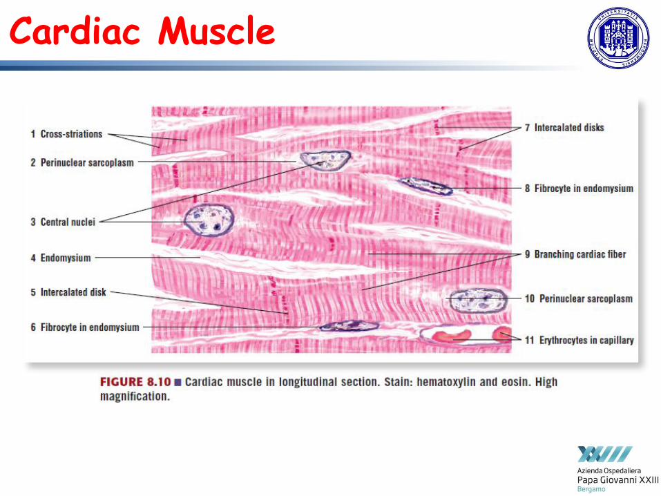
Cardiac Muscle • Located in heart and large vessels with cross-striations of actin and myosin forming similar I bands, A bands, and Z lines as in skeletal muscle • Characterized by dense junctional complexes called intercalated disks, containing gap junctions [coupling all fibers for rhythmic contraction, forming functional syncytium], one or two central nuclei, fibers shorter and showing branching • T tubules located at Z lines and larger + SR less well developed + mitochondria larger and more abundant than in skeletal muscles • For contraction, Ca+ is imported from outside cell and from SR • Exhibit autorhythmicity and spontaneously generate stimuli • Autonomic nervous system innervates heart and influences heart rate and blood pressure

Cardiac Muscle

Cardiac Muscle

Cardiac Muscle
Cardiac Muscle

Cardiac Muscle

Smooth Muscle • Fibers fusiform, containing single central nuclei, found in hollow organs and blood vessels, with actin and myosin filaments without cross-striation, not showing regular arrangement or striations, but forming lattice network, and inserting into dense bodies in sarcoplasm and cytoplasm • Zonula adherens binds muscle cells, whereas gap junctions provide functional coupling • In intestines, muscles are arranged in concentric layers, and in blood vessels in a circular pattern

Smooth Muscle
• Following stimulation, calcium enters sarcoplasm from caveolae and SR • Calmodulin, a calcium-binding protein, stimulates actin and myosin interaction, contract muscle by a sliding mechanism similar to skeletal muscle • Exhibit spontaneous activity and maintain tonus in hollow organs: peristaltic contractions propel contents in the organs; innervated by postganglionic neurons of sympathetic and parasympathetic divisions; involuntary muscles regulated by autonomic nervous system, hormones, and stretching
• SR not well developed for Ca+ storage and sarcolemma containing invaginations called caveolae (controlling influx of Ca+ into cell after stimulation)

Smooth Muscle

Smooth Muscle

Smooth Muscle

Smooth Muscle

Muscle tissues

Muscular System
system to name skeletal muscles: in some cases, the muscle is named by its shape, and in other cases it is named by its location or attachments to the skeleton.
understanding meaning of the name of the muscle, often it will help you remember its location and/or what it does, to describe how skeletal muscles are arranged to accomplish movement, and how other muscles may assist, or be arranged on the skeleton to resist or carry out the opposite movement

To move the skeleton, tension created by the contraction of the fibers in most
skeletal muscles is transferred to the tendons = strong bands of dense, regular connective tissue connecting muscles to bones (bone connection
muscle called skeletal muscle).
Interactions of Skeletal Muscles in the Body

To pull on a bone (=to change angle at synovial joint = moving the skeleton), a skeletal muscle must also be attached to a fixed part of the skeleton
moveable end of the muscle that attaches to the bone being pulled is called
the muscle’s insertion, and the end of the muscle attached to a fixed
(stabilized) bone is called the origin.
Interactions of Skeletal Muscles in the Body
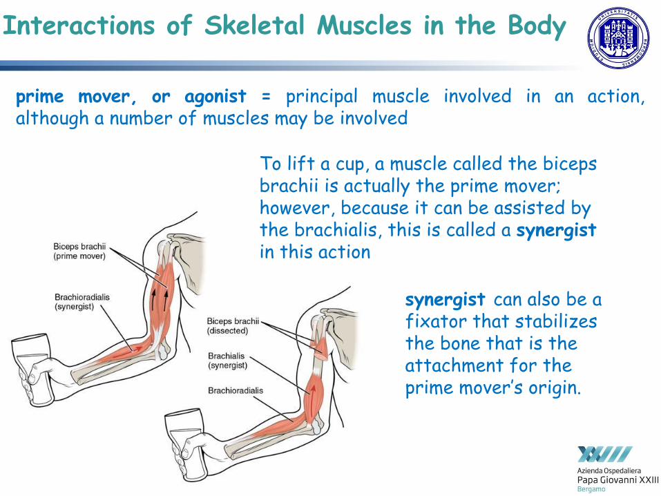
prime mover, or agonist = principal muscle involved in an action, although a number of muscles may be involved
Interactions of Skeletal Muscles in the Body
To lift a cup, a muscle called the biceps brachii is actually the prime mover; however, because it can be assisted by the brachialis, this is called a synergist in this action
synergist can also be a fixator that stabilizes the bone that is the attachment for the prime mover’s origin.

Interactions of Skeletal Muscles in the Body
Antagonist = muscle with the opposite action of the prime mover play 2 important roles in muscle function: (1) maintain body or limb position, such as holding the arm out or standing erect (2) control rapid movement
also be reversed for the opposing action
Interactions of Skeletal Muscles in the Body
Also skeletal muscles not pulling against skeleton for movements: • muscles that produce facial
expressions. • skeletal muscles in the tongue,
and the external urinary and anal sphincters that allow for voluntary regulation of urination and defecation, respectively.
• diaphragm contracts and relaxes to change the volume of the pleural cavities but it does not move the skeleton to do this.

Skeletal muscle enclosed in connective tissue scaffolding at 3 levels: • each muscle fiber (cell) is covered by endomysium • entire muscle is covered by epimysium • when a group of muscle fibers is “bundled” as a unit within the whole muscle (=
fascicle) by an additional covering of a connective tissue called perimysium Fascicle arrangement by perimysia is correlated to the force generated by a muscle; it also affects the range of motion of the muscle.
Patterns of Fascicle Organization

Based on the patterns of fascicle arrangement, skeletal muscles can be classified in several ways most common fascicle arrangements Parallel muscles = fascicles arranged in the same direction as the long axis of the muscle (majority of skeletal muscles): 1. some parallel muscles are flat sheets that expand at the ends to make broad attachments. 2. other parallel muscles are rotund with tendons at one or both ends. 3. muscles that seem to be plump have a large mass of tissue located in the middle of the muscle, between the insertion and the origin, which is known as the central body (= belly). When a parallel muscle has a central, large belly that is spindle-shaped, meaning it tapers as it extends to its origin and insertion, it sometimes is called fusiform.
Patterns of Fascicle Organization

Circular muscles = also called sphincters (= when they relax increase the size of the opening, and when they contract shrink to the point of closure) Convergent muscle = widespread expansion over a sizable area, but then the fascicles come to a single, common attachment point [that could be a tendon, an aponeurosis (=flat, broad tendon), or a raphe (= very slender tendon)]. Pennate muscles (= “feathers”) blend into a tendon that runs through the central region of the muscle for its whole length.
Patterns of Fascicle Organization
Patterns of Fascicle Organization
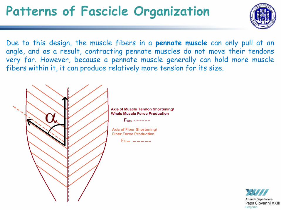
Due to this design, the muscle fibers in a pennate muscle can only pull at an angle, and as a result, contracting pennate muscles do not move their tendons very far. However, because a pennate muscle generally can hold more muscle fibers within it, it can produce relatively more tension for its size.

Patterns of Fascicle Organization
3 subtypes of pennate muscles: 1. unipennate muscle =
fascicles are located on one side of the tendon.
2. bipennate muscle = fascicles on both sides of the tendon.
3. multipennate muscles = muscle fibers wrap around the tendon, sometimes forming individual fascicles

Naming Skeletal Muscles
Greek and Latin

Naming Skeletal Muscles

Naming Skeletal Muscles
naming according to a number of criteria (each describing muscle in some way): • shape • size compared to other muscles in the area • location in the body or location of its attachments to the
skeleton • how many origins it has • action
divided into axial (muscles of the trunk and head) and appendicular (muscles of the arms and legs) categories, reflecting bones

Axial Muscles of the Head, Neck, and Back
origins of the muscles of facial expression = on skull surface (remember, the origin of
a muscle does not move) insertions = fibers intertwined with connective tissue and
the dermis of the skin, thus, when contracting, skin moves to create facial expression
Muscles That Create Facial Expression

Axial Muscles of the Head, Neck, and Back
Muscles That Create Facial Expression • orbicularis oris = circular muscle that moves the lips • orbicularis oculi = circular muscle that closes the eye • occipitofrontalis = moves up scalp and eyebrows (frontal belly + occipital belly: muscle
on the forehead [frontalis]and one on the back of the head [occipitalis], but no muscle across the top of the head, two bellies connected by a broad tendon called the epicranial aponeurosis, or galea (= “apple”)
• buccinator muscle = (majority of the face ) compresses cheek (whistle, blow, and suck + contributes to chew)
• corrugator supercilii = prime mover of the eyebrow • several additional small facial muscles

Axial Muscles of the Head, Neck, and Back
extrinsic eye muscles = movement of the eyeball, originate outside the eye and insert onto the outer surface of the white of the eye, located inside eye socket and cannot be seen on any part of the visible eyeball
Muscles That Move the Eyes

Axial Muscles of the Head, Neck, and Back
Muscles That Move the Eyes

Axial Muscles of the Head, Neck, and Back
chewing = mastication muscles involved in chewing must be able to exert enough pressure to bite through and then chew food before it is swallowed: masseter muscle = main muscle used for chewing, assisted by temporalis muscle (retracts mandible) medial pterygoid and lateral pterygoid muscles provide assistance in chewing and moving food within the mouth.
Muscles That Move the Lower Jaw

Axial Muscles of the Head, Neck, and Back
Muscles That Move the Lower Jaw

Axial Muscles of the Head, Neck, and Back
mastication, deglutition (swallowing), and speech tongue muscles intrinsic (insert into the tongue from origins within it, allow the tongue to change its shape
such as, curling the tongue in a loop or flattening it) + extrinsic (insert into the tongue from outside origins, move the whole tongue in different
directions), all include the word root glossus (glossus = “tongue”), and the muscle names are derived from where the muscle originates: genioglossus (genio = “chin”) originates on mandible and allows the tongue to move downward and forward; styloglossus originates on styloid bone, and allows upward and backward motion; palatoglossus originates on the soft palate to elevate the back of the tongue; hyoglossus originates on the hyoid bone to move the tongue downward and flatten it.
Muscles That Move the Tongue

Axial Muscles of the Head, Neck, and Back
assist in deglutition (swallowing) + speech by controlling the positions of the larynx and hyoid bone (= horseshoe-shaped bone functioning as a solid foundation on which tongue can move), neck muscles categorized according to their position relative to hyoid bone, suprahyoid muscles superior to it, and infrahyoid muscles located inferiorly.
Muscles of the Anterior Neck

Axial Muscles of the Head, Neck, and Back
Muscles of the Anterior Neck • suprahyoid muscles: raise hyoid bone, floor of the mouth, and larynx during
deglutition, including digastric muscle (= has anterior and posterior bellies working to elevate hyoid bone and larynx when one swallows; it also depresses the mandible), stylohyoid muscle (= moves hyoid bone posteriorly, elevating the larynx), mylohyoid muscle (lifts it and helps press the tongue to the top of the mouth), geniohyoid (depresses mandible in addition to raising and pulling the hyoid bone anteriorly)
• infrahyoid muscles (strap-like) generally depress hyoid bone and control position of the larynx: omohyoid muscle (has superior and inferior bellies, depresses hyoid bone in conjunction with thyrohyoid (also elevates the larynx’s thyroid cartilage,) and sternothyroid (depresses it to create different tones of voice)

Axial Muscles of the Head, Neck, and Back
Muscles That Move the Head

Axial Muscles of the Head, Neck, and Back
Sternocleidomastoid =major muscle that laterally flexes and rotates the head,
in addition, both muscles working together are the flexors of the head (divides the
neck into anterior and posterior triangles when viewed from the side)
Muscles That Move the Head

Axial Muscles of the Head, Neck, and Back
posterior muscles of the neck = primarily concerned with head movements, like extension back muscles = stabilize and move the vertebral column (grouped according to lengths and direction of the fascicles) splenius muscles = originating at the midline, run laterally and superiorly to their insertions: splenius capitis inserts onto head region, and splenius cervicis extends onto the cervical region, extending, laterally flexing and rotating head
Muscles of the Posterior Neck and the Back

Axial Muscles of the Head, Neck, and Back
erector spinae group = majority of the muscle mass of the back and primary extensor of vertebral column, controlling flexion, lateral flexion, and rotation and maintaining lumbar curve; comprises: iliocostalis (laterally placed) group:
iliocostalis cervicis, associated with the cervical region; iliocostalis thoracis, associated with the thoracic region; and iliocostalis lumborum, associated with the lumbar region.
longissimus (intermediately placed) group: 3 muscles = longissimus capitis, associated with the head region; longissimus cervicis, associated with the cervical region; longissimus thoracis, associated with the thoracic region.
spinalis (medially placed) group: spinalis capitis (head region), the spinalis cervicis (cervical region), and the spinalis thoracis (thoracic region).
Muscles of the Posterior Neck and the Back

Axial Muscles of the Head, Neck, and Back
transversospinales muscles = from transverse processes to spinous processes of vertebrae, are named for areas of the body with which they are associated: semispinalis muscles include semispinalis capitis, semispinalis cervicis, and semispinalis thoracis. multifidus muscle of lumbar region = extend and laterally flex vertebral column. segmental muscle group = important in stabilization of vertebral column, includes interspinales and intertransversarii muscles (bringing together spinous and transverse processes of each consecutive vertebra) scalene muscles = flex, laterally flex, and rotate the head and also contribute to deep inhalation, including anterior scalene muscle (anterior to the middle scalene), middle scalene muscle (the longest, intermediate between the anterior and posterior scalenes), and posterior scalene muscle (the smallest, posterior to the middle scalene).
Muscles of the Posterior Neck and the Back

muscles of vertebral column,
thorax, and abdominal wall
extend, flex, and stabilize
different parts of the body’s trunk
in order to balance body on two
feet and walk upright
Muscles of the Abdomen 4 pairs of abdominal muscles,
covering anterior and lateral
abdominal region and meet at
anterior midline = muscles of
anterolat abdominal:
• external obliques,
• internal obliques,
• transversus abdominis
• rectus abdominis
Axial Muscles of the Abdominal Wall and Thorax

Muscles of the Abdomen
Axial Muscles of the Abdominal Wall and Thorax

Muscles of the Abdomen
Axial Muscles of the Abdominal Wall and Thorax
3 flat muscles in ant-lat wall of abdomen: external oblique = closest to the surface, extend inferiorly and medially, internal oblique = perpendicular to it (intermediate), extending superiorly and medially, transversus abdominis = (deep muscle) arranged transversely around the abdomen, arrangement of 3 bands of muscles in different orientations allows various movements and rotations of trunk and also help to protect internal abdominal organs in an area where there is no bone. linea alba = white, fibrous band that is made of bilateral rectus sheaths that join at the anterior midline of the body, enclosing rectus abdominis muscles (a pair of long, linear muscles) originating at pubic crest and symphysis, and extending length of body’s trunk and segmented by three transverse bands of collagen fibers called tendinous intersections.

Pyramidalis Muscles
Axial Muscles of the Abdominal Wall and Thorax

Pyramidalis Muscles
Axial Muscles of the Abdominal Wall and Thorax
EPIPUBIC BONE in MARSUPIALS

Muscles of the Abdomen
Axial Muscles of the Abdominal Wall and Thorax
posterior abdominal wall is formed by the lumbar vertebrae, parts of the ilia of the hip bones, psoas major and iliacus muscles, and quadratus lumborum muscle = this part of the core plays a key role in stabilizing the rest of the body and maintaining posture.

Axial Muscles of the Abdominal Wall and Thorax
= to facilitate breathing by changing the size of the thoracic cavity
Muscles of the Thorax

Axial Muscles of the Abdominal Wall and Thorax
Muscles of the Thorax: Diaphragm = change in volume of thoracic cavity during breathing is due to alternate contraction and relaxation; separates the thoracic and abdominal cavities, and is dome-shaped at rest (superior surface convex, creating elevated floor of thoracic cavity, inferior surface is concave, creating curved roof of abdominal cavity).

Axial Muscles of the Abdominal Wall and Thorax
Muscles of the Thorax: Diaphragm Defecating, urination, and even childbirth involve cooperation between the diaphragm and abdominal muscles (= “Valsalva maneuver”): holding breath by a steady contraction of diaphragm with stabilization of volume and pressure of peritoneal cavity + abdominal muscles contract, pressure cannot push the diaphragm up, so it increases pressure on the intestinal tract (defecation), urinary tract (urination), or reproductive tract (childbirth).
Axial Muscles of the Abdominal Wall and Thorax
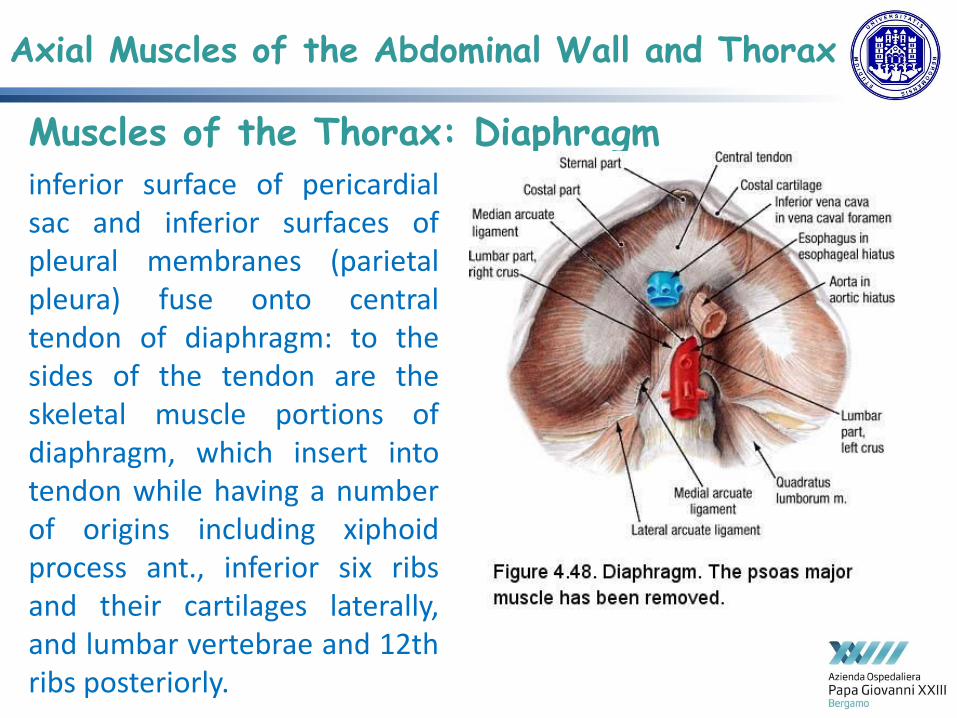
Muscles of the Thorax: Diaphragm inferior surface of pericardial sac and inferior surfaces of pleural membranes (parietal pleura) fuse onto central tendon of diaphragm: to the sides of the tendon are the skeletal muscle portions of diaphragm, which insert into tendon while having a number of origins including xiphoid process ant., inferior six ribs and their cartilages laterally, and lumbar vertebrae and 12th ribs posteriorly.

Axial Muscles of the Abdominal Wall and Thorax
Muscles of the Thorax: Diaphragm
diaphragm also includes 3 openings for passage of structures between thorax and abdomen: 1. inferior vena cava through caval opening, 2. esophagus and attached nerves through esophageal hiatus, 3. aorta, thoracic duct, and azygous vein through aortic hiatus of the posterior diaphragm.

Axial Muscles of the Abdominal Wall and Thorax
3 sets of muscles, = intercostal muscles, which span each of the intercostal spaces: principal role to assist in breathing by changing the dimensions of the rib cage 1. 11 pairs of superficial external intercostal muscles aid in inspiration, raising rib cage,
which expands it 2. 11 pairs of internal intercostal muscles, just under the externals, used for expiration
because they constrict the rib cage 3. innermost intercostal muscles (deepest): act as synergists for action of internal
intercostals.
The Intercostal Muscles

Axial Muscles of the Abdominal Wall and Thorax
pelvic floor = muscular sheet defining inferior portion of pelvic cavity: pelvic diaphragm, spanning ant to post from pubis to coccyx, comprises : levator ani [= consists of 2 muscles, pubococcygeus and iliococcygeus, considered the most important muscle of pelvic floor supporting pelvic viscera and resisting pressure produced by contraction of abd muscles applied to colon to aid in defecation and to uterus to aid in childbirth, also creates skeletal muscle sphincters at the urethra and anus]+ assisted by the ischiococcygeus, which pulls the coccyx anteriorly openings include anal canal and urethra, and vagina in women.
Muscles of the Pelvic Floor and Perineum

Axial Muscles of the Abdominal Wall and Thorax
Muscles of the Pelvic Floor and Perineum perineum = diamond-shaped space between pubic symphysis (ant), coccyx (post), and ischial tuberosities (lat): divided transversely into triangles, ant urogenital triangle = external genitals and post anal triangle = anus

Muscles of the Pectoral Girdle and Upper Limbs
Generality Muscles of the shoulder and upper limb can be divided into 4 groups: 1. muscles that stabilize and position pectoral girdle, 2. muscles that move arm, 3. muscles that move forearm 4. muscles that move wrists, hands, and fingers. pectoral girdle, or shoulder girdle = consists of lateral ends of clavicle and scapula, along with proximal end of humerus, and muscles covering these 3 bones to stabilize shoulder joint. The girdle creates a base from which head of humerus, in its ball-and-socket joint with the glenoid fossa of scapula, can move arm in multiple directions.

Muscles of the Pectoral Girdle and Upper Limbs
located either on ant or on post thorax: anterior muscles 1. subclavius, 2. pectoralis minor 3. serratus anterior posterior muscles 1. trapezius 2. rhomboid major 3. rhomboid minor
1. Muscles That Position the Pectoral Girdle

Muscles of the Pectoral Girdle and Upper Limbs
1. Muscles That Position the Pectoral Girdle

Muscles of the Pectoral Girdle and Upper Limbs
2. Muscles That Move the Humerus 2 axial muscles 1. pectoralis major = thick and fan-shaped, covering much of superior portion of anterior
thorax. 2. latissimus dorsi = broad, triangular latissimus dorsi is located on inferior part of back, where
it inserts into a thick connective tissue shealth called an aponeurosis. 7 muscles originating on scapula 1. deltoid = thick muscle creating rounded lines of shoulder: major abductor of arm (also
facilitates flexing and medial rotation, as well as extension and lateral rotation) 2. Subscapularis = originates on the anterior scapula and medially rotates the arm. 3. supraspinatus = (superior to the spine of the scapula) abduct the arm 4. Infraspinatus = (inferior to the spine of the scapula) laterally rotate the arm 5. teres major = thick and flat is inferior to the teres minor and extends the arm, and assists in
adduction and medial rotation of it 6. teres minor = laterally rotates and extends the arm 7. coracobrachialis = flexes and adducts the arm.
= axial and scapular muscles:

Muscles of the Pectoral Girdle and Upper Limbs
2. Muscles That Move the Humerus rotator cuff (musculotendinous cuff) = circle of tendons around shoulder joint: tendons of
deep subscapularis, supraspinatus, infraspinatus, and teres minor connect scapula to humerus
= axial and scapular muscles:

Muscles of the Pectoral Girdle and Upper Limbs
2. Muscles That Move the Humerus

Muscles of the Pectoral Girdle and Upper Limbs
2. Muscles That Move the Humerus

Muscles of the Pectoral Girdle and Upper Limbs
3. Muscles That Move the Forearm
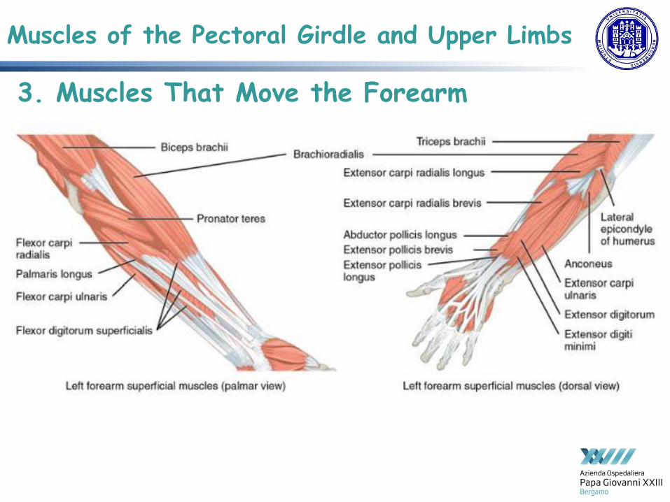
forearm flexors (anterior flexor compartment of the arm) = 1. biceps brachii (two-headed muscle crossing shoulder and elbow joints to flex forearm, also taking part in supinating the forearm at radioulnar joints and flexing arm at shoulder joint), 2. brachialis (provides additional power in flexing forearm), 3. brachioradialis (flex forearm quickly or help lift a load slowly).
extensors = triceps brachii and anconeus. pronators = pronator teres and pronator quadratus, supinator = only one that turns forearm anteriorly
Forearm = 4 main types of action: flexion, extension, pronation, and supination

Muscles of the Pectoral Girdle and Upper Limbs
3. Muscles That Move the Forearm
Muscles of the Pectoral Girdle and Upper Limbs
3. Muscles That Move the Forearm

Muscles of the Pectoral Girdle and Upper Limbs
3. Muscles That Move the Forearm

Muscles of the Pectoral Girdle and Upper Limbs
4. Muscles That Move the Wrist, Hand, and Fingers Wrist, hand, and finger movements are facilitated by 2 groups of muscles: extrinsic muscles of the hand = forearm is origin intrinsic muscles of the hand = palm is origin

Muscles of the Pectoral Girdle and Upper Limbs
4a. Extrinsic muscles superficial anterior flexor compartment of the forearm = originate on humerus and insert
onto different parts of hand: (bulk of forearm), -from lateral to medial- flexor carpi radialis, palmaris longus, flexor carpi ulnaris, and flexor digitorum superficialis (= flexes hand as well as digits at the knuckles, allowing for rapid finger movements, as in typing or playing a musical instrument (Carpal Tunnel Syndrome).
deep anterior compartment = flexion and bends fingers to make a fist: flexor pollicis longus and flexor digitorum profundus.
superficial posterior extensor compartment of the forearm = originate on the humerus: extensor radialis longus, extensor carpi radialis brevis, extensor digitorum, extensor digiti minimi, and extensor carpi ulnaris.
deep posterior extensor compartment of the forearm= originate on radius and ulna: abductor pollicis longus, extensor pollicis brevis, extensor pollicis longus, and extensor indicis.
tendons of forearm muscles attach to wrist and extend into the hand: fibrous bands called retinacula sheath tendons at the wrist flexor retinaculum extends over hand palmar surface, while extensor retinaculum extends over hand dorsal surface

Muscles of the Pectoral Girdle and Upper Limbs
4a. Extrinsic muscles

Muscles of the Pectoral Girdle and Upper Limbs
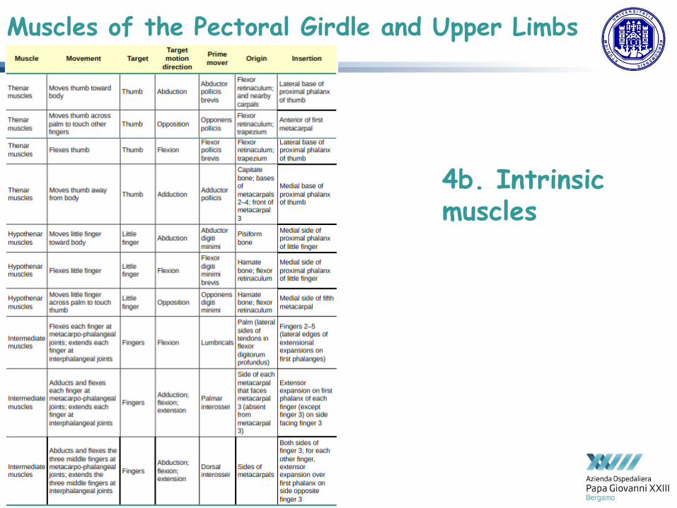
4b. Intrinsic muscles = both originate and insert within hand, allowing to make precise movements for actions, such as typing or writing, divided into 3 groups: 1.thenar muscles (on radial aspect of the palm, abductor pollicis brevis, opponens pollicis, flexor pollicis brevis, and the adductor pollicis - form thenar eminence, rounded contour of thumb base, all act on thumb), 2. hypothenar muscles (on medial aspect, abductor digiti minimi, flexor digiti minimi brevis, opponens digiti minimi - form hypothenar eminence, rounded contour of little finger, all act on little finger), 3.intermediate muscles (midpalmar, act on all fingers, lumbrical, palmar interossei, and dorsal interossei).
Muscles of the Pectoral Girdle and Upper Limbs
4b. Intrinsic muscles

Appendicular Muscles of Pelvic Girdle and Lower Limbs
Gluteal Region Muscles That Move the Femur Most muscles that insert on femur and move it, originate on pelvic girdle.
psoas major and iliacus = iliopsoas group.
gluteal group = some of the largest and most powerful muscles in the body: 1.gluteus
maximus (largest); 2. gluteus medius (deep to gluteus maximus); 3. gluteus minimus
(deep to the gluteus medius, smallest of the trio)
tensor fascia lata = thick, squarish muscle in superior aspect of lateral thigh (acts as a
synergist of gluteus medius and iliopsoas in flexing and abducting the thigh + stabilize lateral
aspect of the knee)
piriformis, obturator internus, obturator externus, superior gemellus, inferior gemellus,
and quadratus femori = deep to gluteus maximus, laterally rotate femur at the hip.
adductor longus, adductor brevis, and adductor magnus = medially and laterally rotate
thigh depending on the placement of the foot (adductor longus flexes thigh, whereas adductor
magnus extends it).
pectineus adducts and flexes femur at hip as well (located in femoral triangle, also includes
the femoral nerve, the femoral artery, the femoral vein, and the deep inguinal lymph nodes
Pelvic girdle = less range of motion because designed to stabilize and support body.
Muscles of the Thigh

Appendicular Muscles of Pelvic Girdle and Lower Limbs
Gluteal Region Muscles That Move the Femur
Muscles of the Thigh

Appendicular Muscles of Pelvic Girdle and Lower Limbs
Gluteal Region Muscles That Move the Femur
Muscles of the Thigh

Appendicular Muscles of Pelvic Girdle and Lower Limbs
Thigh Muscles That Move the Femur, Tibia, and Fibula Deep fascia in thigh separates into: 1.medial (responsible for adducting the femur at the hip) = 1.gracilis (strap-like) along with adductor longus, adductor brevis, adductor magnus, and pectineus, adducts thigh in addition to flexing leg at knee. 2.anterior (flex thigh and extend leg) = 1. quadriceps femoris group (= 4 muscles extending and stabilizing knee: rectus femoris, vastus lateralis, vastus medialis, vastus intermedius patellar tendon common to all four inserting into patella and continuing below it as patellar ligament, attaches to tibial tuberosity 2. sartorius (band-like muscle extending from ant. Sup. iliac spine to medial side of proximal tibia flexes leg at knee and flexes, abducts, and laterally rotates leg at hip = to sit cross-legged) 3.posterior compartments (flex leg and extend thigh) = (hamstring group flexing knee) 1.biceps femoris, 2.semitendinosus, 3.semimembranosus (tendons form popliteal fossa = diamond-shaped space at back of knee).
Muscles of the Thigh

Appendicular Muscles of Pelvic Girdle and Lower Limbs
Thigh Muscles That Move the Femur, Tibia, and Fibula
Muscles of the Thigh

Appendicular Muscles of Pelvic Girdle and Lower Limbs
deep fascia separate muscles of the leg into 3 compartments:
1. Anterior = 1. tibialis anterior (long and thick muscle on lateral surface of tibia), 2. extensor hallucis longus, 3. extensor digitorum longus (all contribute to raising front of foot), 4. fibularis tertius (associated with extensor digitorum longus, but not present in all people) [thick bands of connective tissue = superior extensor retinaculum (transverse ligament of the ankle) and inferior extensor retinaculum hold tendons of these muscles in place during dorsiflexion]
2. Lateral = 1. fibularis longus (peroneus longus) and 2. fibularis brevis (p. brevis) 3. Posterior = • superficial = [all insert onto strong calcaneal tendon (Achilles tendon) inserting
into calcaneal bone] muscles large and strong keeping humans upright = 1. gastrocnemius (most superficial and visible muscle of the calf) 2. soleus (deep to gastrocnemius, wide, flat) 3. plantaris (running obliquely between two) 4. tibialis posterior
• deep = 1. popliteus, 2.flexor digitorum longus, 3. flexor hallucis longus, 4. tibialis posterior
Muscles That Move the Feet and Toes

Appendicular Muscles of Pelvic Girdle and Lower Limbs
Muscles That Move the Feet and Toes

Appendicular Muscles of Pelvic Girdle and Lower Limbs
Muscles That Move the Feet and Toes

Appendicular Muscles of Pelvic Girdle and Lower Limbs
foot also has intrinsic muscles = originate and insert within it,
primarily providing support for foot and its arch and contributing to movements of toes, 2 groups: dorsal group = 1. extensor digitorum brevis plantar group = 4 layers, starting with the most superficial. [principal support for longitudinal arch of foot = deep fascia called plantar aponeurosis, running from calcaneus bone to toes]
Muscles That Move the Feet and Toes

Appendicular Muscles of Pelvic Girdle and Lower Limbs
Muscles That Move the Feet and Toes

Appendicular Muscles of Pelvic Girdle and Lower Limbs
Muscles That Move the Feet and Toes
intrinsic