Embed Size (px)
Citation preview
Murmurs in Children
Adrian Dancea MD, MBA, FRCPC
Division Head, Pediatric Cardiology
Montreal Children’s Hospital of the McGill University Health Center

• I have no financial disclosures
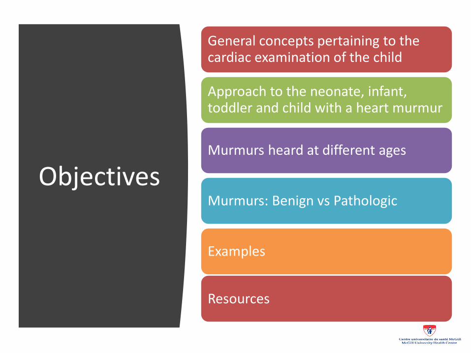
Objectives
General concepts pertaining to the cardiac examination of the child
Approach to the neonate, infant, toddler and child with a heart murmur
Murmurs heard at different ages
Murmurs: Benign vs Pathologic
Examples
Resources
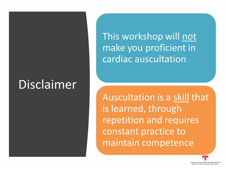
Disclaimer
This workshop will notmake you proficient in cardiac auscultation
Auscultation is a skill that is learned, through repetition and requires constant practice to maintain competence
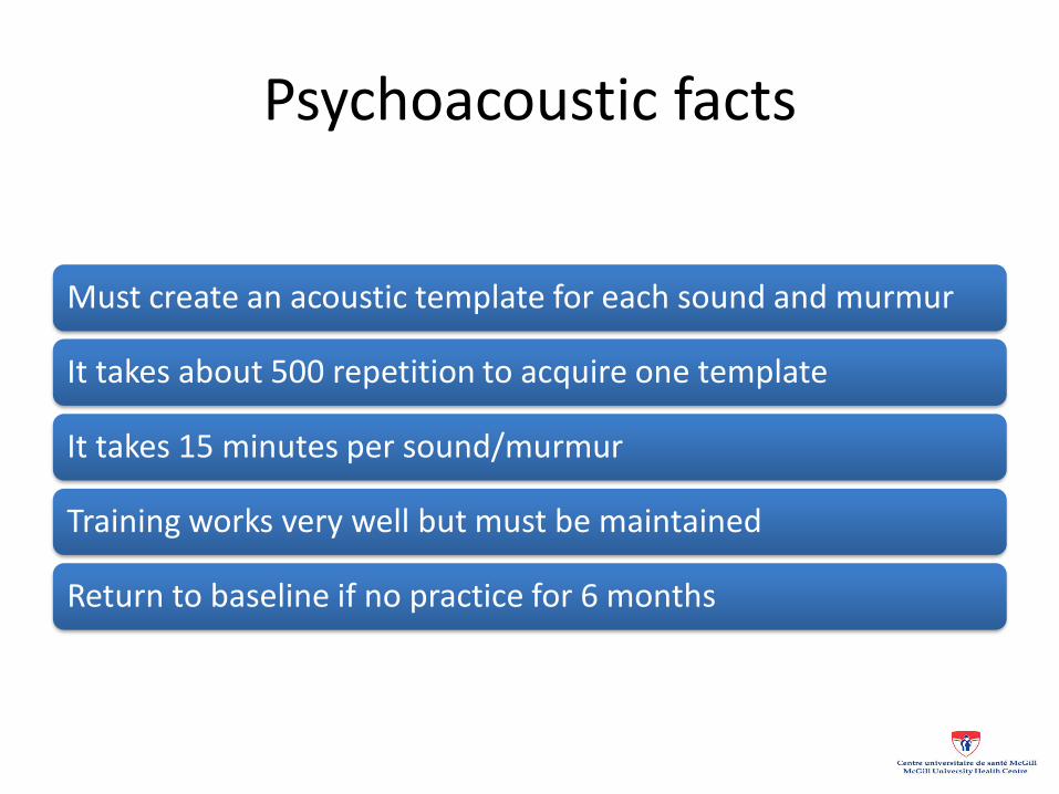
Psychoacoustic facts
Must create an acoustic template for each sound and murmur
It takes about 500 repetition to acquire one template
It takes 15 minutes per sound/murmur
Training works very well but must be maintained
Return to baseline if no practice for 6 months
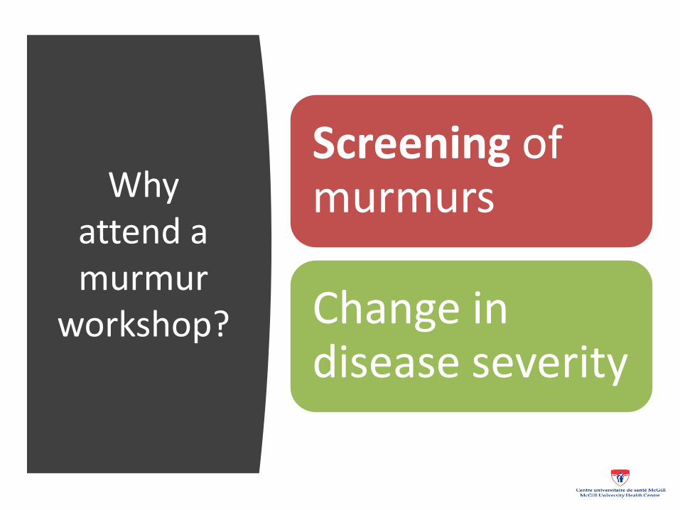
Why attend a murmur
workshop?
Screening of murmurs
Change in disease severity
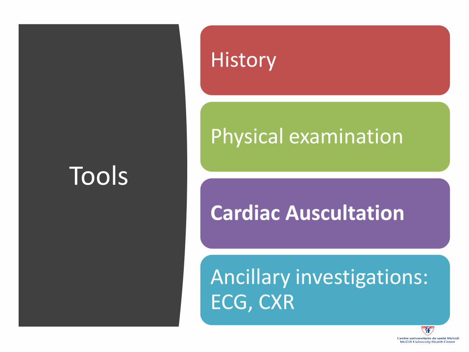
Tools
History
Physical examination
Cardiac Auscultation
Ancillary investigations: ECG, CXR

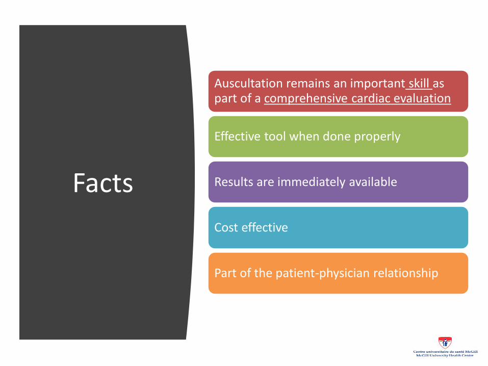
Facts
Auscultation remains an important skill as part of a comprehensive cardiac evaluation
Effective tool when done properly
Results are immediately available
Cost effective
Part of the patient-physician relationship
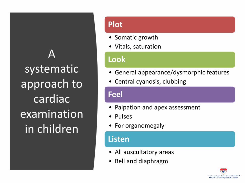
A systematic
approach to cardiac
examination in children
Plot
• Somatic growth
• Vitals, saturation
Look
• General appearance/dysmorphic features
• Central cyanosis, clubbing
Feel
• Palpation and apex assessment
• Pulses
• For organomegaly
Listen
• All auscultatory areas
• Bell and diaphragm

General concepts
• Quiet room
• Quiet child
– Establish report
– Distractions
– Parent’s lap for anxious toddlers
– Warm hands
– Warm stethoscope
• Quiet parent
• Good tools
– Invest in a decent stethoscope
– Shorter than 30” tubing
– Comfortable earpiece
• Be thorough but flexible
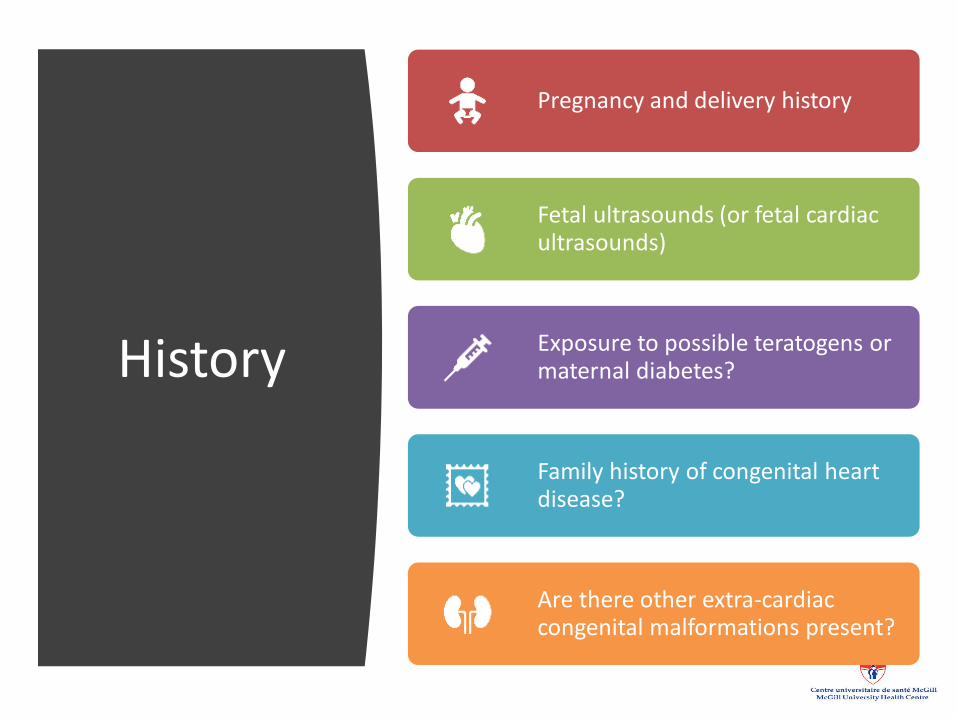
History
Pregnancy and delivery history
Fetal ultrasounds (or fetal cardiac ultrasounds)
Exposure to possible teratogens or maternal diabetes?
Family history of congenital heart disease?
Are there other extra-cardiac congenital malformations present?
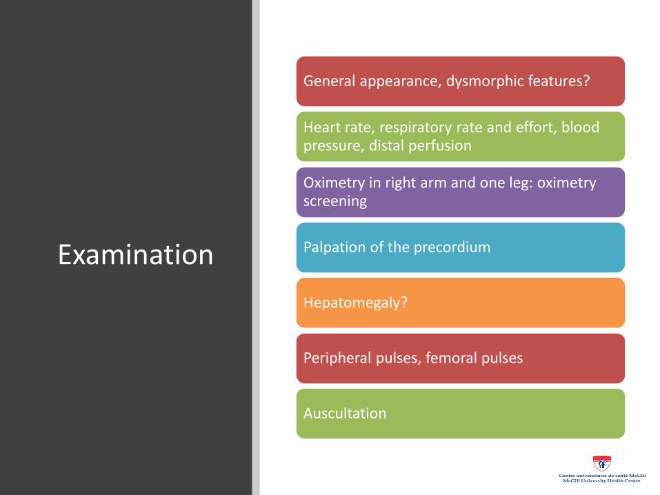
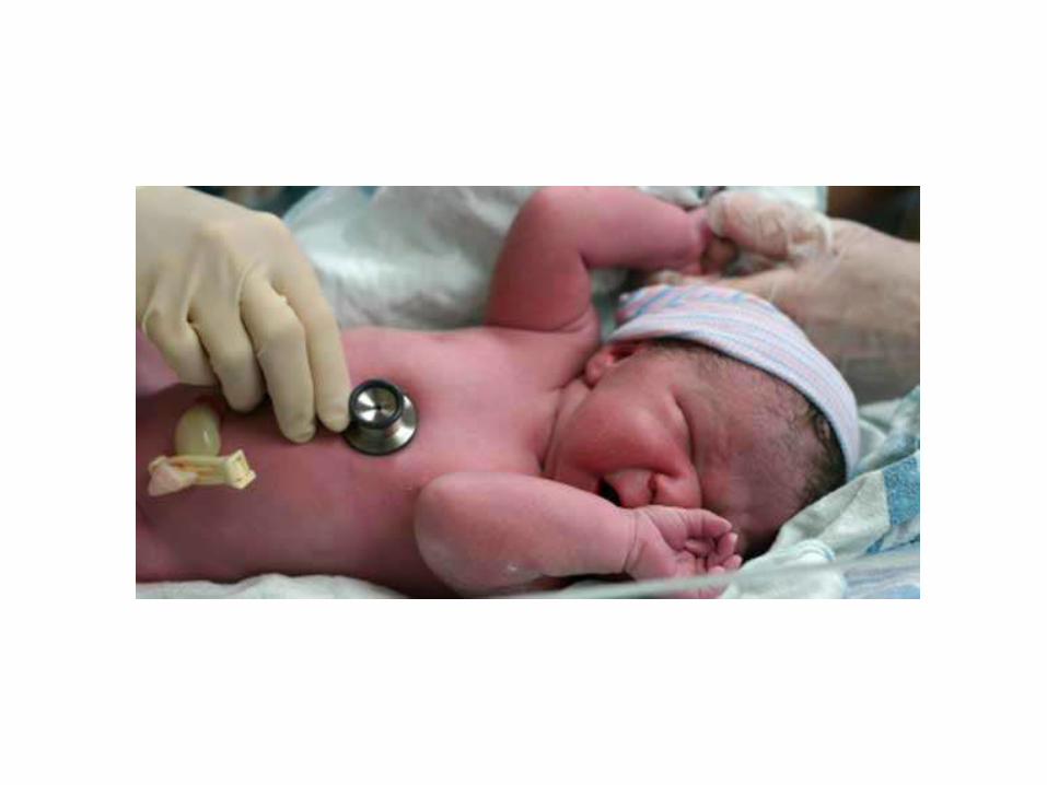
Examination
General appearance, dysmorphic features?
Heart rate, respiratory rate and effort, blood pressure, distal perfusion
Oximetry in right arm and one leg: oximetry screening
Palpation of the precordium
Hepatomegaly?
Peripheral pulses, femoral pulses
Auscultation
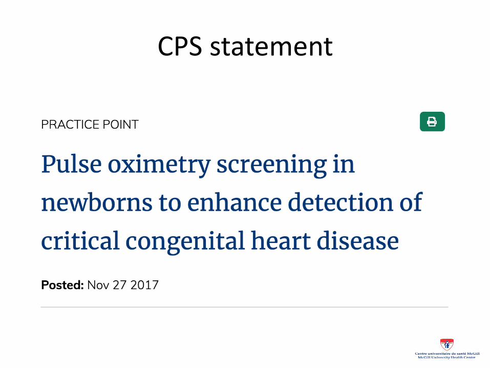
CPS statement
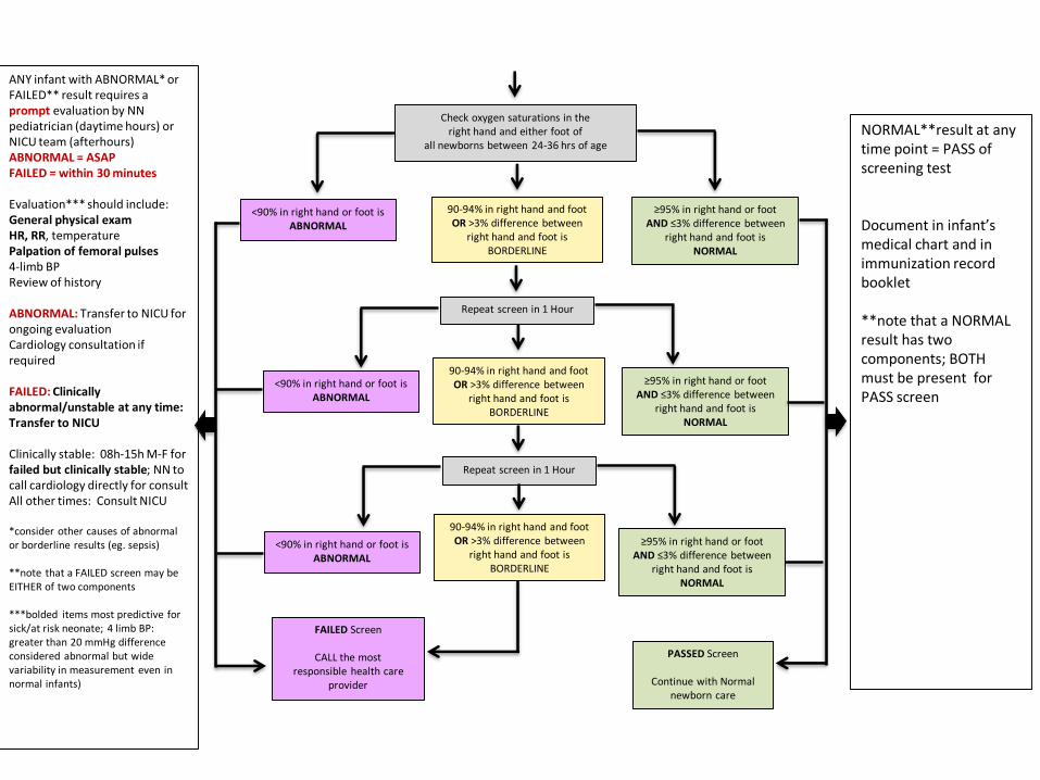
Check oxygen saturations in theright hand and either foot of
all newborns between 24-36 hrs of age
90-94% in right hand and foot OR >3% difference between
right hand and foot isBORDERLINE
90-94% in right hand and foot OR >3% difference between
right hand and foot isBORDERLINE
Repeat screen in 1 Hour
Repeat screen in 1 Hour
90-94% in right hand and foot OR >3% difference between
right hand and foot isBORDERLINE
<90% in right hand or foot isABNORMAL
<90% in right hand or foot isABNORMAL
<90% in right hand or foot isABNORMAL
≥95% in right hand or foot AND ≤3% difference between
right hand and foot isNORMAL
≥95% in right hand or foot AND ≤3% difference between
right hand and foot isNORMAL
≥95% in right hand or foot AND ≤3% difference between
right hand and foot isNORMAL
FAILED Screen
CALL the mostresponsible health care
provider
PASSED Screen
Continue with Normalnewborn care
ANY infant with ABNORMAL* or FAILED** result requires a prompt evaluation by NN pediatrician (daytime hours) or NICU team (afterhours) ABNORMAL = ASAPFAILED = within 30 minutes
Evaluation*** should include:General physical examHR, RR, temperaturePalpation of femoral pulses4-limb BPReview of history
ABNORMAL: Transfer to NICU for ongoing evaluationCardiology consultation if required
FAILED: Clinically abnormal/unstable at any time: Transfer to NICU
Clinically stable: 08h-15h M-F for failed but clinically stable; NN to call cardiology directly for consult All other times: Consult NICU
*consider other causes of abnormal or borderline results (eg. sepsis)
**note that a FAILED screen may be EITHER of two components
***bolded items most predictive for sick/at risk neonate; 4 limb BP: greater than 20 mmHg difference considered abnormal but wide variability in measurement even in normal infants)
NORMAL**result at any time point = PASS of screening test
Document in infant’s medical chart and in immunization record booklet
**note that a NORMAL result has two components; BOTH must be present for PASS screen

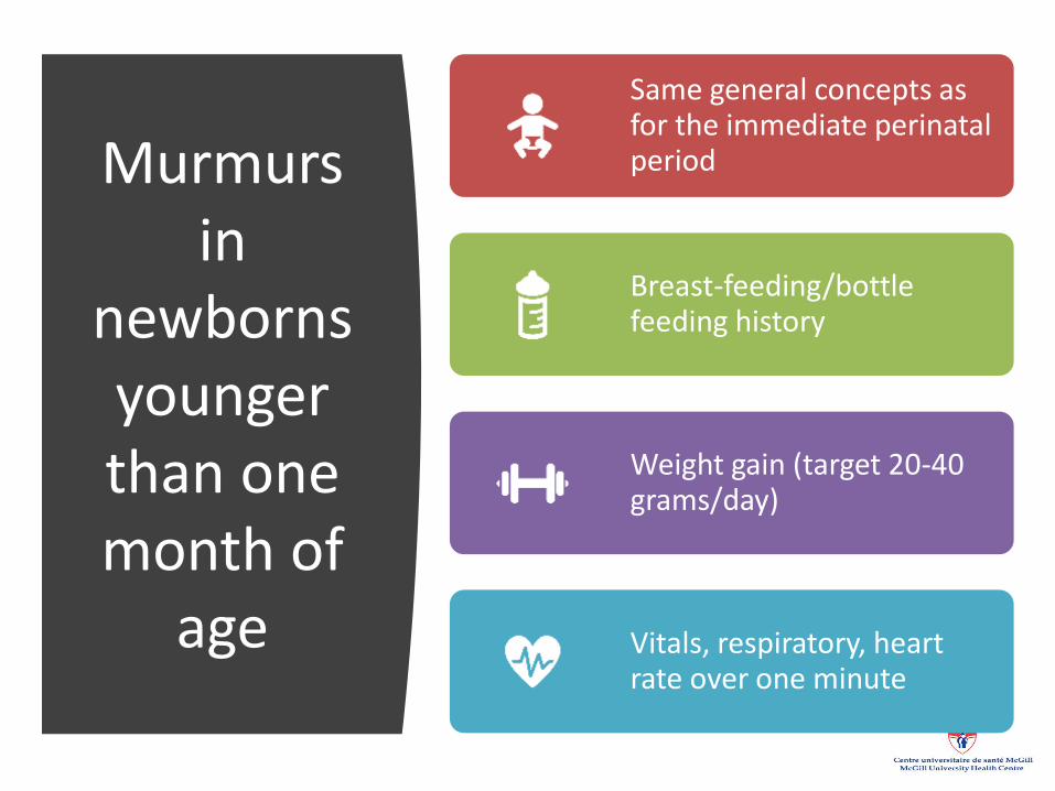
Murmurs in
newborns younger than one month of
age
Same general concepts as for the immediate perinatal period
Breast-feeding/bottle feeding history
Weight gain (target 20-40 grams/day)
Vitals, respiratory, heart rate over one minute
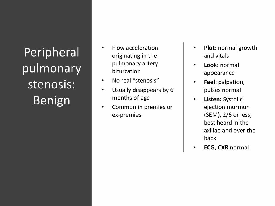
Peripheral pulmonary stenosis: Benign
• Flow acceleration originating in the pulmonary artery bifurcation
• No real “stenosis”
• Usually disappears by 6 months of age
• Common in premies or ex-premies
• Plot: normal growth and vitals
• Look: normal appearance
• Feel: palpation, pulses normal
• Listen: Systolic ejection murmur (SEM), 2/6 or less, best heard in the axillae and over the back
• ECG, CXR normal
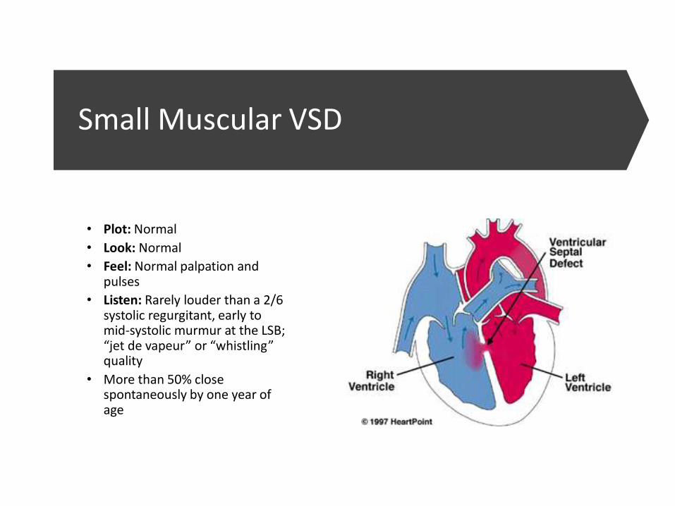
Small Muscular VSD
• Plot: Normal
• Look: Normal
• Feel: Normal palpation and pulses
• Listen: Rarely louder than a 2/6 systolic regurgitant, early to mid-systolic murmur at the LSB; “jet de vapeur” or “whistling” quality
• More than 50% close spontaneously by one year of age
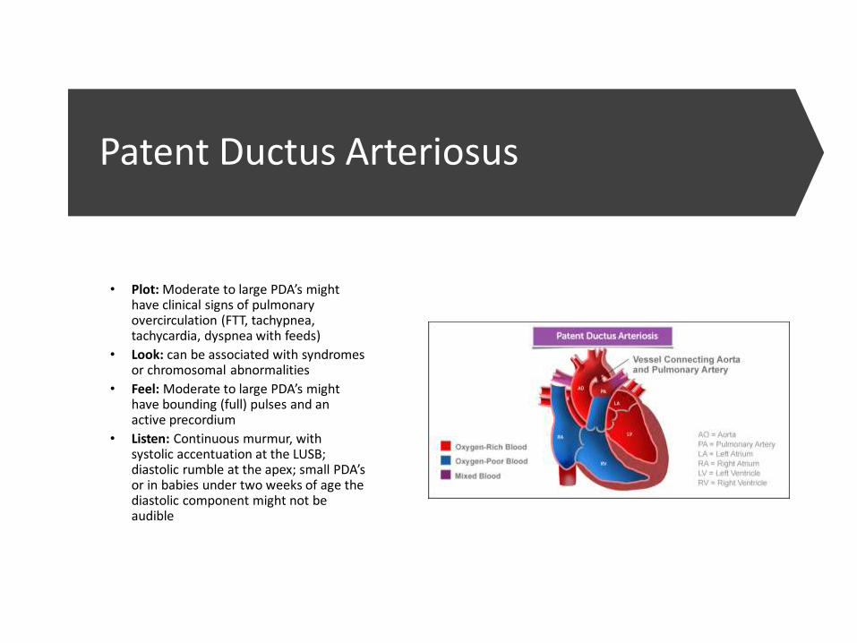
Patent Ductus Arteriosus
• Plot: Moderate to large PDA’s might have clinical signs of pulmonary overcirculation (FTT, tachypnea, tachycardia, dyspnea with feeds)
• Look: can be associated with syndromes or chromosomal abnormalities
• Feel: Moderate to large PDA’s might have bounding (full) pulses and an active precordium
• Listen: Continuous murmur, with systolic accentuation at the LUSB; diastolic rumble at the apex; small PDA’s or in babies under two weeks of age the diastolic component might not be audible
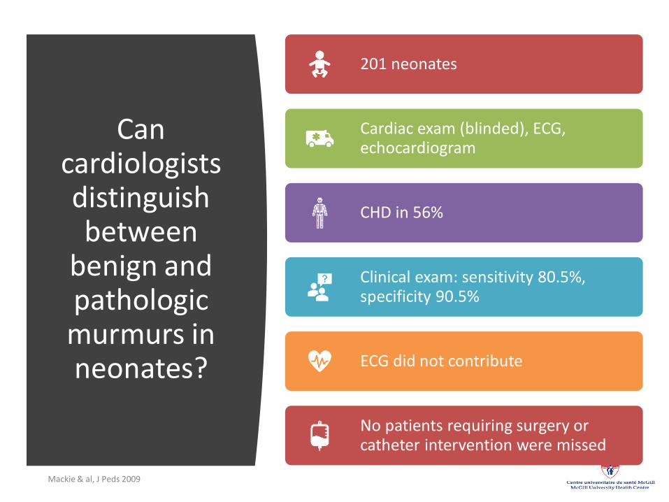
Can cardiologists distinguish between
benign and pathologic murmurs in neonates?
Mackie & al, J Peds 2009
201 neonates
Cardiac exam (blinded), ECG, echocardiogram
CHD in 56%
Clinical exam: sensitivity 80.5%, specificity 90.5%
ECG did not contribute
No patients requiring surgery or catheter intervention were missed

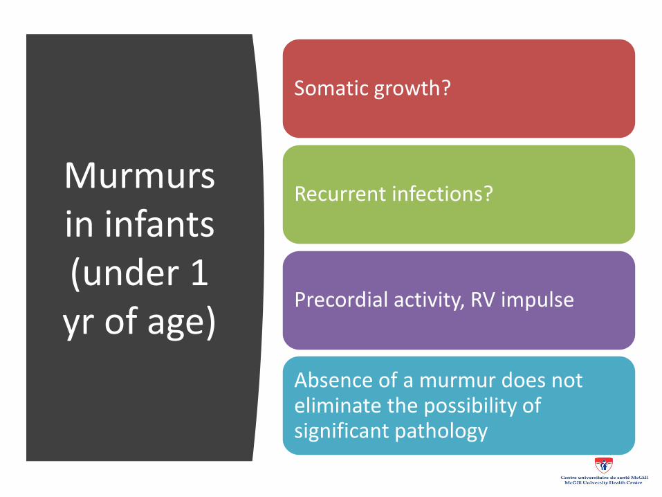
Murmurs in infants (under 1 yr of age)
Somatic growth?
Recurrent infections?
Precordial activity, RV impulse
Absence of a murmur does not eliminate the possibility of significant pathology
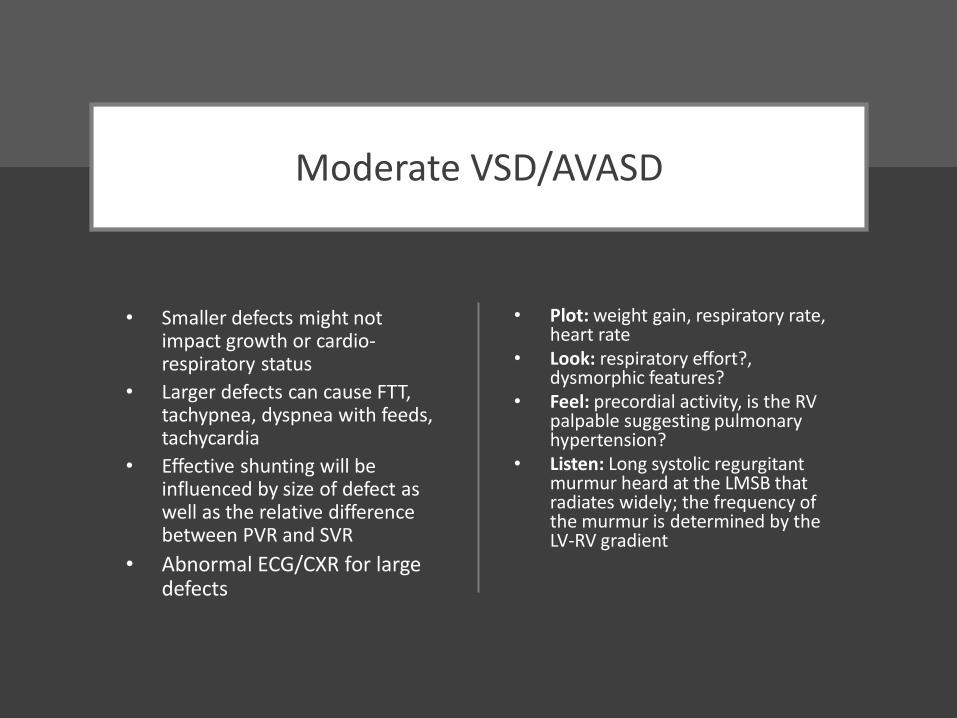
Moderate VSD/AVASD
• Smaller defects might not impact growth or cardio-respiratory status
• Larger defects can cause FTT, tachypnea, dyspnea with feeds, tachycardia
• Effective shunting will be influenced by size of defect as well as the relative difference between PVR and SVR
• Abnormal ECG/CXR for large defects
• Plot: weight gain, respiratory rate, heart rate
• Look: respiratory effort?, dysmorphic features?
• Feel: precordial activity, is the RV palpable suggesting pulmonary hypertension?
• Listen: Long systolic regurgitant murmur heard at the LMSB that radiates widely; the frequency of the murmur is determined by the LV-RV gradient

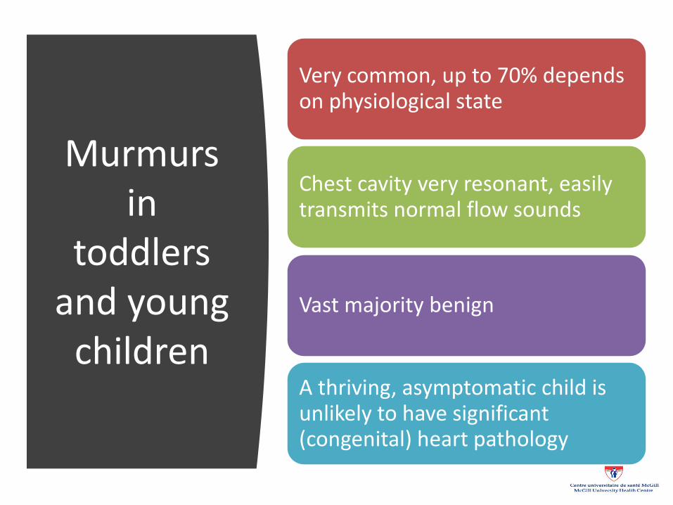
Murmurs in
toddlers and young
children
Very common, up to 70% depends on physiological state
Chest cavity very resonant, easily transmits normal flow sounds
Vast majority benign
A thriving, asymptomatic child is unlikely to have significant (congenital) heart pathology
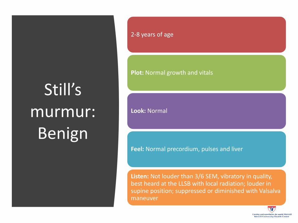
Still’s murmur: Benign
2-8 years of age
Plot: Normal growth and vitals
Look: Normal
Feel: Normal precordium, pulses and liver
Listen: Not louder than 3/6 SEM, vibratory in quality, best heard at the LLSB with local radiation; louder in supine position; suppressed or diminished with Valsalva maneuver
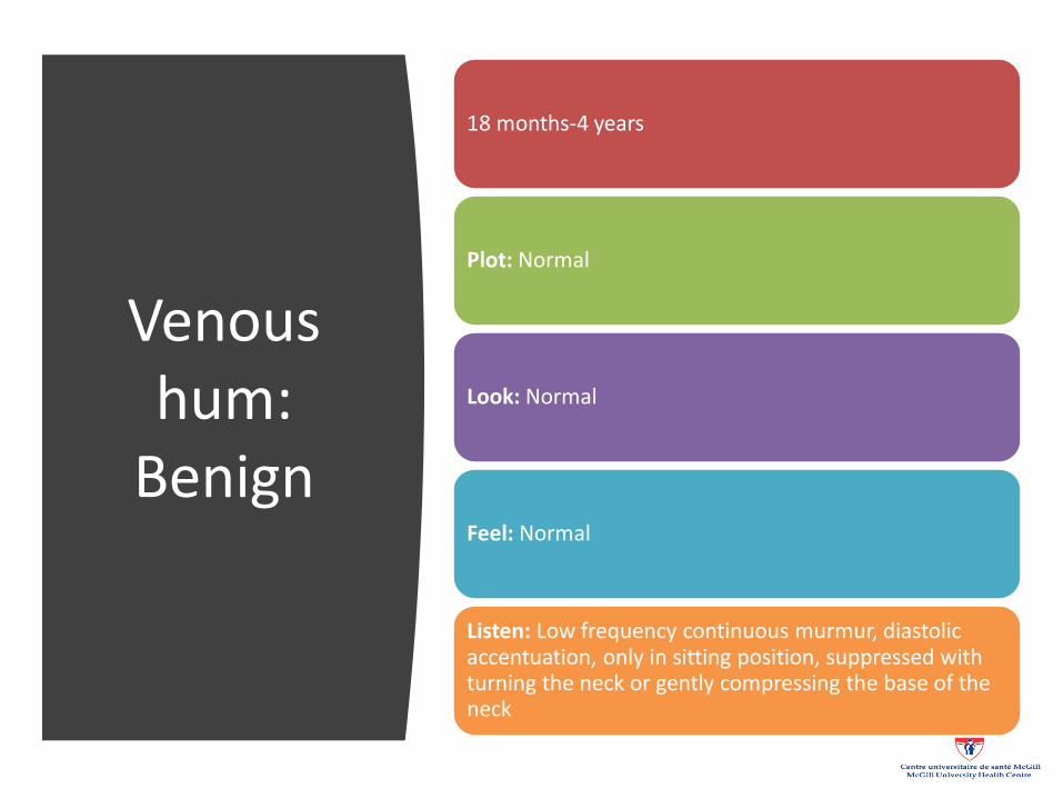
Venous hum:
Benign
18 months-4 years
Plot: Normal
Look: Normal
Feel: Normal
Listen: Low frequency continuous murmur, diastolic accentuation, only in sitting position, suppressed with turning the neck or gently compressing the base of the neck
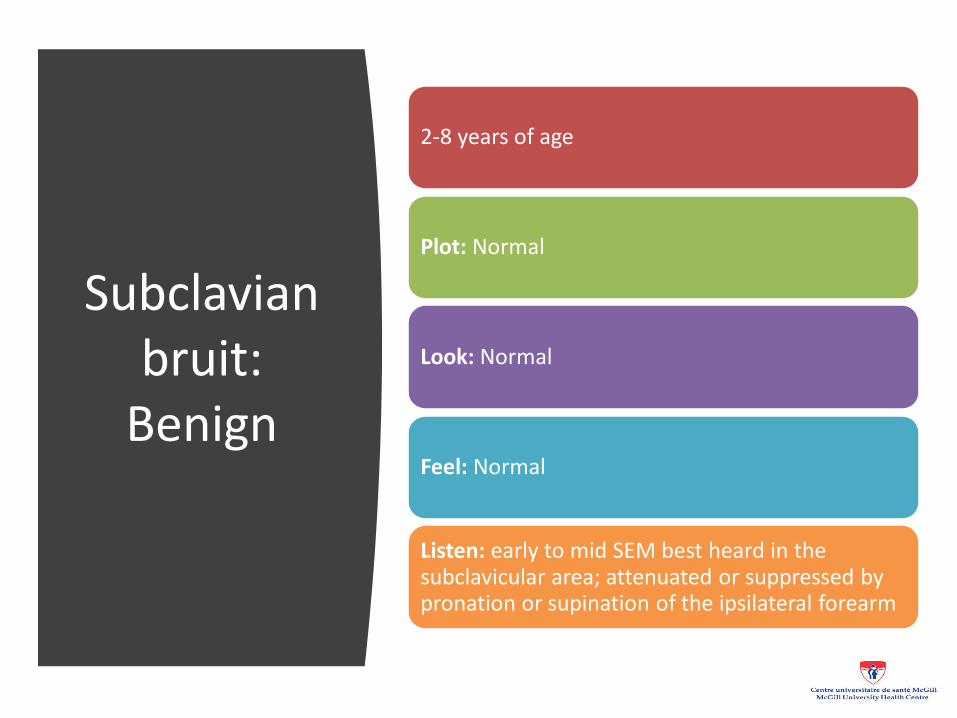
Subclavian bruit:
Benign
2-8 years of age
Plot: Normal
Look: Normal
Feel: Normal
Listen: early to mid SEM best heard in the subclavicular area; attenuated or suppressed by pronation or supination of the ipsilateral forearm

Carotid bruit:
Benign
2 years –adolescence
Plot: Normal
Look: Normal
Feel: can be associated with thrill in neck
Listen: early to mid SEM, right more than left
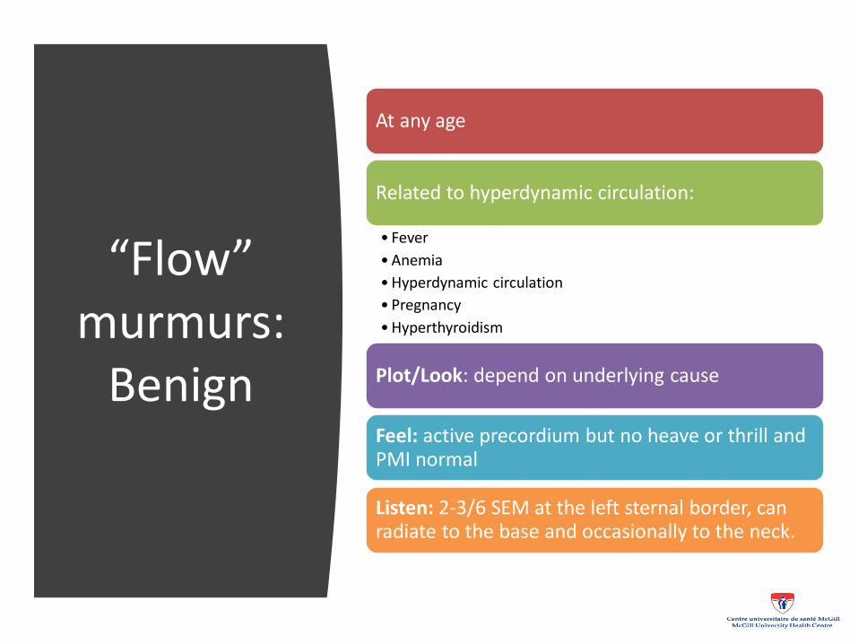
“Flow” murmurs:
Benign
At any age
Related to hyperdynamic circulation:
• Fever
• Anemia
• Hyperdynamic circulation
• Pregnancy
• Hyperthyroidism
Plot/Look: depend on underlying cause
Feel: active precordium but no heave or thrill and PMI normal
Listen: 2-3/6 SEM at the left sternal border, can radiate to the base and occasionally to the neck.

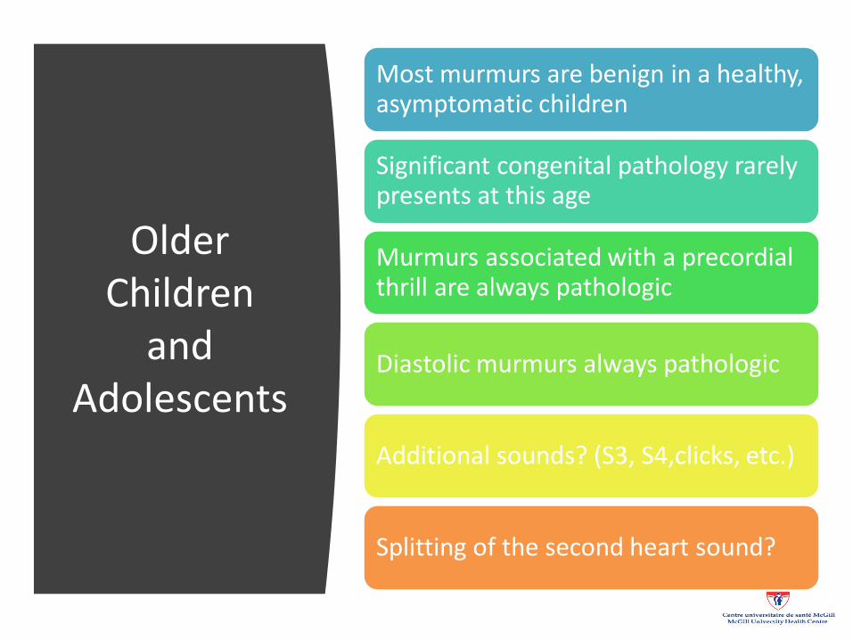
Older Children
and Adolescents
Most murmurs are benign in a healthy, asymptomatic children
Significant congenital pathology rarely presents at this age
Murmurs associated with a precordial thrill are always pathologic
Diastolic murmurs always pathologic
Additional sounds? (S3, S4,clicks, etc.)
Splitting of the second heart sound?
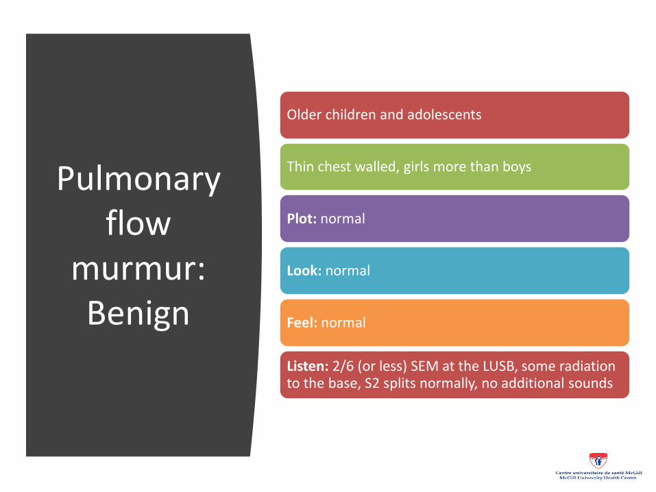
Pulmonary flow
murmur: Benign
Older children and adolescents
Thin chest walled, girls more than boys
Plot: normal
Look: normal
Feel: normal
Listen: 2/6 (or less) SEM at the LUSB, some radiation to the base, S2 splits normally, no additional sounds
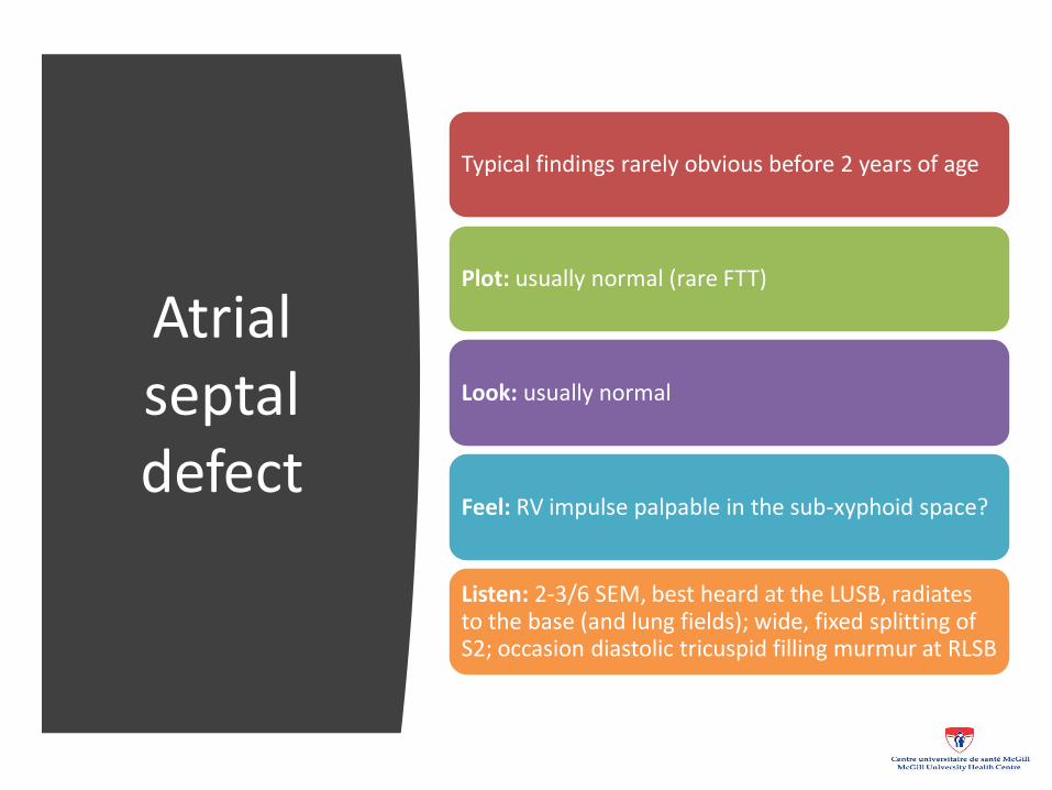
Atrial septal defect
Typical findings rarely obvious before 2 years of age
Plot: usually normal (rare FTT)
Look: usually normal
Feel: RV impulse palpable in the sub-xyphoid space?
Listen: 2-3/6 SEM, best heard at the LUSB, radiates to the base (and lung fields); wide, fixed splitting of S2; occasion diastolic tricuspid filling murmur at RLSB
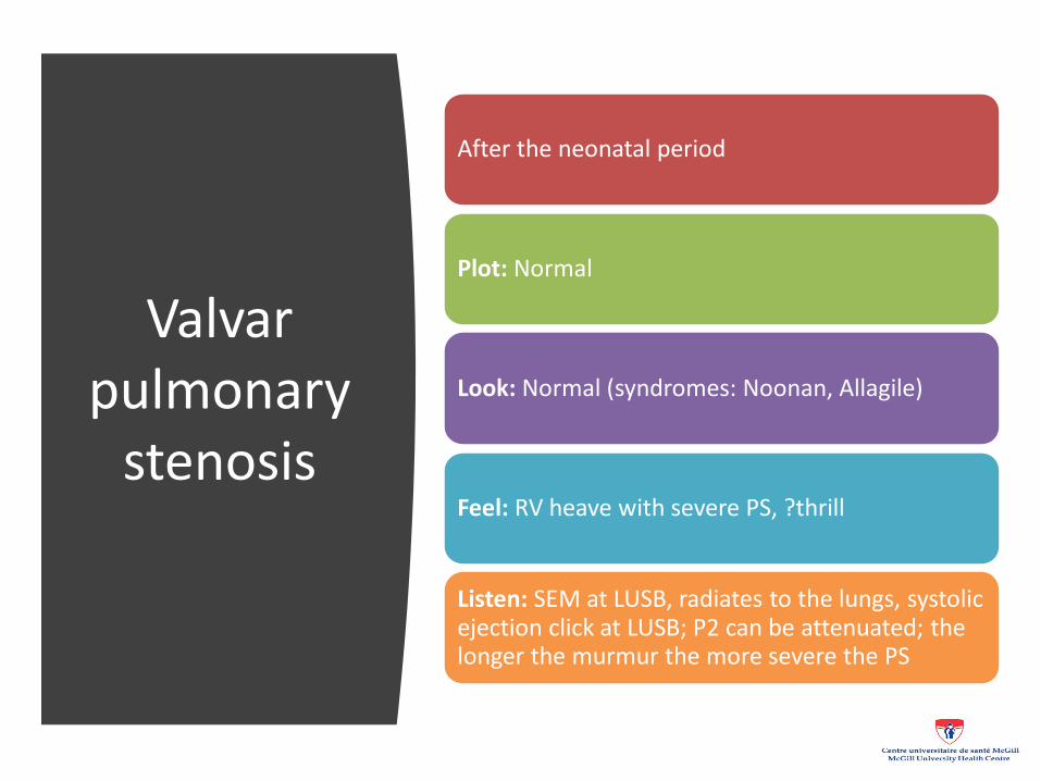
Valvar pulmonary
stenosis
After the neonatal period
Plot: Normal
Look: Normal (syndromes: Noonan, Allagile)
Feel: RV heave with severe PS, ?thrill
Listen: SEM at LUSB, radiates to the lungs, systolic ejection click at LUSB; P2 can be attenuated; the longer the murmur the more severe the PS
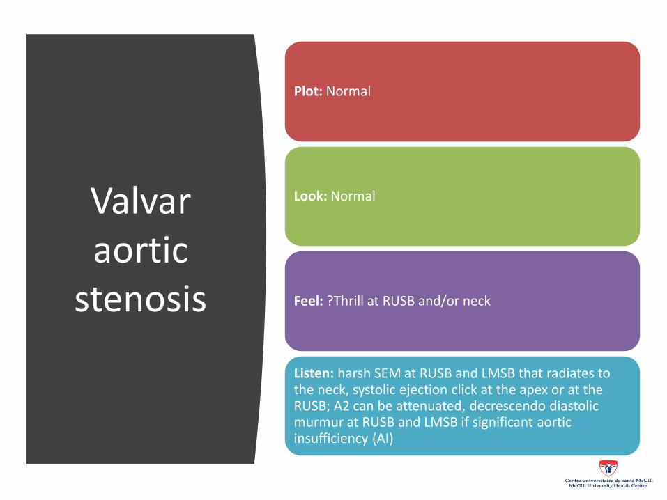
Valvar aortic
stenosis
Plot: Normal
Look: Normal
Feel: ?Thrill at RUSB and/or neck
Listen: harsh SEM at RUSB and LMSB that radiates to the neck, systolic ejection click at the apex or at the RUSB; A2 can be attenuated, decrescendo diastolic murmur at RUSB and LMSB if significant aortic insufficiency (AI)
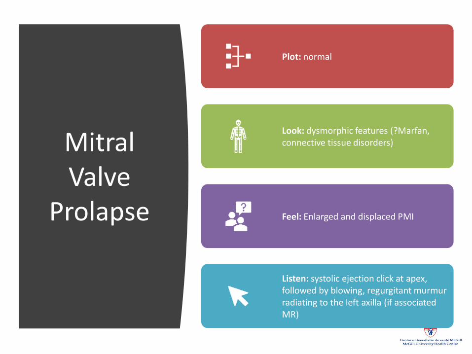
Mitral Valve
Prolapse
Plot: normal
Look: dysmorphic features (?Marfan, connective tissue disorders)
Feel: Enlarged and displaced PMI
Listen: systolic ejection click at apex, followed by blowing, regurgitant murmur radiating to the left axilla (if associated MR)
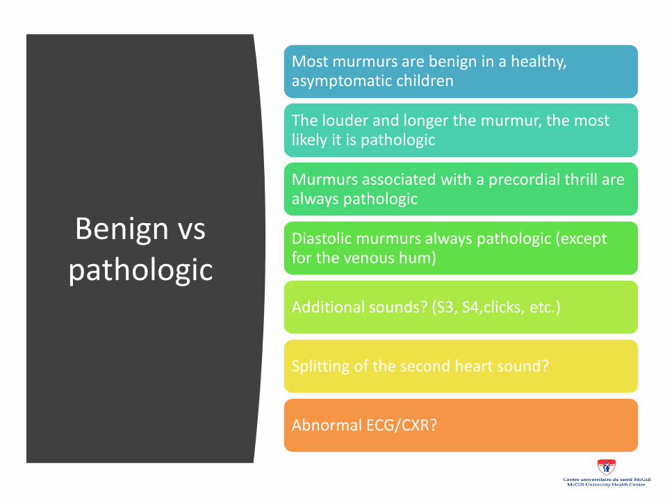
Benign vs pathologic
Most murmurs are benign in a healthy, asymptomatic children
The louder and longer the murmur, the most likely it is pathologic
Murmurs associated with a precordial thrill are always pathologic
Diastolic murmurs always pathologic (except for the venous hum)
Additional sounds? (S3, S4,clicks, etc.)
Splitting of the second heart sound?
Abnormal ECG/CXR?

Go to this link to open up the examples
• https://www.dropbox.com/sh/nwqinbzw34adrt5/AACgYTez5UUbwc_h_KxMiO_Qa?dl=0
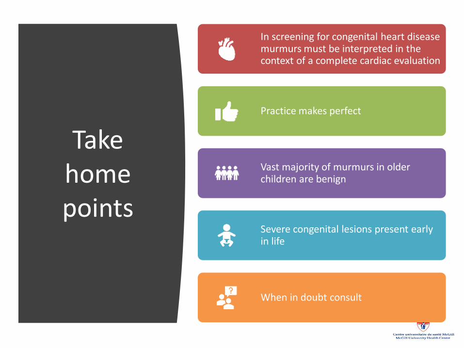
Take home points
In screening for congenital heart disease murmurs must be interpreted in the context of a complete cardiac evaluation
Practice makes perfect
Vast majority of murmurs in older children are benign
Severe congenital lesions present early in life
When in doubt consult
How to reach us
MCH Cardiology:
Phone: 514-412-4423Fax: 514-412-4273
MCH Sub-Specialty Consultation: 514-412-4242
Selected References
and Resources
• “Teaching heart Auscultation to health professionals: methods for improving the practice of an ancient but critical skill” Contributing Editor- John Finley, MD ISBN 978 0 9877400 0 7: endorsed by the Canadian Pediatric Cardiology Society
• https://teachingheartauscultation.com• Pediatric cardiac auscultation. Altman CA &al
Lippincott, Williams and Wilkins 2000.
• Pediatric Heart Sounds. McConnel ME. Springer 2008
• Youtube: “thinklabs, heart sounds”
• Mobile Applications: “emurmur university”, ”emurmur primer”, “heart songs”, “heart murmurs pro”, “3M Littmann Learning”

Questions?