Embed Size (px)
Citation preview
INFECTION AND IMMUNITY, June 1985, p. 850-8520019-9567/85/060850-03$02.00/0Copyright © 1985, American Society for Microbiology
Multiplication of Mycobacterium marinum Within Phagolysosomesof Murine Macrophages
NATAN MORDepartment of Comparative Medicine, Hebrew University-Hadassah Medical School, 91010 Jerusalem, Israel
Received 13 November 1984/Accepted 27 February 1985
Both in vivo and in vitro, Mycobacterium marinum organisms were found to multiply within phagolysosomesof murine macrophages. It thus appears that M. marinum are neither killed nor inhibited from multiplying bylysosomal enzymes.
Mycobacterium marinum is a human pathogen; experi-mental infections with this organism have served as a usefulmodel for immunological studies in which simulation ofmouse footpad infection with M. leprae was needed (12).Inoculation of 5.0 x 10³ M. marinum into mouse hindfootpads produces a localized, self-limited disease processinvolving primarily the footpad tissues and the drainingpopliteal lymph node (10, 12). The organisms appear tomultiply in the macrophages (MO) of mouse footpads for 10to 14 days after inoculation to a maximum of ca. 106 perfootpad, after which they are killed. M. marinum multipliesintracellularly (10) in the cooler parts of the body; however,it is not known whether the organism multiplies extra-phagosomally, as does M. leprae (6, 9), or intraphagosom-ally, as do organisms of other mycobacterial species (2, 7).Accordingly, a study was undertaken to determine theintracellular location of M. marinum, both in vivo in the M4of mouse footpad lesions and in vitro in resident murineperitoneal MO. In addition to normal mice, thymectomizedand irradiated (T900R) mice were employed, because thelarger number of organisms in the latter animals facilitateselectron microscopic (EM) study. Earlier work had shownthat these mice permit enhanced multiplication of M. mari-num but are nevertheless able to limit bacterial multiplica-tion (3).
Locally bred, specific-pathogen-free CBA/Ca mice wereemployed. To produce T900R mice, 6-week-old animalswere subjected to thymectomy by aspiration and 2 weekslater to whole-body X-irradiation in a dose of 900 rads,followed immediately by transfusion with syngeneic bonemarrow. Resident peritoneal M4 were obtained by peritone-al lavage with Hanks balanced salt solution containing 5 IUof heparin per ml and cultured in medium 199 containing40%o horse serum in plastic Leighton tubes. Inocula of M.marinum of the strain previously employed (10-12) wereprepared from fresh cultures in Dubos broth supplementedwith bovine serum albumin. The inoculation of mice with M.marinum, harvest of the organisms from the inoculated footpads, and counting of the acid-fast bacteria were accom-
plished by the methods previously described for work withM. leprae (14, 16). For EM, footpad tissues were fixed inglutaraldehyde, followed by osmium tetroxide, and proc-essed as previously described (9). M4 monolayers werefixed by the simultaneous addition to the Leighton tubes ofglutaraldehyde and osmium tetroxide (8), after which thecells were scraped from the surface with a rubber policeman,collected by centrifugation, dehydrated with acetone, andembedded in Spurr resin. Interrupted thin sections were
stained with uranyl acetate-lead citrate and screened bymeans of a Phillips 300 transmission EM. Results wereanalyzed statistically by calculating exact probabilities (15,17).
In experiments in vivo, normal and T900R mice wereinoculated with 5.0 x 10³ M. marinum in each hind footpad,and organisms were harvested from the footpad tissues atintervals thereafter. A small portion of footpad tissue wasalso taken for EM. M. marinum multiplied to a higher ceilingin T900R than in normal mice (Table 1). Moreover, theorganisms appeared to survive for a longer time in T900Rthan in normal mice. Finally, the immune response of theT900R mice was accompanied by minimal enlargement ofthe popliteal lymph node; in normal mice, enlargement of thepopliteal node is a prominent feature of this process (10, 11).
In both groups of animals, at all intervals, EM revealed(Fig. 1) that the organisms were located within phagosomesor phagolysosomes of the footpad M4; no organisms wereobserved to lie free in the cytoplasm.
In an experiment in vitro, M4, the lysosomes of which hadbeen labeled with ferritin (2), were incubated for 1 h with M.marinum and washed, and the medium was replaced withfresh medium containing 25 ,ug of streptomycin per ml. Atseveral intervals thereafter, cultures were fixed for EM. Twohours after inoculation, the great majority of the organismswere found within phagosomes (Table 2), most of which had
TABLE 1. Multiplication of M. marinum in the foot pads ofnormal and T90OR mice
Test results with:
Time after Normal mice T900R miceinoculation No. (10G) of: Amt of No. (105) of: Amt of
(days) PN PNAFBa CFUa (mg)b AFBa.c CFUa.d (mg)b.e
6 7.5 3.0 6.5 10 5.5 2.010 5.0 2.3 15 11 5.5 5.016 1.1 1.4 21 6.5 2.2 6.021 2.4 0.01 27 3.2 2.5 6.030 4.3 0.003 30 2.8 0.1 6.3
aMean number of acid-fast bacilli (AFB) or colony-forming units (CFU) perfootpad, calculated from harvests of M. marinum performed on the pooledtissues of four footpads.
b Mean weight of the popliteal node, calculated from the weight of a pool offour lymph nodes. PN, Popliteal node.
c T900R > normal; P = (0.5)4 = 0.062.d T900R < normal; P = (0.5)s = 0.031.e T90OR < normal; P = (0.5)5 = 0.031.
850
Vol. 48, No. 3
on August 31, 2018 by guest
http://iai.asm.org/
Dow
nloaded from

NOTES 851
'a..;.
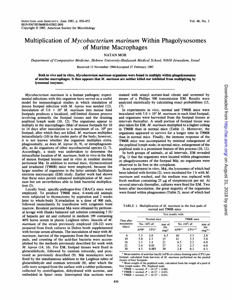
FIG. 1. M. marinum enclosed by phagosomal membrane (indicated by arrow) of a M4~10 days after inoculation into the footpad of a T900Rmouse. Bar, 0.2 pLm.
fused with lysosomes (Fig. 2). At 24 h after inoculation ofM.marinum, more than 95% of the phagosomes were fusedwith lysosomes. The majority of the organisms were foundwithin phagosomes and appeared intact. There were manydisrupted phagosomes, sometimes associated with organ-isms apparently free in the cytoplasm of the macrophages.The proportion of extraphagosomal organisms was higher at24 than at 2 h (P = 0.033); it appears likely that the increaseof extraphagosomal organisms between 2 and 24 h had
resulted from bacterial multiplication within phagosomes,followed by disruption of the phagosomal membrane andrelease of the organisms into the cytoplasm of the M+.
In a second experiment in vitro, M4X were incubated for 1h with viable M. marinum and treated as in the firstexperiment. M. marinum were found to multiply in culturedperitoneal M+; the number of organisms recovered from themonolayer increased approximately 10-fold during the first48 h (Table 3). During this time, the number of organisms
72
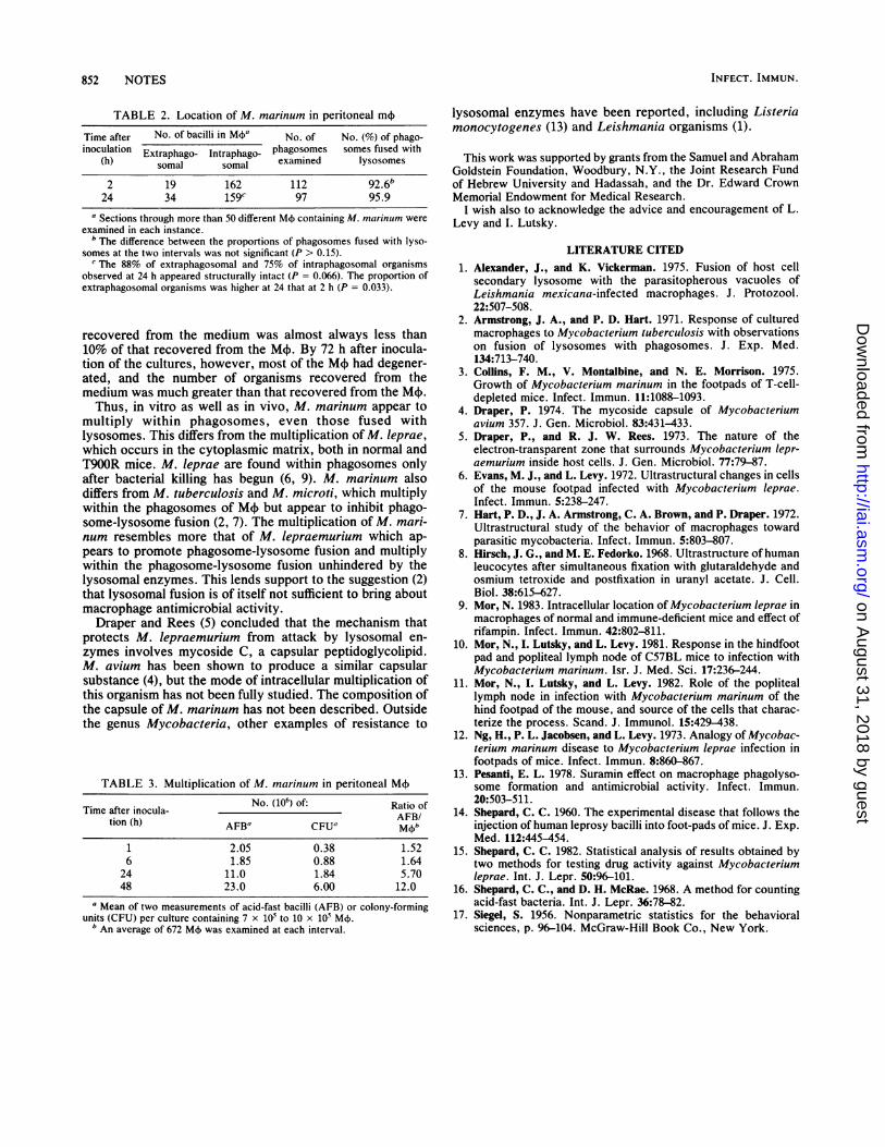
FIG. 2. Phagosome (indicated by short arrow), fused with ferritin-labeled lysosome (indicated by long arrow), containing M. marinum 6h after inoculation of M4 monolayer. Bar, 0.2 ,.m .
VOL. 48, 1985
on August 31, 2018 by guest
http://iai.asm.org/
Dow
nloaded from
852 NOTES
TABLE 2. Location of M. marinum in peritoneal m4)
Time after No. of bacilli in M4+a No. of No. (%) of phago-inoculation Extraphago- Intraphago_ phagosomes somes fused with
(h) somal somal examined lysosomes
2 19 162 112 92.624 34 159C 97 95.9
a Sections through more than 50 different M( containing M. marinum wereexamined in each instance.
b The difference between the proportions of phagosomes fused with lyso-somes at the two intervals was not significant (P > 0.15).
c The 88% of extraphagosomal and 75% of intraphagosomal organismsobserved at 24 h appeared structurally intact (P = 0.066). The proportion ofextraphagosomal organisms was higher at 24 that at 2 h (P = 0.033).
recovered from the medium was almost always less than10% of that recovered from the M+. By 72 h after inocula-tion of the cultures, however, most of the MX had degener-ated, and the number of organisms recovered from themedium was much greater than that recovered from the M+.Thus, in vitro as well as in vivo, M. marinum appear to
multiply within phagosomes, even those fused withlysosomes. This differs from the multiplication of M. leprae,which occurs in the cytoplasmic matrix, both in normal andT900R mice. M. leprae are found within phagosomes onlyafter bacterial killing has begun (6, 9). M. marinum alsodiffers from M. tuberculosis and M. microti, which multiplywithin the phagosomes of M4~but appear to inhibit phago-some-lysosome fusion (2, 7). The multiplication of M. mari-num resembles more that of M. lepraemurium which ap-pears to promote phagosome-lysosome fusion and multiplywithin the phagosome-lysosome fusion unhindered by thelysosomal enzymes. This lends support to the suggestion (2)that lysosomal fusion is of itself not sufficient to bring aboutmacrophage antimicrobial activity.Draper and Rees (5) concluded that the mechanism that
protects M. lepraemurium from attack by lysosomal en-zymes involves mycoside C, a capsular peptidoglycolipid.M. avium has been shown to produce a similar capsularsubstance (4), but the mode of intracellular multiplication ofthis organism has not been fully studied. The composition ofthe capsule of M. marinum has not been described. Outsidethe genus Mycobacteria, other examples of resistance to
TABLE 3. Multiplication of M. marinum in peritoneal M4
Time after inocula- No. (106) of: Ratio oftion (h) AFB CFUa
1 2.05 0.38 1.526 1.85 0.88 1.64
24 11.0 1.84 5.7048 23.0 6.00 12.0
a Mean of two measurements of acid-fast bacilli (AFB) or colony-formingunits (CFU) per culture containing 7 x 10⁵ to 10 X 105 M+.bAn average of 672 M4 was examined at each interval.
lysosomal enzymes have been reported, including Listeriamonocytogenes (13) and Leishmania organisms (1).
This work was supported by grants from the Samuel and AbrahamGoldstein Foundation, Woodbury, N.Y., the Joint Research Fundof Hebrew University and Hadassah, and the Dr. Edward CrownMemorial Endowment for Medical Research.
I wish also to acknowledge the advice and encouragement of L.Levy and I. Lutsky.
LITERATURE CITED1. Alexander, J., and K. Vickerman. 1975. Fusion of host cell
secondary lysosome with the parasitopherous vacuoles ofLeishmania mexicana-infected macrophages. J. Protozool.22:507-508.
2. Armstrong, J. A., and P. D. Hart. 1971. Response of culturedmacrophages to Mycobacterium tuberculosis with observationson fusion of lysosomes with phagosomes. J. Exp. Med.134:713-740.
3. Collins, F. M., V. Montalbine, and N. E. Morrison. 1975.Growth of Mycobacterium marinum in the footpads of T-cell-depleted mice. Infect. Immun. 11:1088-1093.
4. Draper, P. 1974. The mycoside capsule of Mycobacteriumavium 357. J. Gen. Microbiol. 83:431-433.
5. Draper, P., and R. J. W. Rees. 1973. The nature of theelectron-transparent zone that surrounds Mycobacterium lepr-aemurium inside host cells. J. Gen. Microbiol. 77:79-87.
6. Evans, M. J., and L. Levy. 1972. Ultrastructural changes in cellsof the mouse footpad infected with Mycobacterium leprae.Infect. Immun. 5:238-247.
7. Hart, P. D., J. A. Armstrong, C. A. Brown, and P. Draper. 1972.Ultrastructural study of the behavior of macrophages towardparasitic mycobacteria. Infect. Immun. 5:803-807.
8. Hirsch, J. G., and M. E. Fedorko. 1968. Ultrastructure of humanleucocytes after simultaneous fixation with glutaraldehyde andosmium tetroxide and postfixation in uranyl acetate. J. Cell.Biol. 38:615-627.
9. Mor, N. 1983. Intracellular location of Mycobacterium leprae inmacrophages of normal and immune-deficient mice and effect ofrifampin. Infect. Immun. 42:802-811.
10. Mor, N., I. Lutsky, and L. Levy. 1981. Response in the hindfootpad and popliteal lymph node of C57BL mice to infection withMycobacterium marinum. Isr. J. Med. Sci. 17:236-244.
11. Mor, N., I. Lutsky, and L. Levy. 1982. Role of the popliteallymph node in infection with Mycobacterium marinum of thehind footpad of the mouse, and source of the cells that charac-terize the process. Scand. J. Immunol. 15:429-438.
12. Ng, H., P. L. Jacobsen, and L. Levy. 1973. Analogy of Mycobac-terium marinum disease to Mycobacterium leprae infection infootpads of mice. Infect. Immun. 8:860-867.
13. Pesanti, E. L. 1978. Suramin effect on macrophage phagolyso-some formation and antimicrobial activity. Infect. Immun.20:503-511.
14. Shepard, C. C. 1960. The experimental disease that follows theinjection of human leprosy bacilli into foot-pads of mice. J. Exp.Med. 112:445-454.
15. Shepard, C. C. 1982. Statistical analysis of results obtained bytwo methods for testing drug activity against Mycobacteriumleprae. Int. J. Lepr. 50:96-101.
16. Shepard, C. C., and D. H. McRae. 1968. A method for countingacid-fast bacteria. Int. J. Lepr. 36:78-82.
17. Siegel, S. 1956. Nonparametric statistics for the behavioralsciences, p. 96-104. McGraw-Hill Book Co., New York.
INFECT. IMMUN.
on August 31, 2018 by guest
http://iai.asm.org/
Dow
nloaded from