Embed Size (px)
Citation preview

Multiple Myeloma: An Overview
Ola Landgren, M.D., Ph.D.Mary Ann Yancey, RN, MSN, AOCN
Previously healthy 64-year-old man
• Presents with persistent pain in his lower back and fatigue
• CBC reveals a hemoglobin level of 9.6 g/dL
• A monoclonal-(M)-protein is detected on serum protein electrophoresis (IgG kappa)
• Radiologic skeletal bone survey shows lytic bone lesions of the vertebrae and the pelvis
IgG
IgA
IgM
kappa
lambda

Previously healthy 64-year-old man
• Multiple myeloma (MM) is confirmed by bone marrow aspiration showing infiltrate of plasma cells
• Serum calcium and creatinine levels are normal
• Albumin is 3.7 g/dL and beta2- microglobulin is 2.8 mg/L
• Fluorescence in situ hybridization (FISH) of bone marrow plasma cells shows deletion of chromosome 13

Previously healthy 64-year-old man
• Interpretation: – Relatively young age – Absence of coexisting illnesses
• A hematologist recommends:– Induction therapy followed by…– High-dose therapy with autologous
hematopoietic stem-cell transplantation (ASCT) as initial treatment

Clinical dilemma
• 20,580 new cases (11,680 men; 8,980 women) and 10,580 deaths per year
• Average age at dx 65-70 yrs (<40 yrs; ~2%)
• The 2nd most common hematologic malignancy in whites; in Blacks it is #1
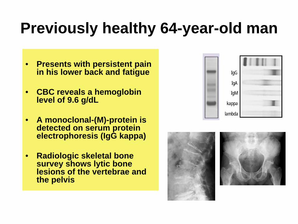
MM is preceded by MGUS
MGUS MMGerminal-center B-cell
Yrs priorMM dx
M-protein,* n/N (%; 95% CI)
Abnormal FLC ratio,‡n/N (%; 95% CI)
MGUS,§n/N (%; 95% CI)
2 25/27 (93; 76–99) 23/27 (85; 66–96) 27/27 (100; 8727/27 (100; 87––100)100)
3 54/58 (93; 83–98) 46/58 (79; 67–89) 57/58 (98; 91–100)
4 45/48 (94; 83–99) 29/46 (63; 48–77) 47/48 (98; 89–100)
5 34/37 (92; 78–98) 25/37 (68; 50–82) 35/37 (95; 82–99)
6 25/25 (100; 86–100) 19/25 (76; 55–91) 25/25 (100; 86–100)
7 14/15 (93; 68–100) 11/15 (73; 45–92) 14/15 (93; 68–100)
> 8 13/17 (77; 50–93) 8/17 (47; 23–72) 14/17 (82; 57–96)
Landgren et al. Blood 2009; Weiss et al. Blood 2009

What causes MM?Support for genetic factors• 3-fold increased relative risk
of developing MM among first-degree relatives of MM and MGUS pts7
• Twice as common among Blacks (compared to whites); earlier age of onset in Blacks
Lynch et al. NEJM 2008; Landgren et al. Blood 2009

What causes MM?Support for environmentalfactors• Exposure to pesticides
and radiation associated with increased risk
• Chronic immune stimulation (e.g. infections, autoimmunity, obesity) associated with increased risk
Alexander et al. Int J Cancer 2007; Brown et al. Blood 2008; Iwanaga et al. Blood 2009; Landgren et al. Blood 2009

Pathophysiology of MM• Clonal B-cell tumor of plasma cells in
the bone marrow
• Most malignant plasma cells express– CD38, CD56/58, CD79a, CD138
• Most malignant plasma cells do not express the pan-B cell antigens CD19 and CD20
• Cytokine and signaling alterations in the bone marrow microenvironment– IL-6, tumor necrosis factor (TNF)-alpha,
IL-1-beta, VEGF, fibroblast growth factor-beta, DDK-1, etc…

Harousseau and Moreau, NEJM 2009
In MM, the bone marrow micro- environment plays a key role!

Harousseau and Moreau, NEJM 2009
Molecularly, MM is not one disease!
Non-hyperdiploid (translocations)
Hyperdioploid (trisomies)

Zhan et al, Blood 2006
Associated with genetic lesions–MF (MAF translocation)–MS (MMSET/FGFR3 translocation)–CD1 (Cyclin D1 or D3 translocation)–CD2 (Cyclin D1 or D3 translocation)–Hyperdiploid
Associated with phenotype–PR (proliferative)–LB (low incidence of bone disease)
Gene expression reveals 7 molecular MM subtypes

Zhan et al, Blood 2006
Gene expression MM subtypes have different outcomes

Common symptoms at MM diagnosis
• Bone pain• Fatigue• Weight loss• Parasthesias
• ~10% are asymptomatic/have only mild symptoms at dx
Kyle and Rajkumar, N Engl J Med 2004

Clinical hallmarks of MM
Kyle and Rajkumar, N Engl J Med 2004
• Hypercalcemia• Renal failure• Anemia• Bone destructions (lytic lesions)• Increased risk of infections• Presence of monoclonal protein
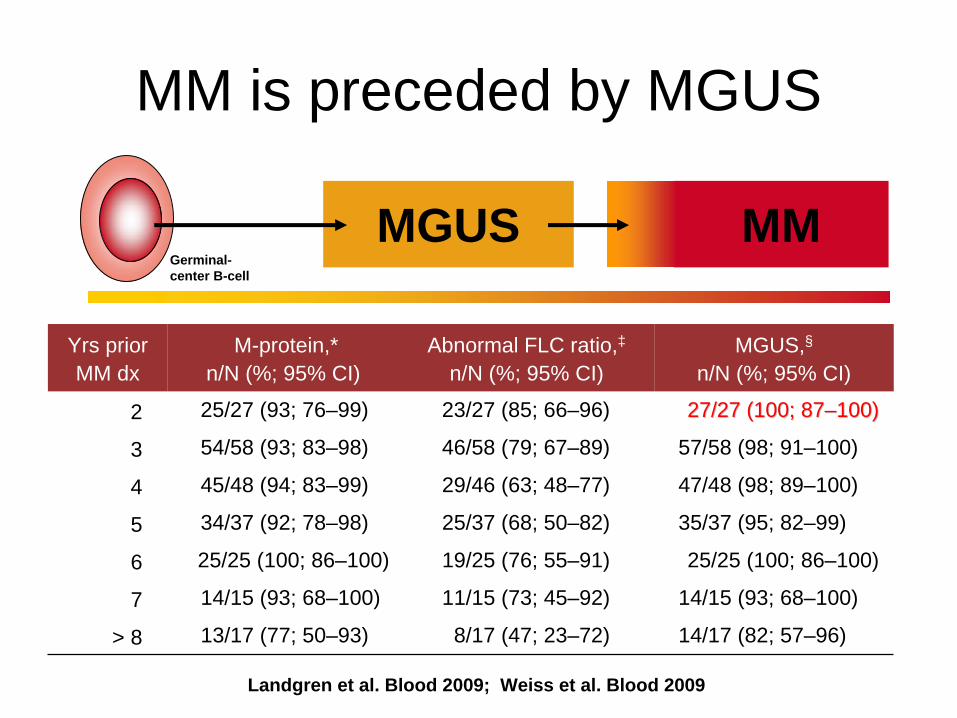
Serum protein electrophoresis
Monoclonal proteinNormal
Immunoglobulin (Ig) G kappa
Immunofixation
IgG
IgA
IgM
kappa
lambda
Katzmann et al, Electrophoresis 1997

Skeletal X-ray shows punched-out lytic lesions, diffuse osteoporosis, and fractures
For MM work-up, bones should be evaluated with a complete “skeletal survey”, including:- Skull - Spine - Pelvis - Extremities (including forearms and legs)


Int’l Myeloma Working Group. Br J Haematol 2003
Monoclonal gammopathy of undetermined significance
(MGUS)
Smoldering myeloma
(SMM)
Multiple myeloma
(MM)
Monoclonal (M)- protein in serum <3 g/dL >3 g/dL Any
Monoclonal plasma cells in bone marrow
AND <10% OR >10% Any
End-organ damage No No Yes
Comment Requires exclusion of all other B-cell lymphoproliferative disorders
Indolent MM is a non- standard term to refer to disease with end- organ damage but minimal symptoms
End-organ damage:• Hypercalcemia• Renal failure• Anemia• Lytic bone lesions
Diagnostic criteria

Differential diagnosis• MGUS• SMM• Solitary plasmacytoma• Amyloidosis• Light chain deposition disease• Waldenström's macroglobulinemia• Lymphoproliferative disorders• Infections (e.g. CMV)• Rheumatologic autoimmune disorders• Certain skin or neurologic disorders

Treatment
• Initial treatment for MM depends if the patient is a candidate for Autologous Stem Cell Transplant (ASCT)
• Typically, eligibility is determined by– Age– Performance status– Comorbidity

• VAD, Dex or Thal/Dex induction• Melphalan 200 mg/m2
• Overall response rate 80%– CR/nCR rate 20%
• Median PFS 20-36 mos• Median OS 48-60 mos
• Overall response rate 40-50%– CR/nCR 5%
• Median PFS 12-15 mos• Median OS 30-36 mos
ASCT
Patient Age
Melphalan and prednisone
>65-70<65-70
Kyle and Rajkumar. Clin Lymphoma & Myeloma 2009
Treatment strategy before novel drugs

• International Staging System (ISS)
• Adverse cytogenetic abnormalities (by FISH)
Stage Criteria Median
I Serum β2 -microglobulin <3.5 mg/L 62 mo.Serum albumin ≥
3.5 g/dL
II Not stage I or III 44 mo.
III Serum β2 -microglobulin ≥
5.5 mg/L 29 mo.
t(4;14)=15% of MM (dysregulation of FGFR3 and MMSET)
p53 deletion=10% of MM(loss of tumor suppressor gene)
Advances in prognosis

Mayo Clinic “mSMART classification”of active MM
Dispenzieri, et al. Mayo Clin Proc 2007 (revised and updated Jan 2009, v5)

Int’l Myeloma Working Group response criteria
Durie, et al. Leukemia 2007; Anderson, et al. Leukemia 2008

Novel agents in MM
Main Toxicities
• Thalidomide
• Bortezomib
• Lenalidomide
• Teratogenicity, peripheral neuropathy, constipation, sedation, rash, venous thromboembolism
__________________________• Fatigue, GI toxicity, peripheral
neuropathy, decrease in platelets and neutrophils
__________________________• Myelosuppression, venous
thromboembolism________________________
Agent

• Add novel agent to melphalan + prednisone
• IMiD + dexamethasone
• 3-4 drug regimens +/- maintenance1-5
1Morgan G, et al. Blood 2007;110: abstract 3593 2Richardson PG, et al. Blood 2008;112: abstract 92; 3Reeder CB, et al. Leukemia 2009;23: 1337-1341; Kumar S et al. Blood 2008;112: abstract 91;
5Offandini M. et al. Br J Haematol 2009;144: 653-659.
Current treatment options for newly dx non-transplant eligible MM pts

Current best outcomes with non- ASCT regimens in phase III trials
Reference Rx Duration of therapy
(wks)
Overall response rate (CR+nCR) (%)
Median PFS
(mos)
Median OS
(mos)
2 year OS(%)
Facon1 MPT 72 76 (18) 27.5* 51.6* 78Palumbo2 MPT 24+ 76 (28) 21.8* 45 82Hulin3 MPT 72 61 (7) 24* 45* 70San Miguel4 VMP 54 71 (35) 24* NYR* 83Rajkumar5 Len+
dexUntil prog 70 (14 CR) ~24 NYR 93
1Facon T, et al. Lancet 2007:370; 1209-1218; 2Palumbo A , et al. Blood 2008;112: 3107-3114; 3 Hulin C, et al. Blood 2007; 110: abstract 75;4San Miguel JF, et al. N Engl J Med 2008;
5 Rajkumar V, et al; Blood 2007; 110: Abstract 74.
* P<0.05 (when compared to MP alone)

Novel 3 or 4-drug Combinations (e.g.,RVD)
Rev + Dex Bortezomib combination at relapse
Bortezomib + MPThalidomide + MP Rev + Dex
Overall survival ?
MPSequential novel agents
+/- steroids
What is the optimal non-ASCT strategy in MM?

VAD
THALIDOMIDEThalDex*
TAD*CTD*
BORTEZOMIB(VELCADE)
Bortez+Dex*VTD*PAD*VCD
RVDD
LENALIDOMIDE(REVLIMID)
RD*Rd*RVD
Stem cell harvestHigh-dose melphalan +
ASCTRD: Lenalidomide + high-dose dexRd: Lenalidomide + low-dose dex
*Studied in phase III trials
Current induction regimens before ASCT

Strategies to improve ASCT results
• Risk stratification– Cytogenetics– Molecular classification
• Improved induction therapy• Improved consolidation therapy
– New regimens– Tandem ASCT
• Maintenance therapy
• ASCT followed by alloSCT

Tandem ASCT reportedly beneficial if <VGPR after first ASCT
EFS Overall survival
Attal M, et al. N Engl J Med 2003;349:2405

N CR/VGPR rate (%)Single Tandem
Median PFS (mo)Single Tandem
Median OS (mo)Single Tandem
Attal, 2003
399 42 50 25 30 48 58
Fermand, 2003
277 39 37 31 33 49 73
Goldschmidt, 2005
268 -- -- 22 NYR 23 NYR
Sonneveld, 2004
303 13 28 20 22 55 50
Cavo, 2007
321 38 48 23 35 65 71
Values highlighted in red indicate p<0.05
Randomized tandem ASCT trials

Reference N Thalidomide dose (mg)/duration
PFS/ EFS
Overall survival
Attal, 2006 597 Thal 200 (median dose) vs. obs /progression
+ +
Spencer, 2006 243 Thal 200 + pred vs. pred/12 months
+ +
Maiolino, 2008 212 Thal 200 + dex vs. dex/12 months
+ NS
Barlogie, 2006* 668 Thal 400/progression
+ NS*
Morgan, 2008* -- Thal 100/progression
+/- NS*
NCIC, 2009 325 Thal 200 + pred vs. obs/48 months
? ?
*Thalidomide also given as part of induction therapy
Thalidomide maintenance after ASCT

• Preceded by novel induction regimens
• Melphalan 200 mg/m2
+/- second ASCT +/-maintenance
• Overall response rate 80-90%• CR/nCR rate 35-50%• 2 year PFS 69-93%• 2 year OS 90-93%
• Overall response rate 65-75%• CR/nCR 20-25%• Median PFS 24-30 mos• Median OS 48-50 mos• 2 year OS 70-93%
ASCTMPT or MPV
or Lenalidomide + Dex
Treatment strategies in 2010

Treatment strategies for relapsed/refractory patients
• Initial treatment can be repeated in selected patients– Commonly used with alkylating agents (cyclophosphamide +
prednisone is alternative to repeated MP)– Also high-dose melphalan + ASCT– Data emerging that novel agents can be used again
• Novel agents can be introduced– As single agents– With steroids– In 3-4 drug regimens with conventional chemotherapy and/or other novel
agents

Clinical myeloma studies at NCI in 2010-
• Precursor disease– Natural history study
(individualized profiling)
• Smoldering myeloma– Early treatment
MGUS MMGerminal-center B-cell
• Relapsed multiple myeloma– MEK inhibitor– HDAC/mTOR inhibitors
• From precursor to multiple myeloma– Imaging study

-Thank you very much for your attention!