Embed Size (px)
Citation preview
Genetics and Molecular Research 6 (3): 510-521 (2007) FUNPEC-RP www.funpecrp.com.br
Multiple antimicrobial resistance in Enterobacteriaceae isolates from pristine freshwater
C.I. Lima-Bittencourt1, L. Cursino1, H. Gonçalves-Dornelas1, D.S. Pontes1, R.M.D. Nardi2, M. Callisto1, E. Chartone-Souza1 and A.M.A. Nascimento1
1Departamento de Biologia Geral, 2Departamento de Microbiologia, Instituto de Ciências Biológicas, Universidade Federal de Minas Gerais, Belo Horizonte, MG, BrasilCorresponding author: A.M.A. NascimentoE-mail: [email protected]
ABSTRACT. A freshwater enterobacterial population (N = 111) was studied for antimicrobial and mercury resistance patterns, and for its possible association with biotic and abiotic factors in that environment. Conventional biochemical tests identified Klebsiella sp, Morganella sp, Serratia sp, Escherichia sp, Enterobacter sp, Edwarsiella sp, Proteus sp, Citrobacter sp, Providencia sp, and Kluyvera sp. There was no correlation between antimicrobial resist-ance patterns of isolates and bacterial genera, but resistance pat-terns varied among water samples and between seasons. Resistance to multiple antimicrobials was common (61%). The percentage of bacteria resistant to at least one antimicrobial differed between the
Genet. Mol. Res. 6 (3): 510-521 (2007)Received June 13, 2007Accepted July 30, 2007Published September 5, 2007

Genetics and Molecular Research 6 (3): 510-521 (2007) www.funpecrp.com.br
Resistant enterobacteria from freshwater streams 511
INTRODUCTION
Antibiotic resistance in bacteria is a contemporary global public health problem (Levy and Marshall, 2004; Wright, 2007). Bacterial resistance to antibiotics arises not only by gene mu-tations, but mostly by the acquisition of heterologous resistance genes from other bacteria (Kapil, 2005). It is widely accepted that antibiotic-resistant strains primarily arise in hospitals and spread further among pathogenic bacterial populations as a result of strong antibiotic selective pressure (Blazquez et al., 2002; O’Brien, 2002). Efforts to reduce this resistance are based on the assump-tion that it will decrease in the absence of positive selection by antibiotics. Control of antibiotic use should therefore restrain the spread of resistance (Barza and Gorbach, 2002).
To date, the majority of the studies of resistance patterns have focused on pathogenic bacterial populations, but it is known that commensal bacteria are also common reservoirs of antibiotic resistance genes (Levy and Marshall, 2004). Therefore, the information derived from studies of pathogenic bacteria may not be representative of all bacteria. In fact, as resistance is studied in bacteria isolated from presumably nonselective environments unrelated to clinical ones, the emergence and spread of antibiotic-resistant bacteria have also been reported (Ash et al., 2002; Whitehead et al., 2003; Salyers et al., 2004). Thus, screening for antibiotic-resistant bacteria in natural ecosystems is highly relevant, and worldwide studies have been carried out to identify environmental reservoirs of bacterial antibiotic-resistance in wild-animal populations and natural water supplies (McKeon et al., 1995; Gõni-Urriza et al., 2000; Sherley et al., 2000; Nascimento et al., 2003). Selection of antibiotic-resistant bacteria in these environments may be a consequence of antibiotic production by other microorganisms. The pressure exercised by these microorganisms would lead to the dispersion of resistance genes carried by plasmids and transposons, which contain not only antibiotic resistance determinants, but also other selectable markers, such as heavy metals (Davison, 1999; Nascimento and Chartone-Souza, 2003).
Members of the family Enterobacteriaceae have a worldwide distribution and are found in various environments and hosts such as water, soil, plants, human beings, and other animals, but they are also significant causes of human disease. Antibiotic resistance among
rainy (100%) and dry seasons (89%). Resistance to β-lactams and chloramphenicol was the most frequent and resistance to amikacin, gentamicin and kanamycin was less frequent. The main water vari-ables examined (abiotic factors pH and temperature; biotic factor chlorophyll a concentration) did not influence antimicrobial resist-ance. Significant impact on freshwater enterobacteria, as evidenced by antimicrobial-multiple resistance and by the presence of blaTEM gene, may point to the fact that it has an important role in horizontal spread of resistance.
Key words: Abiotic factors, Antibiotic resistance, Enterobacteriaceae, Freshwater, Mercury
Genetics and Molecular Research 6 (3): 510-521 (2007) www.funpecrp.com.br
C.I. Lima-Bittencourt et al. 512
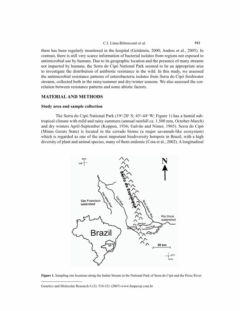
them has been regularly monitored in the hospital (Goldstein, 2000; Andres et al., 2005). In contrast, there is still very scarce information of bacterial isolates from regions not exposed to antimicrobial use by humans. Due to its geographic location and the presence of many streams not impacted by humans, the Serra do Cipó National Park seemed to be an appropriate area to investigate the distribution of antibiotic resistance in the wild. In this study, we assessed the antimicrobial resistance patterns of enterobacteria isolates from Serra do Cipó freshwater streams, collected both in the rainy/summer and dry/winter seasons. We also assessed the cor-relation between resistance patterns and some abiotic factors.
MATERIAL AND METHODS
Study area and sample collection
The Serra do Cipó National Park (19o-20o S; 43o-44o W; Figure 1) has a humid sub-tropical climate with mild and rainy summers (annual rainfall ca. 1,500 mm, October-March) and dry winters April-September (Koppen, 1936; Galvão and Nimer, 1965). Serra do Cipó (Minas Gerais State) is located in the cerrado biome (a major savannah-like ecosystem) which is regarded as one of the most important biodiversity hotspots in Brazil, with a high diversity of plant and animal species, many of them endemic (Cota et al., 2002). A longitudinal
Figure 1. Sampling site locations along the Indaiá Stream in the National Park of Serra do Cipó and the Peixe River.
50 km
Genetics and Molecular Research 6 (3): 510-521 (2007) www.funpecrp.com.br
Resistant enterobacteria from freshwater streams 513
gradient was studied in the Indaiá Stream (1st- up to 5th-order stream reaches, inside the Serra do Cipó National Park) and Peixe River (7th- and 9th-order stream reaches, outside the Serra do Cipó National Park, near cattle farms), both belonging to the Doce River watershed. This hydrological classification based on orders of stream magnitude is in accordance with Strahler (1957).
Water samples were collected under the stream surface in sterilized glass bottles and stored on ice for up to 6 h from the time of collection for transport and subsequent analysis in the laboratory. Collection took place in March (rainy season) and in July (dry season) of 2001. The water temperature, dissolved oxygen concentration, and pH at the sampling sites were measured with a multiprobe model U-10 (Horiba, Kyoto, Japan). The concentration of chloro-phyll a (Chl a), dissolved organic carbon, total nitrogen, and total phosphorus were measured using standard methods (Lorenzen, 1967; Golterman et al., 1978; Margurran, 1988; Benner and Strom, 1993).
Isolation and identification of Enterobacteriaceae
Water samples were plated on eosin methylene blue agar. The isolates were purified by restreaking. Following overnight incubation in nutrient broth at 25°C, the isolates were stored in glycerol at -70°C. Identification of isolates obtained in pure culture was based on Gram staining, respiration-fermentation tests, citrate utilization, and production of H2S, urease, tryptophan deaminase, indole, and lysine decarboxylase, according to the procedures recom-mended in the Bergey’s Manual of Determinative Bacteriology (Holt, 1984). Isolates that could not be identified at the genus level were disregarded.
Susceptibility testing
The minimum inhibitory concentration was determined by the agar dilution method in Mueller-Hinton medium in accordance with the National Committee for Clini-cal Laboratory Standards (NCCLS, 2001) guidelines. Eleven antimicrobial agents were selected as representatives of important classes of antimicrobials: ampicillin, amoxicil-lin-clavulanic acid, tetracycline, chloramphenicol, nalidixic acid, rifampicin, amikacin, gentamicin, kanamycin, streptomycin, and the heavy metal, mercury bichloride. For data analysis, the NCCLS breakpoints for resistance and susceptibility for Enterobacteriaceae were used to reflect their clinical significance. For mercury bichloride, the minimum in-hibitory concentration breakpoint was established as 4 µg/mL, based on a previous study (Nascimento et al., 1999).
Detection of β-lactamase production and blaTEM gene by polymerase chain reaction
β-lactamase activity was tested with nitrocefin (Calbiochem, San Diego, CA, USA) as described by Braga et al. (2005). The blaTEM gene was amplified by polymerase chain re-action (PCR) using the primers TEM-A and TEM-B (Belaaouaj et al., 1994). PCR mixtures (20 μL) consisted of 0.4 mM of each dNTP, 0.4 µM of each primer, 1 unit Taq DNA polymer-ase, and 50 ng bacterial DNA. The thermal cycling conditions used were those described by Belaaouaj et al. (1994).
Genetics and Molecular Research 6 (3): 510-521 (2007) www.funpecrp.com.br
C.I. Lima-Bittencourt et al. 514
Statistical analyses
Statistical analyses were performed with Minitab for Windows version 14.2 (Minitab Inc., Philadelphia, MA, USA). The chi-square test and the Pearson’s correla-tion coefficient were used to test differences between antimicrobials and mercury of resistant bacteria isolated from rainy and dry season samples. Only sample sizes ≥5 were considered in these analyses. The software NTSYSpc version 2.1 (Exeter Software, New York, NY, USA) was used for the principal-component analysis (PCA) of environmental variables. Multiple linear regression by the method of least squares was used to describe the relationship between resistance parameters and abiotic characteristics of interest. These relationships were analyzed with the StatView statistical software package ver-sion 4.51 (Abacus Concepts, Inc., Berkeley, CA, USA). A P value ≤ 0.05 was considered to be statistically significant.
RESULTS
Characterization of biotic and abiotic factors
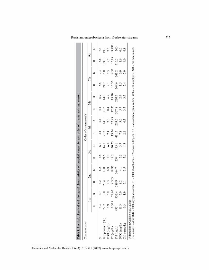
The physical, chemical, and biological characteristics of the orders of stream reaches studied are shown in Table 1. Higher water temperatures were recorded, as ex-pected, in the rainy summer period. The temperature variation between sampled periods reached up to 10°C. The waters were well oxygenated in both seasons. The pH data ob-tained suggested a slightly acidic environment with water values close to 7.0 during the dry season, except for the first-order stream reach. A positive correlation between pH and temperature was found (r = 0.73, P < 0.05) during the rainy season. Higher dissolved or-ganic carbon concentrations were observed in the rainy season. Total nitrogen and total phosphorus concentrations showed no limitations of these essential nutrients in the study area. No clear correlation was detected in Chl a concentration between the two seasons in the different orders of stream reaches.
Aquatic enterobacterium identification
A total of 111 bacterial isolates (45 from the rainy season and 66 from the dry season sam-plings) were identified to at least the genus level: Citrobacter sp (N = 2), Enterobacter sp (N = 9), Edwarsiella sp (N = 4), Escherichia sp (N = 8), Klebsiella sp (N = 52), Kluyvera sp (N = 1), Mor-ganella sp (N = 16), Proteus sp (N = 5), Providentia sp (N = 2), and Serratia sp (N = 12).
Antimicrobial resistance
The percentage of freshwater enterobacteria showing antimicrobial resistance was determined, and resistance was demonstrated for all antimicrobials tested and mercury bichloride. Of the total 102 enterobacteria analyzed (nine were excluded since they be-longed to genera with N < 5) from seven orders of stream reaches, 93% showed resistance to at least one antimicrobial at some level (Table 2). Kluyvera was the only genus suscep-tible to all antimicrobials tested.
Genetics and Molecular Research 6 (3): 510-521 (2007) www.funpecrp.com.br
Resistant enterobacteria from freshwater streams 515
Tabl
e 1.
Phy
sica
l, ch
emic
al a
nd b
iolo
gica
l cha
ract
eris
tics o
f sam
pled
wat
ers f
or e
ach
orde
r of s
tream
reac
h an
d se
ason
.
Cha
ract
eris
tica
Ord
er o
f stre
am re
ach
1st
2nd
3rd
4th
5th
7th
9th
RD
RD
RD
RD
RD
RD
RD
pH4.
34.
74.
26.
24.
56.
14.
46.
44.
46.
95.
57.
35.
87.
3Te
mpe
ratu
re (°
C)
22.7
19.0
23.4
17.0
21.5
14.0
21.3
14.0
21.2
14.0
24.7
15.0
24.3
19.0
TOD
(mg/
L)7.
96.
98.
36.
97.
16.
77.
47.
08.
46.
89.
17.
58.
77.
5TP
(mg/
L)1.
325
24.6
59.
503
19.9
714
.520
.25
13.3
427
.95
12.3
315
.20
13.5
514
.52
15.8
88.
492
TN (m
g/L)
493
432.
458
9.9
264.
723
414
5.1
411.
520
3.6
367.
823
0.5
244.
624
1.2
516.
5N
DD
OC
(mg/
L)11
.37.
08.
24.
17.
33.
57.
83.
65.
52.
41.
50.
91.
60.
6C
hl a
(mg/
L)0.
31.
95.
13.
53.
51.
32.
10.
33.
53.
72.
42.
94.
04.
8a A
dapt
ed f
rom
Cal
listo
et a
l. (2
002)
.R
= r
ainy
; D =
dry
; TO
D =
tota
l oxy
gen
diss
olve
d; T
P =
tota
l pho
spho
rous
; TN
= to
tal n
itrog
en; D
OC
= d
isso
lved
org
anic
car
bon;
Chl
a =
chl
orop
hyll
a; N
D =
not
det
erm
ined
.

Genetics and Molecular Research 6 (3): 510-521 (2007) www.funpecrp.com.br
C.I. Lima-Bittencourt et al. 516
Wide differences in resistance percentages were observed for particular antimicro-bials among bacterial genera, as was the case for tetracycline and for genera such as Ser-ratia and Klebsiella. Moreover, resistance percentages for different antimicrobials greatly differed within the same bacteria even within the same season, e.g., among Klebsiella sp isolates, none was resistant to amikacin, whereas 83% were resistant to ampicillin. Resist-ance percentages were significantly different between seasons within the same bacterial genus, with the exception of Serratia sp and Klebsiella sp isolates which were resistant to amoxicillin-clavulanic acid and chloramphenicol, respectively (Table 2). Ampicillin resist-ance was the most frequent (84%) irregardless of the season, reaching 97% during summer and 76% during winter. The lowest resistance percentages were detected for aminoglyco-sides and ranged from 3 to 17%.
Multiple resistant enterobacteria were frequent (61%), especially in the rainy season samplings (77%), and less frequent in the dry season samplings (52%) (Table 2). Overall, an isolate was most frequently resistant to four antimicrobials than to one antimicrobial (Figure 2). Proteus was the genus that showed resistance to more antimicrobials simultaneously. On the other hand, Escherichia was the most sensitive genus to antimicrobials because only 12.5% of the isolates demonstrated multiple resistance. Klebsiella isolates from the dry season sam-plings exhibited the largest number of resistance markers, amounting to eight of the 11 antimi-crobial agents investigated. Furthermore, the seasonal analysis revealed that the percentage of resistance found in bacterial isolates recovered from the rainy season samplings differed from those of the dry season samplings (P < 0.05). This was evidenced by the percentage of nalidixic acid- and tetracycline-resistant bacteria. Resistance percentages were higher in the rainy season isolates than in the dry season isolates. All bacterial isolates from rainy season samplings were resistant to at least one antimicrobial agent. Resistance to mercury was more frequent in the dry season samplings, and it was always associated with at least two other resistance markers. The highest percentage of mercury resistance was observed for the genus Serratia (Table 2). There was a highly significant correlation between antimicrobial markers and mercury, which was more evident for nalidixic acid (r = 0.934, P < 0.001), chloramphenicol (r = 0.900, P < 0.001) and ampicillin (r = 0.771, P < 0.001).
Ampicillin-resistant bacteria were predominant, and we therefore considered it im-portant to determine β-lactamase production. The colorimetric assay in the ampicillin-resistant population revealed a high frequency of β-lactamase producers in both seasons: 97% in the rainy season samplings and 90% in the dry season isolates. β-lactamase was not detected for five Klebsiella sp isolates and one Serratia sp isolate, and therefore, the mechanism of resist-ance to ampicillin could not be identified. All bacterial genera, except for Citrobacter harbored the blaTEM gene, as revealed by PCR.
The influence of biotic and abiotic factors on antimicrobial resistance
A PCA was performed on all environmental variables described in Table 1 to reduce data and to obtain an overview of the biotic and abiotic conditions in the investigated freshwa-ter sites. Multiple regression was also performed in order to relate environmental variables with changes in the composition of the bacterial communities and with the antimicrobial resistance patterns observed. This analysis was restricted to the environmental variables that strongly cor-

Genetics and Molecular Research 6 (3): 510-521 (2007) www.funpecrp.com.br
Resistant enterobacteria from freshwater streams 517
Tabl
e 2.
Ant
imic
robi
al re
sist
ance
and
mul
tiple
resi
stan
ce p
erce
ntag
e in
bac
teria
l gen
era
sam
pled
dur
ing
the
rain
y an
d dr
y se
ason
s.
Ant
imic
robi
al
a Bac
teria
l gen
usEn
t. (N
= 9
)E.
(N =
8)
Kl.
(N =
52)
Mo.
(N =
16)
Pr. (
N =
5)
Ser.
(N =
12)
Tota
l (N
= 1
02)
RD
RD
RD
RD
RD
RD
RD
Am
830
100
050
3592
010
00
7575
8230
50A
p10
033
100
7183
7810
033
100
010
075
9776
84A
k17
00
00
08
020
00
08
03
Gm
170
00
00
80
200
00
80
3K
m0
00
1417
27
00
00
05
34
Sm17
010
00
3315
80
400
370
2611
17C
m17
010
00
5050
80
800
3750
3340
37N
x0
010
00
1752
00
200
1250
1067
29R
f33
00
1417
2639
040
062
5039
2429
Tc33
010
00
5015
380
800
5075
4916
28H
g0
00
5717
4315
00
037
100
1544
33To
tal
100
6710
010
010
091
100
3310
00
100
100
100
8993
Mul
tiple
resi
stan
ce25
6771
100
7588
77
52
61
a Onl
y ge
nera
with
sam
ple
size
≥5
wer
e in
clud
ed. E
nt. =
Ent
erob
acte
r; E
. = E
sche
rich
ia; K
l. =
Kle
bsie
lla; M
o. =
Mor
gane
lla; P
r. =
Pro
teus
; Ser
. = S
erra
tia; R
= r
ainy
; D =
dry
; Am
=
amox
icill
in-c
lavu
lani
c ac
id; A
p =
ampi
cilli
n; A
k =
amik
acin
; Gm
= g
enta
mic
in; K
m =
kan
amyc
in; S
m =
stre
ptom
ycin
; Cm
, chl
oram
phen
icol
; Nx
= na
lidix
ic a
cid;
Rf =
rifa
mpi
cin;
Tc
= te
tracy
clin
e; H
g =
mer
cury
bic
hlor
ide.

Genetics and Molecular Research 6 (3): 510-521 (2007) www.funpecrp.com.br
C.I. Lima-Bittencourt et al. 518
Figure 2. Differences in multiple antimicrobial resistance by bacterial genus. Only genera with sample size ≥5 were included.

Genetics and Molecular Research 6 (3): 510-521 (2007) www.funpecrp.com.br
Resistant enterobacteria from freshwater streams 519
related with the three most relevant components obtained by PCA, Chl a, pH and temperature, which altogether explained 91% of the variance for both seasons (data not shown). Multiple regression results showed that pH, temperature and Chl a concentration were not correlated to antimicrobial resistance patterns according to the seasons.
DISCUSSION
This study shows that members of the family Enterobacteriaceae are only a fraction of the total number of culturable bacteria in pristine freshwater (data not shown). Among the clinically important Enterobacteriaceae observed were: Klebsiella, Proteus, Enterobacter, Morganella, and Serratia. The high resistance percentage to at least one antimicrobial agent (93%) and to multiple-antimicrobial agents (61%) for freshwater enterobacteria observed herein have also been found in bacteria isolated from rural groundwater supplies (McKeon et al., 1995) and wild mammals (Salyers et al., 2004). These studies also noted that ampicil-lin-resistance was common. In contrast, Gõni-Urriza et al. (2000) reported that ampicillin and multiple-antimicrobial resistance percentages in bacteria from an urban effluent were much lower than the one we found. Therefore, the present study has important implications for the interpretation of results from polluted sites. Additionally, studies of clinical isolates also showed that the β-lactam-resistance was as high as in our findings (Bantar et al., 2000; Brisse and Duijkeren, 2005). Our data also agree with the cited studies that revealed uncom-mon resistance to aminoglycosides.
The high incidence of resistance to antimicrobials herein observed could be related to some well-known resistance mechanisms. Nitrocefin hydrolysis data suggest that β-lactamase production is the main ampicillin-resistance mechanism in these isolates. Ash et al. (2002) also observed the same phenomenon in Gram-negative bacteria from rivers. In addition, the presence of the blaTEM gene will be important in determining transmissibility of β-lactamase resistance since this gene is known to be associated with transposons and plasmids (Gootz, 2004). On the other hand, in accordance to Rahman et al. (2004) and Andres et al. (2005), the resistance to amoxicillin-clavulanic acid suggests that the isolates can produce extended-spec-trum β-lactamases.
The observed variations in the percentages and patterns of resistance among the isolates and the lower resistance percentage during the dry season probably reflect changes in the composition of the bacterial populations sampled. The data also suggest that sus-ceptible bacteria may have a higher survival rate than antimicrobial-resistant bacteria in surface waters during the dry season. According to Andersson and Levin (1999), the ac-quisition of an antimicrobial-resistant phenotype reduces bacterial fitness and, therefore, replacement of resistant populations by susceptible populations may occur in the absence of selection and/or nutrient.
The abiotic factors pH and temperature and the biotic factor Chl a concentration did not influence the antimicrobial resistance patterns. Our study demonstrates that resistance pat-terns change over space and time. It is tempting to suggest that there are constant changes in bacterial genomes through mutations, horizontal gene transfer and other recombinational events. The present results show the presence of multiple antimicrobial resistance in pristine freshwater bacteria with dynamic resistance profiles. In addition, the resistance profiles do not

Genetics and Molecular Research 6 (3): 510-521 (2007) www.funpecrp.com.br
C.I. Lima-Bittencourt et al. 520
seem to result from changes in environmental conditions. The findings here obtained are quite alarming, when considering the detection of drug-resistant bacteria from hospitals and animal sources, and hence may have clinical implications.
ACKNOWLEDGMENTS
We appreciate the financial support given by CNPq (Brazil), FAPEMIG (Brazil) and PROGRAD-UFMG (Brazil) in the form of a scholarship to C.I. Lima-Bittencourt. The authors are especially grateful to students from the Laboratory of Benthic Ecology in the Department of Biologia Geral (ICB-UFMG) for field assistance and to Andréa Reis for labo-ratory assistance.
REFERENCES
Andersson DI and Levin BR (1999). The biological cost of antibiotic resistance. Curr. Opin. Microbiol. 2: 489-493.Andres P, Petroni A, Faccone D, Pasterán F, et al. (2005). Extended-spectrum β-lactamases in Shigella flexneri from
Argentina: first report of TOHO-1 outside Japan. Int. J. Antimicrob. Agents 25: 501-507.Ash RJ, Mauck B and Morgan M (2002). Antibiotic resistance of Gram-negative bacteria in rivers, United States.
Emerg. Infect. Dis. 8: 713-716.Bantar C, Famiglietti A and Goldberg M (2000). Three-year surveillance study of nosocomial bacterial resistance
in Argentina. The Antimicrobial Committee; and the National Surveillance Program (SIR) Participants Group. Int. J. Infect. Dis. 4: 85-90.
Barza M and Gorbach SL (2002). The need to improve antimicrobial use in agriculture: ecological and human health consequences. Clin. Infect. Dis. 34: 71-77.
Belaaouaj A, Lapoumeroulie C, Caniça MM, Vedel G, et al. (1994). Nucleotide sequences of the genes coding for the TEM-like/3-lactamases IRT-1 and IRT-2 (formerly called TRI-1 and TRI-2). FEMS Microbiol. Lett. 120: 75-80.
Benner R and Strom M (1993). A critical evaluation of the analytical blank associated with DOC measurements by high-temperature catalytic oxidation. Mar. Chem. 41: 153-160.
Blazquez J, Oliver A and Gomez-Gomez JM (2002). Mutation and evolution of antibiotic resistance: antibiotics as promoters of antibiotic resistance? Curr. Drug Targets 3: 345-349.
Braga LC, Leite AA, Xavier KG, Takahashi JA, et al. (2005). Synergic interaction between pomegranate extract and antibiotics against Staphylococcus aureus. Can. J. Microbiol. 51: 541-547.
Brisse S and Duijkeren E (2005). Identification and antimicrobial susceptibility of 100 Klebsiella animal clinical isolates. Vet. Microbiol. 105: 307-312.
Callisto M, Moreno P, Goulart M, Medeiros A, et al. (2002). The assessment of aquatic biodiversity along an altitu-dinal gradient at the Serra do Cipó (south-eastern Brazil). Verh. Internat. Verein. Limnol. 28: 1-4.
Cota BB, Oliveira AB, Ventura CP, Mendonça MP, et al. (2002). Antimicrobial activity of plant species from a Bra-zilian hotspot for conservation priority. Pharm. Biol. 40: 542-547.
Davison J (1999). Genetic exchange between bacteria in the environment. Plasmid 42: 73-91.Galvão MV and Nimer E (1965). Clima. In: Geografia do Brasil-Grande Região Leste (Galvão MV and Nimer E,
eds.). IBGE, Rio de Janeiro, 91-139.Goldstein FW (2000). Antibiotic susceptibility of bacterial strains isolated from patients with community-acquired
urinary tract infections in France. Multicentre Study Group. Eur. J. Clin. Microbiol. Infect. Dis. 19: 112-117.Golterman HL, Clymo RS and Ohnstad MAM (1978). Methods for chemical analysis of fresh waters. Blackwell
Scientific Publications, Oxford.Gõni-Urriza M, Capdepuy M, Arpin C, Raymond N, et al. (2000). Impact of an urban effluent on antibiotic resis-
tance of riverine Enterobacteriaceae and Aeromonas spp. Appl. Environ. Microbiol. 66: 125-132.Gootz TD (2004). Global dissemination of beta-lactamases mediating resistance to cephalosporins and carbapen-
ems. Expert. Rev. Anti. Infect. Ther. 2: 317-327.Holt JG (1984). Bergey’s Manual of Systematic Bacteriology. Williams and Wilkins, Baltimore.Kapil A (2005). The challenge of antibiotic resistance: need to contemplate. Indian J. Med. Res. 121: 83-91.
Genetics and Molecular Research 6 (3): 510-521 (2007) www.funpecrp.com.br
Resistant enterobacteria from freshwater streams 521
Koppen W (1936). Das geographisca System der Klimate. In: Handbuch der Klimatologie (Köppen W and Geiger R, eds.). Gebrüder Borntraeger, Berlin, 1-46.
Levy SB and Marshall B (2004). Antibacterial resistance worldwide: causes, challenges and responses. Nat. Med. 10: S122-S129.
Lorenzen CJ (1967). Determination of chlorophyll and pheopigments: spectrophotometric equations. Limnol. Oceanogr. 12: 343-346.
Margurran AE (1988). Ecological diversity and its measurements. Princeton University Press, Princeton.McKeon DM, Calabrese JP and Bissonnette GK (1995). Antibiotic-resistant gram-negative bacteria in rural ground-
water supplies. Water Res. 29: 1902-1908.Nascimento AMA and Chartone-Souza E (2003). Operon mer: bacterial resistance to mercury and potential for
bioremediation of contaminated environments. Genet. Mol. Res. 2: 92-101.Nascimento AMA, Campos CE, Campos EP, Azevedo JL, et al. (1999). Re-evaluation of antibiotic and mercury
resistance in Escherichia coli populations isolated in 1978 from Amazonian rubber tree tappers and Indians. Res. Microbiol. 150: 407-411.
Nascimento AMA, Cursino L, Gonçalves-Dornelas H, Reis A, et al. (2003). Antibiotic-resistant Gram-negative bacteria in birds from the Brazilian Atlantic Forest. Condor 105: 358-363.
National Committee for Clinical Laboratory Standards (NCCLS) (2001). Methods for dilution antimicrobial sus-ceptibility tests for bacteria that grow aerobically, approved standard M7-A5. National Committee for Clinical Laboratory Standards, Wayne.
O’Brien TF (2002). Emergence, spread, and environmental effect of antimicrobial resistance: how use of an antimi-crobial anywhere can increase resistance to any antimicrobial anywhere else. Clin. Infect. Dis. 34: S78-S84.
Rahman M, Shoma S, Rashid H, Siddique AK, et al. (2004). Extended-spectrum β-lactamase-mediated third-gen-eration cephalosporin resistance in Shigella isolates in Bangladesh. J. Antimicrob. Chemother. 54: 846-847.
Salyers AA, Gupta A and Wang Y (2004). Human intestinal bacteria as reservoirs for antibiotic resistance genes. Trends Microbiol. 12: 412-416.
Sherley M, Gordon DM and Collignon PJ (2000). Variations in antibiotic resistance profile in Enterobacteriaceae isolated from wild Australian mammals. Environ. Microbiol. 2: 620-631.
Strahler HN (1957). Quantitative analysis of watershed geomorphology. Am. Geophys. Union Trans. 3: 913-920.Whitehead TR, Cotta MA, Whittle G, Shoemaker N, et al. (2003). The commensal bacterial populations of swine
feces and stored swine manure: reservoirs of antibiotic resistance? J. Anim. Sci. 81: 461.Wright GD (2007). The antibiotic resistome: the nexus of chemical and genetic diversity. Nat. Rev. Microbiol. 5:
175-186.