Embed Size (px)
Citation preview

materials
Article
Multi Dynamic Extraction: An Innovative Method toObtain a Standardized Chemically and BiologicallyReproducible Polyphenol Extract from Poplar-TypePropolis to Be Used for Its Anti-Infective Properties
Vincenzo Zaccaria 1 , Emanuele Ugo Garzarella 2, Carmen Di Giovanni 2, Fabio Galeotti 3,Lucia Gisone 1, Davide Campoccia 4 , Nicola Volpi 3, Carla Renata Arciola 4,5,*and Maria Daglia 2,6,*
1 Department of Drug Sciences, Medicinal Chemistry and Pharmaceutical Technology Section,Pavia University, Viale Taramelli 12, 27100 Pavia, Italy; [email protected] (V.Z.);[email protected] (L.G.)
2 Department of Pharmacy, Nutraceutical Lab, University of the Naples, Federico II, Via D. Montesano 49,80131 Napoli, Italy; [email protected] (E.U.G.); [email protected] (C.D.G.)
3 Department of Life Sciences, University of Modena and Reggio Emilia, Via Campi 213/D,41121 Modena, Italy; [email protected] (F.G.); [email protected] (N.V.)
4 Laboratorio di Patologia delle Infezioni Associate all’Impianto, IRCCS Istituto Ortopedico Rizzoli,via di Barbiano 1/10, 40136 Bologna, Italy; [email protected]
5 Department of Experimental, Diagnostic and Specialty Medicine, University of Bologna, via San Giacomo 14,40126 Bologna, Italy
6 International Research Center for Food Nutrition and Safety, Jiangsu University, Zhenjiang 212013, China* Correspondence: [email protected] (C.R.A.); [email protected] (M.D.); Tel.: +39-051-636-6599
(C.R.A.); Tel.: +39-081-678-644 (M.D.)
Received: 15 September 2019; Accepted: 6 November 2019; Published: 13 November 2019 �����������������
Abstract: Antimicrobial activity is a well-known property of propolis, making it a candidatefor antimicrobial surfaces in biomedical devices. Nevertheless, large-scale use of propolis asan anti-infective agent is limited by the heterogeneity of its chemical composition and consequentvariation in antimicrobial activity. The aim of this study was to demonstrate that the multi dynamicextraction (M.E.D.) method produces standardized polyphenolic mixtures from poplar-type propolis,with reproducible chemical composition and anti-microbial activity, independently from the chemicalcomposition of the starting raw propolis. Three raw propolis samples, from Europe, America,and Asia, were analyzed for their polyphenol chemical composition by means of HPLC–UV andthen combined to obtain three mixtures of propolis, which werme submitted to the M.E.D. extractionmethod. The chemical composition and the antimicrobial activity of M.E.D. propolis against bacteriaand fungi were determined. The three M.E.D. propolis showed similar chemical compositionsand antimicrobial activities, exhibiting no relevant differences against antibiotic-susceptible andantibiotic-resistant strains. The batch-to-batch reproducibility of propolis extracts obtained withthe M.E.D. method encourages the design of drugs alternative to traditional antibiotics and thedevelopment of anti-infective surface-modified biomaterials.
Keywords: multi dynamic extraction (M.E.D.) method; poplar-type propolis; standardized polyphenolicmixture; antimicrobial activity; antibiotic-resistant bacteria; anti-infective agent; anti-infective biomaterials
Materials 2019, 12, 3746; doi:10.3390/ma12223746 www.mdpi.com/journal/materials

Materials 2019, 12, 3746 2 of 16
1. Introduction
Propolis is a natural resinous product, processed by bees from a range of plants for use inconstruction of their hives [1,2]. Its composition varies depending on its botanical and geographicalorigins [3], although the different types of propolis do have a shared chemical nature [4]. In fact,in addition to resin (50%), wax (30%), essential oils (10%), pollen (5%), and mineral salts (2%), the maincomponents of propolis are polyphenols, including flavonoids, phenolic acids, and their esters [5].
The analysis of the chemical composition and biological properties of propolis, as well as its usein drugs, foods, and cosmetics requires an extraction process so as to remove impurities and inertmaterials while preserving the majority of the plant secondary metabolites, especially the polyphenolfraction [4–6]. Until now, this has been generally achieved by extraction with solvents, in particularwith 70% ethanol. Hydro-alcoholic extraction results in wax-free tinctures containing variable amountsof bioactive compounds, including the phenolic substances [7,8]. Nevertheless, while simple andeffective, this method suffers from some limits in application due to the potential presence of ethanolresidues in the resultant pharmaceutical products, foods, and cosmetics.
Therefore, the production of non-ethanolic propolis extracts represents an important prospect,with progress focused on solubilization of the active molecules of propolis in aqueous or oily solvents,as phenolic compounds were found at 10-fold lower concentrations in these conditions comparedto ethanolic extracts [9,10]. A new, patented propolis extraction method, called multi dynamicextraction (M.E.D.), has recently been proposed [11,12]. M.E.D. allows the preparation of non-ethanolicpropolis extracts (M.E.D. extracts) with reliable polyphenolic contents, in contrast to the majorityof other extraction methods that yield a poorly replicable chemical composition. On the contrary,M.E.D. extracts possess a standardized polyphenol composition containing 5–20% phenolic acidsand 50–80% flavonoids (20–30% flavones and flavonols, 30–50% flavanones and dihydroflavonols,5–20% glycosilated flavonoids and terpenoids), with these extracts being rich in six active compounds,i.e., galangin, chrysin, pinocembrin, apigenin, pinobanksin, and quercetin, with a relative concentrationof about 40% (w/w) [13]. From its composition, M.E.D. extract can be defined as a poplar-typepropolis polyphenolic mixture, rich in bioactive compounds, and highly purified from impurities andinert materials [12].
Propolis demonstrates many healthy properties, of which the most studied is antimicrobialactivity [5,14–17], which has been described against multiple bacterial strains and fungi [18,19].The complex mechanism by which propolis exerts its antimicrobial activity is yet to be elucidatedand could involve synergisms between different polyphenols [20]. The major problems regardingthese studies on the antimicrobial properties of propolis consist in the variation in composition of thestudied propolis extracts, which give different results in terms of antimicrobial activity and make itimpossible to compare results obtained from different propolis extracts. A further limit of the literaturedata is that the chemical composition of the tested extracts is different from that of the extracts currentlyused as drug, food, or cosmetics ingredients and so these results have limited practical applications.In summary, several factors such as origin, composition, and extraction method can influence thebiological behavior of the product. On the basis of these considerations, following a request from theEuropean Commission about a list of propolis health claims, the opinion of the European Food SafetyAuthority (EFSA) was that a cause–effect relationship cannot be established between the consumptionof propolis and the claimed effects, due to the fact that “type and content of flavonoids in propolis may varydepending on the specific propolis raw material as well as the extraction and preparation methods”.
The aim of our study is to demonstrate the chemical and biological reproducibility of poplar-typepropolis extracts obtained using a M.E.D. method on a combination of poplar-type propolis of differentgeographical origins. Thus, the chemical composition of nine hydroalcoholic propolis extracts and threenon-ethanolic M.E.D. propolis was evaluated by high-performance liquid chromatography coupledwith UV detection and mass spectrometry (HPLC–UV–MSn) and these were compared. In addition,the antimicrobial activity was used as a validation method of the extractive process M.E.D. to show thebiological reproducibility of M.E.D. propolis.

Materials 2019, 12, 3746 3 of 16
2. Materials and Methods
2.1. Materials
HPLC-grade water was obtained from a LC-Pak™Millex system (Millipore Corporation, Billerica,MA, USA). Formic acid, MS grade methanol, quercetin, apigenin, pinobaskin, crysin, pinocembrin,and galangin were obtained from PhytoLab, Vestenbergsgreuth, Germany.
2.2. Hydroalcoholic Propolis Extract Preparation
Three poplar-type raw propolis samples were obtained from as many European regions (Italy (E1),Spain (E2), and Turkey (E3)), three further poplar-type raw propolis samples were obtained from threedifferent Southern American regions (Uruguay (sA1), Mexico (sA2), and Argentina (sA3)), and the lastthree poplar-type raw propolis samples were collected from distinct Asian regions (Mongolia (A1),Kazakhstan (A2), and north China (A3)). Samples (20 mg each) were dissolved in 2 mL of 70% ethanol.After vigorous mixing and sonication for 30 min, polyphenols were extracted at 70 ◦C in a water bathfor 2 hours under continuous mixing. After centrifugation at 10,000 RPM for 10 min, hydroalcoholicpropolis extracts were analyzed by RP-HPLC–PDA–ESI–MSn.
2.3. M.E.D. Propolis Preparation
In order to determine the chemical composition and test the antibacterial activity of M.E.D.propolis, three mixtures (mix A, mix B, and mix C) were prepared combining a European, an American,and an Asian poplar-type raw propolis sample (Eu + Am + As). M.E.D. propolis A, B, and C, respectively,were obtained from each raw mixture using M.E.D. (multi dynamic extraction), as reported in [11].In brief, raw propolis mixtures were submitted to the M.E.D. extraction process, comprising severalsteps. These steps consisted of an initial aqueous extraction from dewaxed raw propolis, followed bya series of extractions on the residue using an ethanol/water mixture, with each extraction being carriedout on the residue from the previous extraction using a higher percentage of ethanol. The combinedextracts were mixed and concentrated by distillation to a residual humidity value ranging from 15 to20% (w/w). The concentrated extracts were then analyzed by RP-HPLC–PDA–ESI–MSn and submittedto an antimicrobial assay.
2.4. Analyses of Hydroalcoholic Propolis Extracts and M.E.D. Propolis by RP-HPLC–PDA–ESI–MSn
The chromatographic analyses were performed by means of the RP-HPLC-PAD-ESI-MSn method,set up by Cui-ping et al., with some modifications [21]. These were performed using an Agilent1100 VL series mass spectrometer (Agilent Technologies, Inc., Santa Clara, CA, USA), which wasfurther used on-line with HPLC equipment. The electrospray interface was set in negative ionizationmode with the capillary voltage at 3500 V and a temperature source of 350 ◦C in full scan spectra(200–2200 Da, 10 full scans/s). Nitrogen was used as a drying (9 L/min) and nebulizing gas (11 p.s.i.).Software versions were 4.0 LC/MSD trap control 4.2 and Data Analysis 2.2 (Agilent Technologies,Inc., Santa Clara, CA, USA). Compound separation was obtained with an analytical Synergi FusionRP-18 column (150_4.6 mm, 5_m), equipped with a Hypersil Gold C18 precolumn (10 _2.1 mm,5_m), all produced by Phenomenex (Torrance, CA, USA). The mobile phase used was acidifiedwater, with 0.1% formic acid (eluent A) and methanol (eluent B). The run time was 110 min in total,including the reconditioning of the column. The flow rate was maintained at 1.00 mL/min, and thetemperatures of the autosampler and column were kept at 4 and 33 ◦C. The volume of injection wasset to 5 L. The elution method is described in Table 1. Chromatograms were registered at 260 nm.The HPLC-ESI-MSn data were collected using Xcalibur software (Xcalibur 2.0, Thermo Fisher Scientific,Waltham, MA, USA).

Materials 2019, 12, 3746 4 of 16
Table 1. RP-HPLC–PDA–ESI–MSn analysis elution method.
Time (min) % Eluent A % Eluent B
0 85 1530 60 4065 45 5570 38 6285 0 10090 0 100
100 85 15110 85 15
2.5. Antimicrobial Assays
In order to determine the inhibitory effects of M.E.D. propolis against different microorganisms,these experiments were carried out using the broth dilution method according to the procedures ofthe Clinical and Laboratory Standards Institute (CLSI), so as to determine the minimum inhibitoryconcentration (MIC), defined as the lowest concentration of an antimicrobial agent that can inhibit thegrowth of microorganisms.
Each dry M.E.D. propolis extract (A, B, or C) was resuspended in 50% (v/v) ethanol/water to obtaina final concentration of 50 mg/mL. A blank sample was also prepared, without adding any extract.Then, the three samples were serially diluted 1:2 in 50% ethanol, and 0.8 mL of each dilution weremixed with 7.2 mL of the specific agar culture medium, previously equilibrated at 70 ◦C, to finallycover a polyphenol concentration range between 0.007 mg/mL and 0.872 mg/mL. Once perfectly mixedby vortexing, agar culture medium was added to each propolis extract at different concentrationsand then poured into a 6 mm Petri plate, and a cell suspension from a frozen vial was plated atabout 5 × 10³ CFU/spot. As a positive control, some plates were prepared with the culture mediumcontaining 0.8 mL of the blank stock. The growing media and conditions were chosen according tomicroorganism species (Table 2).
Table 2. The growing conditions for the microorganisms selected to test propolis antimicrobial activity.
Microbial Strain Media Conditions
S. aureus methicillin-sensitive ATCC25923(MSSA)(L1280)
S. aureus methicillin-resistant (MRSA) (L4064)S. aureus MSSA + glycopeptide-interMediate
resistant (GISA) (L3797)S. aureus MRSA + GISA (L3798)
S. aureus clinda-inducible erm(A)+ (ND053410)S. aureus community acquired USA300 MRSA
(ND054910)S. aureus MRSA + macrolides-resistant (ND060411)
S. hominis ATCC27844 (L323)S. epidermidis (L147; ND052110; ND051710)
S. epidermidis teicoplanin-resistant (ND042409)S. capitis MRSA (ND021008)S. xylosus MRSA (ND026108)
S. simulans (ND029808)S. haemolyticus MRSA (L1730, ND040809; L1729)
E. coli hyperpermeable (G1640)E. coli ATCC25922 (L1281)
E. coli hyperpermeable (L4242; L47)P. aeruginosa ATCC27853 (L1367)
M. catarrhalis (L3292)A. baumannii (L3030)
L. monocytogenes ATCC13932 (L1450)B. cereus ATCC10702 (L85)
Mueller Hinton Agar Aerobic, 24 h, 37 ◦C
Materials 2019, 12, 3746 5 of 16
Table 2. Cont.
Microbial Strain Media Conditions
S. pneumoniae penicillin-susceptible (L44)S. pneumoniae penicillin-resistant (L3917)
S. pneumoniae clindamycin and erythromycinresistant (L1542)
S. pneumoniae macrolide and erythromycin resistant(L1402)
Todd Hewitt Agar Aerobic, 24 h, 37 ◦C
C. parapsilosis ATCC90018 (L3022)C. parapsilosis ATCC22019 (L4119)
C. albicans ATCC24443 (L4120)C. albicans ATCC90028 (L3023)
C. guillermondii ATCC6260 (L2065)C. kruzei (L2880)
A. niger ATCC10535 (L53)
SabouraudDextrose Agar Aerobic, 48 h, 37 ◦C
G. vaginalis (L1629; L1622; L1630)A. vaginae (ND736; ND737)
B. fragilis ATCC25285 (L1011)L. paracasei (L1693)L. plantarum (L19)L. gasseri (ND787)
L. acidophilus (ND786)C. difficile (L1365; L1366)
C. difficile ATCC17858 (L4013)
Brucella Agar with 5%laked horse blood and 1%
hemin and vitamin KAnaerobic, 72 h, 37 ◦C
N. gonorrhoeae (L1600; L1601; L1599)
Brucella Agar with 5%laked horse blood, 1%
hemin and vitamin K and1% isovitalex
Anaerobic, 72 h, 37 ◦C
C. perfrigens (L4053)C. perfrigens ATCC13124 (L3697)
P. acnes ATCC25746 (L1016)
Brucella Agar with 5%laked horse blood and 1%
hemin and vitamin KAnaerobic, 48 h, 37 ◦C
A large panel of bacterial and fungal species (Table 2) were used to test the antimicrobial activity ofM.E.D. propolis, sourced from the American Type Culture Collection (ATCC) and clinical isolates (CodeL) provided by Naicos srl, Milan, Italy (IRCCS Policlinico San Donato, San Donato Milanese, Italy; CentreHospitalier Universitaire de Limoges, Limoges, France; International Health Management Associates, Inc.,Shaumburg, IL, USA; Micromyx, LLC, Kalamazoo, MI, USA; Ospedale Busto Arstizio, Busto Arsizio,Italy; MM: IRCCS Multimedica, Milan, Itay; Rockville, MD, USA; S. Raffaele Hospital, Milan, Italy).The tested microorganisms included both drug-resistant species, showing resistance against one or moreantibiotics, and sensitive species, i.e., non-resistant (Table 7). Tables 7 and 8 present the MIC values ofselected antibiotics as reported by the literature where available, these were used as positive control.
2.6. Statistical Analysis
The values represent mean values of at least 3 replications. Data were analyzed by analysis ofvariance (ANOVA) with the statistical package GraphPad PRISM v 6.0 (2015) (GraphPad Software Inc.,San Jose, CA, USA). Means were separated with the Tukey’s HSD method.
3. Results
3.1. RP-HPLC–PDA–ESI–MSn Analyses of Hydroalcoholic Propolis Extracts
Raw poplar-type propolis materials, obtained from three European regions (Eu1, Eu2, Eu3), threeSouthern American regions (Am1, Am2, Am3), and three Asian regions (As1, As2, As3) were submittedto hydroalcoholic extraction. The extracts were analyzed by means of RP-HPLC–PDA–ESI–MSn.The main flavonoid species, flavonols (galangin, quercetin), flavones (chrysin, apigenin), and flavonones(pinocembrin, pinobanksin), were identified on the basis of their UV and mass spectra, checking themolecular ion and fragment ions against the fragmentation patterns of standard molecules (Table 3) [12].

Materials 2019, 12, 3746 6 of 16
Table 3. Chromatographic and spectral properties of polyphenol compounds detected in propolis samples.
Peak number RT(min)
UVabsorption
(λmax)
m/z[M-H]
Fragments(m/z) Proposed Structure
1 31.8 256 301 151, 179, 257, 273 Quercetin2 35.2 325 271 151, 165, 225, 253 Pinobaskin3 35.5 267, 338 269 117, 149, 225 Apigenin4 44.7 270 253 209 Chrysin5 45.5 290 255 151, 187, 213 Pinocembri6 46.2 261, 351 269 227 Galangin
After the initial identification of quercetin, apigenin, pinobaskin, chrysin, pinocembrin, and galangin,they were determined by an on-line HPLC–UV according to methods previously described (Table 4).
The results (Table 4) showed that while the total polyphenol contents of the Asian hydroalcoholicpropolis extracts were similar (mean value: 46.3% w/w, standard deviation: 0.8), the total polyphenolcontent of the American propolis samples ranged from 37.8 to 59.3 (mean value: 50.1%, standarddeviation: 11.1), as was the case for the total polyphenol contents of the European propolis samples,which ranged from 38.2 to 42.6 (mean value: 40.6%, standard deviation 2.2).
As far as the relative percentage of each main flavonoid compound is concerned, the analysisof variance (ANOVA) was used to evaluate whether the concentration of each compound wasstatistically different between the hydroalcoholic propolis extracts, considering their different origins.The results, reported in Table 5, showed that the relative percentages of the polyphenols were oftenstatistically different, confirming that the high variability of propolis raw materials of differentorigin leads to propolis extracts with different compositions when using common extraction methods(i.e., hydroalcoholic extraction) (Figure 1).
Table 4. Relative percentage (% w/w) of the specific polyphenols occurring in the nine hydroalcoholicpropolis extracts of different geographical origins, determined by HPLC-UV.
Polyphenols Eu1 Eu2 Eu3
1-Quercetin 1.4 ± 0.6 0.8 ± 0.2 0.7 ± 0.42-Pinobanksin 1.5 ± 0.1 1.0 ± 0.2 1.5 ± 0.3
3-Apigenin 1.6 ± 0.3 1.1 ± 0.3 1.2 ± 0.34-Chrysin 18.3 ± 0.3 21.4 ± 0.2 24.1 ± 0.4
5-Pinocembrin 2.8 ± 0.3 4.8 ± 0.1 3.1 ± 0.26-Galangin 12.6 ± 0.1 12.0 ± 0.1 12.0 ± 0.2
Sum of percentages 38.2 41.1 42.6
- Am1 Am2 Am3
1-Quercetin 0.5 ± 0-6 0.5 ± 0.1 0.9 ± 0.52-Pinobanksin 1.0 ± 0.2 0.9 ± 0.2 3.0 ± 0.8
3-Apigenin 1.5 ± 0.9 0.9 ± 0.3 3.5 ± 1.24-Chrysin 30.3 ± 3.3 22.2 ± 1.1 28.6 ± 0.6
5-Pinocembrin 4.4 ± 0.4 1.8 ± 0.3 13.9 ± 1.16-Galangin 15.4 ± 0.6 11.5 ± 0.4 9.4 ± 1.3
Sum of percentages 53.1 37.8 59.3
- As1 As2 As3
1-Quercetin 0.4 ± 0.4 0.4 ± 0.5 0.9 ± 0.42-Pinobanksin 1.0 ± 0.2 1.8 ± 0.4 10.0 ± 2.0
3-Apigenin 2.2 ± 0.8 2.0 ± 1.7 1.2 ± 0.14-Chrysin 25.0 ± 2.5 24.4 ± 0.8 19.6 ± 1.4
5-Pinocembrin 2.0 ± 0.1 2.4 ± 0.1 1.7 ± 0.26-Galangin 16.1 ± 0.6 15.9 ± 0.5 12.0 ± 1.0
Sum of percentages 46.7 46.9 45.4

Materials 2019, 12, 3746 7 of 16
Table 5. Analysis of Variance: contribution of geographical origin (country) on the relative percentage(% w/w) of each main flavonoid species: flavonols (galangin, quercetin), flavones (chrysin, apigenin),and flavonones (pinocembrin, pinobanksin).
Comparisons Significance
Quercetin Pinobanksin Apigenin Chrysin Pinocembrin Galangin
EU 1 vs EU 2 Yes * No ** No No Yes NoEU 1 vs EU 3 Yes No No Yes No NoEU 1 vs AM 1 Yes No No Yes Yes YesEU 1 vs AM 2 Yes No No No No NoEU 1 vs AM 3 No No Yes Yes Yes YesEU 1 vs AS 1 Yes No No Yes No YesEU 1 vs AS 2 Yes No No Yes No YesEU 1 vs AS 3 No Yes No No No No
EU 2 vs EU 3 No No No No Yes NoEU 2 vs AM 1 No No No Yes No YesEU 2 vs AM 2 No No No No Yes NoEU 2 vs AM 3 No No Yes Yes Yes YesEU 2 vs AS 1 No No Yes No Yes YesEU 2 vs AS 2 No No Yes No Yes YesEU 2 vs AS 3 No Yes No No Yes No
EU 3 vs AM 1 No No No Yes Yes YesEU 3 vs AM 2 No No No No Yes NoEU 3 vs AM 3 No No Yes Yes Yes YesEU 3 vs AS 1 No No Yes No No YesEU 3 vs AS 2 No No No No No YesEU 3 vs AS 3 No Yes No Yes Yes No
AM 1 vs AM 2 No No No Yes Yes YesAM 1 vs AM 3 No No Yes No Yes YesAM 1 vs AS 1 No No No Yes Yes NoAM 1 vs AS 2 No No No Yes Yes NoAM 1 vs AS 3 No Yes No Yes Yes Yes
AM 2 vs AM 3 No No Yes Yes Yes YesAM 2 vs AS 1 No No Yes No No YesAM 2 vs AS 2 No No Yes No No YesAM 2 vs AS 3 No Yes No No No No
AM 3 vs AS 1 No No Yes No Yes YesAM 3 vs AS 2 No No Yes No Yes YesAM 3 vs AS 3 No Yes Yes Yes Yes Yes
AS 1 vs AS 2 No No No No No NoAS 1 vs AS 3 No Yes Yes Yes No Yes
AS 2 vs AS 3 No Yes No Yes No Yes
* Yes means statistically significant difference between the relative percentage (% w/w) of each flavonoid inhydroalcoholic propolis extracts from different origins. ** No means no statistically significant difference betweenthe relative percentage (% w/w) of each flavonoid in hydroalcoholic propolis extracts from different origins.
Materials 2019, 12, 3746 8 of 16
Materials 2019, 12, x FOR PEER REVIEW 8 of 16
AM 1 vs AS 2 No No No Yes Yes No AM 1 vs AS 3 No Yes No Yes Yes Yes AM 2 vs AM 3 No No Yes Yes Yes Yes AM 2 vs AS 1 No No Yes No No Yes AM 2 vs AS 2 No No Yes No No Yes AM 2 vs AS 3 No Yes No No No No AM 3 vs AS 1 No No Yes No Yes Yes AM 3 vs AS 2 No No Yes No Yes Yes AM 3 vs AS 3 No Yes Yes Yes Yes Yes AS 1 vs AS 2 No No No No No No AS 1 vs AS 3 No Yes Yes Yes No Yes AS 2 vs AS 3 No Yes No Yes No Yes
* Yes means statistically significant difference between the relative percentage (% w/w) of each flavonoid in hydroalcoholic propolis extracts from different origins. ** No means no statistically significant difference between the relative percentage (% w/w) of each flavonoid in hydroalcoholic propolis extracts from different origins.
Figure 1. Analysis of Variance: contribution of geographical origin (country) on the relative percentage (% w/w) of each main flavonoid species: flavonols (galangin, quercetin), flavones (chrysin, apigenin), and flavonones (pinocembrin, pinobanksin).
3.2. RP-HPLC–PDA–ESI–MSn Analyses of Non-Ethanolic M.E.D. Propolis
Raw propolis materials obtained from European (Eu1, Eu2, Eu3), Southern American (Am1, Am2, Am3), and Asian regions (As1, As2, As3) were combined to obtain three mixtures of European, American, and Asian poplar-type propolis (Eu + Am + As). Each mixture was submitted to the M.E.D. extraction process to give the extracts A, B, and C, respectively.
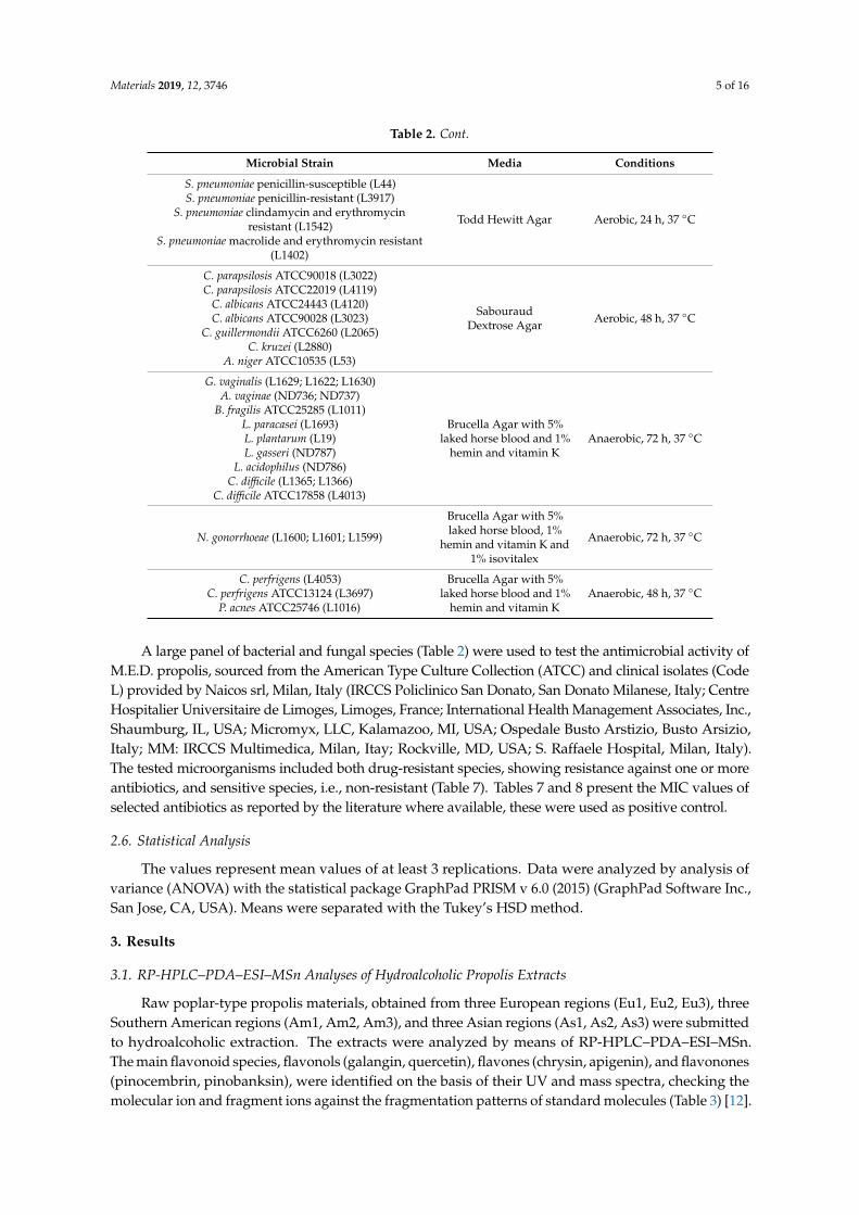
After the identification of quercetin, apigenin, pinobaskin, chrysin, pinocembrin, and galangin on the basis of their UV and mass spectra (Table 3 and Figure 2D), they were quantified by means of HPLC–UV analyses (Table 6). The chromatograms acquired at λ 260 nm for each M.E.D. propolis extract are reported in Figure 2A–C. The relative percentages of each flavonoid were tested with ANOVA, showing that no differences were found in flavonoid composition between the non-ethanolic M.E.D. propolis extracts (Figure 3).
Figure 1. Analysis of Variance: contribution of geographical origin (country) on the relative percentage(% w/w) of each main flavonoid species: flavonols (galangin, quercetin), flavones (chrysin, apigenin),and flavonones (pinocembrin, pinobanksin).
3.2. RP-HPLC–PDA–ESI–MSn Analyses of Non-Ethanolic M.E.D. Propolis
Raw propolis materials obtained from European (Eu1, Eu2, Eu3), Southern American (Am1,Am2, Am3), and Asian regions (As1, As2, As3) were combined to obtain three mixtures of European,American, and Asian poplar-type propolis (Eu + Am + As). Each mixture was submitted to the M.E.D.extraction process to give the extracts A, B, and C, respectively.
After the identification of quercetin, apigenin, pinobaskin, chrysin, pinocembrin, and galanginon the basis of their UV and mass spectra (Table 3 and Figure 3D), they were quantified by meansof HPLC–UV analyses (Table 6). The chromatograms acquired at λ 260 nm for each M.E.D. propolisextract are reported in Figure 3A–C. The relative percentages of each flavonoid were tested withANOVA, showing that no differences were found in flavonoid composition between the non-ethanolicM.E.D. propolis extracts (Figure 2).
Table 6. Relative percentage (% w/w) of the specific polyphenols occurring in the three propolis extractsdetermined by high-performance liquid chromatography–UV (HPLC–UV).
Polyphenols M.E.D. Propolis A M.E.D. Propolis B M.E.D. Propolis C
1-Quercetin 1.1 ± 0.05 1.2 ± 0.10 0.9 ± 0.062-Pinobanksin 1.2 ± 0.40 0.8 ± 0.11 1.6 ± 0.36
3-Apigenin 1.2 ± 0.30 1.0 ± 0.20 1.4 ± 0.044-Chrysin 23.2 ± 0.60 22.0 ± 0.71 22.0 ± 1.02
5-Pinocembrin 1.17 ± 0.04 1.4 ± 0.06 1.4 ± 0.046-Galangin 13.4 ± 0.15 14.7 ± 0.11 14.3 ± 0.10

Materials 2019, 12, 3746 9 of 16
Materials 2019, 12, x FOR PEER REVIEW 9 of 16
Figure 2. Chromatograms registered at λ 260 nm. (A) Multi dynamic extraction (M.E.D.) propolis extract A; (B) M.E.D. propolis extract B; (C) M.E.D. propolis extract C; (D) Standard compounds: 1—quercetin; 2—pinobainskin; 3—apigenin; 4—chrysin; 5—pinocembrin; 6—galangin.
Table 6. Relative percentage (% w/w) of the specific polyphenols occurring in the three propolis extracts determined by high-performance liquid chromatography–UV (HPLC–UV).
Polyphenols M.E.D. Propolis A M.E.D. Propolis B M.E.D. Propolis C 1-Quercetin 1.1 ± 0.05 1.2 ± 0.10 0.9 ± 0.06
2-Pinobanksin 1.2 ± 0.40 0.8 ± 0.11 1.6 ± 0.36 3-Apigenin 1.2 ± 0.30 1.0 ± 0.20 1.4 ± 0.04 4-Chrysin 23.2 ± 0.60 22.0 ± 0.71 22.0 ± 1.02
5-Pinocembrin 1.17 ± 0.04 1.4 ± 0.06 1.4 ± 0.04 6-Galangin 13.4 ± 0.15 14.7 ± 0.11 14.3 ± 0.10
Figure 2. Analysis of Variance: contribution of M.E.D. extraction process on the relative percentage(% w/w) of each main flavonoid species: quercetin, pinobanksin, apigenin, chrysin, pinocembrin,and galangin.
Materials 2019, 12, x FOR PEER REVIEW 9 of 16
Figure 2. Chromatograms registered at λ 260 nm. (A) Multi dynamic extraction (M.E.D.) propolis extract A; (B) M.E.D. propolis extract B; (C) M.E.D. propolis extract C; (D) Standard compounds: 1—quercetin; 2—pinobainskin; 3—apigenin; 4—chrysin; 5—pinocembrin; 6—galangin.
Table 6. Relative percentage (% w/w) of the specific polyphenols occurring in the three propolis extracts determined by high-performance liquid chromatography–UV (HPLC–UV).
Polyphenols M.E.D. Propolis A M.E.D. Propolis B M.E.D. Propolis C 1-Quercetin 1.1 ± 0.05 1.2 ± 0.10 0.9 ± 0.06
2-Pinobanksin 1.2 ± 0.40 0.8 ± 0.11 1.6 ± 0.36 3-Apigenin 1.2 ± 0.30 1.0 ± 0.20 1.4 ± 0.04 4-Chrysin 23.2 ± 0.60 22.0 ± 0.71 22.0 ± 1.02
5-Pinocembrin 1.17 ± 0.04 1.4 ± 0.06 1.4 ± 0.04 6-Galangin 13.4 ± 0.15 14.7 ± 0.11 14.3 ± 0.10
Figure 3. Chromatograms registered at λ 260 nm. (A) Multi dynamic extraction (M.E.D.) propolisextract A; (B) M.E.D. propolis extract B; (C) M.E.D. propolis extract C; (D) Standard compounds:1—quercetin; 2—pinobainskin; 3—apigenin; 4—chrysin; 5—pinocembrin; 6—galangin.
3.3. Antimicrobial Activity of M.E.D. Propolis Extracts
The antimicrobial activity of the three M.E.D. propolis extracts was first tested against microorganismstrains representing the major families: Gram-positive or Gram-negative bacteria and fungi.
As expected, MIC values showed that the three M.E.D. propolis extracts exerted antimicrobialactivity, confirming literature data on this property of propolis [18,19]. In particular, low MIC values(ranging between 20 and 156 µg/mL) were found against Aspergillus niger, Streptococcus pneumoniapenicillin-susceptible, Moraxella catarrhalis, Atopobium vaginae, and Neisseria gonorrhoeae. Moderateactivity was found against Staphylococcus spp and Gardnerella vaginalis, (MIC value = 312 µg/mL). Poor

Materials 2019, 12, 3746 10 of 16
effects were registered on the growth of Candida spp and Clostridium spp, shown by MIC values above1250 µg/mL. No activity could be detected against Bacteroides fragilis and Lactobacillus spp (Table 7).
The results obtained by our experiments gave comparable MIC values for each extract obtainedusing the M.E.D. method against the same microorganisms, despite the different geographical originsof the three samples.
Table 7. Minimum inhibitory concentration (MIC) values of the M.E.D. propolis extracts A, B, and C againstthe tested microbial strains, and of the antimicrobial drugs against their susceptible microrganisms.
Microbial StrainMIC (µg/mL) MIC (µg/mL)
CODE A B C Antimicrobial agent
Staphylococcus aureus MSSA ATCC25923 L1280 312 312 312 -Staphylococcus epidermidis ATCC12228 L147 312 312 312 -
Escherichia coli hyperpermeable G1640 312 625 625 0.078, trimethroprimMoraxella catarrhalis L3292 39 78 78 0.3, ampicillin
Streptococcus pneumoniaepenicillin-susceptible L44 20 39 39 2.0, ampicillin
Candida albicans ATCC24443 L4120 1250 1250 1250 0.75, fluconazoleCandida albicans ATCC90028 L3023 1250 2500 2500 1.0, fluconazole
Candida parapsilosis ATCC90018 L3022 2500 2500 2500 4.0, fluconazoleCandida kruzei L2280 2500 2500 2500 10.0, fluconazole
Aspergillus niger ATCC10535 L53 78 156 156 1000, fluconazoleBacteroides fragilis ATCC25285 L1011 5000 >5000 >5000 6.0, cefoxitin
Propionebacterium acnes ATCC25746 L1016 >5000 >5000 >5000 1.8, clindamycinClostridium difficile L1365 2500 2500 2500 0.6, vancomycin
Clostridium difficile ATCC17858 L4013 5000 2500 2500 1.6, vancomycinAtopobium vaginae ND736 156 156 156 0.478, ampicillinLactobacillus gasseri ND787 5000 >5000 >5000 0.25, ampicillin
Lactobacillus acidophilus ND786 >5000 >5000 >5000 1.0, clindamycinNeisseria gonorrhoeae L1600 156 156 156 16.0, ampicillinNeisseria gonorrhoeae L1601 156 78 78 -Gardnerella vaginalis L1629 312 312 156 0.020 ampicillinGardnerella vaginalis L1630 312 312 312 -
Therefore, since the chemical composition and the antimicrobial activity values of the three M.E.D.propolis extracts were found to be comparable, we randomly selected extract A to investigate theantimicrobial activity more widely, including against antibiotic resistant microbial strains, as reportedin Table 8. In this case, MIC values confirmed the antimicrobial activity of the extract against certaintypes of microorganisms, revealing the same effects of propolis on both sensitive and resistant species.
Table 8. MIC values of M.E.D. propolis extract A and antimicrobial drugs on a wider microbial set.
Microbial Strain CODEPropolisExtract A
MIC (µg/mL)
Antimicrobial AgentMIC (µg/mL)
Escherichia coli L4242 312 0.12, ampicillinStaphylococcus aureus GISA MSSA L3797 625 -Staphylococcus aureus GISA MRSA L3798 312 -Staphylococcus haemolyticus MRSA L1730 312 -Staphylococcus hominis ATCC27844 L323 625 0.046, ampicillin
Staphylococcus capitis MRSA ND021008 156 -Staphylococcus xylosus MRSA ND026108 625 -
Staphylococcus simulans ND029808 1250 -Staphylococcus haemolyticus MRSA ND040809 625 -Staphylococcus haemolyticus MRSA L1729 1250 -
Staphylococcus aureus Clinda-inducible erm(A)+ ND053410 625 64.0, ampicillin
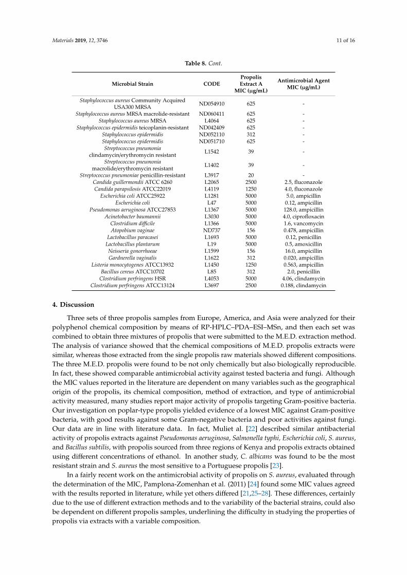
Materials 2019, 12, 3746 11 of 16
Table 8. Cont.
Microbial Strain CODEPropolisExtract A
MIC (µg/mL)
Antimicrobial AgentMIC (µg/mL)
Staphylococcus aureus Community AcquiredUSA300 MRSA ND054910 625 -
Staphylococcus aureus MRSA macrolide-resistant ND060411 625 -Staphylococcus aureus MRSA L4064 625 -
Staphylococcus epidermidis teicoplanin-resistant ND042409 625 -Staphylococcus epidermidis ND052110 312 -Staphylococcus epidermidis ND051710 625 -Streptococcus pneumonia
clindamycin/erythromycin resistant L1542 39 -
Streptococcus pneumoniamacrolide/erythromycin resistant L1402 39 -
Streptococcus pneumoniae penicillin-resistant L3917 20 -Candida guillermondii ATCC 6260 L2065 2500 2.5, fluconazoleCandida parapsilosis ATCC22019 L4119 1250 4.0, fluconazole
Escherichia coli ATCC25922 L1281 5000 5.0, ampicillinEscherichia coli L47 5000 0.12, ampicillin
Pseudomonas aeruginosa ATCC27853 L1367 5000 128.0, ampicillinAcinetobacter baumannii L3030 5000 4.0, ciprofloxacin
Clostridium difficile L1366 5000 1.6, vancomycinAtopobium vaginae ND737 156 0.478, ampicillin
Lactobacillus paracasei L1693 5000 0.12, penicillinLactobacillus plantarum L19 5000 0.5, amoxicillinNeisseria gonorrhoeae L1599 156 16.0, ampicillinGardnerella vaginalis L1622 312 0.020, ampicillin
Listeria monocytogenes ATCC13932 L1450 1250 0.563, ampicillinBacillus cereus ATCC10702 L85 312 2.0, penicillin
Clostridium perfringens HSR L4053 5000 4.06, clindamycinClostridium perfringens ATCC13124 L3697 2500 0.188, clindamycin
4. Discussion
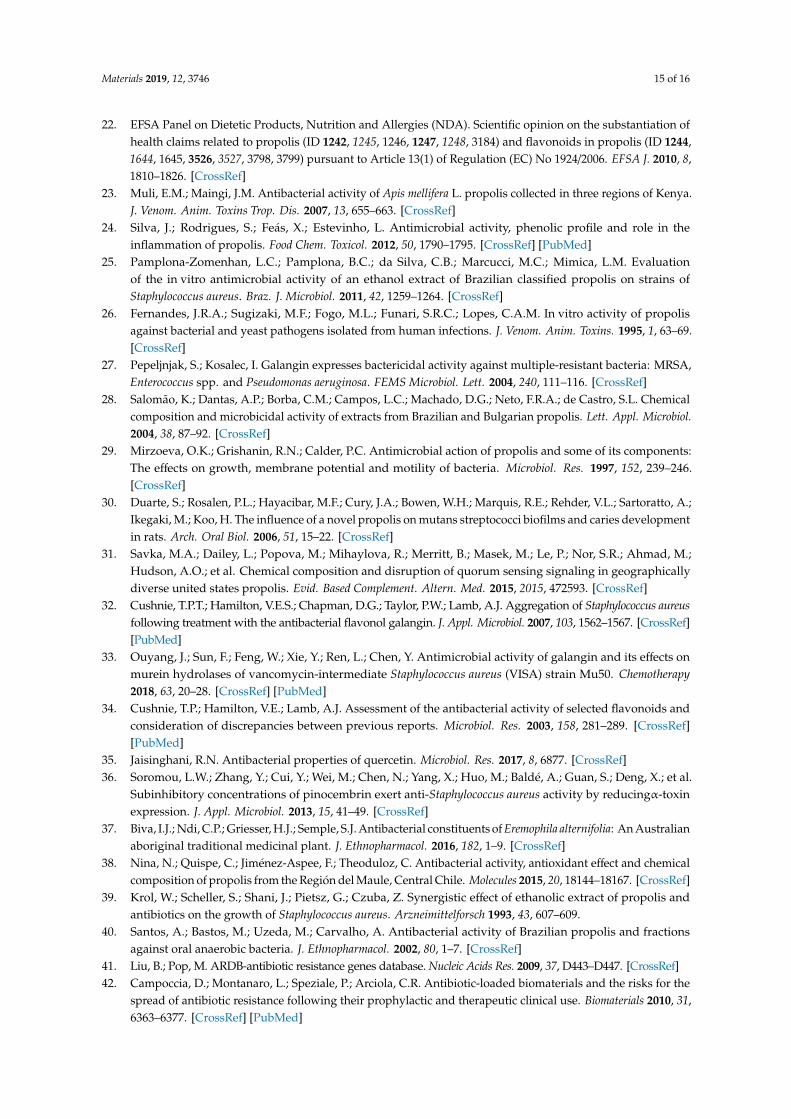
Three sets of three propolis samples from Europe, America, and Asia were analyzed for theirpolyphenol chemical composition by means of RP-HPLC–PDA–ESI–MSn, and then each set wascombined to obtain three mixtures of propolis that were submitted to the M.E.D. extraction method.The analysis of variance showed that the chemical compositions of M.E.D. propolis extracts weresimilar, whereas those extracted from the single propolis raw materials showed different compositions.The three M.E.D. propolis were found to be not only chemically but also biologically reproducible.In fact, these showed comparable antimicrobial activity against tested bacteria and fungi. Althoughthe MIC values reported in the literature are dependent on many variables such as the geographicalorigin of the propolis, its chemical composition, method of extraction, and type of antimicrobialactivity measured, many studies report major activity of propolis targeting Gram-positive bacteria.Our investigation on poplar-type propolis yielded evidence of a lowest MIC against Gram-positivebacteria, with good results against some Gram-negative bacteria and poor activities against fungi.Our data are in line with literature data. In fact, Muliet al. [22] described similar antibacterialactivity of propolis extracts against Pseudomonas aeruginosa, Salmonella typhi, Escherichia coli, S. aureus,and Bacillus subtilis, with propolis sourced from three regions of Kenya and propolis extracts obtainedusing different concentrations of ethanol. In another study, C. albicans was found to be the mostresistant strain and S. aureus the most sensitive to a Portuguese propolis [23].
In a fairly recent work on the antimicrobial activity of propolis on S. aureus, evaluated throughthe determination of the MIC, Pamplona-Zomenhan et al. (2011) [24] found some MIC values agreedwith the results reported in literature, while yet others differed [21,25–28]. These differences, certainlydue to the use of different extraction methods and to the variability of the bacterial strains, could alsobe dependent on different propolis samples, underlining the difficulty in studying the properties ofpropolis via extracts with a variable composition.

Materials 2019, 12, 3746 12 of 16
With the commonly used extraction methods (i.e., hydroalcoholic extraction), propolis extractsshow poorly replicable chemical composition and biological properties due to the variability in theraw material. Nevertheless, in view of the uses for propolis extracts in drugs, foods, and cosmetics,it is important to have non-ethanolic propolis extracts with a standardized chemical composition andthe guarantee of final products with the same biological properties. In our study, we are the first toreport the antimicrobial activities of three mixtures of poplar-type propolis from different geographicalareas with the same polyphenol content, obtained using the M.E.D. method as described in thepaper by Zaccaria et al. [11]. The extracts show the presence of the main flavonoid species, flavonols(galangin, quercetin), flavones (chrysin, apigenin), and flavonones (pinocembrin, pinobanksin), that canbe involved in different inhibitory mechanisms of microbial activity, such as inhibition of the mobilityof the bacteria or peptidoglycan synthesis [29,30], pinocembrin-mediated inhibition of quorumsensing [31], and adhesion blocking by galangin [32]. Galangin and apigenin are present in M.E.D.propolis extracts with a relative average percentage w/w of 14.15% and 1.19%, respectively. In theliterature, galangin showed a MIC = 32 µg/mL against different types of S. aureus (ATCC25293, N315,and Mu50), significantly suppressing bacterial growth at concentrations of 4, 8, and 16 µg/mL [33].In another paper, apigenin and galangin were investigated against sensitive and antibiotic resistantstrains of S. aureus, Enterococcus faecalis, Enterococcus faecium, E. coli, and P. aeruginosa. Galangin wasshown to have a MIC ranging from 25 to 50 µg/mL against six strains of S. aureus, but negligible activityagainst other species, partially inhibiting the growth of all six enterococci. Apigenin only displayedmarginal activity against S. aureus and did not show any activity against enterocci [34]. Quercetin ispresent in M.E.D. propolis extracts at an average percentage w/w of 1.11%. A recent study revealedantibacterial activity against S. aureus, E. coli, P. vulgaris, Shigella, P. aeruginosa, and Lactobacillus caseivar. shirota. S. aureus and P. aeruginosa were inhibited, with an MIC value = 20 µg/mL, while moderateactivity was seen against E. coli and P. vulgaris (MIC = 30 µg/mL) and no activity against Shigella flexneriand Lactobacillus casei with MIC > 300 µg/mL [35]. Other compounds, pynocembrin, and pinobanksin,revealed weak antibacterial activities against Gram-positive and Gram-negative strains. Pynocembrin,present at a 1.30% average percentage in M.E.D. propolis extract, did not inhibit the growth ofS. aureus, as described in a paper by Soromou et al. [36]. Pinobanskin (1.20% average percentage inM.E.D. propolis extracts) showed MIC > 515 µg/mL against four strains of S. aureus, but no data werereported against E. coli. [37]. Finally, chrysin showed MIC > 50 µg/mL against S. aureus ATCC25923methicillin-sensitive, S. aureus ATCC43300 methicillin-resistant, and different strains of E. coli [38].The comparison between MIC values reported in the literature for each flavonoid and the concentrationsof these compounds present in M.E.D. propolis extracts, suggests that the antimicrobial activity ofM.E.D. propolis is not purely due to these flavonoids, but is influenced in a more complex matrixeffect. With this view, previous papers have reported that the antimicrobial activity of propolis can beascribed to the synergistic effects of phenolics and flavonoids [14,39] and other compounds such asditerpenic acids [40].
Our study also evidenced interesting activities of M.E.D. propolis extracts against antibioticresistant bacteria such as Streptococcus pneumoniae clindamycin/erythromycin resistant and to a lesserextent against different strains of Staphylococcus. Antibiotic resistance is a serious and urgent problemfor public health. As published by the CDC web site, at least 2 million people are infected withantibiotic-resistant bacteria each year in the U.S., and at least 23,000 people die as a result. Infectionscaused by antibiotic-resistant microorganisms are very difficult to treat and, in most cases, requirehospitalization and expensive therapeutic alternatives. The Antibiotic Resistant Gene Database (ARDB)evidences about 20,000 genes potentially able to mutate and acquire antibiotic resistance by multiplemechanisms of action [41]. Our results are encouraging and useful for the development of newantimicrobial agents in the near future, targeting multi-resistant bacteria strains in support of ongoingtherapies. Our results also encourage the design of new anti-infective biomaterials alternative toantibiotic-loaded materials [42]. Since the 1990s, the search for biomaterials able to thwart bacterialcontamination and infection has been exponential in terms of surface modifications and coatings [43–45].

Materials 2019, 12, 3746 13 of 16
Recently, this research has been focusing on making the material surfaces not only bactericidal butalso functionalized with other favorable properties [46,47]. It is expected that the new generation ofanti-infective biomaterials will be able to integrate with host tissues, exert anti-inflammatory activities,and promote wound healing, in addition to counteracting bacterial colonization [48]. Propolis isa bactericidal compound worthy of recognition as a candidate for preparing new anti-infective materials.It meets the expectations and corresponds with the newly emerging concepts of biocompatibility, beingendowed with multifaceted therapeutic bioactivities, mainly attributable to its polyphenol content [1,5].In a very recent study, cornstarch incorporated with propolis and hyaluronic acid was found to improvewound dressing [49]. Films based on polysaccharides loaded with propolis and vitamin C have beenshown to be an effective bio-coating to accelerate the healing of diabetic wounds [50]. Polyvinylalcohol (PVA) hydrogels loaded with a Brazilian propolis have been shown to be effective in promotingburn wound healing [51]. Recent advances in therapeutic strategies to control the immune responseto implants have highlighted the role of propolis in modulating the behavior of neutrophils and inharmonizing the cellular responses to biomaterials. Indeed, propolis seems to be able to favor thetransition of macrophages from the M1 to M2 phenotype, extinguishing inflammation [51]. A safe andlong-term use of polymeric materials requires the application of anti-degrading agents with a widerange of actions. Raw propolis originating from two geographic regions of Europe was used to protectnatural rubber materials from degradation by oxygen, ozone, and microorganisms [52]. The additionof propolis to a rubber mixture made the material resistant to thermo-oxidative aging and ozoneand protected it from biodegradation [53]. As for the new and interesting field of nanomaterials,chitosan–propolis nanoparticles, prepared using propolis from Asia, exhibited the abilities to alter thezeta potential of S. epidermidis, inhibit biofilm formation by modulating gene expression, and synergizewith antibiotics [54]. Propolis/polyurethane composite nanofibers and electrospun materials havebeen proposed for biomedical applications [55]. Propolis polyphenols, although beneficially bioactivemolecules, pose problems of low bioavailability, and thus may benefit from targeted nanodeliverysystems via nanocarriers [56]. In particular, permeation through the buccal mucosa appears to bea promising approach to bypass liver metabolism and release propolis-based nano-formulations andniosomes locally or systemically [57,58].
As far as the in vitro cytotoxicity, it has been reported in the literature that propolis extracts expresstoxic effects on tumor cells (the half-maximal inhibitory concentration IC50 sometimes approachingvalues as low as 2 µg/mL) and, to a lesser extent, even on normal cells (IC50: 58 µg/mL) [59,60]. In theearly stages of the study of new biomaterials, investigations are required to assess their cytocompatibilityunder conditions close to the final clinical application. The propolis extracts here presented will beused as nanostructured coatings or nanoformulated drugs. Nanocoatings and nanoformulations canbe expected to amplify the antibacterial properties while minimizing the side effects towards host cells.
In conclusion, this study suggests the use of a non-ethanolic mixture of poplar-type propolis witha standardized polyphenol content obtained by an extraction method such as M.E.D., so as to producecomparable activity data. Moreover, the analysis of the antimicrobial data suggests new potentialmedical applications for M.E.D. poplar-type propolis against antibiotic resistant microbial strains thatwill need to be further investigated in the future.
Author Contributions: V.Z., C.D.G., E.U.G., and M.D. conceived and designed the experiments; V.C. and E.U.G.performed the antimicrobial assays; F.G. and N.V. performed HPLC analyses; L.G. participated in the investigation;D.C. joined the preparation of the revision; M.D. and C.R.A. formulated ideas on the evolution of the research field;M.D., C.D.G., V.Z., and C.R.A. wrote the paper; all authors revised the paper and approved the final manuscript.
Funding: This research was funded by Regione Lombardia, Direzione Generale Istruzione, Formazione e Lavoro,Decreto dirigenziale no. 9752, published on 21 October 2014, grant number no. 1012.
Acknowledgments: The authors would like to thank B Natural S.r.l. and Alfredo Fachini for the preparation anddelivery of M.E.D. extract of poplar-type propolis. Contribution by “5 per mille” for Health Research to IOR isalso acknowledged.
Conflicts of Interest: The authors declare no conflict of interest.

Materials 2019, 12, 3746 14 of 16
References
1. Pasupuleti, V.R.; Sammugam, L.; Ramesh, N.; Gan, S.H. Honey, propolis, and royal jelly: A comprehensivereview of their biological actions and health benefits. Oxid. Med. Cell. Longev. 2017, 2017, 1259510. [CrossRef][PubMed]
2. Ghisalberti, E.L. Propolis: A review. Bee World 1979, 60, 59–84. [CrossRef]3. Bankova, V.; Bertelli, D.; Borba, R.; Conti, B.J.; da Silva Cunha, I.B.; Danert, C.; Eberlin, M.N.; Falcão, S.I.;
Isla, M.I.; Papotti, G.; et al. Standard methods for Apis mellifera propolis research. J. Apic. Res. 2016, 58, 1–49.[CrossRef]
4. Gómez-Caravaca, A.M.; Gómez-Romero, M.; Arráez-Román, D.; Segura-Carretero, A.; Fernández-Gutiérrez, A.Advances in the analysis of phenolic compounds in products derived from bees. J. Pharm. Biomed. Anal. 2006, 41,1220–1234. [CrossRef]
5. Zabaiou, N.; Fouache, A.; Trousson, A.; Baron, S.; Zellagui, A.; Lahouel, M.; Lobaccaro, J.A. Biologicalproperties of propolis extracts: Something new from an ancient product. Chem. Phys. Lipids 2017, 207,214–222. [CrossRef]
6. Bankova, V.; Popova, M.; Trusheva, B. Propolis volatile compounds: Chemical diversity and biologicalactivity: A review. Chem. Cent. J. 2014, 8, 28. [CrossRef]
7. Woisky, R.G.; Salatino, A. Analysis of propolis: Some parameters and procedures for chemical quality control.J. Apic. Res. 1998, 37, 99–105. [CrossRef]
8. Pietta, P.G.; Gardana, C.; Pietta, A.M. Analytical methods for quality control of propolis. Fitoterapia 2002, 73,S7–S20. [CrossRef]
9. Kubiliene, L.; Laugaliene, V.; Pavilonis, A.; Maruska, A.; Majiene, D.; Barcauskaite, K.; Kubilius, R.;Kasparaviciene, G.; Savickas, A. Alternative preparation of propolis extracts: Comparison of their compositionand biological activities. BMC Complement. Altern. Med. 2015, 15, 156. [CrossRef]
10. Mello, B.C.B.S.; Petrus, J.C.C.; Hubinger, M.D. Concentration of flavonoids and phenolic compounds inaqueous and ethanolic propolis extracts through nanofiltration. J. Food Eng. 2010, 96, 533–539. [CrossRef]
11. Zaccaria, V.; Curti, V.; Di Lorenzo, A.; Baldi, A.; Maccario, C.; Sommatis, S.; Mocchi, R.; Daglia, M. Effect ofgreen and brown propolis extracts on the expression levels of microRNAs, mRNAs and proteins, related tooxidative stress and inflammation. Nutrients 2017, 9, 1090. [CrossRef] [PubMed]
12. Galeotti, F.; Maccari, F.; Fachini, A.; Volpi, N. Chemical composition and antioxidant activity of propolisprepared in different forms and in different solvents useful for finished products. Foods 2018, 7, 41. [CrossRef]
13. Volpi, N.; Fachini, A. Procedimento Per L’ottenimento di Estratti Integrali di Propoli Ricchi in Polifenolie Dot, ati di Attività Antibatterica e Sua Applicazione Nella Prevenzione e Trattamento di Processi Infettividi Origine Batterica. UfficioItalianoBrevetti e Marchi No. 0001425516, 2 February 2017.
14. Castaldo, S.; Capasso, F. Propolis, an old remedy used in modern Medicine. Fitoterapia 2002, 73, S1–S6.[CrossRef]
15. Kocot, J.; Kiełczykowska, M.; Luchowska-Kocot, D.; Kurzepa, J.; Musik, I. Antioxidant potential of propolis,bee pollen, and royal jelly: Possible medical application. Oxid. Med. Cell. Longev. 2018, 2018, 7074209.[CrossRef] [PubMed]
16. Havsteen, B.H. The biochemistry and medical significance of the flavonoids. Pharmacol. Ther. 2002, 96,67–202. [CrossRef]
17. Bankova, V. Recent trends and important developments in propolis research. Evid. Based. Complement.Alternat. Med. 2005, 2, 29–32. [CrossRef]
18. Chee, H.Y. In vitro evaluation of the antifungal activity of propolis extract on Cryptococcus neoformans andCandida albicans. Mycobiology 2002, 30, 93–95. [CrossRef]
19. Ota, C.; Unterkircher, C.; Fantinato, V.; Shimizu, M.T. Antifungal activity of propolis on different species ofCandida. Mycoses 2001, 44, 375–378. [CrossRef]
20. Kujumgiev, A.; Tsvetkova, I.; Serkedjieva, Y.; Bankova, V.; Christov, R.; Popov, S. Antibacterial, antifungal andantiviral activity of propolis of different geographic origin. J. Ethnopharmacol. 1999, 64, 235–240. [CrossRef]
21. Miorin, P.L.; Levy Junior, N.C.; Custodio, A.R.; Bretz, W.A.; Marcucci, M.C. Antibacterial activity of honeyand propolis from Apis mellifera and Tetragonisca angustula against Staphylococcus aureus. J. Appl. Microbiol.2003, 95, 913–920. [CrossRef]
Materials 2019, 12, 3746 15 of 16
22. EFSA Panel on Dietetic Products, Nutrition and Allergies (NDA). Scientific opinion on the substantiation ofhealth claims related to propolis (ID 1242, 1245, 1246, 1247, 1248, 3184) and flavonoids in propolis (ID 1244,1644, 1645, 3526, 3527, 3798, 3799) pursuant to Article 13(1) of Regulation (EC) No 1924/2006. EFSA J. 2010, 8,1810–1826. [CrossRef]
23. Muli, E.M.; Maingi, J.M. Antibacterial activity of Apis mellifera L. propolis collected in three regions of Kenya.J. Venom. Anim. Toxins Trop. Dis. 2007, 13, 655–663. [CrossRef]
24. Silva, J.; Rodrigues, S.; Feás, X.; Estevinho, L. Antimicrobial activity, phenolic profile and role in theinflammation of propolis. Food Chem. Toxicol. 2012, 50, 1790–1795. [CrossRef] [PubMed]
25. Pamplona-Zomenhan, L.C.; Pamplona, B.C.; da Silva, C.B.; Marcucci, M.C.; Mimica, L.M. Evaluationof the in vitro antimicrobial activity of an ethanol extract of Brazilian classified propolis on strains ofStaphylococcus aureus. Braz. J. Microbiol. 2011, 42, 1259–1264. [CrossRef]
26. Fernandes, J.R.A.; Sugizaki, M.F.; Fogo, M.L.; Funari, S.R.C.; Lopes, C.A.M. In vitro activity of propolisagainst bacterial and yeast pathogens isolated from human infections. J. Venom. Anim. Toxins. 1995, 1, 63–69.[CrossRef]
27. Pepeljnjak, S.; Kosalec, I. Galangin expresses bactericidal activity against multiple-resistant bacteria: MRSA,Enterococcus spp. and Pseudomonas aeruginosa. FEMS Microbiol. Lett. 2004, 240, 111–116. [CrossRef]
28. Salomão, K.; Dantas, A.P.; Borba, C.M.; Campos, L.C.; Machado, D.G.; Neto, F.R.A.; de Castro, S.L. Chemicalcomposition and microbicidal activity of extracts from Brazilian and Bulgarian propolis. Lett. Appl. Microbiol.2004, 38, 87–92. [CrossRef]
29. Mirzoeva, O.K.; Grishanin, R.N.; Calder, P.C. Antimicrobial action of propolis and some of its components:The effects on growth, membrane potential and motility of bacteria. Microbiol. Res. 1997, 152, 239–246.[CrossRef]
30. Duarte, S.; Rosalen, P.L.; Hayacibar, M.F.; Cury, J.A.; Bowen, W.H.; Marquis, R.E.; Rehder, V.L.; Sartoratto, A.;Ikegaki, M.; Koo, H. The influence of a novel propolis on mutans streptococci biofilms and caries developmentin rats. Arch. Oral Biol. 2006, 51, 15–22. [CrossRef]
31. Savka, M.A.; Dailey, L.; Popova, M.; Mihaylova, R.; Merritt, B.; Masek, M.; Le, P.; Nor, S.R.; Ahmad, M.;Hudson, A.O.; et al. Chemical composition and disruption of quorum sensing signaling in geographicallydiverse united states propolis. Evid. Based Complement. Altern. Med. 2015, 2015, 472593. [CrossRef]
32. Cushnie, T.P.T.; Hamilton, V.E.S.; Chapman, D.G.; Taylor, P.W.; Lamb, A.J. Aggregation of Staphylococcus aureusfollowing treatment with the antibacterial flavonol galangin. J. Appl. Microbiol. 2007, 103, 1562–1567. [CrossRef][PubMed]
33. Ouyang, J.; Sun, F.; Feng, W.; Xie, Y.; Ren, L.; Chen, Y. Antimicrobial activity of galangin and its effects onmurein hydrolases of vancomycin-intermediate Staphylococcus aureus (VISA) strain Mu50. Chemotherapy2018, 63, 20–28. [CrossRef] [PubMed]
34. Cushnie, T.P.; Hamilton, V.E.; Lamb, A.J. Assessment of the antibacterial activity of selected flavonoids andconsideration of discrepancies between previous reports. Microbiol. Res. 2003, 158, 281–289. [CrossRef][PubMed]
35. Jaisinghani, R.N. Antibacterial properties of quercetin. Microbiol. Res. 2017, 8, 6877. [CrossRef]36. Soromou, L.W.; Zhang, Y.; Cui, Y.; Wei, M.; Chen, N.; Yang, X.; Huo, M.; Baldé, A.; Guan, S.; Deng, X.; et al.
Subinhibitory concentrations of pinocembrin exert anti-Staphylococcus aureus activity by reducingα-toxinexpression. J. Appl. Microbiol. 2013, 15, 41–49. [CrossRef]
37. Biva, I.J.; Ndi, C.P.; Griesser, H.J.; Semple, S.J. Antibacterial constituents of Eremophila alternifolia: An Australianaboriginal traditional medicinal plant. J. Ethnopharmacol. 2016, 182, 1–9. [CrossRef]
38. Nina, N.; Quispe, C.; Jiménez-Aspee, F.; Theoduloz, C. Antibacterial activity, antioxidant effect and chemicalcomposition of propolis from the Región del Maule, Central Chile. Molecules 2015, 20, 18144–18167. [CrossRef]
39. Krol, W.; Scheller, S.; Shani, J.; Pietsz, G.; Czuba, Z. Synergistic effect of ethanolic extract of propolis andantibiotics on the growth of Staphylococcus aureus. Arzneimittelforsch 1993, 43, 607–609.
40. Santos, A.; Bastos, M.; Uzeda, M.; Carvalho, A. Antibacterial activity of Brazilian propolis and fractionsagainst oral anaerobic bacteria. J. Ethnopharmacol. 2002, 80, 1–7. [CrossRef]
41. Liu, B.; Pop, M. ARDB-antibiotic resistance genes database. Nucleic Acids Res. 2009, 37, D443–D447. [CrossRef]42. Campoccia, D.; Montanaro, L.; Speziale, P.; Arciola, C.R. Antibiotic-loaded biomaterials and the risks for the
spread of antibiotic resistance following their prophylactic and therapeutic clinical use. Biomaterials 2010, 31,6363–6377. [CrossRef] [PubMed]

Materials 2019, 12, 3746 16 of 16
43. Arciola, C.R.; Radin, L.; Alvergna, P.; Cenni, E.; Pizzoferrato, A. Heparin surface treatment ofpoly(methylmethacrylate) alters adhesion of a Staphylococcus aureus strain: Utility of bacterial fatty acidanalysis. Biomaterials 1993, 14, 1161–1164. [CrossRef]
44. Tiller, J.C.; Liao, C.J.; Lewis, K.; Klibanov, A.M. Designing surfaces that kill bacteria on contact.Proc. Natl. Acad. Sci. USA 2001, 98, 5981–5985. [CrossRef] [PubMed]
45. Bazaka, K.; Jacob, M.V.; Crawford, R.J.; Ivanova, E.P. Efficient surface modification of biomaterial to preventbiofilm formation and the attachment of microorganisms. Appl. Microbiol. Biotechnol. 2012, 95, 299–311.[CrossRef] [PubMed]
46. Marchese, A.; Arciola, C.R.; Coppo, E.; Barbieri, R.; Barreca, D.; Chebaibi, S.; Sobarzo-Sánchez, E.; Nabavi, S.F.;Nabavi, S.M.; Daglia, M. The natural plant compound carvacrol as an antimicrobial and anti-biofilm agent:Mechanisms, synergies and bio-inspired anti-infective materials. Biofouling 2018, 34, 630–656. [CrossRef]
47. Russo, N.; Cassinelli, C.; Torre, E.; Morra, M.; Iviglia, G. Improvement of the physical properties of guidedbone regeneration membrane from porcine pericardium by polyphenols-rich pomace extract. Materials 2019,12, 2564. [CrossRef]
48. Williams, D.F. Challenges with the development of biomaterials for sustainable tissue engineering.Front. Bioeng. Biotechnol. 2019, 7, 127. [CrossRef]
49. Eskandarinia, A.; Kefayat, A.; Rafienia, M.; Agheb, M.; Navid, S.; Ebrahimpour, K. Cornstarch-based wounddressing incorporated with hyaluronic acid and propolis: In vitro and in vivo studies. Carbohydr. Polym.2019, 216, 25–35. [CrossRef]
50. Voss, G.T.; Gularte, M.S.; Vogt, A.G.; Giongo, J.L.; Vaucher, R.A.; Echenique, J.V.Z.; Soares, M.P.; Luchese, C.;Wilhelm, E.A.; Fajardo, A.R. Polysaccharide-based film loaded with vitamin C and propolis: A promisingdevice to accelerate diabetic wound healing. Int. J. Pharm. 2018, 552, 340–351. [CrossRef]
51. Oliveira, R.N.; McGuinness, G.B.; Rouze, R.; Quilty, B.; Cahill, P.; Soares, G.D.A.; Thiré, R.M.S.M. PVAhydrogels loaded with a Brazilian propolis for burn wound healing applications. J. Appl. Polym. Sci. 2015,132, 42129. [CrossRef]
52. Boni, B.O.O.; Lamboni, L.; Souho, T.; Gauthier, M.; Yang, G. Immunomodulation and cellular response tobiomaterials: The overriding role of neutrophils in healing. Mater. Horiz. 2019, 6, 1122–1137. [CrossRef]
53. Kmiotek, M.; Bielinski, D.; Piotrowska, M. Propolis as an antidegradant and biocidal agent for natural rubber.J. Appl. Polym. Sci. 2018, 135, 45911. [CrossRef]
54. Ong, T.H.; Chitra, E.; Ramamurthy, S.; Ling, C.C.S.; Ambu, S.P.; Davamani, F. Cationic chitosan-propolisnanoparticles alter the zeta potential of S. epidermidis, inhibit biofilm formation by modulating gene expressionand exhibit synergism with antibiotics. PLoS ONE 2019, 14, e0213079. [CrossRef] [PubMed]
55. Kim, J.I.; Pant, H.R.; Sim, H.J.; Lee, K.M.; Kim, C.S. Electrospun propolis/polyurethane composite nanofibersfor biomedical applications. Mater. Sci. Eng. C Mater. Biol. Appl. 2014, 44, 52–57. [CrossRef] [PubMed]
56. Patra, J.K.; Das, G.; Fraceto, L.F.; Campos, E.V.R.; Rodriguez-Torres, M.D.P.; Acosta-Torres, L.S.;Diaz-Torres, L.A.; Grillo, R.; Swamy, M.K.; Sharma, S.; et al. Nano based drug delivery systems: Recentdevelopments and future prospects. J. Nanobiotechnol 2018, 16, 71. [CrossRef]
57. Reinholz, J.; Landfester, K.; Mailänder, V. The challenges of oral drug delivery via nanocarriers. Drug Deliv.2018, 25, 1694–1705. [CrossRef]
58. Arafa, M.G.; Ghalwash, D.; El-Kersh, D.M.; Elmazar, M.M. Propolis-based niosomes as oromuco-adhesivefilms: A randomized clinical trial of a therapeutic drug delivery platform for the treatment of oral recurrentaphthous ulcers. Sci. Rep. 2018, 8, 18056. [CrossRef]
59. Chen, C.N.; Weng, M.S.; Wu, C.L.; Lin, J.K. Comparison of radical scavenging activity, cytotoxiceffects and apoptosis induction in human melanoma cells by Taiwanese propolis from different sources.Evid. Based Complement. Altern. Med. 2004, 1, 175–185. [CrossRef]
60. Sadeghi-Aliabadi, H.; Hamzeh, J.; Mirian, M. Investigation of Astragalus honey and propolis extract’scytotoxic effect on two human cancer cell lines and their oncogen and proapoptotic gene expression profiles.Adv. Biomed. Res. 2015, 4, 42. [CrossRef]
© 2019 by the authors. Licensee MDPI, Basel, Switzerland. This article is an open accessarticle distributed under the terms and conditions of the Creative Commons Attribution(CC BY) license (http://creativecommons.org/licenses/by/4.0/).


![Innovative Ergonomic Solutions in Manufacturing “Cost ... · PDF filein Manufacturing “Cost effective ergonomic improvements ... Dust Extraction ... Snow 227.ppt [Read-Only]](https://img.pdfslide.us/doc/110x75/5aaac1897f8b9a86188e7061/innovative-ergonomic-solutions-in-manufacturing-cost-manufacturing-cost.jpg)