Embed Size (px)
Citation preview
… this newsle�er represents our opinion about
current periodontal technologies / procedures...
So� Tissue Gra�ing Overview
Perio News
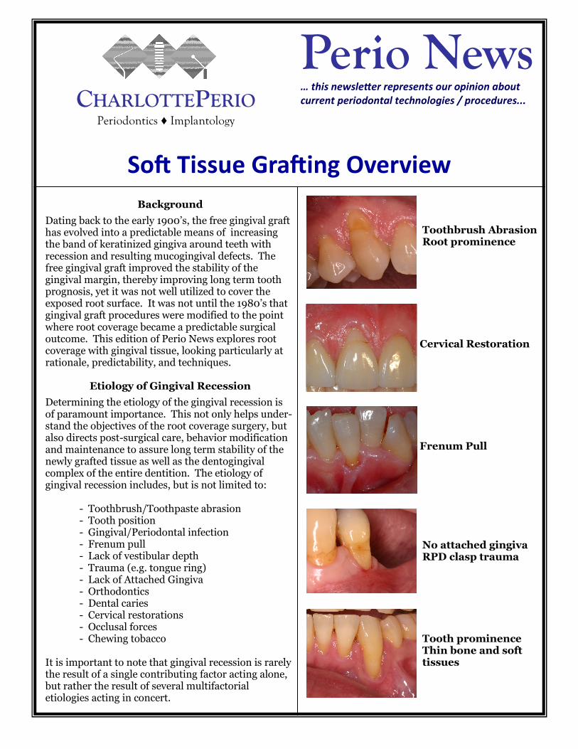
Background
Dating back to the early 1900’s, the free gingival graft has evolved into a predictable means of increasing the band of keratinized gingiva around teeth with recession and resulting mucogingival defects. The free gingival graft improved the stability of the gingival margin, thereby improving long term tooth prognosis, yet it was not well utilized to cover the exposed root surface. It was not until the 1980’s that gingival graft procedures were modified to the point where root coverage became a predictable surgical outcome. This edition of Perio News explores root coverage with gingival tissue, looking particularly at rationale, predictability, and techniques.
Etiology of Gingival Recession
Determining the etiology of the gingival recession is of paramount importance. This not only helps under-stand the objectives of the root coverage surgery, but also directs post-surgical care, behavior modification and maintenance to assure long term stability of the newly grafted tissue as well as the dentogingival complex of the entire dentition. The etiology of gingival recession includes, but is not limited to: - Toothbrush/Toothpaste abrasion - Tooth position - Gingival/Periodontal infection - Frenum pull - Lack of vestibular depth - Trauma (e.g. tongue ring) - Lack of Attached Gingiva - Orthodontics - Dental caries - Cervical restorations - Occlusal forces - Chewing tobacco It is important to note that gingival recession is rarely the result of a single contributing factor acting alone, but rather the result of several multifactorial etiologies acting in concert.
Toothbrush Abrasion Root prominence
Cervical Restoration
Frenum Pull
No attached gingiva RPD clasp trauma
Tooth prominence Thin bone and soft tissues
CHARLOTTEPERIO
Periodontics ♦ Implantology

Rationale for Root Coverage
Although improved esthetics may be the most common rationale, there are multiple reasons (alone or collectively) that coverage of the exposed root surface is a desired surgical outcome. These include, but are not limited to:
- Eliminate dentin hypersensitivity - Prevent root surface plaque adherence and food packing - Eliminate need for root surface restoration
When considering the root coverage graft, the outcome objectives are discussed with the patient, keeping in mind that stabilization of the gingival tissue and prevention of further progression of the recession lesion is still the primary goal.
Root Coverage Predictability
For decades, covering the exposed root surfaces was unpredictable and despite the introduction of multiple surgical techniques it was considered elusive and largely unattainable. In clinical situations where the exposed root surface needed to be resolved, most dentists relied upon the tradi-tional free gingival graft to stabilize the attached gingiva and subsequently placed a cervical tooth restoration or crown. Then, in the 1980’s, new advances in surgical techniques along with an improved understanding of the potential to achieve root coverage with a soft tissue graft were discovered. In 1985, Miller (1) published a landmark article which still today serves as the benchmark for classifying gingival recession in relation to the ability to achieve predictable root coverage.
Miller’s Classification of Gingival Recession
This classification (Class I—IV) has several factors that determine the amount of root coverage: - The amount of recession - The amount of attached gingiva - Recession relative to the mucogingival junction - Interproximal bone levels - Tooth position (in or outside the bony housing)
Class II: Marginal tissue recession which extends to or beyond the mucogingival junction. There is no periodontal loss (bone or soft tissue) in the interdental area, and 100% root coverage can be achieved.
Class I: Marginal tissue recession which does not extend to the mucogingival junction. No perio-dontal loss (bone or soft tissue) in the interdental area and 100% root coverage can be achieved.
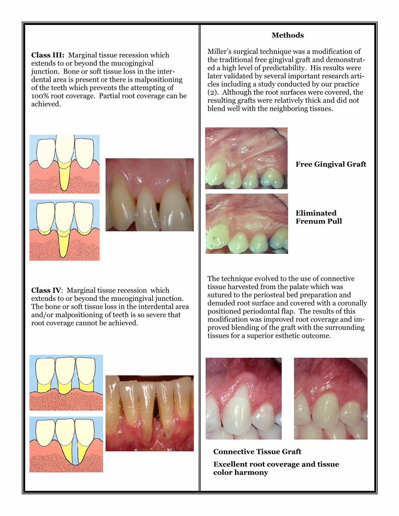
Class III: Marginal tissue recession which extends to or beyond the mucogingival junction. Bone or soft tissue loss in the inter-dental area is present or there is malpositioning of the teeth which prevents the attempting of 100% root coverage. Partial root coverage can be achieved.
Class IV: Marginal tissue recession which extends to or beyond the mucogingival junction. The bone or soft tissue loss in the interdental area and/or malpositioning of teeth is so severe that root coverage cannot be achieved.
Methods Miller’s surgical technique was a modification of the traditional free gingival graft and demonstrat-ed a high level of predictability. His results were later validated by several important research arti-cles including a study conducted by our practice (2). Although the root surfaces were covered, the resulting grafts were relatively thick and did not blend well with the neighboring tissues.
The technique evolved to the use of connective tissue harvested from the palate which was sutured to the periosteal bed preparation and denuded root surface and covered with a coronally positioned periodontal flap. The results of this modification was improved root coverage and im-proved blending of the graft with the surrounding tissues for a superior esthetic outcome.
Free Gingival Graft Eliminated Frenum Pull
Connective Tissue Graft
Excellent root coverage and tissue color harmony

Conclusion
Conventional free gingival grafts and connective tissue grafts have evolved to a level of high predictability in achieving root coverage with excellent esthetic outcomes. While other treatment modalities exist, including the use of allografts, autogenous tissue grafting remains the gold standard by which all other treatments are measured. As with all periodontal surgical procedures, a complete understanding of the etiologic factors, along with the elimination/control of these factors, is paramount to long term success. A proper diagnosis and classification of the recession defect is imperative in predicting the success of the root coverage graft surgery and must be communicated fully to the patient in order for surgical outcomes to be appropriately understood and anticipated.
Futures editions of Perio News will explore some of the alternatives to the conventional free gingival and connective tissue graft procedures (including graft surgery technique modifications and allografts like AlloDerm).
References: 1. Miller, P.D., A Classification of Marginal Tissue Recession. International Journal of Periodontics and Restorative Dentistry. 1985; 5 (2): 9-13. 2. Tolmie,PN, Rubins, RP, Buck, GS, Vagianos, V, Lanz, JC. The Predictability of Root Coverage By Way of Free Gingival Autografts and Citric Acid Application: An Evaluation By Multiple Clinicians. International Journal of Periodontics and Restorative Dentistry. 1991; 11: 261-271.
Example from page 1: Toothbrush abrasion and root prominence Connective Tissue Graft Excellent root coverage and tissue color harmony
Example from page 1: No attached gingiva and RPD clasp trauma Free Gingival Graft 7 mm band of attached gingiva Root coverage not possible due to distal bone loss
Please call or email if you have questions or comments. We appreciate your feedback and
will be happy to discuss in further detail any thoughts or questions you may have.
Bob Rubins, DDS Paul Tolmie, DDS
[email protected] [email protected]
Ken Corsig, DMD Eric Kerr, DDS
[email protected] [email protected]
3535 Randolph Road, Suite 103R ♦ Charlotte, NC 28211 ♦ Telephone: 704.365.0123