Embed Size (px)
Citation preview
mRNA pseudoknot structures can act as ribosomalroadblocksJesper Tholstrup1, Lene B. Oddershede2 and Michael A. Sørensen1,*
1Department of Biology, Ole Maaløes vej 5, University of Copenhagen, DK-2200 Copenhagen and 2Niels BohrInstitute, Blegdamsvej 17, University of Copenhagen, DK-2100 Copenhagen, Denmark
Received March 24, 2011; Revised August 5, 2011; Accepted August 7, 2011
ABSTRACT
Several viruses utilize programmed ribosomalframeshifting mediated by mRNA pseudoknots incombination with a slippery sequence to producea well defined stochiometric ratio of the upstreamencoded to the downstream-encoded protein. Acorrelation between the mechanical strength ofmRNA pseudoknots and frameshifting efficiencyhas previously been found; however, the physicalmechanism behind frameshifting still remains to befully understood. In this study, we utilized syntheticsequences predicted to form mRNA pseudoknot-like structures. Surprisingly, the structurespredicted to be strongest lead only to limited frame-shifting. Two-dimensional gel electrophoresis ofpulse labelled proteins revealed that a significantfraction of the ribosomes were frameshifted butunable to pass the pseudoknot-like structures.Hence, pseudoknots can act as ribosomal road-blocks, prohibiting a significant fraction of theframeshifted ribosomes from reaching the down-stream stop codon. The stronger the pseudoknotthe larger the frameshifting efficiency and thelarger its roadblocking effect. The maximal amountof full-length frameshifted product is produced froma structure where those two effects are balanced.Taking ribosomal roadblocking into account is aprerequisite for formulating correct frameshiftinghypotheses.
INTRODUCTION
The reading frame of the vast majority of mRNAs isdetermined by the start codon after which the downstreamcistron is translated in the same frame. Maintenance of thereading frame occurs without further signals to theribosome. However, examples of genes containing infor-mation for programmed frameshifts can be found in most
organisms, or in some of their IS sequences, transposableelements, retroelement-derived sequences or viruses. Thesequence-information needed for programmed ribosomalframeshift varies and both+1 and �1 frameshifts can beinduced (1–3).Here, we focus on the frameshifting signal found in
several viruses (1), including infectious bronchitis virus(IBV) and SARS-CoV. The signal leads to programmedribosomal �1 frameshift, whereby multiple proteins areproduced from a single polycistronic messenger RNA(mRNA) (4,5). The frameshift efficiency, i.e. the fractionof ribosomes, which change reading frame, is important toensure a correct stoichiometric relationship between thedifferent products of translation. It has been shown thataltered frameshift efficiency has detrimental effects on theproliferation of HIV-I and the yeast L-A viruses (6,7). Inorder to induce �1 frameshift, these viruses rely on threephysical features on the mRNA: a heptanucleotidesequence, a spacer and a downstream structure (8). Theheptanucleotide sequence, called the slippery sequence, iswhere the �1 frameshift occurs and typically has the fol-lowing sequence: X XXY YYZ, where X, Y and Z denotenucleotide species and spaces indicate initial readingframe. The spacer is a stretch of 6–9 nt positioning theribosome correctly at the slippery site when encounteringthe downstream structure. The downstream structure ismost often found to be a pseudoknot. The pseudoknotstructure probably functions as a physical barrier deform-ing upon approach of the translating ribosome (9),thereby assisting the frameshifting process; however,geometry and surface charge of the structure may alsoplay a role for the frameshifting (10).In bacteria and yeast, programmed frameshift signals
can have rather different elements, as, e.g. the upstreamShine–Dalgarno binding element in the autoregulatoryRF2 gene frameshift site first described in Escherichiacoli (11) or the different pattern of the +1 frameshiftstimulating heptanucleotide sequences present inSaccharomyces Ty elements (2). However, many frame-shift signals deviate little from those described for thevirus-derived system used here and many signals are of
*To whom correspondence should be addressed. Tel: +45 3532 3711; Fax +45 3532 2128; Email: [email protected]
Published online 8 September 2011 Nucleic Acids Research, 2012, Vol. 40, No. 1 303–313doi:10.1093/nar/gkr686
� The Author(s) 2011. Published by Oxford University Press.This is an Open Access article distributed under the terms of the Creative Commons Attribution Non-Commercial License (http://creativecommons.org/licenses/by-nc/3.0), which permits unrestricted non-commercial use, distribution, and reproduction in any medium, provided the original work is properly cited.
at Det K
ongelige Bibliotek on February 7, 2012
http://nar.oxfordjournals.org/D
ownloaded from
such general character that ribosomes from differentkingdoms of life will respond to them by shifting frame(12). This happens not always with the same efficiency asin the original organism (12,13) and there are evenexamples found where a frameshift element can directthe ribosomes into �2 or +1 frameshift depending onthe test organism (14). Here, we challenged E. coli ribo-somes by constructing artificial frameshifting signals con-taining pseudoknot-like structures with strong stems.Using a refined frameshift assay, involvingtwo-dimensional (2D) gel electrophoresis of pulselabelled proteins, we show that a significant amount offrameshifted ribosomes permanently stall within thestrongest pseudoknots which therefore efficiently act asroadblocks.The small ribosomal subunits have been shown to be
sensitive towards mRNA secondary structure in theprocess of translation initiation and mRNA structurescan exclude initiation both in eukaryotes during thescanning process (15) and in prokaryotes for bindingbetween the mRNA and the 30-end of 16S RNA (16).The fully assembled and translating 70S or 80S ribosomesseem to be more robust. It is, however, broadly acceptedthat mRNA secondary structures can function as obs-tacles to translating ribosomes (17,18) althoughexamples exists of large secondary structures in mRNAthat are translated without any ribosomal delay (19).Nevertheless, there is compelling evidence from in vitroexperiments showing that ribosomes may pauseupstream to such structures, most pronounced if the struc-tures form pseudoknots (20–22). Possibly the lack of ro-tational freedom in the helix of stem 1, due to the pairingin stem 2, makes pseudoknot structures harder to ‘unzip’by the ribosome than simple stem–loop structures (23).This may explain why pseudoknots can pause ribosomes.Examples from nature show the existence of diversepeptide sequences, often present in regulatory circuits,which will stall ribosomes (24), but to our knowledge, apermanent halt of ribosomes caused by mRNA structureshas not been shown previously.Recent single molecule investigations suggest that the
mechanical strength of pseudoknots correlate with theability of the pseudoknot to stimulate frameshift (25–27), at least in a certain interval. However, the calculatedGibbs free energy does not always correlate with frame-shift efficiency. Not only the strength of the stems, butalso the interaction between the loop and the stemsmight be of importance for the ability to induce frameshiftand for the overall mechanical strength and brittleness ofthe structure. If the pseudoknot becomes too strong theribosome, frameshifted or not, might not be able to openit and continue translation, whereby the pseudoknot actsas a roadblock. Often in literature (25–31) frameshiftingassays were performed on constructs exhibiting thecommon feature that the stop codon for the normalreading frame was located at the entrance of thepseudoknot (or inside the pseudoknot) and the stopcodon for the successful �1 frameshift was located down-stream of the pseudoknot. In most frameshiftingassays, the amount of frameshifting is determined byquantifying the amount of full-length frameshifted
versus non-frameshifted products. However, for this tobe a correct measure, the frameshifted ribosome mustcontinue translation through the pseudoknot andbeyond to the �1 frameshifted stop codon. If the �1frameshifted ribosome permanently stalls inside thepseudoknot, it would falsely be interpreted as if theribosome did not frameshift. Therefore, there is a seriouspitfall in the classical methods which renders the amountof frameshifted ribosomes to be non-correctly determined,i.e. be underestimated, potentially leading to falsehypotheses regarding the physical mechanism offrameshifting.
The observation that strong pseudoknot-like structurescan stop translation lead to the hypothesis that the largestamount of frameshifted product will be produced if thepseudoknot is mechanically strong but without a signifi-cant roadblocking effect. Most likely, this is exactly thebalance exhibited by naturally occurring viralpseudoknots.
MATERIALS AND METHODS
Bacterial growth
Escherichia coli strain MAS90 [E. coli K-12, recA1D(pro-lac) thi ara F0: lacIq1 lacZ::Tn5 proAB+]. Liquidcultures were grown in minimal MOPS media (32) usingglycerol as carbon source. Cultures were incubated withshaking at 37�C for at least 10 generations in the log phaseprior to being used in frameshift assays.
Plasmid construction
Pseudoknots were designed using custom-made software,which ensued that the codon usage was appropriate forexpression in E. coli and that the sequences were likely tofold into the correct structure as determined by pknotsRG(33). Hence, the resulting sequences are artificialpseudoknot-like structures and there is always a riskthat the structure does not fold as anticipated. Theselected sequences were synthesized by GeneScript andwere subsequently inserted into plasmid OFX302 [con-taining slippery sequence, spacer and pseudoknot (25)]between HindIII and ApaI restriction sites.
Frameshift assay
The in vivo frameshift assays were performed asdescribed previously (25). Briefly, 1ml of an exponentiallygrowing culture was induced with Isopropyl b-D-Thiogalactopyranoside (IPTG) to a final concentrationof 1mM at an optical density of 0.4–0.7 measured at436 nm (OD436). After induction for 15min, the culturewas pulse-labelled with �10 mCi L-[35S]-methionine for20 s and chased with 100mg L-methionine for 2minbefore being transferred to 25 ml of chloramphenicol(100 mg/ml) on ice. The cells were harvested by centrifuga-tion and proteins were boiled in SDS buffer and separatedby 9% SDS–PAGE. The gel was dried and placed on aphosphor imager screen (Molecular Dynamics) and left toexpose for 1–3 days. Relative amount of protein of the
304 Nucleic Acids Research, 2012, Vol. 40, No. 1
at Det K
ongelige Bibliotek on February 7, 2012
http://nar.oxfordjournals.org/D
ownloaded from
relevant polypeptides was quantified using ImageQuantsoftware and the frameshift efficiency (e) was determinedas follows:
e ¼VFS=nmet,FS
VFS=nmet,FS+VSTOP=nmet,STOP
where VFS is the relative radioactivity in the frameshiftproduct, nmet,FS is the number of methionines in theframeshift product, VSTOP is the relative radioactivity inthe in-frame stop product and nmet,STOP is the number ofmethionines in the in-frame stop product.
Two-dimensional SDS–PAGE
Two-dimensional SDS–gels were performed as described(34) with a few modifications (35) using samples from theframeshift assay described above. The frameshift effi-ciency was determined as described for the frameshiftassay above, although polygonal shapes were used toencircle the polypeptides of interest and quantify therelative amount of radioactivity in them.
Polypeptides originating from stalled ribosomes werefound as radioactive polypeptides with appropriate iso-electric point and molecular weight appearing on gelswhen the translated transcript contained a pseudoknot.These polypeptides were absent when a transcriptwithout a pseudoknot was translated. The weakeststalled protein spots were difficult to distinguish fromspots originating from endogenous gene expression onthese gels (compare to the 0 construct in SupplementaryFigure S5) and their determination is connected with someuncertainty. The statistical analysis used to compare thestalling efficiency between pseudoknot 22/6a and 22/6bwas an unpaired one-tailed Student’s t-test with a signifi-cance level of 0.05.
Northern blots
Total RNA was extracted from 1.5ml culture samples bythe ‘Hot–phenol’ extraction method and separated ac-cording to size by electrophoresis on 1.2% agarose, 6%formaldehyde gels in recirculating 1xMOPS buffer.Capillary blots were performed onto Hybond-N+
(Perkin Elmer) membranes, and the RNA was crosslinkedto the membrane by 0.12 J/cm2 UV light in a Stratalinker1800. Riboprobes covering mRNA sequences as describedin Figure 4 were made by T7 RNA polymerase transcriptsfrom the pMAS39 ‘downstream’ template (19) or fromtemplates made by PCR where one primer included‘hanging out’ T7 promoter sequences (gene10 and lacZ50 probes). The riboprobes were synthesized in thepresence of 32-P-UTP and the final specific activity wasabout 40Ci/mmol of nucleotide. Hybridization andstripping of membranes were performed followingstandard protocols (Amersham, Hybond-N+ booklet,2006). The membranes were wrapped in Saran wrap andplaced on a phosphor imager screen (MolecularDynamics) and left to expose over night. Signals werevisualized using ImageQuant software.
RESULTS
mRNA pseudoknot constructs to separate programmedstop from ribosome stalling
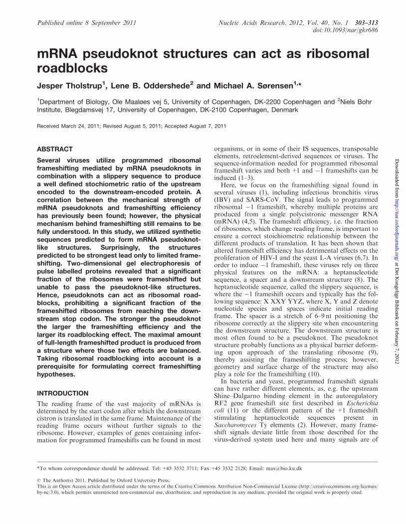
We created a series of plasmids containing differentpseudoknots and where the in-frame stop codon wasplaced either immediately upstream (‘Upstream stop’) or�150 nt downstream (‘Downstream stop’) from thepseudoknot (Figure 1A). The ‘Upstream stop’ constructshad an in-frame stop codon in the spacer between theslippery sequence and the pseudoknot. This causednon-frameshifted ribosomes to produce a 28 kDa polypep-tide (gene10 from phage T7) while ribosomes undergoinga �1 frameshift continued through the pseudoknot andinto lacZ producing a 148 kDa fusion protein of the T7gene10 and lacZ sequences. In the ‘Downstream Stop’constructs we replaced the UAA stop codon immediatelyupstream from the pseudoknot with a lysine codon(AAA). This change caused non-frameshifting ribosomesto continue through the pseudoknot and terminate at adownstream UGA codon producing a 37 kDa polypep-tide. The pseudoknot constructs based on the plasmidOFX302 (25) are detailed in Figure 1B. We systematicallyincreased the length of stem 1 and in pseudoknot 22/6athrough 22/6c, we exchanged GC with UA base pairs,thus, gradually decreasing the stability of stem 1.Often, the number of ribosomes which undergo �1
frameshift has been determined from constructs such asour ‘Upstream stop’ constructs, by separating radio-actively labelled proteins by SDS–PAGE and quantifyingthe relative amount of protein in each of the two polypep-tides (28 versus 148 kDa). Given the limited resolution ofSDS–PAGE, it is, however, impossible to clearly differen-tiate between polypeptides produced by ribosomes thatterminate at the in-frame UAA stop codon and ribosomesthat undergo �1 frameshift but stall within thepseudoknot. In order to overcome this problem, weinvoked 2D SDS–PAGE (34) whereby polypeptides wereseparated not only by molecular weight but also by theirisoelectric point (pI).While polypeptides originating from ribosomes stalled
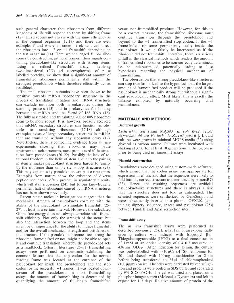
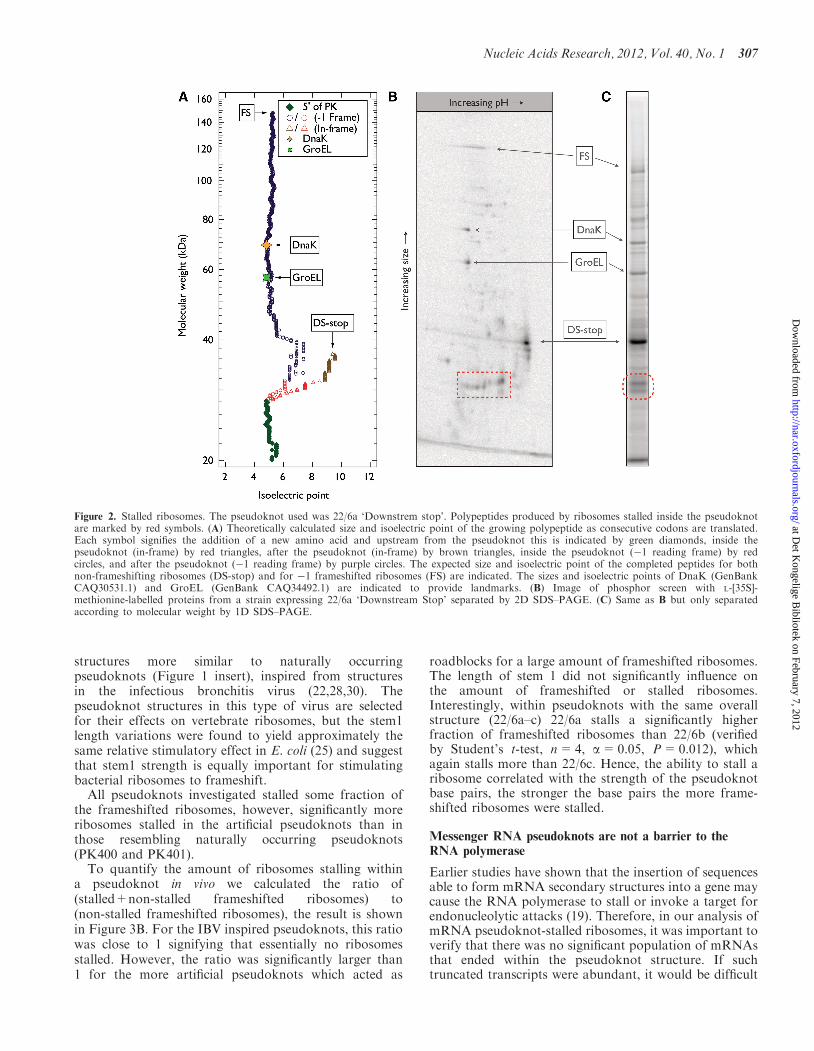
in the pseudoknot varied only slightly in molecular weight,they varied significantly in their pI. Based on the‘Downstream Stop’ construct, we calculated a theoretical2D SDS–PAGE assay of a growing polypeptide as con-secutive codons are translated (shown in Figure 2A). Ataround 28 kDa, the trace splits into two, the trianglesdenote the non-frameshifted product and the circlesdenote the �1 frameshifted product. Red symbolsdenote codons inside the pseudoknot. Experimental dataoriginating from the ‘Downstream Stop’ construct isshown in Figure 2B, the theoretically expected featuresare indeed present, e.g. both the non-frameshifted(DS-stop) and the �1 frameshifted (FS) products arevisible. The heat shock proteins GroEL and DnaK serveas landmarks on the gel. Interestingly, a series of polypep-tides originating from ribosomes stalled inside thepseudoknot appeared (inside dashed red line). For com-parison, a standard SDS–PAGE of the same sample isshown in Figure 2C, here, the second level of information
Nucleic Acids Research, 2012, Vol. 40, No. 1 305
at Det K
ongelige Bibliotek on February 7, 2012
http://nar.oxfordjournals.org/D
ownloaded from
(isoelectric point) is lost and the relative blurry bands aredifficult to interpret.
Quantification of ribosome stalling and correlation withstem strength
The results shown in Figure 2 revealed that a 1D SDS–PAGE assay could not firmly identify polypeptidesoriginating from a �1 frameshifted ribosome stalled inthe pseudoknot from the non-frameshifted product in a‘Downstream Stop’ construct. In order to quantify theamount of �1 frameshifted ribosomes stalled inside thepseudoknot, we performed a 2D SDS–PAGE separationof the radioactively labelled proteins originating from the‘Upstream Stop’ construct (Supplementary Figures S4 andS5), which is the type of construct most commonly usedthroughout literature. The advantage of a 2D-gel analysisis that all the unfinished protein chains with differentlengths concentrate in a common spot when they havethe same pI. This made it possible to identify randomlystalled translation products inside the pseudoknotsequence and we quantified the amount of radioactivity
in all identified additional spots. This produced a conser-vative estimate of the amount of stalled translations.
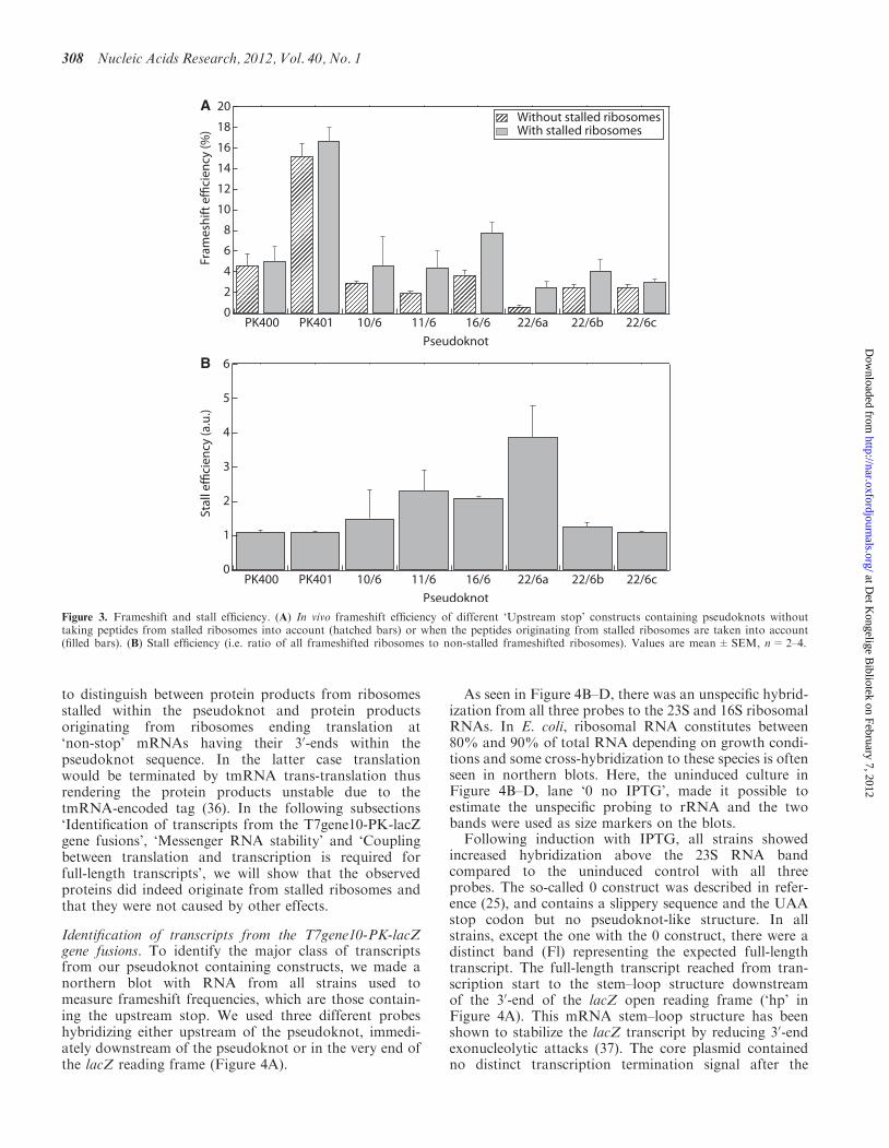
The result of quantifying the fraction of in vivo �1frameshifted ribosomes, both those which made it all theway to the lacZ stop codon (gene10/lacZ fusion) and thosewhich stalled inside the pseudoknot, is shown inFigure 3A. The hatched bars denote the �1 frameshiftefficiency taking into account only the end product of�1 frameshift (148 kDa gene10/lacZ fusion). This frame-shift efficiency was calculated as (intensity of FS product)/(intensity of non-FS product+intensity of FS product).The filled bars denote the �1 frameshift efficiency whenboth the end product (148 kDa gene10/lacZ fusion) andthe products originating from stalled ribosomes are takeninto account. This frameshift efficiency was calculated as(intensity of FS product+intensity of stalled product)/(in-tensity of non-FS product+intensity of FS product+in-tensity of stalled product).
In addition to the six artificial pseudoknot-like struc-tures, we also analysed two earlier investigatedpseudoknots PK400 and PK401 (25), with over-all
A
B
Figure 1. Frameshift assay and pseudoknot structures. (A) All plasmid constructs contain an IPTG inducible promoter in front of T7 gene10 (lightgrey), a complete frameshift signal, and lacZ (dark grey). The frame shift stimulating pseudoknot-like structure is inserted downstream of gene10.Immediately, downstream from the pseudoknot lacZ is inserted in the �1 reading frame relative to gene10. In the ‘Upstream Stop’ construct thenon-frameshifting ribosomes will translate gene10 and terminate at a UAA stop codon in the spacer sequence and produce a 28 kDa polypeptide.Ribosomes undergoing �1 frameshift at the slippery sequence translate lacZ thus producing �148 kDa polypeptide. In the ‘Downstream Stop’construct the UAA stop codon is replaced by an AAA lysine codon thus resulting in �37 kDa polypeptide being produced by non-frameshiftingribosomes which terminate at an UGA stop codon downstream from the pseudoknot. (B) Sequence and structure of the inserted pseudoknots, theslippery sequence and the spacer. In pseudoknot, 10/6, 22/6a, 22/6b and 22/6c the first base in loop 2 has been removed in order to maintain thedownstream reading frame (underlined). The boxed insert in panel B shows the structure and sequence of previously described constructs (25).
306 Nucleic Acids Research, 2012, Vol. 40, No. 1
at Det K
ongelige Bibliotek on February 7, 2012
http://nar.oxfordjournals.org/D
ownloaded from
structures more similar to naturally occurringpseudoknots (Figure 1 insert), inspired from structuresin the infectious bronchitis virus (22,28,30). Thepseudoknot structures in this type of virus are selectedfor their effects on vertebrate ribosomes, but the stem1length variations were found to yield approximately thesame relative stimulatory effect in E. coli (25) and suggestthat stem1 strength is equally important for stimulatingbacterial ribosomes to frameshift.
All pseudoknots investigated stalled some fraction ofthe frameshifted ribosomes, however, significantly moreribosomes stalled in the artificial pseudoknots than inthose resembling naturally occurring pseudoknots(PK400 and PK401).
To quantify the amount of ribosomes stalling withina pseudoknot in vivo we calculated the ratio of(stalled+non-stalled frameshifted ribosomes) to(non-stalled frameshifted ribosomes), the result is shownin Figure 3B. For the IBV inspired pseudoknots, this ratiowas close to 1 signifying that essentially no ribosomesstalled. However, the ratio was significantly larger than1 for the more artificial pseudoknots which acted as
roadblocks for a large amount of frameshifted ribosomes.The length of stem 1 did not significantly influence onthe amount of frameshifted or stalled ribosomes.Interestingly, within pseudoknots with the same overallstructure (22/6a–c) 22/6a stalls a significantly higherfraction of frameshifted ribosomes than 22/6b (verifiedby Student’s t-test, n=4, a=0.05, P=0.012), whichagain stalls more than 22/6c. Hence, the ability to stall aribosome correlated with the strength of the pseudoknotbase pairs, the stronger the base pairs the more frame-shifted ribosomes were stalled.
Messenger RNA pseudoknots are not a barrier to theRNA polymerase
Earlier studies have shown that the insertion of sequencesable to form mRNA secondary structures into a gene maycause the RNA polymerase to stall or invoke a target forendonucleolytic attacks (19). Therefore, in our analysis ofmRNA pseudoknot-stalled ribosomes, it was important toverify that there was no significant population of mRNAsthat ended within the pseudoknot structure. If suchtruncated transcripts were abundant, it would be difficult
Figure 2. Stalled ribosomes. The pseudoknot used was 22/6a ‘Downstrem stop’. Polypeptides produced by ribosomes stalled inside the pseudoknotare marked by red symbols. (A) Theoretically calculated size and isoelectric point of the growing polypeptide as consecutive codons are translated.Each symbol signifies the addition of a new amino acid and upstream from the pseudoknot this is indicated by green diamonds, inside thepseudoknot (in-frame) by red triangles, after the pseudoknot (in-frame) by brown triangles, inside the pseudoknot (�1 reading frame) by redcircles, and after the pseudoknot (�1 reading frame) by purple circles. The expected size and isoelectric point of the completed peptides for bothnon-frameshifting ribosomes (DS-stop) and for �1 frameshifted ribosomes (FS) are indicated. The sizes and isoelectric points of DnaK (GenBankCAQ30531.1) and GroEL (GenBank CAQ34492.1) are indicated to provide landmarks. (B) Image of phosphor screen with L-[35S]-methionine-labelled proteins from a strain expressing 22/6a ‘Downstream Stop’ separated by 2D SDS–PAGE. (C) Same as B but only separatedaccording to molecular weight by 1D SDS–PAGE.
Nucleic Acids Research, 2012, Vol. 40, No. 1 307
at Det K
ongelige Bibliotek on February 7, 2012
http://nar.oxfordjournals.org/D
ownloaded from
to distinguish between protein products from ribosomesstalled within the pseudoknot and protein productsoriginating from ribosomes ending translation at‘non-stop’ mRNAs having their 30-ends within thepseudoknot sequence. In the latter case translationwould be terminated by tmRNA trans-translation thusrendering the protein products unstable due to thetmRNA-encoded tag (36). In the following subsections‘Identification of transcripts from the T7gene10-PK-lacZgene fusions’, ‘Messenger RNA stability’ and ‘Couplingbetween translation and transcription is required forfull-length transcripts’, we will show that the observedproteins did indeed originate from stalled ribosomes andthat they were not caused by other effects.
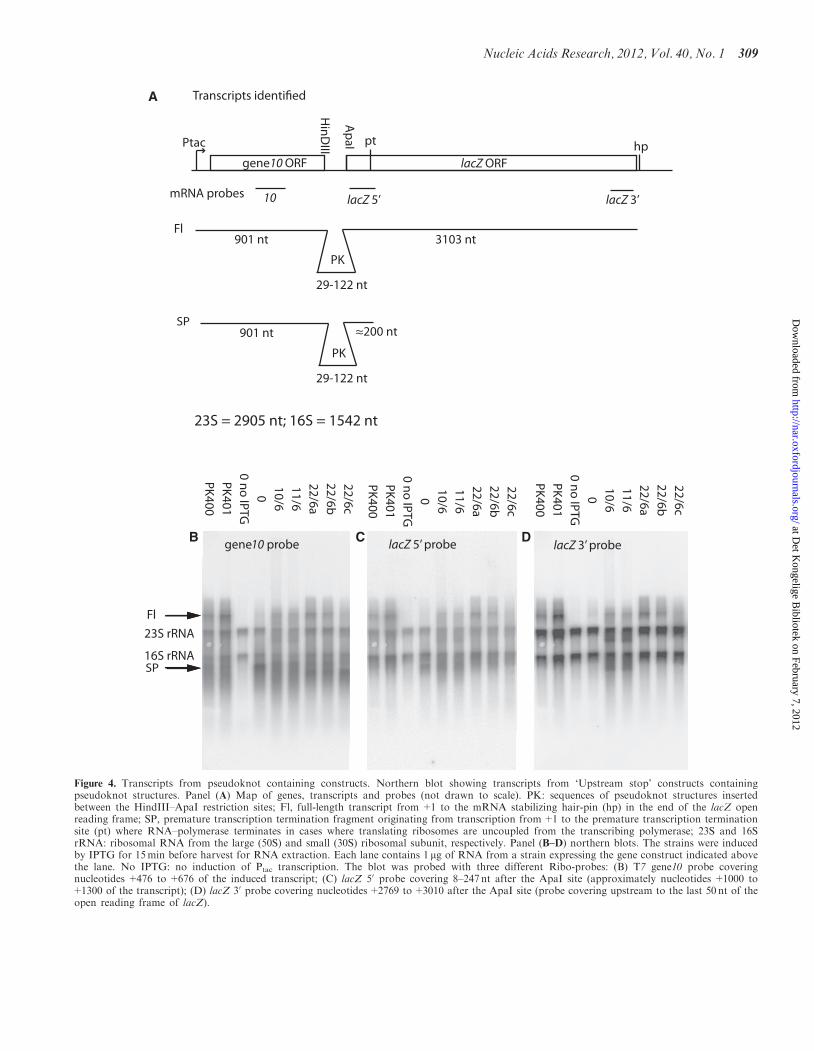
Identification of transcripts from the T7gene10-PK-lacZgene fusions. To identify the major class of transcriptsfrom our pseudoknot containing constructs, we made anorthern blot with RNA from all strains used tomeasure frameshift frequencies, which are those contain-ing the upstream stop. We used three different probeshybridizing either upstream of the pseudoknot, immedi-ately downstream of the pseudoknot or in the very end ofthe lacZ reading frame (Figure 4A).
As seen in Figure 4B–D, there was an unspecific hybrid-ization from all three probes to the 23S and 16S ribosomalRNAs. In E. coli, ribosomal RNA constitutes between80% and 90% of total RNA depending on growth condi-tions and some cross-hybridization to these species is oftenseen in northern blots. Here, the uninduced culture inFigure 4B–D, lane ‘0 no IPTG’, made it possible toestimate the unspecific probing to rRNA and the twobands were used as size markers on the blots.
Following induction with IPTG, all strains showedincreased hybridization above the 23S RNA bandcompared to the uninduced control with all threeprobes. The so-called 0 construct was described in refer-ence (25), and contains a slippery sequence and the UAAstop codon but no pseudoknot-like structure. In allstrains, except the one with the 0 construct, there were adistinct band (Fl) representing the expected full-lengthtranscript. The full-length transcript reached from tran-scription start to the stem–loop structure downstreamof the 30-end of the lacZ open reading frame (‘hp’ inFigure 4A). This mRNA stem–loop structure has beenshown to stabilize the lacZ transcript by reducing 30-endexonucleolytic attacks (37). The core plasmid containedno distinct transcription termination signal after the
A
B
Figure 3. Frameshift and stall efficiency. (A) In vivo frameshift efficiency of different ‘Upstream stop’ constructs containing pseudoknots withouttaking peptides from stalled ribosomes into account (hatched bars) or when the peptides originating from stalled ribosomes are taken into account(filled bars). (B) Stall efficiency (i.e. ratio of all frameshifted ribosomes to non-stalled frameshifted ribosomes). Values are mean±SEM, n=2–4.
308 Nucleic Acids Research, 2012, Vol. 40, No. 1
at Det K
ongelige Bibliotek on February 7, 2012
http://nar.oxfordjournals.org/D
ownloaded from
A
B C D
Figure 4. Transcripts from pseudoknot containing constructs. Northern blot showing transcripts from ‘Upstream stop’ constructs containingpseudoknot structures. Panel (A) Map of genes, transcripts and probes (not drawn to scale). PK: sequences of pseudoknot structures insertedbetween the HindIII–ApaI restriction sites; Fl, full-length transcript from +1 to the mRNA stabilizing hair-pin (hp) in the end of the lacZ openreading frame; SP, premature transcription termination fragment originating from transcription from+1 to the premature transcription terminationsite (pt) where RNA–polymerase terminates in cases where translating ribosomes are uncoupled from the transcribing polymerase; 23S and 16SrRNA: ribosomal RNA from the large (50S) and small (30S) ribosomal subunit, respectively. Panel (B–D) northern blots. The strains were inducedby IPTG for 15min before harvest for RNA extraction. Each lane contains 1 mg of RNA from a strain expressing the gene construct indicated abovethe lane. No IPTG: no induction of Ptac transcription. The blot was probed with three different Ribo-probes: (B) T7 gene10 probe coveringnucleotides +476 to +676 of the induced transcript; (C) lacZ 50 probe covering 8–247 nt after the ApaI site (approximately nucleotides +1000 to+1300 of the transcript); (D) lacZ 30 probe covering nucleotides+2769 to+3010 after the ApaI site (probe covering upstream to the last 50 nt of theopen reading frame of lacZ).
Nucleic Acids Research, 2012, Vol. 40, No. 1 309
at Det K
ongelige Bibliotek on February 7, 2012
http://nar.oxfordjournals.org/D
ownloaded from
lacZ gene, and accordingly we found transcriptsthat exceeded far beyond the full-length Fl band(Figure 4B–D).In the beginning of lacZ, �200 nt into the open reading
frame, there is a site, called ‘pt’ in Figure 4A, where theRNA polymerase is caused to terminate if there is ineffi-cient translation initiation of the lacZ gene (38). In the 0construct there is no pseudoknot to stimulate frameshift atthe slippery site. Therefore, virtually no ribosomes wereexpected to follow the RNA polymerase from gene 10 intothe lacZ part of our gene fusion. As expected, Figure 4Band C, lane ‘0’ shows a prominent band (‘SP’ for stoppolymerase) corresponding in size and probe-ability tothis premature termination product. Also, correspondinglow amounts of high molecular weight transcripts aredetected for this construct. All the other constructsshown in Figure 4 contained frameshift stimulatingpseudoknots and a inspection of the northern blotshowed that the ‘SP’ bands probed with both gene10and lacZ50 sequences were present in sizes which corres-pond to the sizes of the pseudoknots inserted.
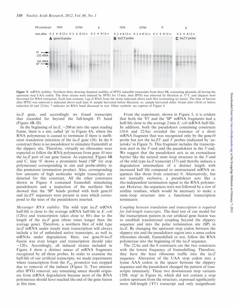
Messenger RNA stability. The wild type lacZ mRNAhalf-life is close to the average mRNA half-life in E. coli(120 s) and transcription takes close to 80 s due to thelength of the lacZ gene (three times longer than theaverage gene). Therefore, a northern blot of wild typelacZ mRNA under steady state transcription will alwaysinclude a lot of unfinished native transcripts, as well asmRNAs under degradation. Here, our gene10-lacZfusion was even longer and transcription should take�120 s. Accordingly, all induced strains included inFigure 4 show a distinct smear of mRNA fragmentsrecognized by all three probes. In order to examine thehalf-life of our artificial transcripts, we made experimentswhere transcription from the Ptac promotor was stoppeddue to removal of the inducer (Figure 5). Two minutesafter IPTG removal, any remaining smear should origin-ate from mRNA degradation because most of the RNApolymerase should have reached the end of the gene fusionat this time.
From the experiment, shown in Figure 5, it is evidentthat both the ‘Fl’ and the ‘SP’ mRNA fragments had ahalf-life close to the average 2min E. coli mRNA half-life.In addition, both the pseudoknot containing constructs(10/6 and 22/6a) revealed the existence of a shortmRNA fragment that was recognized only by the gene10probe but not the lacZ50 and 30 probes (indicated by ‘as-terisks’ in Figure 5). This fragment includes the transcrip-tion start in the 50-end and the pseudoknot in the 30-end.We suggest that the pseudoknot acts as an exonucleasebarrier like the natural stem–loop structure in the 30-endof the wild type lacZ transcript (37) and thereby induces adegradation intermediate of a distinct length withincreased half life compared to unstructured mRNA se-quences like those from construct 0. Alternatively, butnot mutually exclusive, a pseudoknot acts like arho-independent termination signal to the RNA polymer-ase. However, the sequences were not followed by a row ofuridine residues, which would be necessary to make astem–loop structure into a functional transcriptionterminator.
Coupling between translation and transcription is requiredfor full-length transcripts. The final test of our model forthe transcription pattern in our artificial gene fusion wasto establish translational coupling beyond the slipperysequence and into the polar termination site (SP) inlacZ. By changing the upstream stop codon between theslippery site and the pseudoknot region into a sense codonribosomes should, frameshifted or not, follow the RNApolymerase into the beginning of the lacZ sequence.
The 22/6a and the 0 constructs are the two constructswith the lowest frequency of frameshifting. Therefore,they have the least ribosome traffic into the lacZsequence. Alteration of the UAA stop codon into alysine AAA codon in the spacer between the slipperysequence and the pseudoknot changed the pattern of tran-scripts immensely. These two downstream stop variants(‘DS. stop’ in Figure 6), which did not contain a stopcodon upstream from the structure, expressed significantlymore full-length (‘Fl’) transcript and only insignificant
A B C
Figure 5. mRNA stability. Northern blots showing chemical stability of IPTG inducible transcripts from three PK containing plasmids all having theupstream stop UAA codon. The three strains were induced by IPTG for 15min, then IPTG was removed by filtration at 37�C and aliquots wereharvested for RNA extraction. Each lane contains 1 mg of RNA from the strain indicated above each blot (containing six lanes). The time of harvestafter IPTG was removed is indicated above each lane; 0: sample harvested before filtration; 1: sample harvested either 16min after (10/6) or beforeinduction (0 and 22/6a). * indicates an RNA band discussed in text. Other symbols: see caption of Figure 4.
310 Nucleic Acids Research, 2012, Vol. 40, No. 1
at Det K
ongelige Bibliotek on February 7, 2012
http://nar.oxfordjournals.org/D
ownloaded from
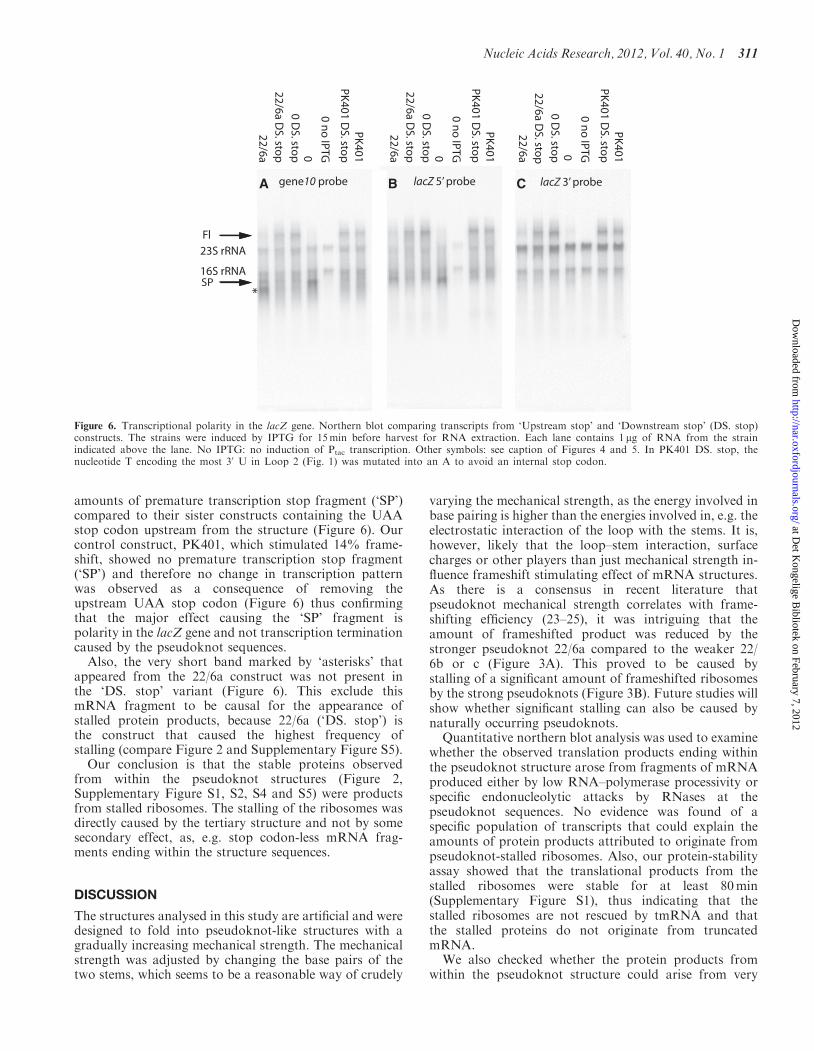
amounts of premature transcription stop fragment (‘SP’)compared to their sister constructs containing the UAAstop codon upstream from the structure (Figure 6). Ourcontrol construct, PK401, which stimulated 14% frame-shift, showed no premature transcription stop fragment(‘SP’) and therefore no change in transcription patternwas observed as a consequence of removing theupstream UAA stop codon (Figure 6) thus confirmingthat the major effect causing the ‘SP’ fragment ispolarity in the lacZ gene and not transcription terminationcaused by the pseudoknot sequences.
Also, the very short band marked by ‘asterisks’ thatappeared from the 22/6a construct was not present inthe ‘DS. stop’ variant (Figure 6). This exclude thismRNA fragment to be causal for the appearance ofstalled protein products, because 22/6a (‘DS. stop’) isthe construct that caused the highest frequency ofstalling (compare Figure 2 and Supplementary Figure S5).
Our conclusion is that the stable proteins observedfrom within the pseudoknot structures (Figure 2,Supplementary Figure S1, S2, S4 and S5) were productsfrom stalled ribosomes. The stalling of the ribosomes wasdirectly caused by the tertiary structure and not by somesecondary effect, as, e.g. stop codon-less mRNA frag-ments ending within the structure sequences.
DISCUSSION
The structures analysed in this study are artificial and weredesigned to fold into pseudoknot-like structures with agradually increasing mechanical strength. The mechanicalstrength was adjusted by changing the base pairs of thetwo stems, which seems to be a reasonable way of crudely
varying the mechanical strength, as the energy involved inbase pairing is higher than the energies involved in, e.g. theelectrostatic interaction of the loop with the stems. It is,however, likely that the loop–stem interaction, surfacecharges or other players than just mechanical strength in-fluence frameshift stimulating effect of mRNA structures.As there is a consensus in recent literature thatpseudoknot mechanical strength correlates with frame-shifting efficiency (23–25), it was intriguing that theamount of frameshifted product was reduced by thestronger pseudoknot 22/6a compared to the weaker 22/6b or c (Figure 3A). This proved to be caused bystalling of a significant amount of frameshifted ribosomesby the strong pseudoknots (Figure 3B). Future studies willshow whether significant stalling can also be caused bynaturally occurring pseudoknots.Quantitative northern blot analysis was used to examine
whether the observed translation products ending withinthe pseudoknot structure arose from fragments of mRNAproduced either by low RNA–polymerase processivity orspecific endonucleolytic attacks by RNases at thepseudoknot sequences. No evidence was found of aspecific population of transcripts that could explain theamounts of protein products attributed to originate frompseudoknot-stalled ribosomes. Also, our protein-stabilityassay showed that the translational products from thestalled ribosomes were stable for at least 80min(Supplementary Figure S1), thus indicating that thestalled ribosomes are not rescued by tmRNA and thatthe stalled proteins do not originate from truncatedmRNA.We also checked whether the protein products from
within the pseudoknot structure could arise from very
A B C
Figure 6. Transcriptional polarity in the lacZ gene. Northern blot comparing transcripts from ‘Upstream stop’ and ‘Downstream stop’ (DS. stop)constructs. The strains were induced by IPTG for 15min before harvest for RNA extraction. Each lane contains 1 mg of RNA from the strainindicated above the lane. No IPTG: no induction of Ptac transcription. Other symbols: see caption of Figures 4 and 5. In PK401 DS. stop, thenucleotide T encoding the most 30 U in Loop 2 (Fig. 1) was mutated into an A to avoid an internal stop codon.
Nucleic Acids Research, 2012, Vol. 40, No. 1 311
at Det K
ongelige Bibliotek on February 7, 2012
http://nar.oxfordjournals.org/D
ownloaded from
slow rather than permanently stalled ribosomes. A pulsechase experiment (Supplementary Figure S2) revealed thatwithin 16min there was no sign of a redistribution of labelbetween the stalled spots and the stop codon-terminateddownstream stop product, thus proving the possibility ofvery slow ribosomes to be unlikely.It is possible that the newly discovered ribosome rescue
factor, ArfA (39) could be active at pseudoknot-stalledribosomes and that nascent proteins would be morestable than if saved by tmRNA. However, as can beseen in Supplementary Figure S3, the growth of strainsexpressing pseudoknot 22/6a was severely affected by in-duction and showed a decrease in growth rate correlatingto the amount of stall product observed. Because ribo-somes are limiting in growing cells (40), the sequestrationof ribosomes by engagement in induced overexpression ofa gene from a plasmid will often cause a strain to growslower than the uninduced counterpart. The enhanced re-duction in growth rate upon induction of 22/6a comparedto the 0 construct (Supplementary Figure S3) couldindicate that stalled ribosomes were not rescued at a suf-ficiently high rate and we suggest that either the ribosomalrescue systems were titrated by the large amount ofmRNA induced from the plasmid alleles, or alternatively,that no rescue is possible for pseudoknot-stalledribosomes.Our results are in agreement with the observation that
the amount of protein produced from an mRNA can bereduced when a pseudoknot is located upstream (29).Also, they provide a possible explanation for the reductionin frameshift efficiency observed by, e.g. Napthine et al.(30) when increasing the thermodynamic stability of stem1 above a certain threshold. This apparent reduction inframeshift efficiency (observed by 1D SDS–PAGE) couldbe caused by the fact that a significant fraction of the‘frameshifted’ ribosomes permanently stalled within thepseudoknot.We propose that pseudoknot induced frameshifting
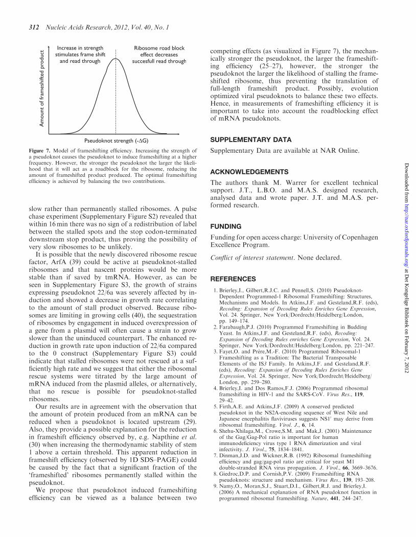
efficiency can be viewed as a balance between two
competing effects (as visualized in Figure 7), the mechan-ically stronger the pseudoknot, the larger the frameshift-ing efficiency (25–27), however, the stronger thepseudoknot the larger the likelihood of stalling the frame-shifted ribosome, thus preventing the translation offull-length frameshift product. Possibly, evolutionoptimized viral pseudoknots to balance these two effects.Hence, in measurements of frameshifting efficiency it isimportant to take into account the roadblocking effectof mRNA pseudoknots.
SUPPLEMENTARY DATA
Supplementary Data are available at NAR Online.
ACKNOWLEDGEMENTS
The authors thank M. Warrer for excellent technicalsupport. J.T., L.B.O. and M.A.S. designed research,analysed data and wrote paper. J.T. and M.A.S. per-formed research.
FUNDING
Funding for open access charge: University of CopenhagenExcellence Program.
Conflict of interest statement. None declared.
REFERENCES
1. Brierley,I., Gilbert,R.J.C. and Pennell,S. (2010) Pseudoknot-Dependent Programmed-1 Ribosomal Frameshifting: Structures,Mechanisms and Models. In Atkins,J.F. and Gesteland,R.F. (eds),Recoding: Expansion of Decoding Rules Enriches Gene Expression,Vol. 24. Springer, New York/Dordrecht/Heidelberg/London,pp. 149–174.
2. Farabaugh,P.J. (2010) Programmed Frameshifting in BuddingYeast. In Atkins,J.F. and Gesteland,R.F. (eds), Recoding:Expansion of Decoding Rules enriches Gene Expression, Vol. 24.Springer, New York/Dordrecht/Heidelberg/London, pp. 221–247.
3. Fayet,O. and Prere,M.-F. (2010) Programmed Ribosomal-1Frameshifting as a Tradition: The Bacterial TransposableElements of the IS3 Family. In Atkins,J.F. and Gesteland,R.F.(eds), Recoding: Expansion of Decoding Rules Enriches GeneExpression, Vol. 24. Springer, New York/Dordrecht/Heidelberg/London, pp. 259–280.
4. Brierley,I. and Dos Ramos,F.J. (2006) Programmed ribosomalframeshifting in HIV-1 and the SARS-CoV. Virus Res., 119,29–42.
5. Firth,A.E. and Atkins,J.F. (2009) A conserved predictedpseudoknot in the NS2A-encoding sequence of West Nile andJapanese encephalitis flaviviruses suggests NS10 may derive fromribosomal frameshifting. Virol. J., 6, 14.
6. Shehu-Xhilaga,M., Crowe,S.M. and Mak,J. (2001) Maintenanceof the Gag/Gag-Pol ratio is important for humanimmunodeficiency virus type 1 RNA dimerization and viralinfectivity. J. Virol., 75, 1834–1841.
7. Dinman,J.D. and Wickner,R.B. (1992) Ribosomal frameshiftingefficiency and gag/gag-pol ratio are critical for yeast M1double-stranded RNA virus propagation. J. Virol., 66, 3669–3676.
8. Giedroc,D.P. and Cornish,P.V. (2009) Frameshifting RNApseudoknots: structure and mechanism. Virus Res., 139, 193–208.
9. Namy,O., Moran,S.J., Stuart,D.I., Gilbert,R.J. and Brierley,I.(2006) A mechanical explanation of RNA pseudoknot function inprogrammed ribosomal frameshifting. Nature, 441, 244–247.
Figure 7. Model of frameshifting efficiency. Increasing the strength ofa pseudoknot causes the pseudoknot to induce frameshifting at a higherfrequency. However, the stronger the pseudoknot the larger the likeli-hood that it will act as a roadblock for the ribosome, reducing theamount of frameshifted product produced. The optimal frameshiftingefficiency is achieved by balancing the two contributions.
312 Nucleic Acids Research, 2012, Vol. 40, No. 1
at Det K
ongelige Bibliotek on February 7, 2012
http://nar.oxfordjournals.org/D
ownloaded from
10. Pallan,P.S., Marshall,W.S., Harp,J., Jewett,F.C., Wawrzak,Z.,Brown,B.A., Rich,A. and Egli,M. (2005) Crystal Structure of aLuteoviral RNA Pseudoknot and Model for a MinimalRibosomal Frameshifting Motif. Biochemistry, 44, 11315–11322.
11. Weiss,R.B., Dunn,D.M., Dahlberg,A.E., Atkins,J.F. andGesteland,R.F. (1988) Reading frame switch caused by base-pairformation between the 30 end of 16S rRNA and the mRNAduring elongation of protein synthesis in Escherichia coli.EMBO J., 7, 1503–1507.
12. Weiss,R.B., Dunn,D.M., Shuh,M., Atkins,J.F. and Gesteland,R.F.(1989) E. coli ribosomes re-phase on retroviral frameshift signalsat rates ranging from 2 to 50 percent. New Biol., 1, 159–169.
13. Garcia,A., van Duin,J. and Pleij,C.W.A. (1993) Differentialresponse to frameshift signals in eukaryotic and prokaryotictranslational systems. Nucleic Acids Res., 21, 401–406.
14. Ivanov,I.P., Gesteland,R.F., Matsufuji,S. and Atkins,J.F. (1998)Programmed frameshifting in the synthesis of mammalianantizyme is +1 in mammals, predominantly +1 in fission yeast,but -2 in budding yeast. RNA, 4, 1230–1238.
15. Kozak,M. (1989) Circumstances and mechanisms of inhibitionof translation by secondary structure in eucaryotic mRNAs.Mol. Cell. Biol., 9, 5134–5142.
16. Hall,M.N., Gabay,J., Debarbouille,M. and Schwartz,M. (1982) Arole for mRNA secondary structure in the control of translationinitiation. Nature, 295, 616–618.
17. Von Heijne,G., Nilsson,L. and Blomberg,C. (1977) Translationand messenger RNA secondary structure. J. Theor. Biol., 68,321–329.
18. Qu,X., Wen,J.-D., Lancaster,L., Noller,H.F., Bustamante,C. andTinoco,I. (2011) The ribosome uses two active mechanisms tounwind messenger RNA during translation. Nature, 475, 118–121.
19. Sørensen,M.A., Kurland,C.G. and Pedersen,S. (1989) Codonusage determines the translation rate in Escherichia coli.J. Mol. Biol., 207, 365–377.
20. Tu,C., Tzeng,T.H. and Bruenn,J.A. (1992) Ribosomal movementimpeded at a pseudoknot required for frameshifting.Proc. Natl Acad. Sci. USA, 89, 8636–8640.
21. Somogyi,P., Jenner,A.J., Brierley,I. and Inglis,S.C. (1993)Ribosomal pausing during translation of an RNA pseudoknot.Mol. Cell. Biol., 13, 6931–6940.
22. Kontos,H., Napthine,S. and Brierley,I. (2001) Ribosomal pausingat a frameshifter RNA pseudoknot is sensitive to reading phasebut shows little correlation with frameshift efficiency.Mol. Cell. Biol., 21, 8657–8670.
23. Plant,E.P. and Dinman,J.D. (2005) Torsional restraint: a newtwist on frameshifting pseudoknots. Nucleic Acids Res., 33,1825–1833.
24. Ito,K., Chiba,S. and Pogliano,K. (2010) Divergent stallingsequences sense and control cellular physiology. Biochem. Biophys.Res. Commun., 393, 1–5.
25. Hansen,T.M., Reihani,S.N., Oddershede,L.B. and Sorensen,M.A.(2007) Correlation between mechanical strength of messengerRNA pseudoknots and ribosomal frameshifting.Proc. Natl Acad. Sci. USA, 104, 5830–5835.
26. Chen,G., Chang,K.Y., Chou,M.Y., Bustamante,C. andTinoco,I. Jr (2009) Triplex structures in an RNA pseudoknotenhance mechanical stability and increase efficiency of -1ribosomal frameshifting. Proc. Natl Acad. Sci. USA, 106,12706–12711.
27. Green,L., Kim,C.-H., Bustamante,C. and Tinoco,I. Jr (2008)Characterization of the Mechanical Unfolding of RNAPseudoknots. J. Mol. Biol., 375, 511–528.
28. Brierley,I., Digard,P. and Inglis,S.C. (1989) Characterization of anefficient coronavirus ribosomal frameshifting signal: requirementfor an RNA pseudoknot. Cell, 57, 537–547.
29. Plant,E.P., Rakauskaite,R., Taylor,D.R. and Dinman,J.D. (2010)Achieving a golden mean: mechanisms by which coronavirusesensure synthesis of the correct stoichiometric ratios of viralproteins. J. Virol., 84, 4330–4340.
30. Napthine,S., Liphardt,J., Bloys,A., Routledge,S. and Brierley,I.(1999) The role of RNA pseudoknot stem 1 length in thepromotion of efficient -1 ribosomal frameshifting. J. Mol. Biol.,288, 305–320.
31. Liphardt,J., Napthine,S., Kontos,H. and Brierley,I. (1999)Evidence for an RNA pseudoknot loop-helix interaction essentialfor efficient-1 ribosomal frameshifting. J. Mol. Biol., 288,321–335.
32. Neidhardt,F.C., Bloch,P.L. and Smith,D.F. (1974) Culturemedium for enterobacteria. J. Bacteriol., 119, 736–747.
33. Reeder,J., Steffen,P. and Giegerich,R. (2007) pknotsRG: RNApseudoknot folding including near-optimal structures and slidingwindows. Nucleic Acids Res., 35, W320–W324.
34. O’Farrell,P.H. (1975) High resolution two-dimensionalelectrophoresis of proteins. J. Biol. Chem., 250, 4007–4021.
35. Sørensen,M.A. and Pedersen,S. (1997) Determination of thePeptide Elongation rate In Vivo. In Martin,R. (ed.), Methods inMolecular Biology: Protein Synthesis: Methods and protocols, Vol.77. The Humana Press, Inc., Totowa, New Jersey, NJ, USA,pp. 129–142.
36. Farrell,C.M., Grossman,A.D. and Sauer,R.T. (2005) Cytoplasmicdegradation of ssrA-tagged proteins. Mol. Microbiol., 57,1750–1761.
37. Cannistraro,V.J., Subbarao,M.N. and Kennell,D. (1986) Specificendonucleolytic cleavage sites for decay of Escherichia colimRNA. J. Mol. Biol., 192, 257–274.
38. Stanssens,P., Remaut,E. and Fiers,W. (1986) Inefficienttranslation initiation causes premature transcription terminationin the lacZ gene. Cell, 44, 711–718.
39. Chadani,Y., Ono,K., Ozawa,S., Takahashi,Y., Takai,K.,Nanamiya,H., Tozawa,Y., Kutsukake,K. and Abo,T. (2010)Ribosome rescue by Escherichia coli ArfA (YhdL) in the absenceof trans-translation system. Mol. Microbiol., 78, 796–808.
40. Vind,J., Sørensen,M.A., Rasmussen,M.D. and Pedersen,S. (1993)Synthesis of proteins in Escherichia coli is limited by theconcentration of free ribosomes: expression from reporter genesdoes not always reflect functional mRNA levels. J. Mol. Biol.,231, 678–688.
Nucleic Acids Research, 2012, Vol. 40, No. 1 313
at Det K
ongelige Bibliotek on February 7, 2012
http://nar.oxfordjournals.org/D
ownloaded from