Embed Size (px)
Citation preview
August-2021 1
MRI Safety Policies
August-2021 2
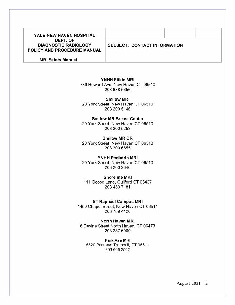
YNHH Fitkin MRI 789 Howard Ave, New Haven CT 06510
203 688 5656
Smilow MRI 20 York Street, New Haven CT 06510
203 200 5146
Smilow MR Breast Center 20 York Street, New Haven CT 06510
203 200 5253
Smilow MR OR 20 York Street, New Haven CT 06510
203 200 6655
YNHH Pediatric MRI 20 York Street, New Haven CT 06510
203 200 2646
Shoreline MRI 111 Goose Lane, Guilford CT 06437
203 453 7181
ST Raphael Campus MRI
1450 Chapel Street, New Haven CT 06511 203 789 4120
North Haven MRI
6 Devine Street North Haven, CT 06473 203 287 6969
Park Ave MRI
5520 Park ave Trumbull, CT 06611 203 666 3562
YALE-NEW HAVEN HOSPITAL
DEPT. OF DIAGNOSTIC RADIOLOGY
POLICY AND PROCEDURE MANUAL
MRI Safety Manual
SUBJECT: CONTACT INFORMATION
August-2021 3
GENERAL SAFETY INFORMATION MRI CONTACT INFORMATION 2 INDEX 3 ACRONYMS 5 INTRODUCTION 6 GENERAL POLICY 7 SAFETY TERMINOLOGY 8 ACR ZONES (SITE ACCESS RESTRICTION) 11 TIME VARYING MAGNETIC FIELD RELATED ISSUES 17 INCIDENT REPORTING INFORMATION 19 EAR PLUG 20 FIREFIGHTERS, POLICE AND SECURITY CONSIDERATIONS 21 CRYOGEN RELATED ISSUES/QUENCH 27
MRI SCREENING
PERSONNEL 29 FAMILY/PREGNANT COMPANIONS 31 GUN SHOT WOUNDS 98 OUTPATIENTS 33 INPATIENTS 35 UNRESPONSIVE PATIENTS 37 OXYGEN CYLINDERS 39 PENILE IMPLANT 40 FOREIGN BODY ORBITS 41 PLAIN FILM XRAYS FOR MRI STUDY CLEARANCE 42 CARDIAC IMPLANTATIONS: STENTS, VALVES, LEADS 43 PACEMAKER PROCESS 44 PASSIVE VASCULAR IMPLANT COILS, FILTERS, STENTS 46 ANEURYSM CLIPS 47 BRAIN STIMULATORS 48 OTHER NERVE STIMULATORS, INTRATHECAL PUMPS 49 ORTHODONTIC DEVICES 52 TRANSDERMAL MEDICATION PATCHES AND WOUND DRESSINGS 54 EAR IMPLANT INFORMATION 56 MISCELLANEOUS IMPLANTS 57
MRI PREGNANCY RELATED ISSUES PREGNANCY EMPLOYEES, PATIENTS, COMPANIONS AND CONTRAST ISSUES 62 PRE PROCEDURE PREGNANCY TESTING 63 ADMINISTRATION CONTRAST WHILE BREAST FEEDING 64
.
YALE-NEW HAVEN HOSPITAL
DEPT. OF DIAGNOSTIC RADIOLOGY
POLICY AND PROCEDURE MANUAL
MRI Safety Manual
Reviewed/Updated 2021
Policy No.
SUBJECT: INDEX
August-2021 4
MRI CONTRAST RELATED ISSUES IV AND ORAL CONTRAST AGENTS 65 EGFR SCREENING 66 GAD QUICK REFERENCE SHEET 71 DOTAREM DOSING INFORMATION 72 PREMEDICATION 74 PHYSICIAN CONTRAST COVERAGE 75
MRI SCHEDULING RELATED AND OTHER POLICIES ED AND STAT EXAMS 76 DEFINITION OF LIFE THREATENING 77 HYPER ACUTE STROKE 79 SRC ON CALL AND OVERNIGHT PROCESS 80 ACUTE CORD COMPRESSION 81 MAGNET ROOM CLEANING 83
NURSING RELATED AND OTHER POLICIES LATEX 84 MONITORING OF PATIENTS 85 CODE PROCEDURE and WORKFLOW/ CODE CART LOCATION 87 INPATIENT THERAPIES AND MRI 92 FERROUS OBJECT IN MR SCAN ROOM 93 MRI SAFETY OFFICER RESPONSIBILITIES 94 MRI MEDICAL DIRECTOR RESPONSIBILITIES 95 WANDING 96 POLICY EXEMPTION 97 PROGRAMMABLE SHUNTS 98 GUN SHOT WOUNDS 102 POST GRID 105 CLAUSTROPHIBIA AND EMOTIONAL DISTRESS 106 TATTOOS JEWELRY BODY PIERCINGS AND HAIR EXTENSIONS 107
Appendix A YNHH Position on SFG Appendix B Contrast Doses, Company Contact info Appendix C MRI Safety Website Information Appendix D Useful Links Appendix E Techniques to Lower SAR and B1RMS Appendix F Biomet Bone Stimulator Reference Sheet Appendix G Passive Vascular Implant Policy Reference information Appendix H Medtronic 8637 scheduling flow sheet Appendix I Common Chest Implants
August-2021 5
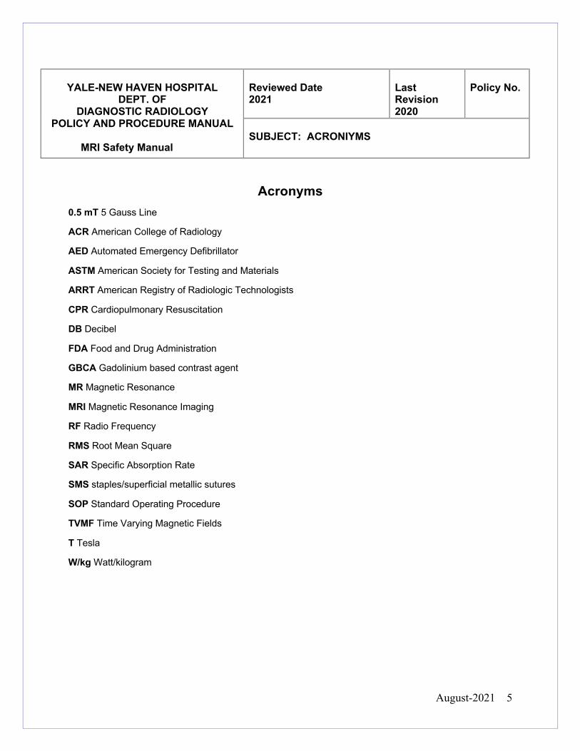
Acronyms
0.5 mT 5 Gauss Line
ACR American College of Radiology
AED Automated Emergency Defibrillator
ASTM American Society for Testing and Materials
ARRT American Registry of Radiologic Technologists
CPR Cardiopulmonary Resuscitation
DB Decibel
FDA Food and Drug Administration
GBCA Gadolinium based contrast agent
MR Magnetic Resonance
MRI Magnetic Resonance Imaging
RF Radio Frequency
RMS Root Mean Square
SAR Specific Absorption Rate
SMS staples/superficial metallic sutures
SOP Standard Operating Procedure
TVMF Time Varying Magnetic Fields
T Tesla
W/kg Watt/kilogram
YALE-NEW HAVEN HOSPITAL
DEPT. OF DIAGNOSTIC RADIOLOGY
POLICY AND PROCEDURE MANUAL
MRI Safety Manual
Reviewed Date 2021
Last Revision 2020
Policy No.
SUBJECT: ACRONIYMS
August-2021 6
Magnetic Resonance Imaging is an ever changing, evolving technology. There are
potential risks in the MR environment, not only for the patient but also for the
accompanying family members, attending health care professionals and others who find
themselves only occasionally or rarely in the magnetic fields of MR scanners, such as
security, housekeeping personnel, firefighters, police, etc. This manual has been
developed to help guide the MR staff regarding these issues.
It is the intent of the Yale New Haven Hospital safety manual to:
• Protect and educate all patients, direct and ancillary personal about the possible
risks, associated with the MR Suite including but not limited to static, time-varying
magnetic fields and RF pulses.
• To be in compliance with the most up to date MR safety information provided by
the Joint Commission and the ACR
• Prove helpful as the field of MRI continues to evolve and mature, providing MR
services that are among the most powerful, yet safest, of all diagnostic
procedures to be developed in the history of modern medicine.
YALE-NEW HAVEN HOSPITAL
DEPT. OF DIAGNOSTIC RADIOLOGY
POLICY AND PROCEDURE MANUAL
MRI Safety Manual
Reviewed Date 2021
Last Revision 2020
Policy No.
SUBJECT: INTRODUCTION
August-2021 7
1.All clinical and research MR sites, irrespective of magnet format or field strength,
including installations for diagnostic, research, interventional, and/or surgical
applications, should maintain MR safety policies
2. These policies and procedures will be regularly reviewed by the MR Safety Officer
and the Medical Director to account for the significant changes in the MR center
environment. This will take into account ACR, Joint Commission and international
standards.
3. The responsibility for implementation and maintenance of these policies and
procedures belong to the Medical Director of the YNHH MRI Centers.
4. Annually, all MR personnel will review safety within the MR environment
5. Provide all non-MR staff, patients and their families with appropriate materials (e.g.,
guidelines, brochure, and poster) that explain the potential for accidents and adverse
events in the MRI environment.
6. Provide Access to all updated safety policies to all MR staff online and/or an updated
hard copy in every MR area.
7. MR safety incidents or "near incidents" that occur in the MRI center are to be
reported to the Manager of the center, the MR safety officer, and the Medical Director in
a timely manner, an Event Report (RL solutions) should be documented by the
technologist via the intranet and to the FDA Maude website if any equipment was
involved www.fda.gov/medwatch.
YALE-NEW HAVEN HOSPITAL
DEPT. OF DIAGNOSTIC RADIOLOGY
POLICY AND PROCEDURE MANUAL
MRI Safety Manual
Reviewed Date 2021
Last Revision 2020
Policy No.
SUBJECT: GENERAL POLICY
August-2021 8
The MR task group of the American Society for Testing and Materials (ASTM)
International has developed a set of MR safety terms. This terminology is NOT being
applied retrospectively to implants and devices that previously received FDA approved
labeling using the terms "MR safe" or "MR compatible". This applies to those objects
tested prior to December 2005.
Particularly with regard to nonclinical and incidental equipment, current products
marketed with ill-defined terminology such as ‘‘nonmagnetic’’, or the outdated
classifications described above (‘‘MR compatible’’), should NOT be presumed to
conform to a particular current ASTM classification.
To go along with the new terminology, the ASTM introduced corresponding icons
consistent with international standards for colors and shapes of safety signs. They are
intended for use on items that may be brought into or near the MRI environment as well
as in product labeling.
YALE-NEW HAVEN HOSPITAL
DEPT. OF DIAGNOSTIC RADIOLOGY
POLICY AND PROCEDURE MANUAL
MRI Safety Manual
Reviewed Date 2020
Last Revision 2020
Policy No.
SUBJECT: SAFETY TERMINOLOGY
August-2021 9
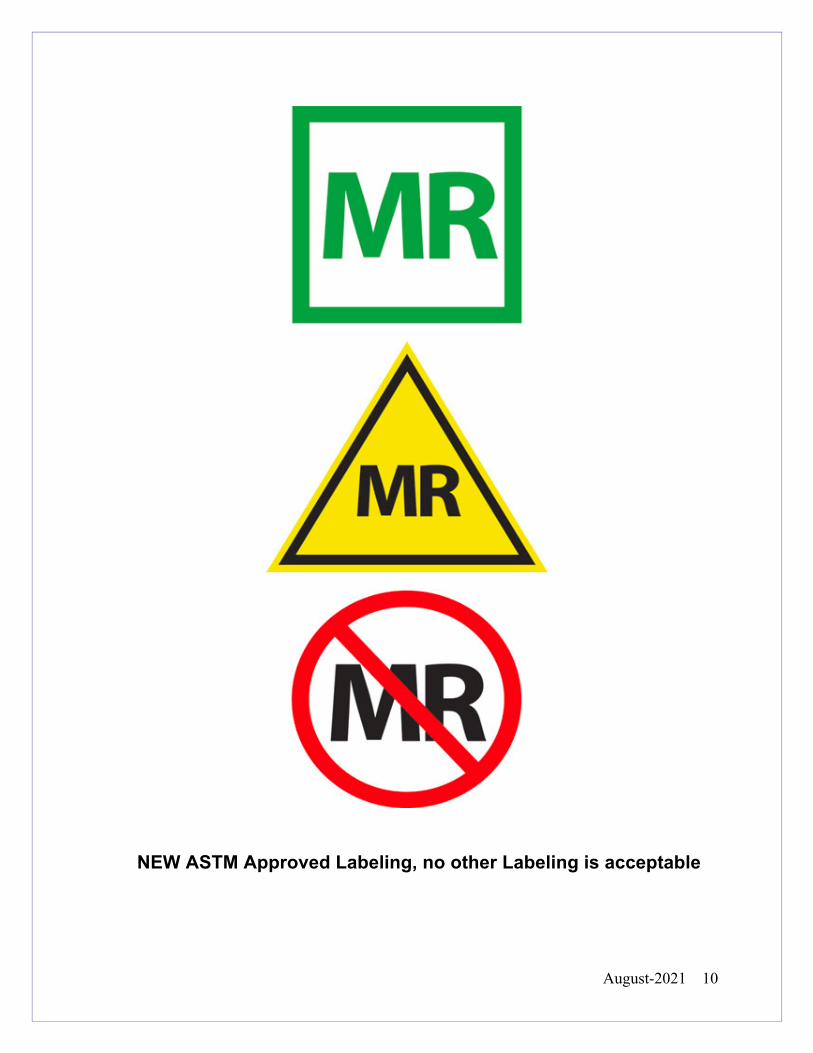
• MR SAFE - is an item that poses no known hazards in all MRI environments.
Using the new terminology, "MR Safe" items include non-conducting, non-
metallic, non-magnetic items such as a plastic Petri dish. The "MR Safe" icon
consists of the of the letters "MR" in green in a white square with a green border -
or - the letters "MR" in white in a green square.
• MR CONDITIONAL - is an item that has been demonstrated to pose no known
hazards in a specified MR environment as long as specified conditions of use are
met. The "MR Conditional" icon consists of the letter "MR" in black inside a
yellow triangle with a black border. The item labeling must include the results of
testing and the specific conditions of use sufficient to characterize the behavior of
the item in the MRI environment.
• MR UNSAFE - is an item that is known to pose hazards in all MRI environments.
MR Unsafe items include magnetic items such as a pair of ferromagnetic
scissors. The "MR Unsafe" icon consists of the letters "MR" in black in a white
field inside a red circle with a diagonal red band.
• Safety in MRI Not Evaluated- For devices that have historically not provided any
information about MRI safety
August-2021 10
NEW ASTM Approved Labeling, no other Labeling is acceptable
August-2021 11
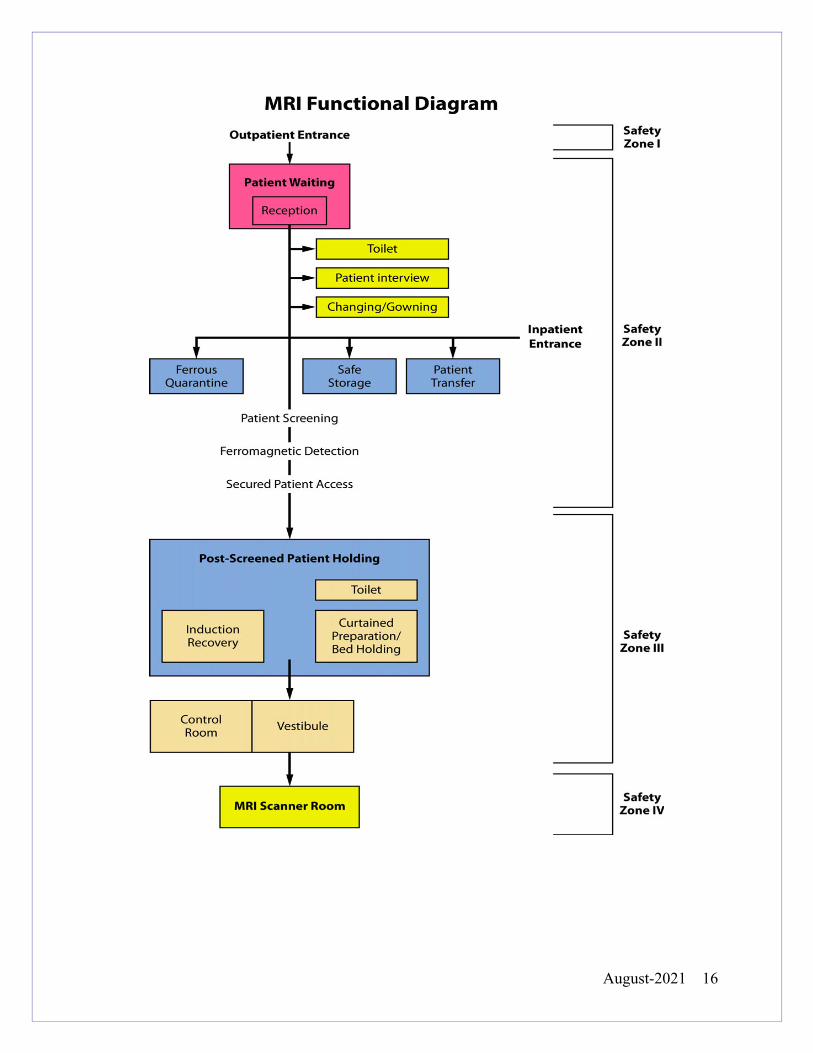
The ACR established the 4 zone concept as defined in the ACR Guidance Document
for Safe MR Practices: 2007. The four zone concept provides for progressive
restrictions in access to the MRI scanner. All MRI Suites are marked with Zone signs
• Zone I: General public freely accessible to the public. This area is typically outside the
MR environment.
• Zone II: Limited Access: This is the Zone located between the public uncontrolled
Zone 1 and the strictly controlled Zone 3. This area has limited access - available to
patients, family members and hospital personnel who have been safety trained or safety
screened by Level 2 MR personnel. It is in Zone II that the answers to MR screening
questions, patient histories, medical insurance questions, etc. are typically obtained.
YALE-NEW HAVEN HOSPITAL
DEPT. OF DIAGNOSTIC RADIOLOGY
POLICY AND PROCEDURE MANUAL
MRI Safety Manual
Reviewed Date 2021
Last Revision 2020
Policy No.
SUBJECT: ACR ZONES: Site Access Restrictions
August-2021 12
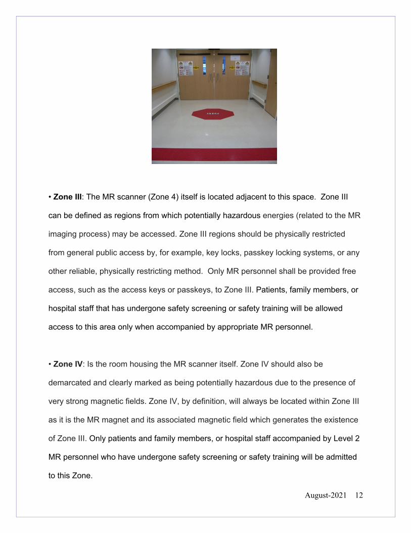
• Zone III: The MR scanner (Zone 4) itself is located adjacent to this space. Zone III
can be defined as regions from which potentially hazardous energies (related to the MR
imaging process) may be accessed. Zone III regions should be physically restricted
from general public access by, for example, key locks, passkey locking systems, or any
other reliable, physically restricting method. Only MR personnel shall be provided free
access, such as the access keys or passkeys, to Zone III. Patients, family members, or
hospital staff that has undergone safety screening or safety training will be allowed
access to this area only when accompanied by appropriate MR personnel.
• Zone IV: Is the room housing the MR scanner itself. Zone IV should also be
demarcated and clearly marked as being potentially hazardous due to the presence of
very strong magnetic fields. Zone IV, by definition, will always be located within Zone III
as it is the MR magnet and its associated magnetic field which generates the existence
of Zone III. Only patients and family members, or hospital staff accompanied by Level 2
MR personnel who have undergone safety screening or safety training will be admitted
to this Zone.
August-2021 13
Non-MR Personnel should be accompanied by, or under the immediate supervision of
and visual contact with, one specifically identified MR person for the entirety of their
duration within Zone III and a level 2 MR person in Zone IV restricted regions.
SITE ACCESS RESTRICTIONS:
MRI Center, Fitkin Basement
The MRI outpatient center is located at 789 Howard Avenue. Stretcher/wheelchair
bound patients will be transferred to MR safe equipment in the prep hold area located
adjacent to MR 5 and MR 1. The amount of additional hospital staff for any procedural
MRI’s will be restricted to 3 people per service; this will help alleviate overcrowding and
potential safety incidents.
August-2021 14
Smilow (Level 2), MRI Suite
The Smilow MRI Suite is located at 20 Park Street, second floor of the Smilow Cancer
Hospital. Stretcher and wheelchair bound patients will be transferred to MR safe
equipment in the prep hold area. The amount of additional hospital staff for any
procedural MRI’s will be restricted to 3 people per service; this will help alleviate
overcrowding and potential safety incidents in the suite
Smilow (Level 1) Breast Center MRI Suite
The Breast Center MRI Suite is located at 20 Park Street; first floor of the Smilow
Cancer Hospital is divided into four zones. All stretcher bound patients will attempt to be
scheduled on the second floor MRI suite. Wheelchair bound patients will be transferred
to MR safe equipment in the changing area.
North Haven, MRI Suite
The North Haven MRI suite located at 6 Devine Street in North Haven is divided into
four zones. Stretcher and wheel chair bound patients will be transferred to MR safe
equipment in the transfer area.
Pediatric Suite West Pavilion:
The Pediatric MRI Suite is located on the second floor of the YNHH Children’s Hospital.
Due to limited space in the suite, stretcher bound patients will be transferred to MR safe
stretchers in the MRI intake room. The amount of additional hospital staff for procedural
August-2021 15
MRI will be restricted to 3 people per service; this will help alleviate overcrowding and
potential safety incidents. For the safety of patients, families and staff, the detachable
table at this location must be used.
Shoreline Medical Center Guildford, CT MRI Suite
The YNHH Temple Street MRI suite located at 111 Goose Lane Guilford CT. The
amount of additional hospital staff for procedural MRI will be restricted to 3 people per
service; this will help alleviate overcrowding and potential safety incidents. The
detachable table will be used to transfer wheelchair or stretcher patients from MR safe
area to MR scan room.
St Raphael, MRI Center
The St Raphael’s Campus MRI center located 1450 Chapel Street New Haven CT. Due
to limited space in the suite, stretcher bound and wheel chair patients will be transferred
to MR safe equipment in the nursing area. The amount of additional hospital staff for
procedural MRI will be restricted to 3 people per service; this will help alleviate
overcrowding and potential safety incidents.
August-2021 16
August-2021 17
The time varying magnetic fields in MRI produce auditory, induced voltage and thermal
issues that we should be aware of.
Induced Voltage Considerations:
Implanted wires pose a possibility of creating a current along the wire inside the MRI
• Patients with implanted wires in anatomically and/or functionally sensitive area
should be considered at a higher risk. The decision to perform imaging and/or
limit the rate of magnetic field change and strength of the magnetic field should
be reviewed by the radiologist supervising the case.
Thermal Considerations:
SAR-Specific Absorption Rate is defined as the RF power absorbed per unit of mass of
an object. It is measured in watts per kilogram (w/kg). The SAR describes the potential
for heating of the patient’s tissues due to application of the RF necessary to produce the
MR signal. Technologists will monitor SAR levels introduced to the patient, and will stay
within appropriate levels. Electric currents can be created during MR imaging which
could cause burns to the patient.
• All electrical connections such as surface coil leads, monitoring devices, etc.,
must be physically checked by the scanning technologist before beginning the
scan to ensure the integrity of the thermal and electrical insulation.
YALE-NEW HAVEN HOSPITAL
DEPT. OF DIAGNOSTIC RADIOLOGY
POLICY AND PROCEDURE MANUAL
MRI Safety Manual
Reviewed Date 2021
Last Revision 2020
Policy No.
SUBJECT: TIME VARYING GRADIENT MAGNETIC FIELD-RELATED ISSUES
August-2021 18
• All unnecessary or unused electrically conductive materials external to the
patient should be completely removed from the MR system before scanning
starts.
• For electrically conductive material, wires, leads, implants, etc., that are required
to remain in the bore of the magnet with the patient during imaging, pads, etc.
should be placed between the patient and the electrically conductive material
during imaging to keep the electrical conductor from directly contacting the
patient. Pads can also be placed between the conductive material and the wall of
the magnet if the body coil is being used, no loops should be created.
• Care is needed to ensure that the patients' tissues do not directly come into
contact with the inner bore if the scanner during the imaging process. Pads
should be placed between the patient and the magnet walls. It is also important
to ensure that the patients' own tissues do not form large conductive loops.
Therefore, care should be taken to ensure that the patients' arms and legs not be
positioned in such a way as to form a loop within the bore of the magnet.
• There have been rare reports of thermal injuries/burns associated with clothing
that contained electrically conductive materials, such as metallic threads and
silver impregnated clothing. As such, all patients remove their own clothing and
instead change into provided gowns.
August-2021 19
• All unconscious/unresponsive patients should have attached leads insulated from
their skin during scanning.
• It is important to follow established product MR Conditional labeling and safety
guidelines carefully and precisely, applying them to and only to the static
magnetic field strengths at which they had been tested. MR scanning at either
stronger and/or weaker magnetic field strengths than those tested may result in
significant heating where none had been observed at the tested field strength(s).
• The patient should immediately report any burning/or discomfort to the MR
technologist, the scan should be stopped and the situation accessed. A RL
solution should be documented by the technologist via the Intranet and to the
FDA Maude website if any equipment was involved www.fda.gov/medwatch
Auditory Considerations:
Patients ALWAYS need to have hearing protection as well as their family members who
accompany them into the scan room. Please review our Ear plug policy
MRI Imaging with Ear Plugs:
YALE-NEW HAVEN HOSPITAL
DEPT. OF DIAGNOSTIC RADIOLOGY
POLICY AND PROCEDURE MANUAL
MRI Safety Manual
Reviewed Date 2021
Last Revision 2020
Policy No.
SUBJECT: EAR PLUGS DURING AN MRI EXAM
August-2021 20
The noise generated by scanning may reach a level in the scan room and in the bore of the
magnet that can result in temporary (and occasionally) permanent hearing loss.
Properly inserted earplugs will limit the level of the noise that reach the inner ear.
Any patient who undergoes an MRI, as well as anyone in Zone 4 during a scan, MUST wear
earplugs.*
The earplugs will be inserted by the MRI staff. The earplugs are Latex Free and have an
acceptable NNR rating.
In pediatric patients, and patients with unusual shaped ear canals, the earplugs may not fit
properly to limit the noise level. In these instances, MRI staff will use another form of hearing
protection such as headphones or ear muffs that will hold the earplugs in place and further
dampen the noise level.
If a patient has their own custom ear plugs, designed for their ear canals it is acceptable to use
after they have been wanded for metal. MR staff need to check they are inserted.
*A patient who is deaf in one or both ears, do not need to use hearing protection in the affected
ear canal. Please document in EPIC
YALE-NEW HAVEN HOSPITAL DEPT. OF
DIAGNOSTIC RADIOLOGY POLICY AND PROCEDURE MANUAL
MRI Safety Manual
Reviewed Date 2021
Last Revision 11/2020
Policy No.
SUBJECT: FIREFIGHTERS, POLICE AND SECURITY CONSIDERATIONS
August-2021 21
A.) All MR sites should arrange to prospectively educate their local fire marshals, police
and security personnel about the potential hazards of responding to emergencies in the
MR suite.
B.) It should be stressed that even in the presence of a fire or other emergency the
magnetic field may be present and fully operational. Free access to Zone IV by
firefighters and other non-MRI personnel with air tanks, axes, crowbars, guns, etc. can
prove to be catastrophic or even lethal. Helium is not flammable and does not pose a
fire hazard directly; however, the liquid oxygen that can result from the super cooled air
might well increase the fire hazard in this area. If there are appropriately trained MRI
personnel available during the emergency who are able to keep the emergency
responders from the magnet room and the five gauss line, then quenching the magnet
should not be a requirement. As part of the Zone III and IV restrictions, all MR sites
must have clearly marked, readily accessible MR Conditional or MR Safe fire
extinguishing equipment physically stored within Zone III or IV.
C.) If the fire or emergency is in the magnet room, and the emergency response
personnel and their equipment must enter the room, a decision to quench the magnet
should be made to protect the health and lives of the emergency responders. Should a
quench be performed, appropriately designated MRI personnel still need to ensure that
all non MRI personnel continue to be restricted from the magnet room until the
designated MRI personnel have verified that the static field is either no longer
detectable or at least sufficiently attenuated so as to no longer present a potential
hazard (see Cryogen related policy)
August-2021 22
In the event of a fire at any YNHH MRI suites and please follow the following
procedure:
R.A.C.E. RESPONSE PROTOCOL:
R.A.C.E. Stands for RESCUE, ALARM, CONFINE AND EXTINGUISH.
• RESCUE: Injured visitors, employees or staff must rapidly be rescued from the
immediate area of the fire/smoke origin.
• ALARM: At the sight of flames or smoke, immediately activate a Fire Alarm Pull
Station.
• CONFINE: Fire, Smoke and Toxic combustion products must be confined to the
area of fire origin as much as possible. Close the door to the room of fire origin
as soon as any rescue is accomplished.
• EXTINGUISH: If at all possible, staff should make one attempt to extinguish the
fire with a hand-held fire extinguisher. They are to be used only after Rescue,
Alarm and Confine have been completed. A fire extinguisher is not a
replacement for activating the fire alarm system. Any fire that a fire extinguisher
has been used on is to be reported to the Governing Police Department and/or
Fire Marshal.
To Use a fire Extinguisher, use the PASS Protocol
P.A.S.S. FIRE EXTINGUISHER PROTOCOL:
P.A.S.S.: method is used for the proper operation of a hand held fire extinguisher.
• P.A.S.S.: Stands for Pull, Aim, Squeeze, Sweep.
• PULL: Pull the safety ring/pin at the top of the fire extinguisher.
• AIM: Aim the discharge nozzle at the base of the fire.
• SQUEEZE: Squeeze the handle of the fire extinguisher together to discharge the
agent.
• SWEEP: Sweep the agent side to side at the base of the fire.
August-2021 23
• Whenever using a fire extinguisher, the following is to be remembered.
• Maintain a clear exit
• Keep your back to that clear exit.
• If at any time you feel as if you are in danger evacuate the area and close the door
behind you.
YNHH MRI CENTERS GENERAL EVACUATION PLAN
IN CASE FIRE OR SMOKE
1. Stay calm.
2. Always sound the building fire alarm immediately. If the alarm fails to operate,
warn other occupants by knocking on doors and shouting warnings.
3. Call your centers Emergency number. (Temple ST 911, YNHH 155, St.
Raphael’s 155, Guilford 911, North Haven 911, Yale University Office of the Fire
Marshal @ (203) 4329923) from a safely located telephone. Give as much
information as possible to the dispatcher. Do not assume that someone else has
already notified them. They will immediately notify the Fire Department and
dispatch officers to the scene. Do not hang up until told to do so by the
dispatcher.
4. Before opening the door, feel it with the back of your hand. If it is not hot skip to
STEP 5. If it is hot, do the following
• Seal cracks around the door with towels, tape, bed clothing or similar items to keep
out the smoke. Shout for help. Call your centers Emergency number. (Temple ST
August-2021 24
911, YNHH 155, St. Raphael’s 155, Guilford 911, North Haven 911 Yale University
Office of the Fire Marshal 203-432-9923) and tell them that you are unable to get
out of your room. They will be in contact with officers at the fire. Remain calm until
firefighters reach you from the hallway or window. Their first duty upon arriving at
a fire is to search for persons trapped in the burning building.
5. If you are able to leave your room, do so immediately and:
• Take your key with you in case you are forced to return. Close all
doors behind you as you exit. This will lessen the spread of smoke
and damage.
• Go to the nearest exit or stairway. Do not use an elevator.
• If smoke, heat or fire blocks your exit, go to an alternate exit.
• If all exits from a floor are blocked go back to your room and follow
the procedures described above in step 4
6. If smoke is present, keep low to the floor. Take short breaths to avoid inhaling
any more smoke than necessary.
7. Leave the building immediately. When the Police and/or Firefighters arrive, direct
them to the fire.
Non-essential staff should follow exit signs and or the directions of Police and Firefighters.
Do not re-enter the building for any reason until the Fire Department has declared it safe.
FIRE ALARM ACTIVATION PROCEDURE
In the event of a fire alarm activation alarm or other emergency, visitors,
employees and non-essential staff are to evacuate the building using the nearest
stairway or exit. Essential staff and patients undergoing procedures should be aware of
their surroundings and be vigilant in checking for smoke or fire. An immediate
evacuation order may be issued by Governing Fire, Security, Police Department, or a
representative from the Fire Marshal’s Office. If an evacuation order is issued, it must
be followed immediately.
August-2021 25
Elevators are not to be used during a fire alarm or smoke/fire condition. During a fire
alarm evacuation, non-essential staff should assist ambulatory patients in evacuating
the building by following the exit signs which lead to the building exits. Once outside,
everyone should move away from the exit discharge doors of the building and to avoid
congregating close to the building where they may hamper emergency operations. Do
not block the exits or fire department access to the building.
The onsite administrator or business manager of each department will be responsible
for the management of the evacuation process. They are to have a list of all employees
in their department with them and are to advise the fire department of any employee
that is missing. If the onsite administrator or business manager is not available, they
are to have designated someone who is to manage the evacuation process for their
department or group.
Any disabled individual who cannot evacuate using the outside means of egress are to
be moved to the other side of the smoke barrier doors and wait for the fire department
to remove them safely from the building. The buddy system is to be used for these
employees. YNHH staff is to notify the fire department when they reach the street that
there may be disabled individuals still in the building and where they are located.
The fire alarm system is not to be silenced or reset without the permission of the fire
department.
Occupants are not to reenter the building until the "All Clear" is given by the fire department.
MRI EMERGENCY PROCEDURES
• When a call has been placed for a fire or police emergency in the MRI Center,
MR technologists at all scanners should immediately stop scanning and remove
patients from the scanner room.
• The MR safety officer should be called and informed of the emergent situation so
that they can be on site prior to the arrival of emergency personnel.
• The MR safety officer and a designated MR staff member should monitor the
scanner room doors to prevent free access by emergency personnel. (NOTE:
Even in the event of a fire or other emergency the magnetic fields are likely to be
present and fully operational.)
August-2021 26
• In the case of a fire that is not in the scanner room, quenching the magnet should
not become necessary.
• If the fire is in a location that fire fighters and their equipment (oxygen, canisters,
crowbars, axes, defibrillators, ETC.) need to enter the scanner room, a decision to
quench the magnet may become necessary to protect the health and lives of the
emergency personnel.
• If a quench is performed the MR safety officer and MR technologists need to ensure
that all emergency personnel are restricted from the scanner room until the static
magnetic field is no longer present.
FIRE PREVENTION PLAN:
Accumulations of flammable or combustible waste material are not to be left in the
building. Computer rooms are not to be used for storage. All combustible waste
materials are to be removed each day. Smoking is not allowed anywhere in the building.
Corridors and stairs are not to be used for storage or equipment areas since they
become an obstruction during an emergency. Storage and equipment can cause a fire
if it is energized equipment, or contribute fuel to a fire, which will fill the corridor with
smoke and toxic fumes. The building has a fire alarm system with consists of smoke
detectors and pull stations and a fire sprinkler system. The building has also been
provided with emergency lighting and exit signs to locate the exit stairwells. Hand held
fire extinguishers have been provided throughout the building.
Questions regarding Yale University building be directed to the Yale University Office of
the Fire Marshal @ (203) 4329923
In the event of a system quench it is imperative that all personnel/patients be evacuated
from the MR scan room as quickly and as safely feasible.
YALE-NEW HAVEN HOSPITAL
DEPT. OF DIAGNOSTIC RADIOLOGY
POLICY AND PROCEDURE MANUAL
MRI Safety Manual
Reviewed Date 2021
Last Revision 2020
Policy No.
SUBJECT: CRYOGEN RELATED ISSUES
August-2021 27
• Stop all scanning and open the scan room door immediately. If the door to the
scan room is closed the pressure may build up making it impossible to open the
door. In this event, it may become necessary to break the glass window to allow
the gasses to escape and the pressure to lessen so that the scan room door
may be opened.
• The access to the scan room should be immediately restricted to all individuals
until the arrival of the MR equipment service personnel.
• Do not rely upon the oxygen sensors in the room to warn of low oxygen levels in
the room. This technology is now considered by industry experts not to be
sufficiently reliable to allow for continued operations during situations of power
outage, etc.
• It is especially important to ensure that all police and fire response personnel are
restricted from entering the MR scan room with their equipment (axes, air tanks,
guns, etc.) until it can be confirmed that the magnetic field has been successfully
dissipated, as there may still be considerable static magnetic field present
despite a quench or partial quench of the magnet.
• MR Safety Officer and MR Medical Director need to be informed immediately
• In a Quench or Emergency Situation
August-2021 28
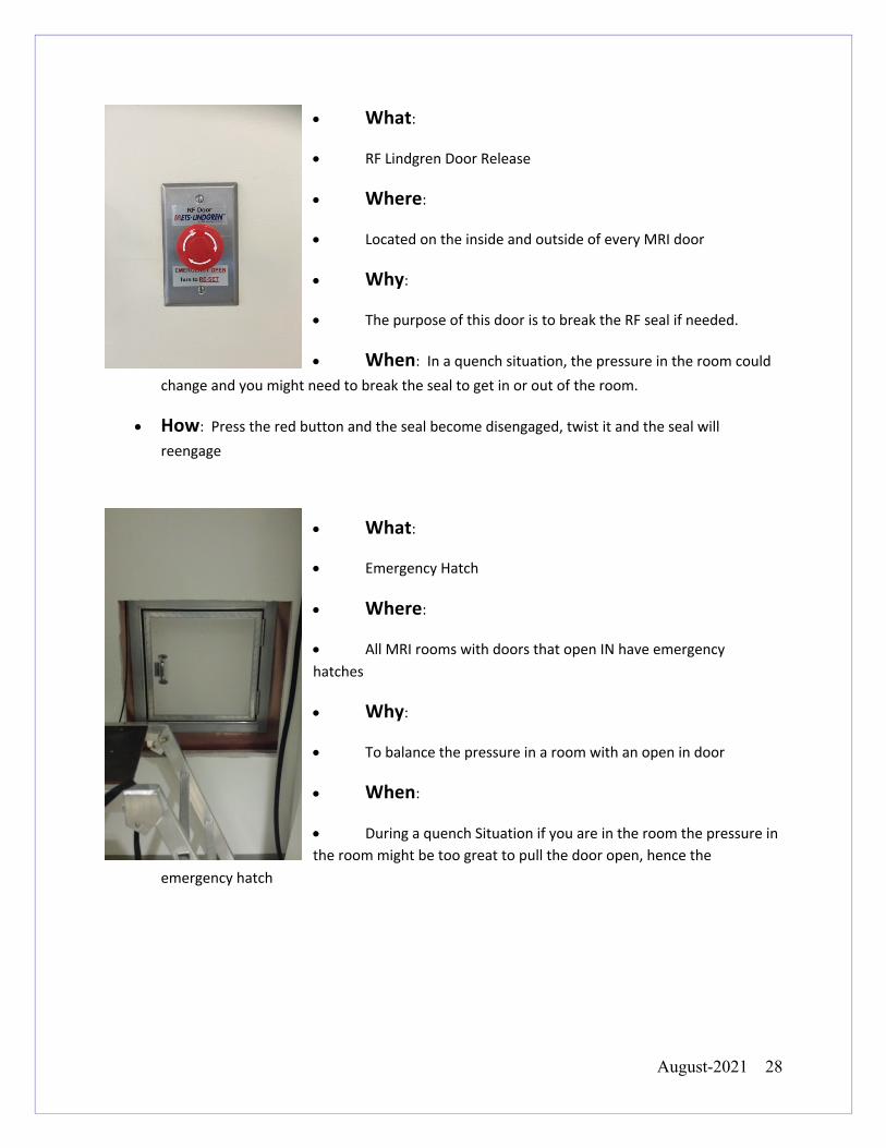
• What:
• RF Lindgren Door Release
• Where:
• Located on the inside and outside of every MRI door
• Why:
• The purpose of this door is to break the RF seal if needed.
• When: In a quench situation, the pressure in the room could change and you might need to break the seal to get in or out of the room.
• How: Press the red button and the seal become disengaged, twist it and the seal will reengage
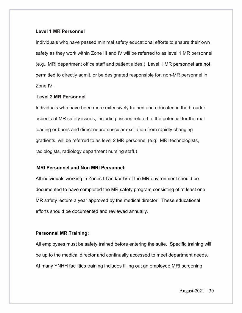
• What:
• Emergency Hatch
• Where:
• All MRI rooms with doors that open IN have emergency hatches
• Why:
• To balance the pressure in a room with an open in door
• When:
• During a quench Situation if you are in the room the pressure in the room might be too great to pull the door open, hence the
emergency hatch
August-2021 29
MRI Personnel Screening:
All MR personnel are to undergo an MR Screening process as part of their
employment interview to ensure their own safety in the MR environment. At this
time, it is the employees' responsibility to fully disclose any trauma, procedure or
surgery that they have experienced or undergone in which ferromagnetic objects or
devices may have become introduced within them or on them. This will permit an
appropriate screening to be performed upon the employee to determine the safety of
introducing them to the MR environment.
Personnel Definitions:
Non-MR Personnel
Patients, visitors or facility staff who do not meet the criteria of level 1 or level 2 MR
personnel will be referred to as non-MR personnel. Specifically, non-MR personnel
will be the terminology used to refer to any individual or group who has not within the
previous 12 months undergone the designated formal training in MR safety issues
defined by the MR safety director of that installation.
YALE-NEW HAVEN HOSPITAL
DEPT. OF DIAGNOSTIC RADIOLOGY
POLICY AND PROCEDURE MANUAL
MRI Safety Manual
Reviewed Date 2020
Last Revision 2018
Policy No.
SUBJECT: MRI SAFETY SCREENING: PERSONNEL, FAMILY MEMBERS
August-2021 30
Level 1 MR Personnel Individuals who have passed minimal safety educational efforts to ensure their own
safety as they work within Zone III and IV will be referred to as level 1 MR personnel
(e.g., MRI department office staff and patient aides.) Level 1 MR personnel are not
permitted to directly admit, or be designated responsible for, non-MR personnel in
Zone IV.
Level 2 MR Personnel Individuals who have been more extensively trained and educated in the broader
aspects of MR safety issues, including, issues related to the potential for thermal
loading or burns and direct neuromuscular excitation from rapidly changing
gradients, will be referred to as level 2 MR personnel (e.g., MRI technologists,
radiologists, radiology department nursing staff.)
MRI Personnel and Non MRI Personnel:
All individuals working in Zones III and/or IV of the MR environment should be
documented to have completed the MR safety program consisting of at least one
MR safety lecture a year approved by the medical director. These educational
efforts should be documented and reviewed annually.
Personnel MR Training:
All employees must be safety trained before entering the suite. Specific training will
be up to the medical director and continually accessed to meet department needs.
At many YNHH facilities training includes filling out an employee MRI screening
August-2021 31
sheet, watching a designated MR safety video and speaking with a level 2 MR
employee.
Family Members and Non MR Personnel:
All family members and companions entering Zones III and/or Zone IV will be
required to complete an MRI safety form. Any positive responses and possible
contraindications to these areas will be discussed with the MR technologist. If the
technologist cannot resolve the issues the radiologist will be consulted.
In addition to the screening form, pregnant family members who want to go into zone
IV will also be given information about pregnancy and MRI. It is their choice to stay
in the room during the MRI.
All non-MR Personnel and family members who are going into Zone IV - the scanner
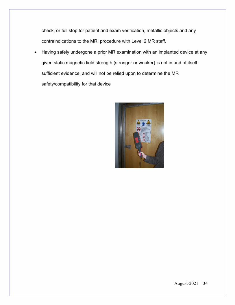
room - will remove all metal objects and personal belongings, and will be wanded by
a ferrous metal detector in Zone II before given permission to enter Zone III and
Zone IV..
Device/Object Screening:
• MRI staff members should pay particular attention to the stretchers and beds
of inpatient and remove all oxygen tanks and any other potential hazards.
• All portable metallic or partially metallic devices that are on or external to the
patient (e.g., oxygen cylinders) are to be positively identified in writing as non-
ferromagnetic and either MR safe or MR compatible prior to admitting them
into Zone III.
August-2021 32
• As part of the Zone III site restriction and equipment testing and clearing
responsibilities, all sites should have ready access to a strong handheld
magnet (1000-Gauss) and/or a handheld ferromagnetic detection device. This
will enable the site to test external, and even some superficial internal devices
or implants for the presence of grossly detectable ferromagnetic attractive
forces.
• If external devices/objects are demonstrated to be ferromagnetic and non-MR
safe/conditional, they may still, under specific circumstances, be brought into
Zone IV if deemed by MR personnel to be necessary and appropriate for the
care of the patient (ex. arterial line, catheter bag with clip). These
devices/objects must be appropriately secured at all times. The safe
utilization of these devices is the responsibility of MR personnel to ensure that
they do not inadvertently become introduced too close to the MRI scanner
and become a hazardous projectile or no longer accurately function.
• Never assume MR safety information about any device if it’s not clearly
documented in writing and following ASTM testing standards. If a device’s
MR safety status is unknown, it should not be permitted in the magnet field.
• A prior MR examination with an implanted device at any given static magnetic
field strength (stronger or weaker) is not in and of itself sufficient evidence,
and will not be relied upon to determine the MR safety
August-2021 33
• Outpatients will be checked in by the receptionist at the front desk of the MRI
department. Two Patient identification markers will be used. (Example Full
Name, DOB, Address). They will be given a wrist band and be handed a safety
sheet to fill out (if not done beforehand). Anyone accompanying the patient will
also fill out a safety sheet.
• The MR personnel (level 1 or 2) will review the safety sheet to ensure that there
are no positive responses. Any positive responses will be discussed between
the patient and a level 2 staff member to confirm that the patient has understood
the safety form and understands any risks that are involved with the MRI
procedure.
• If the patient is getting contrast two additional steps should be taken before
scheduling the patient. The patients eGFR and pregnancy status (if applicable)
should be known.
• The patient will be instructed to remove any metal objects and secure their
belongings in the lockers. All patients will be requested to change into hospital
attire.
• After the patient has changed, the patient and any accompanying companions or
facility staff members will be wanded with a ferromagnetic detector have a final
YALE-NEW HAVEN HOSPITAL
DEPT. OF
DIAGNOSTIC RADIOLOGY POLICY AND PROCEDURE MANUAL
MRI Safety Manual
Reviewed Date 2021
Last Revision 2018
Policy No.
SUBJECT: MRI SCREENING PROCESS FOR GENERAL OUTPATIENTS
August-2021 34
check, or full stop for patient and exam verification, metallic objects and any
contraindications to the MRI procedure with Level 2 MR staff.
• Having safely undergone a prior MR examination with an implanted device at any
given static magnetic field strength (stronger or weaker) is not in and of itself
sufficient evidence, and will not be relied upon to determine the MR
safety/compatibility for that device
August-2021 35
MRI Screening Process for all Inpatients
• Inpatient safety needs to be reviewed every time a patient has an MRI. They
will be screened utilizing the inpatient screening form located in Epic. The
form will be filled out on the floor with the patient. The patient needs to be
alert and oriented x3 to be able to fill out their own screening sheet. If the
patient is unable to complete the form (i.e. due to altered mental status) a
nurse, physician, and/or family member may complete the form on the
patient’s behalf. (Unresponsive patients with no family members please see
page 37)
• If the patient is getting contrast two additional steps should be taken before
scheduling the patient. The patients eGFR and pregnancy status (if
applicable) should be known
• Inpatients will have their safety form reviewed for any contraindications to the
MR procedure by the MR personnel before an appointment will be scheduled.
Inpatients will not be scheduled without a complete and signed safety form.
• After these steps the patient will be scheduled.
YALE-NEW HAVEN HOSPITAL
DEPT. OF DIAGNOSTIC RADIOLOGY
POLICY AND PROCEDURE MANUAL
MRI Safety Manual
Reviewed Date 2021
Last Revision 2021
Policy No.
SUBJECT: MRI SCREENING INPATIENTS
August-2021 36
• The patient will be put into the transport system. The inpatient transporter will
transport the patient to the designated Zone II area, where staff will inform the
technologists of the patient arrival
• Inpatients including emergency room patients should be prepared for their
MRI exam before they leave their floor: all personal belongings (including
clothing) should remain at bedside and not travel with patients to the MR
procedure area.
• In Zone 2 in the MRI facility, MR unsafe medical equipment will be replaced
with MR Conditional equipment.
• If the patient is on oxygen they will be put on walled oxygen.
• Before entering Zone III the patient will be wanded with a ferromagnetic
detector and have a final check, or full stop for patient identification, exam
verification and all safety concerns with level 2 MR staff
• For mechanically ventilated patient please follow that specific process.
• Any accompanying companions or facility staff members will also be wanded
and safety screened by MR staff for any ferromagnetic objects or safety
concerns
• Having safely undergone a prior MR examination with an implanted device at
any given static magnetic field strength (stronger or weaker) is not in and of
itself sufficient evidence, and will not be relied upon to determine the MR
safety/compatibility for that device
August-2021 37
YALE-NEW HAVEN HOSPITAL
DEPT. OF DIAGNOSTIC RADIOLOGY
POLICY AND PROCEDURE MANUAL
MRI Safety Manual
Reviewed Date 2020
Last Revision 2018
Policy No.
SUBJECT: Unresponsive Patients in MRI
I. Policy Screening of patients for whom an MR examination is deemed necessary but who are unconscious, unresponsive, not AOx3 or for other reasons unable to provide their own reliable histories regarding prior surgery or exposure to metallic foreign objects, and for whom such histories cannot be reliably obtained from others: The patient should be physically examined for evidence of possible surgery or trauma involving metal, and these areas should be subject to plain-film radiography (if recently obtained imaging of such areas is not already available) . The patient should undergo plain film radiography of the chest and skull or orbits to exclude potentially dangerous metallic foreign objects/devices. Final determination of whether or not to scan any given patient will be made by the supervising radiologist. II. Procedure If a patient is unconscious, unresponsive or for other reasons unable to provide their own reliable histories, the responsible health care professional or supervising radiologist will order plain-film radiographs of the chest and skull or orbits. A verbal order to MRI technologist is acceptable. The ordering health care professional will examine the patient for any additional areas of scars or deformities that might be anatomically indicative of an implant, or foreign body. This examination will be documented in Epic (progress notes) before the patient will be scheduled for their MRI exam. If any areas of concern are found, they can be imaged with plain film radiography based on the ordering physician and/or the radiologist’s request. A radiologist from the service (i.e. Neuro, MSK, Body, Peds) that will interpret the ordered MRI examination will determine whether there are any contraindications for the MRI. The same service will interpret and dictate the plain-film radiographs obtained for MRI screening.
August-2021 38
Prior to the patient being scheduled, the supervising radiologist may review the safety sheet with the MRI technologist and provide verbal approval, but such approval must be documented and scanned into the patient’s chart After the patient has been cleared by the radiologist, the standard inpatient process will continue.
August-2021 39
• No oxygen cylinders of any kind will be allowed in Zone IV - the scan room
• When the patient enters Zone IV - the scan room - the patient will be hooked
up to wall mounted oxygen
• Under no circumstances will standard ferromagnetic oxygen tanks be brought
into Zones III or Zone IV.
• MRI conditional oxygen cylinders are allowed in Zone III with strict
supervision on a case by case basis. NEVER Zone IV
• Hospital staff transporting inpatients with standard ferrous oxygen cylinders
will exchange the O2 from tank to wall O2. All ferromagnetic oxygen
cylinders will be stored in a holder. While the patient waits for their exam or
waits to be transported back to their room the wall mounted oxygen will be
used
• MRI conditional oxygen cylinders are silver with a light green top.
The "Oxytote" oxygen cylinders are not MRI safe. They indeed have ferromagnetic
components. They are to be treated as any other ferrous oxygen cylinder and will
be exchanged for our traditional aluminum cylinders as outlined above.
YALE-NEW HAVEN HOSPITAL
DEPT. OF DIAGNOSTIC RADIOLOGY
POLICY AND PROCEDURE MANUAL
MRI Safety Manual
Reviewed Date 2020
Last Revision 2020
Policy No.
SUBJECT: OXYGEN CYLINDERS
August-2021 40
YALE-NEW HAVEN HOSPITAL
DEPT. OF DIAGNOSTIC RADIOLOGY
POLICY AND PROCEDURE MANUAL
MRI Safety Manual
Reviewed Date 2020
Last Revision 2020
Policy No.
SUBJECT: Penile Implants
There are 2 penile Implants that are considered unsafe for MRI. They are the
Omniphase by Dacomed, which was discontinued and replaced with the Duraphase
by Dacomed. The Duraphase model was discontinued in 1995.
YNHH all penile implants implanted after 1997 are considered MR Conditional on
our 1.5 and 3T systems.
August-2021 41
YALE-NEW HAVEN HOSPITAL
DEPT. OF DIAGNOSTIC RADIOLOGY
POLICY AND PROCEDURE MANUAL
MRI Safety Manual
Reviewed Date 2021
Last Revision 2021
Policy No.
SUBJECT: Foreign Body in Orbits
Policy “All patients who have a history of orbit trauma by a potential ferromagnetic foreign body for which they sought medical attention are to have their orbits cleared by either plain X-ray orbit films (2 views) or by a radiologist’s review and assessment of contiguous cut prior CT or MR images (obtained since the suspected traumatic event) if available”. -ACR white paper 2013 Procedure At YNHH a patient will have orbital x-rays for foreign body if the following criteria are met:
• They have sought out medical attention in the past for a foreign body in the orbit. • No available CT or MRI for a radiologist to verify they are cleared.
If a patient arrives for an MRI and needs orbital x-rays, it is appropriate for the technologist to take a verbal or written order from the radiology attending physician. There is no need to obtain an order from the patient’s referring physician, inpatient or outpatient. Radiologist Responsibility A radiologist from the service (i.e. Neuro, MSK, Body, Peds) that will interpret the ordered MRI examination will determine whether there are any contraindications for the MRI. This service will interpret and dictate the plain-film radiographs obtained for MRI screening. Ordering & Scheduling Foreign Body X-ray of Orbits:
• From the Technologist Worklist: From tech worklist select the patient, right click and select Order Entry
• Change Order Mode to cosign required OR per protocol:no cosign required • Under New Order select XR EYE Foreign Body as the exam • Enter a Reason example “per radiologist, MRI clearance” • Click the Provider Button Change the Ordering DR to Radiology Referring MED
Physician • Click Sign Order • Schedule as appropriate
August-2021 42
YALE-NEW HAVEN HOSPITAL DEPT. OF
DIAGNOSTIC RADIOLOGY POLICY AND PROCEDURE MANUAL
MRI Safety Manual
Reviewed Date 2021
Last Revision 2021
Policy No.
SUBJECT: Plain Film Xrays for MRI Clearance
If a patient has an MRI ordered and the radiologist requires further imaging to safely perform the MRI, it is appropriate for the technologist to take a verbal or written order from the radiology attending physician. There is no need to obtain an order from the patient’s referring physician, inpatient or outpatient. Radiologist Responsibility A radiologist from the service (i.e. Neuro, MSK, Body, Peds) that will interpret the ordered MRI examination will determine whether there are any contraindications for the MRI. This service will interpret and dictate the plain-film radiographs obtained for MRI screening. Ordering & Scheduling Foreign Body X-rays for MRI Studies:
Please use Order entry in Epic.
• From the Technologist Worklist: From tech worklist select the patient, right click
and select Order Entry • Change Order Mode to cosign required OR per protocol:no cosign required • Under New Order select the appropriate exam. • Enter a Reason example “per radiologist, MRI clearance” • Click the Provider Button Change the Ordering DR to Radiology Referring MED
Physician • Click Sign Order • Schedule as appropriate
August-2021 43
CARDIAC VALVES All cardiac valves are considered immediately safe to scan on a 1.5T and 3T. CORONARY ARTERY STENTS All coronary stents are considered immediately safe to scan on a 1.5T and 3T CARDIAC LEADS Abandoned epicardial leads are considered safe to scan on 1.5 and 3T. These are small leads often placed during cardiac surgery that can be pulled or left in place. They are attached to the epicardial surface and are not burrowed into the myocardium or within the heart chambers. They are short in length and usually do not loop and therefore are considered MRI safe. For typical xray appearance see link below https://radiopaedia.org/images/885691 Abandoned intracardiac pacing or ICD leads should be brought to an attending radiologist’s attention for further evaluation. If it is unclear if there are abandoned leads a recent chest x-ray should be reviewed or a new chest x-ray ordered if needed. Any MR scan where the intracardiac leads are in the RF field is not recommended due to potential of significant heating and conduction and should only be done after attending radiologist approval after risk/benefit discussion. Informed patient consent is required. Scans where the retained leads are not within the RF field for the ordered MRI are at very low risk for heating. These require attending radiologist approval but do not require informed consent from the patient. Note, abandoned/retained intracardiac leads may be present when the patient still has a working pacemaker/ICD. These abandoned leads can also effect leads of a MRI conditional pacemaker and should be handled in similar fashion.
IMPLANTED CARDIAC MONITORS CARDIAC LOOP RECORDERS
Make and model need to be known, follow company guidelines.
YALE-NEW HAVEN HOSPITAL
DEPT. OF DIAGNOSTIC RADIOLOGY
POLICY AND PROCEDURE MANUAL
MRI Safety Manual
Reviewed Date 2020
Last Revision 2019
Policy No.
SUBJECT: CARDIAC IMPLANTATIONS: VALVES, STENTS, LEADS, PACEMAKERS
August-2021 44
YALE-NEW HAVEN HOSPITAL
DEPT. OF DIAGNOSTIC RADIOLOGY
POLICY AND PROCEDURE MANUAL
MRI Safety Manual
Reviewed Date 2020
Last Revision 2019
Policy No.
SUBJECT: PACEMAKER PROCESS
Performing Cardiac Pacemaker/Defibrillator Studies- Radiologist Guide
Workflow/Process- Multiple departments involved
1. Radiologist Approves the Case based on indication and necessity of obtaining MRI (see steps below)
2. EP approves, (A) device and (B) patient’s clinical status being fit more MRI 3. Patient scheduled with 3 resources (1) 1.5T MRI machine, (2) Radiology Nursing and (3)
EP team.
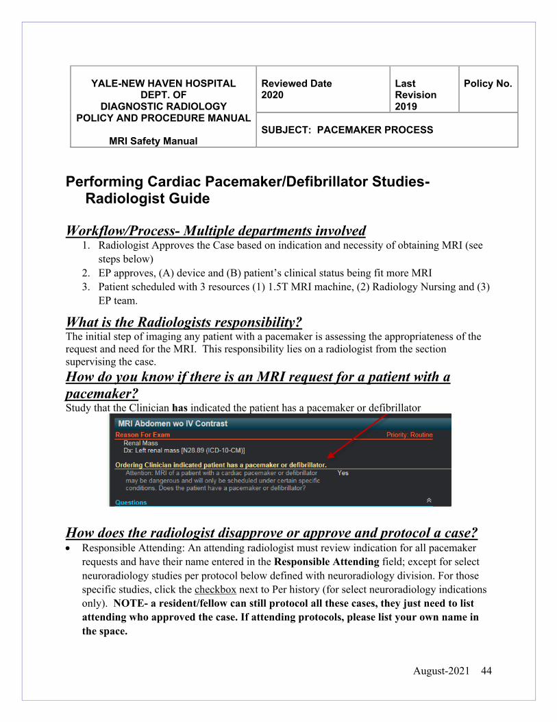
What is the Radiologists responsibility? The initial step of imaging any patient with a pacemaker is assessing the appropriateness of the request and need for the MRI. This responsibility lies on a radiologist from the section supervising the case. How do you know if there is an MRI request for a patient with a pacemaker? Study that the Clinician has indicated the patient has a pacemaker or defibrillator
How does the radiologist disapprove or approve and protocol a case? • Responsible Attending: An attending radiologist must review indication for all pacemaker
requests and have their name entered in the Responsible Attending field; except for select neuroradiology studies per protocol below defined with neuroradiology division. For those specific studies, click the checkbox next to Per history (for select neuroradiology indications only). NOTE- a resident/fellow can still protocol all these cases, they just need to list attending who approved the case. If attending protocols, please list your own name in the space.
August-2021 45
o If radiologist agrees with the Indication for the MRI order, click Medically Necessary and protocol exam as you normally would. NOTE- our job is to assess if the MRI is needed. The EP team assess the pacemaker system and determines MR Conditional or Non MR Conditional status and if they can be scanned off-label when needed. For some studies they will re-consult the radiologist to discuss.
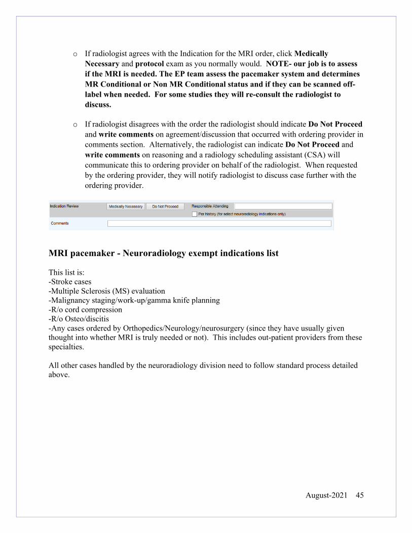
o If radiologist disagrees with the order the radiologist should indicate Do Not Proceed and write comments on agreement/discussion that occurred with ordering provider in comments section. Alternatively, the radiologist can indicate Do Not Proceed and write comments on reasoning and a radiology scheduling assistant (CSA) will communicate this to ordering provider on behalf of the radiologist. When requested by the ordering provider, they will notify radiologist to discuss case further with the ordering provider.
MRI pacemaker - Neuroradiology exempt indications list This list is: -Stroke cases -Multiple Sclerosis (MS) evaluation -Malignancy staging/work-up/gamma knife planning -R/o cord compression -R/o Osteo/discitis -Any cases ordered by Orthopedics/Neurology/neurosurgery (since they have usually given thought into whether MRI is truly needed or not). This includes out-patient providers from these specialties. All other cases handled by the neuroradiology division need to follow standard process detailed above.
August-2021 46
All patients with passive vascular devices such as coils, stents, and filters (see separate
policy on aneurysm clips, and coronary stents in YNHH Safety Manual …) implanted in
the USA can be imaged immediately after implantation, at 1.5 OR 3T if they meet
guidelines below:
The passive device needs to be:
• FDA approved for use
• Tested with ASTM standards with a rating of MR Safe or MR Conditional on
a 1.5T or higher. Many devices are not tested at 3T, but we will allow them to
be performed at 3T if imaging at 1.5T is not possible or an exam on 3T is
preferred.
• Minimum SAR for diagnostic clinical images will be used. Technologist has
flexibility to edit scanner parameters. If needed, image quality can be
reviewed with radiologist to ensure diagnostic quality.
• A patient with any device with a rating of MR Unsafe will not undergo MRI
scan
Radiologists have the ability to alter any policies in the best interest of the patient after a
risk verse benefit decision. For more information see separate policy “Exemptions of
MR Safety Policies” located in YNHH Safety Manual
YALE-NEW HAVEN HOSPITAL DEPT. OF
DIAGNOSTIC RADIOLOGY POLICY AND PROCEDURE MANUAL
MRI Safety Manual
Reviewed Date 2021
Last Revision 9/2019
Policy No.
SUBJECT: PASSIVE VASCULAR IMPLANTS COILS, STENTS, FILTERS –CARDIAC STENTS SEE PAGE 43
August-2021 47
YALE-NEW HAVEN HOSPITAL
DEPT. OF DIAGNOSTIC RADIOLOGY
POLICY AND PROCEDURE MANUAL
MRI Safety Manual
Reviewed Date 2021
Last Revision 9/2018
Policy No.
SUBJECT: ANEURYSM CLIPS
• In the event that it is unclear whether a patient does or does not have an aneurysm clip in place, plain films should be obtained.
• If the patient is identified to have an aneurysm clip the type of aneurysm clip must be documented. All documentation must be in writing; phone or verbal histories are not permitted.
• Having safely undergone a prior MR examination with an aneurysm clip at any given static magnetic field strength is not in and of itself sufficient evidence, and will not be relied upon to determine the MR safety/compatibility or that aneurysm clip.
• All Aneurysm clips surgically placed here at YNHH Main Campus after and including 1986 are MR Conditional and can be scanned on a 3T. (Sugita aneurysm Clip, Yasargil Phynox aneurysm clip (FE), Yasargil Titanium aneurysm clip (FT)).
• Other aneurysm clips may acceptable on a 3T as long as guidelines are met. To image an aneurysm, clip on 3T, specific information (i.e., manufacturer, type or model, and material) about the aneurysm clip must be known and documented. The technologist will check the implant against Dr. Shellocks, “The List” located at www.mrisafety.com for confirmation, or the company’s site if ASTM guidelines for testing have been followed.
• If the type of aneurysm clip cannot be identified and documented the MRI will not be done unless approved by a radiologist.
• If the artifact is great, moving the patient to a 1.5 might be considered if possible
August-2021 48
The lead of a stimulator may heat and cause injury during an MRI scans using the body
coil. This may occur even when a part of the body remote from the head or neck is
scanned. Magnetic and RF fields produced by MRI may change the pulse generator
settings or activate the device. Always identify the device and follow its specific
instructions.
LIVA NOVA (formally CYBERONICS) VAGAL NERVE STIMULATOR
• The vagal nerve stimulator must be turned off by qualified personnel prior to
starting the exam. The patient should have it turned back on after the exam.
• MRI areas and parameters are restricted based on VNS model and implant
location.
https://vnstherapy.com/healthcare-professionals/mri
SCHEDULING LIVA NOVA (CYBERONICS) VNS
• Before the procedure starts, identify the make/model and confirm it is eligible for
the exam ordered. Contact the adult or pediatric epilepsy fellow. The Continuous
Auditory and Visual EKG Department (C.A.V.E 688 3269) can provide current
adult or pediatric epilepsy fellow pager numbers.
• PLEASE CONTACT AS EARLY AS POSSIBLE TO CONFIRM AVAILABILITY
YALE-NEW HAVEN HOSPITAL
DEPT. OF DIAGNOSTIC RADIOLOGY
POLICY AND PROCEDURE MANUAL
MRI Safety Manual
Reviewed Date 2021
Last Revision 2020
Policy No.
SUBJECT: BRAIN STIMULATORS
August-2021 49
BRAIN STIMULATORS (Continued)
.
SCHEDULING MEDTRONIC DBS
• http://manuals.medtronic.com/manuals/main/en_US/manual/therapy?therapy=DB
S+for+Epilepsy
• A qualified person should be made aware of the time and location of the scan to
coordinate the turning on/off of the Medtronic DBS. DBS Rep 405 659 1643
SCHEDULING ST Jude/Abbott DBS
• https://manuals.sjm.com/Search-Form?re=North-
America&cc=US&ln=EN&ct=professional&qry=infinity&ipp=10
PLEASE CONTACT AS EARLY AS POSSIBLE TO CONFIRM AVAILABILITY
NEUROPACE RNS therapy
• The RNS therapy system by Neuropace can be imaged based on model number
• https://www.neuropace.com/manuals/MRI_Guidelines.pdf
August-2021 50
All active devices should be researched and the manufacturer’s current
recommendations should always be followed. Medtronic’s Resource: 1- 800 505 INFO
MEDTRONIC Technical Support 8007070933
Medtronic Bladder Stimulator:
http://manuals.medtronic.com/manuals/main/en_US/manual/therapy?therapy=SN
M+for+Bowel+Control Local bladder stimulator rep Matt @585-506-8640.
Medtronic Intrathecal Pump
http://manuals.medtronic.com/manuals/main/en_US/manual/index
8637 Medtronic Synchomed II intrathecal pump allowed on a 1.5T and 3T needs to
be checked by a device programmer after the MRI *Flowsheet Appendix H**
Outpatients:
Call Melanie (203)-464-5887
Smilow Inpatients Only:
Call Neurosurgery Residents before Reps, Smilow MRI has a pump controller
YALE-NEW HAVEN HOSPITAL
DEPT. OF DIAGNOSTIC RADIOLOGY
POLICY AND PROCEDURE MANUAL
MRI Safety Manual
Reviewed Date 2021
Last Revision 2020
Policy No.
SUBJECT: OTHER SPINAL STIMULATORS, INTRATHECAL PUMPS
August-2021 51
Medtronic Spinal Cord Stimulators
Intellis 97715 RestoreSensor 97714
RestoreUltra 97712 PrimeAdvanced 97702 RestoreAdvanced 97713
SCS Melanie (203)-464-5887 [email protected]
http://manuals.medtronic.com/manuals/mri/en_US/home
Abbott (Saint Jude Medical) Resources
Prodigy MR Protégé MRI Proclaim Elite
• https://www.sjm.com/en/professionals/resources-and-reimbursement/technical-
resources/mri-ready-resources/resources-for-radiology-professionals
• https://manuals.sjm.com/
• https://mri.merlin.net/ #18007277846 toll free number
Boston Scientific Resources
Precision Montage Precision Spectra
• http://www.bostonscientific.com/manuals/manuals/landing-page/US-english.html
Chris Schroder 8607984529 Jared area rep 860-558-3713
Nevro Resources
Senza
Impendence check valid for 7 days (email correspondence Hausner-Reynolds 2/15/19)
• https://www.nevro.com/English/Physicians/manuals/default.aspx
Heather.Lozowski [email protected]
ZIMMER Biomet
August-2021 52
1 (800) 447-3625 (guidance document appendix E)
Orthodontic Appliances Policy Most dental braces/orthodontic hardware is non-ferromagnetic, but some exhibit measurable deflection in a strong magnetic field, and others include magnetic components. Patients may experience vibrations. Loosening is possible if the dental implant is not firmly bonded or ligated. Artifacts from metal components may interfere with assessment of certain parts of the brain or cervical spine, especially at 3T. MRI PROCESS Prior to appointment During the pre-appointment call, the MRI tech aid1 will alert patients with orthodontic appliances that they may feel slight vibrations during MRI. The patient will be advised that they are not required to remove fixed appliances (including wires) prior to any spine/chest/abdominal/pelvic/extremity MRI. However, any components that are easily detachable should be removed before the exam, and loose components should be tightened or secured prior to the exam. Patients undergoing MRI of the brain should be informed that their MRI images will be monitored for significant MRI artifact, which may necessitate a repeat exam following removal of orthodontic appliance. Outpatients who require general anesthesia (intubation) to undergo MRI are required to have orthodontic appliances removed prior to exam. Patients who are intubated for other reasons (e.g. from the ED) do not require hardware removal. Patients undergoing MRI with conscious/moderate sedation do not require orthodontic hardware removal. Immediately prior to exam
YALE-NEW HAVEN HOSPITAL
DEPT. OF DIAGNOSTIC RADIOLOGY
POLICY AND PROCEDURE MANUAL
MRI Safety Manual
Reviewed Date 2020
Last Revision 2018
Policy No.
SUBJECT: Orthodontic Dental Devices
August-2021 53
Confirm with patient that their orthodontic appliance is not loose, does not employ magnets, and is not otherwise MRI conditional or unsafe. During MRI If there is interfering artifact, the images should be reviewed by the supervising radiologist to ensure the scan is diagnostic. If necessary, sequences may be repeated after swap of phase and frequency encoding directions to move artifact away from area of interest and/or moving patient to a 1.5 Tesla scanner, if available. If the radiologist determines that the artifact is serious enough to request removing the wires from the braces, the patient will reschedule after removal. In truly emergent (hospitalized or ED patients) cases where the scan must be repeated for patient care, the referring service (not MRI or radiologist) should contact pediatric or adult dentistry services via page operator to assist with hardware removal prior to attempting re-scan. -------------------------------------------------------------------- 1 Recommended script for tech aid for a patient that has orthodontic hardware/braces that is undergoing a brain MRI: “Almost all dental appliances such as braces decrease the MRI scan quality, but the test
is usually still diagnostic. We do not recommend that you have them removed unless you need general anesthesia to complete the MRI, but if they make it difficult for the radiologist to evaluate certain parts of the brain, we may ask that you return for a repeat MRI after having them removed. Any parts that are easily detachable should be removed before the exam, and any that are loose should be tightened or secured prior to the exam.”
Recommended script for tech aid for a patient that has orthodontic appliance that is undergoing MRI of any body part besides MRI brain: “Your braces/dental hardware, including wires, should not affect the quality of your MRI and do not need to be removed, but you may feel some vibration or mild tugging during the test. Any parts that are easily detachable should be removed before the exam, and any that are loose should be tightened or secured prior to the exam.”
August-2021 54
YALE-NEW HAVEN HOSPITAL
DEPT. OF DIAGNOSTIC RADIOLOGY
POLICY AND PROCEDURE MANUAL
MRI Safety Manual
Reviewed Date 2021
Last Revision 2018
Policy No.
SUBJECT: Trans-Dermal Medication Patches and Wound Dressings
Wound Dressings and Trans-Dermal Medication Patches There are thousands of medical dressings and trans-dermal medication patches that are available and have never been tested for MRI safety. Some wound dressings may contain silver, and some Trans dermal medication patches have a metallic backing. Even clear medication patches could contain metallic particles that are invisible to the naked eye. Exposing these dressings or patches to excessive (RF) can increase their temperature. Increased temperature of metallic particles could cause burns to the patient. Also an increase temperature of a trans dermal patch could in theory alter the dosage given by the transdermal patch. Transdermal Medication Patches Transdermal Medication patches like (fentanyl patches) contain prescribed medication. It is out of a technologist’s scope of practice to manage these medication patches. ANY patch located in the Radio Frequency (RF) field needs to be removed as we can not reliably determine if a patch has a metal backing. If a patch is known to have metal backing, it needs to be removed before the MRI even if not location in the RF field. Outpatients: Outpatients will remove their own transdermal drug delivery patches that need to be removed per guidelines above. After the exam the patient will be given the original patch back and should contact their prescribing physician for a replacement if needed. Inpatients Any medication patches that need to be removed should be removed by the floor RN before the patient is sent for. DI nursing will remove any inpatient medication patches per standard hospital protocol. Wound Dressings that do not contain metallic components or medication Wounds dressings that don’t contain silver or any medication have no MRI restrictions. Wounds Dressings that possibly contain metallic components or medication Wound dressings that contain or possibly contain silver /medication are evaluated on an individual basis before having an MRI by the MRI team and the Radiologist Some of the questions to consider are: Is the dressing in the RF field? Can the exam be shortened? Is it urgent? Will the chemical makeup of the dressing degrade image quality?
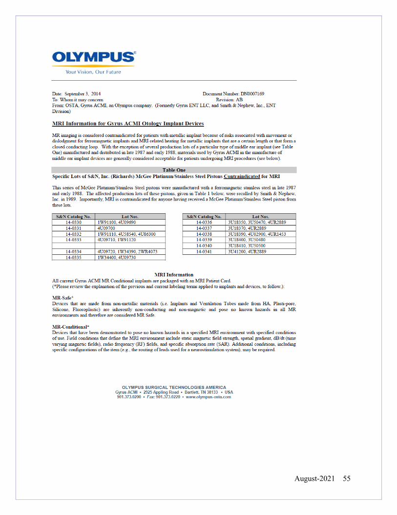
August-2021 55
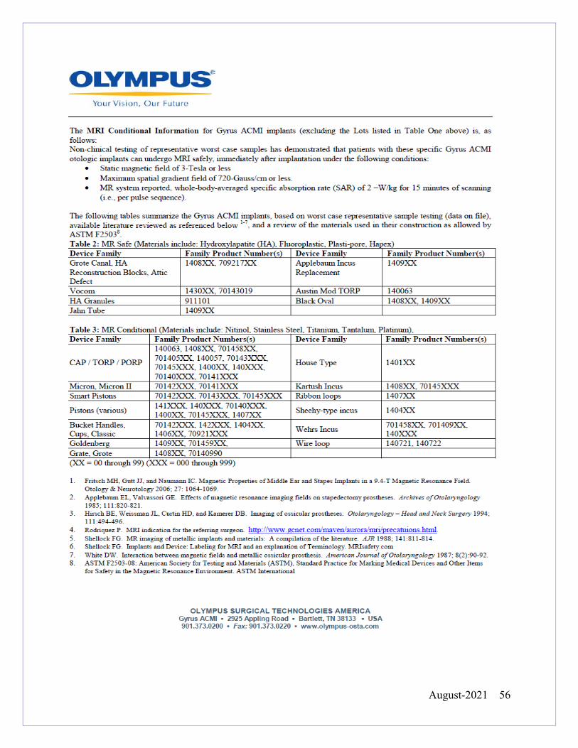
August-2021 56
August-2021 57
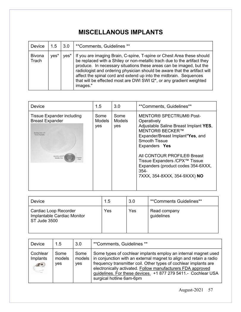
MISCELLANOUS IMPLANTS Device 1.5 3.0 **Comments, Guidelines **
Bivona Trach
yes* yes* If you are imaging Brain, C-spine, T-spine or Chest Area these should be replaced with a Shiley or non-metallic trach due to the artifact they produce. In necessary situations these areas can be imaged, but the radiologist and ordering physician should be aware that the artifact will affect the spinal cord and extend up into the midbrain. Sequences that will be effected most are DWI SWI t2*, or any gradient weighted images."
Device 1.5 3.0 **Comments, Guidelines**
Tissue Expander including Breast Expander
Some Models yes
Some Models yes
MENTOR® SPECTRUM® Post-Operatively Adjustable Saline Breast Implant YES, MENTOR® BECKER™ Expander/Breast Implant*Yes, and Smooth Tissue Expanders Yes All CONTOUR PROFILE® Breast Tissue Expanders /CPX™ Tissue Expanders (product codes 354-6XXX, 354- 7XXX, 354-8XXX, 354-9XXX) NO
Device 1.5 3.0 **Comments Guidelines**
Cardiac Loop Recorder Implantable Cardiac Monitor ST Jude 3500
Yes Yes
Read company guidelines
Device 1.5 3.0 **Comments, Guidelines **
Cochlear Implants
Some models yes
Some models yes
Some types of cochlear implants employ an internal magnet used in conjunction with an external magnet to align and retain a radio frequency transmitter coil. Other types of cochlear implants are electronically activated. Follow manufacturers FDA approved guidelines. For these devices. +1 877 279 5411.- Cochlear USA surgical hotline 6am-6pm
August-2021 58
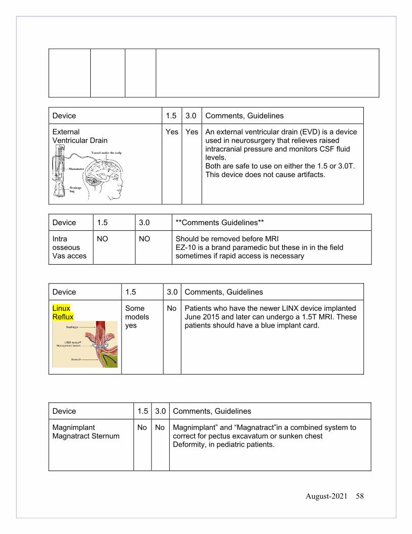
Device 1.5 3.0 Comments, Guidelines
External Ventricular Drain
Yes Yes An external ventricular drain (EVD) is a device used in neurosurgery that relieves raised intracranial pressure and monitors CSF fluid levels. Both are safe to use on either the 1.5 or 3.0T. This device does not cause artifacts.
Device 1.5 3.0 **Comments Guidelines**
Intra osseous Vas acces
NO NO
Should be removed before MRI EZ-10 is a brand paramedic but these in in the field sometimes if rapid access is necessary
Device 1.5 3.0 Comments, Guidelines
Linux Reflux
Some models yes
No Patients who have the newer LINX device implanted June 2015 and later can undergo a 1.5T MRI. These patients should have a blue implant card.
Device 1.5 3.0 Comments, Guidelines
Magnimplant Magnatract Sternum
No No Magnimplant” and “Magnatract”in a combined system to correct for pectus excavatum or sunken chest Deformity, in pediatric patients.
August-2021 59
Device 1.5 3.0 **Comments Guidelines**
Paraguard
Yes Yes
ALL copper IUD, Paraguards can be imaged on 1.5,3T
Device 1.5 3.0 Comments, Guidelines
Perifix Polyamide Catheter
Yes Yes This Catheter is used in epidural procedures. It is made of polyamide nylon and tungeston powder. It has been tested for use on 1.5 and 3 Tesla magnets. All Perfix catheters, gauges and length and tip configurations are considered safe and these strengths
Device 1.5 3.0 **Comments Guidelines**
Reveal XT 9529 DX 9528
Yes Yes
6 week waiting period, Its best practice to send the data collected on the device before the MRI. If an appointment was never set up it is not necessary to delay care. The technologist should continue with the MRI. For more information, contact (800) 742-0884 ask for the representative in the area Check the most up to date Specific MRI instructions on the site below or call 800 505 INFO http://manuals.medtronic.com/manuals/main/us/en_US/home
Device 1.5 3.0 **Comments Guidelines**
Reveal LINQ
Yes Yes
NO waiting period, Its best practice to send the data collected on the device before the MRI. Patients are able do this themselves if they have the MyCareLink Patient Monitor. If the patient doesn’t have it, there is no need to delay care. The technologist should continue with the MRI. For more information, contact (800) 742-0884 ask for the representative in the area Check the most up to date Specific MRI instructions on the site below or call 800 505 INFO http://manuals.medtronic.com/manuals/main/us/en_US/home
August-2021 60
Device 1.5 3.0 **Comments Guidelines**
Scleral Buckle
AFTER WANDING
AFTER WANDING
Tantalum Clips used in scleral buckle surgery are acceptable on 1.5 and 3T Please Wand the orbital area with the ferromagnetic wand.
Device 1.5 3.0 **Comments Guidelines**
Swanz-Ganz Catheters
Some models yes
Some models yes
Some brands are safe for 1.5 and 3T Check the manufacturers FDA approved guidelines Ex. Edwards Life Sciences model numbers Pedi catheter 040F4, 040HF4, 015F4, 015HF4 flow directed catheter 111F7, 114F7, 115F7, 123F6 are SAFE on 1.5 and 3T
Device 1.5 3.0 **Comments Guidelines**
Temperature Foley Catheters
Some models yes
Some models yes
Some brands are conditional for 1.5 and 3T Check the updated manufacturers FDA approved guidelines Ex. All Bard Temp Foleys are OK on 1.5 and 3T. they should be ran straight down the center or the table no loops wires and disconnected from any temp monitoring devices
Device 1.5 3.0 **Comments Guidelines**
X-stop Vertebrae implant
Yes Yes
May cause increased artifact
August-2021 61
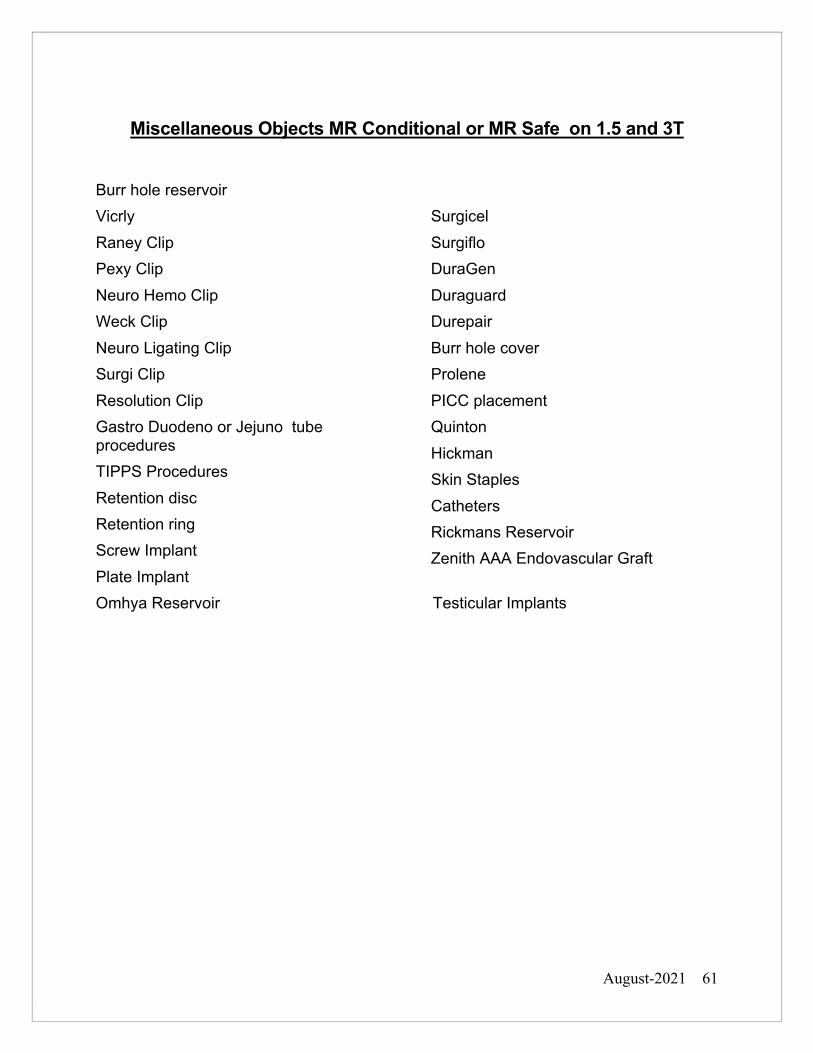
Miscellaneous Objects MR Conditional or MR Safe on 1.5 and 3T
Burr hole reservoirVicrly Raney Clip Pexy Clip Neuro Hemo Clip Weck Clip Neuro Ligating Clip Surgi Clip Resolution Clip Gastro Duodeno or Jejuno tube procedures TIPPS Procedures Retention disc Retention ring Screw Implant Plate Implant
Surgicel Surgiflo DuraGen Duraguard Durepair Burr hole cover Prolene PICC placement Quinton Hickman Skin Staples Catheters Rickmans Reservoir Zenith AAA Endovascular Graft
Omhya Reservoir Testicular Implants
August-2021 62
.
a.) Pregnant Health Care Employees:
All pregnant health care practitioners are permitted to work in and around the MR environment
throughout all stages of their pregnancy. Although permitted to work in and around the MR
environment, pregnant healthcare practitioners are requested not to remain within the MR scanner
room during actual data acquisition.
b.) Pregnant Patients 1.5 and 3T
Pregnant patients may undergo MR scans at any stage of their pregnancy if the referring physician
and the supervising radiologist determine that the risk-benefit ratio to the patient warrants that the
study be performed.
1.) Pregnant patients may be allowed to have an MRI exam on a 3 Tesla magnet, if the area
being imaged is a brain or extremity. Pelvis, abdomen or spine exams should be performed
at 1.5 when imaging at 3T is unlikely to provide additional diagnostic value.
2.) The technologist is required to give the patient an information sheet about MRI and
pregnancy. This should be documented.
3.) Fetal MRI can be performed on a 1.5 or 3T.
YALE-NEW HAVEN HOSPITAL
DEPT. OF DIAGNOSTIC RADIOLOGY
POLICY AND PROCEDURE MANUAL
MRI Safety Manual
Reviewed Date 2021
Last Revision 2020
Policy No.
SUBJECT: PREGNANT MRI HEALTHCARE PROVIDERS AND PREGNANT PATIENTS
August-2021 63
4.) Intravenous gadolinium-based contrast agents should generally NOT be administered for
MRI to pregnant patients. Exceptions may be made at the radiologist discretion, but the
reason for the exception must be documented by the radiologist in the EMR.
c.) Possible Pregnant Patients and NON Contrast
Patients will not be given a urine hCG test unless they are going to receive a gadolinium-based
contrast agent for the MRI exam. In the event the patient is scheduled for a non-contrast exam and
is unsure of pregnancy, the department will assume the patient is pregnant and follow the guidelines
above for pregnant patients.
d.) Pregnant Companions
The companion will be given an information sheet on MRI and pregnancy. It is their choice to stay in
the room during imaging. (Repeated occupational exposure to TVMF is not an issue).
e.) Pre procedure Pregnancy Testing
For Pre procedure Pregnancy Testing please follow the
August-2021 64
Review of the literature shows no evidence to suggest that oral ingestion by an infant of the
tiny amount of gadolinium contrast medium excreted into breast milk would cause toxic
effects [8]. We believe, therefore, that the available data suggest that it is safe for the mother
and infant to continue breast-feeding after receiving such an agent.
If the mother remains concerned about any potential ill effects, she should be given the
opportunity to make an informed decision as to whether to continue or temporarily abstain from
breast-feeding after receiving a gadolinium contrast medium. If the mother so desires, she may
abstain from breast-feeding for 24 hours with active expression and discarding of breast milk
from both breasts during that period. In anticipation of this, she may wish to use a breast pump
to obtain milk before the contrast study to feed the infant during the 24-hour period following
the examination.
References
1. Kubik-Huch RA, Gottstein-Aalame NM, Frenzel T, et al. Gadopentetate dimeglumine excretion into human breast milk
during lactation. Radiology 2000; 216:555-558.
2.. Rofsky NM, Weinreb JC, Litt AW. Quantitative analysis of gadopentetate dimeglumine excreted in breast milk. J Magn Reson Imaging 1993; 3:131-132. 3. Schmiedl U, Maravilla KR, Gerlach R, Dowling CA. Excretion of gadopentetate dimeglumine in human breast milk. AJR
Am J Roentgenol 1990; 154:1305-1306. 4. Weinmann HJ, Brasch RC, Press WR, Wesbey GE. Characteristics of gadolinium-DTPA complex: a potential NMR
contrast agent.
YALE-NEW HAVEN HOSPITAL
DEPT. OF DIAGNOSTIC RADIOLOGY
POLICY AND PROCEDURE MANUAL
MRI Safety Manual
Reviewed Date 2021
Last Revision 2018
Policy No.
SUBJECT: ADMINISTRATION OF CONTRAST TO BREAST FEEDING PATIENTS
August-2021 65
IV Contrast Agents No patient is to be administered MR contrast agents without orders from a licensed
physician. Intravenous injection-qualified MR technologists may start and attend to
peripheral intravenous lines if they have undergone the requisite training.
Injections may be performed through an appropriately sized IV line, which may be removed
after the exam. The IV line will remain in place during the examination should IV drug
therapy be required. This will apply to all patients.
For a patient with a history of contrast reactions please follow the premedication policy. if a
patient experiences a contrast reaction the technologist should immediately contact the
radiologist and nurse so that appropriate action may be taken. An incident report (R/L
solution) is to be filled out via the intranet under Event Reporting. The FDA and the
manufacturer of the contrast should also be contacted.
Oral Contrast
Please note for exams the patient may be asked to drink Breeza oral contrast. There are no significant contraindications for this agent and the agent can be given safely in patients with reported Sulfa Allergies.
YALE-NEW HAVEN HOSPITAL
DEPT. OF DIAGNOSTIC RADIOLOGY
POLICY AND PROCEDURE MANUAL
MRI Safety Manual
Reviewed Date
2021
Last Revision 2018
Policy No.
SUBJECT: IV AND ORAL CONTRAST AGENTS
August-2021 66
The ACR has categorized GBCA agents into groups of risk factors related to Nephrogenic
System Fibrosis (NSF). Dotarem, Gadavist, Prohance and Multihance are Group II agents
and Eovist is a Group III agent. Group I (highest risk) agents are not currently available/on
formulary at YNHH. YNHH patient contrast screening policies has been tailored to the
agents and the potential risk of adverse effect related to low eGFR values and risk of NSF
when given standard weight based dosing. For a complete list of contrast agent groups and
any additional info please visit the ACR website and read the current ACR Manual on
Contrast Media https://www.acr.org/Quality-Safety/Resources/Contrast-Manual.
OUTPATIENTS
Contrast from Group II (such as Dotarem)
Outpatients receiving a GBCA from group II do not need to be screened for eGFR.
Contrast from Group III (Eovist)
Outpatients receiving a GBCA agent from groups III who answer YES to one or more of
the contrast related questions on the MR Safety Screening Form (diabetic, hypertensive,
and renal disease) will be screened for renal function. If an eGFR (or Cr value to calculate
eGFR) is on file < 6 week old this can be used otherwise a value will be obtained day of
exam with Point of Care meter.
YALE-NEW HAVEN HOSPITAL
DEPT. OF DIAGNOSTIC RADIOLOGY
POLICY AND PROCEDURE MANUAL
MRI Safety Manual
Reviewed Date 2021
Last Revision 2018
Policy No.
SUBJECT: Gadolinium and eGFR Screening
August-2021 67
EGFR only required for Eovist or higher than weight based dosing. If valid eGFR is < 30
ml/min/1.73m2 GBCA from group III will be administered only if all of the following
conditions are met;
-Documented approval by the supervising radiologist by assigning/signing an
appropriate protocol
-Informed consent obtained by supervising radiologist (or his/her designated
resident/fellow) and signed by the patient. (Note: Informed consent and reason for
exam should be documented in the report)
INPATIENTS
All inpatient requests for contrast-enhanced MRI exams require the ordering health
professional to answer a series of ordering screen questions designed to identify patients
who might be at risk for NSF. Risk factors include deteriorating renal function.
Contrast from Group II (such as Dotarem)
Inpatients receiving a GBCA from group II do not need to be screened for eGFR.
Contrast from Group III (Eovist)
A documented eGFR > 30 ml/min/1.73m2 obtained within 48 hours prior GBCA
administration is required.
EGFR only required for Eovist or higher than weight based dosing. If valid eGFR is < 30
ml/min/1.73m2 Eovist will be administered only if all of the following conditions are met;
August-2021 68
-Documented approval by the supervising radiologist by assigning and signing an
appropriate protocol
-Informed consent obtained by supervising radiologist (or his/her designated
resident/fellow) and signed by the patient. (Note: Informed consent and reason for
exam should be documented in the report)
Patients on Dialysis or with Acute Kidney Injury (AKI)
If the patient is on dialysis or has known AKI, laboratory testing and calculation of eGFR is
not useful or necessary (i.e., eGFR is not accurate in this setting).
Contrast from Group II
- Patients who are on dialysis or with AKI and receiving GBCA from group II do not need
informed consent given that current data has not shown any unconfounded cases of NSF
with these agents.
- Patients on dialysis should be scheduled for dialysis as close as possible following
conclusion of the MRI exam. Hemodialysis is preferred over peritoneal dialysis whenever
possible due presumed higher efficiency infiltration of GBCAs.
Contrast from Group III (Eovist)
-Patients who are on dialysis or with AKI and receiving GBCA from group III (Eovist) than
informed consent should be obtained.
Off Label Usage
Radiologists commonly use contrast media for a clinical purpose not contained in the FDA
labeling and thus commonly use contrast media off-label. Physicians have latitude in using
August-2021 69
gadolinium chelates off label as guided by clinical indication. This also includes pediatric
usage in patients under the age of 2.
Non-standard Contrast Dosing (ie- double dosing or multiple doses of contrast in 24
hours)
1. Higher than standard weight based dosing
This is occasionally needed for some group II agents is with some MR angiography, brain
MRI, and cardiac MRI applications. In this setting, the Group II agent should be treated like a
group III agent (where there is potential for NSF in at risk populations) as there is limited data
on utilization of GBCA’s with non-standard dosing in this population. The rules of Group III
agents as detailed above would apply in this setting.
2. Receiving more than one GBCA dose in 24 hours.
Occasionally patients need more than one dose of GBCA within a 24-hour period. The NSF
risk of more than one (standard) dose of a group II or III agents in patients is likely negligible
in patients who have eGFR > 30. If eGFR is < 30 (and patient is not on dialysis), then all non-
stat or non-urgent studies should be delayed for 24 hours to allow for adequate clearance. If
on dialysis, dialysis should be performed as soon as possible after the MRI. Therefore, there
is no contraindication to undergoing an urgent or stat contrast enhanced MRI for a patient
who has already been administered one dose of GBCA within a 24-hour period. Truly urgent
or stat studies should not be delayed 24-hours when eGFR <30 if benefits of exam outweigh
NSF risks (radiologist-clinical team joint decision).
August-2021 70
Exceptions regarding Contrast Usage in Patients
Exceptions to the above policies may be made at the discretion of the supervising radiologist,
such as in the rare instance of an acute, life-threatening condition, and after consultation with
the referring health care professional if consent cannot be obtained from the patient or
surrogate when needed. However, the rationale for the exception must be documented by
the supervising radiologist.
Documentation of Contrast Usage
1. MRI technologists will record the specific GBCA and the dose administered to each
patient by annotating the MRI exam and documenting dose in EPIC.
2. The radiologist reporting the exam will include the specific GBCA and dose in their report
for every contrast-enhanced MRI procedure
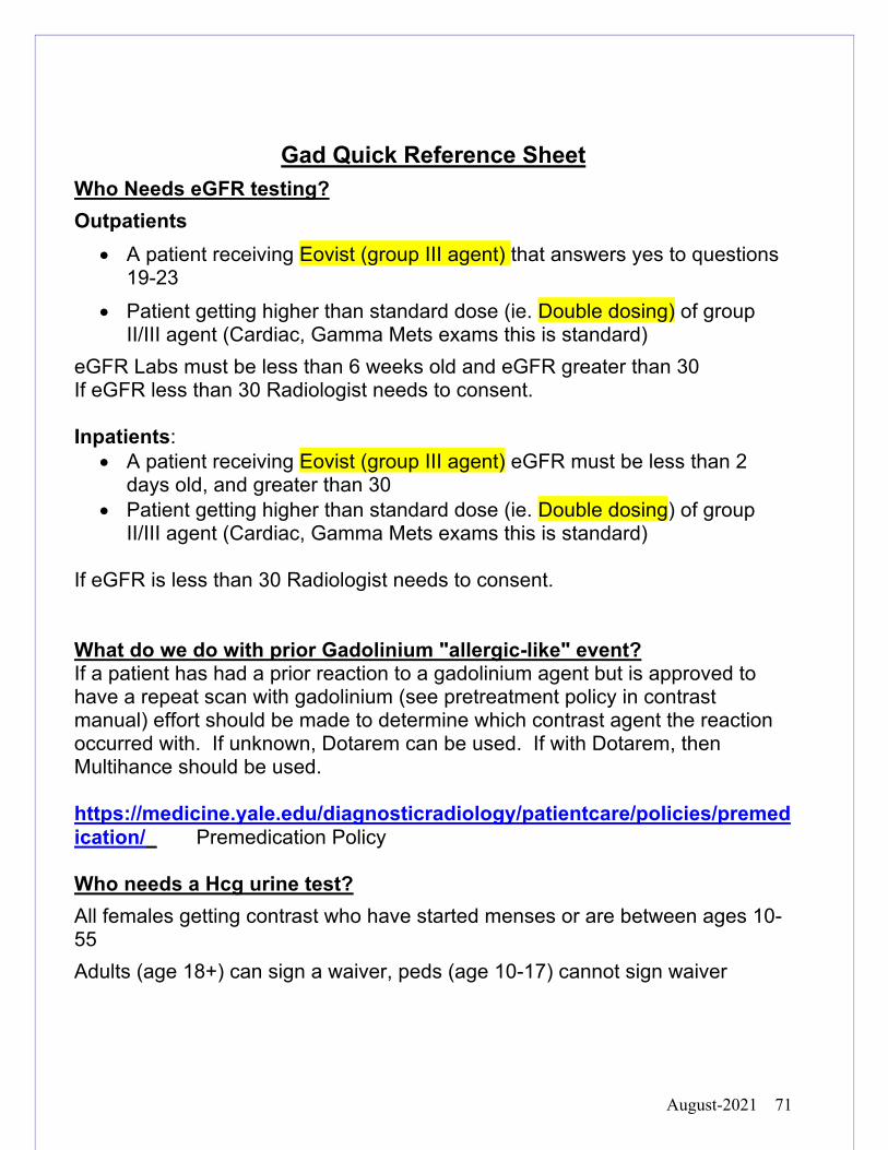
August-2021 71
Gad Quick Reference Sheet Who Needs eGFR testing? Outpatients
• A patient receiving Eovist (group III agent) that answers yes to questions 19-23
• Patient getting higher than standard dose (ie. Double dosing) of group II/III agent (Cardiac, Gamma Mets exams this is standard)
eGFR Labs must be less than 6 weeks old and eGFR greater than 30 If eGFR less than 30 Radiologist needs to consent. Inpatients:
• A patient receiving Eovist (group III agent) eGFR must be less than 2 days old, and greater than 30
• Patient getting higher than standard dose (ie. Double dosing) of group II/III agent (Cardiac, Gamma Mets exams this is standard)
If eGFR is less than 30 Radiologist needs to consent. What do we do with prior Gadolinium "allergic-like" event? If a patient has had a prior reaction to a gadolinium agent but is approved to have a repeat scan with gadolinium (see pretreatment policy in contrast manual) effort should be made to determine which contrast agent the reaction occurred with. If unknown, Dotarem can be used. If with Dotarem, then Multihance should be used. https://medicine.yale.edu/diagnosticradiology/patientcare/policies/premedication/ Premedication Policy Who needs a Hcg urine test? All females getting contrast who have started menses or are between ages 10-55 Adults (age 18+) can sign a waiver, peds (age 10-17) cannot sign waiver
August-2021 72
YALE-NEW HAVEN HOSPITAL DEPT. OF
DIAGNOSTIC RADIOLOGY POLICY AND PROCEDURE MANUAL
MRI Safety Manual
Reviewed Date 2021
Last
Revision 2020
Policy No.
SUBJECT: Gadolinium Dosing Information
I. Purpose To standardize dosing of gadolinium-based contrast agents (GBCAs) in adult and pediatric patients to allow radiology technicians to administer GBCA when ordered by a provider. This protocol will be reviewed annually by the Yale New Haven Health System (YNHHS) Formulary Integration Radiology Subcommittee, local site Pharmacy and Therapeutics Committees and YNHHS Formulary Integration Committee.
II. Background Gadolinium-based contrast agents (GBCAs) are intravenous drugs used in diagnostic imaging procedures to enhance the quality of magnetic resonance imaging (MRI) or magnetic resonance angiography (MRA).
III. Patient Population Adult and pediatric patients undergoing MRI procedures.
IV. Procedure Provider shall order MRI procedure. Radiologist receiving procedure order will protocol the MRI. Once the procedure is protocoled by the Radiologist, the radiology technician will follow the Dosing Protocol outlined in the table below. All dosing will be in mL/kg.
REFERENCES:
1. Dotarem® (gadoterate meglumine) [prescribing information]. Bloomington, IN: Guerbet LLC; July 2016
American College of Radiology Committee on Drugs and Contrast Media. Manual on Contrast
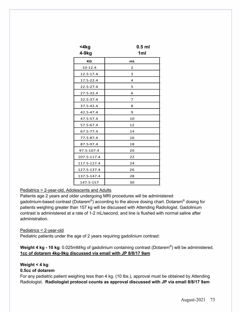
August-2021 73
<4kg 0.5 ml 4-9kg 1ml
Pediatrics > 2-year-old, Adolescents and Adults Patients age 2 years and older undergoing MRI procedures will be administered gadolinium-based contrast (Dotarem®) according to the above dosing chart. Dotarem® dosing for patients weighing greater than 157 kg will be discussed with Attending Radiologist. Gadolinium contrast is administered at a rate of 1-2 mL/second, and line is flushed with normal saline after administration. Pediatrics < 2-year-old Pediatric patients under the age of 2 years requiring gadolinium contrast: Weight 4 kg - 10 kg: 0.025mM/kg of gadolinium containing contrast (Dotarem®) will be administered. 1cc of dotarem 4kg-9kg discussed via email with JP 8/8/17 9am Weight < 4 kg: 0.5cc of dotarem For any pediatric patient weighing less than 4 kg. (10 lbs.), approval must be obtained by Attending Radiologist. Radiologist protocol counts as approval discussed with JP via email 8/8/17 9am
KG mL
10-12.4 2
12.5-17.4 3
17.5-22.4 4
22.5-27.4 5
27.5-32.4 6
32.5-37.4 7
37.5-42.4 8
42.5-47.4 9
47.5-57.4 10
57.5-67.4 12
67.5-77.4 14
77.5-87.4 16
87.5-97.4 18
97.5-107.4 20
107.5-117.4 22
117.5-127.4 24
127.5-137.4 26
137.5-147.4 28
147.5-157 30
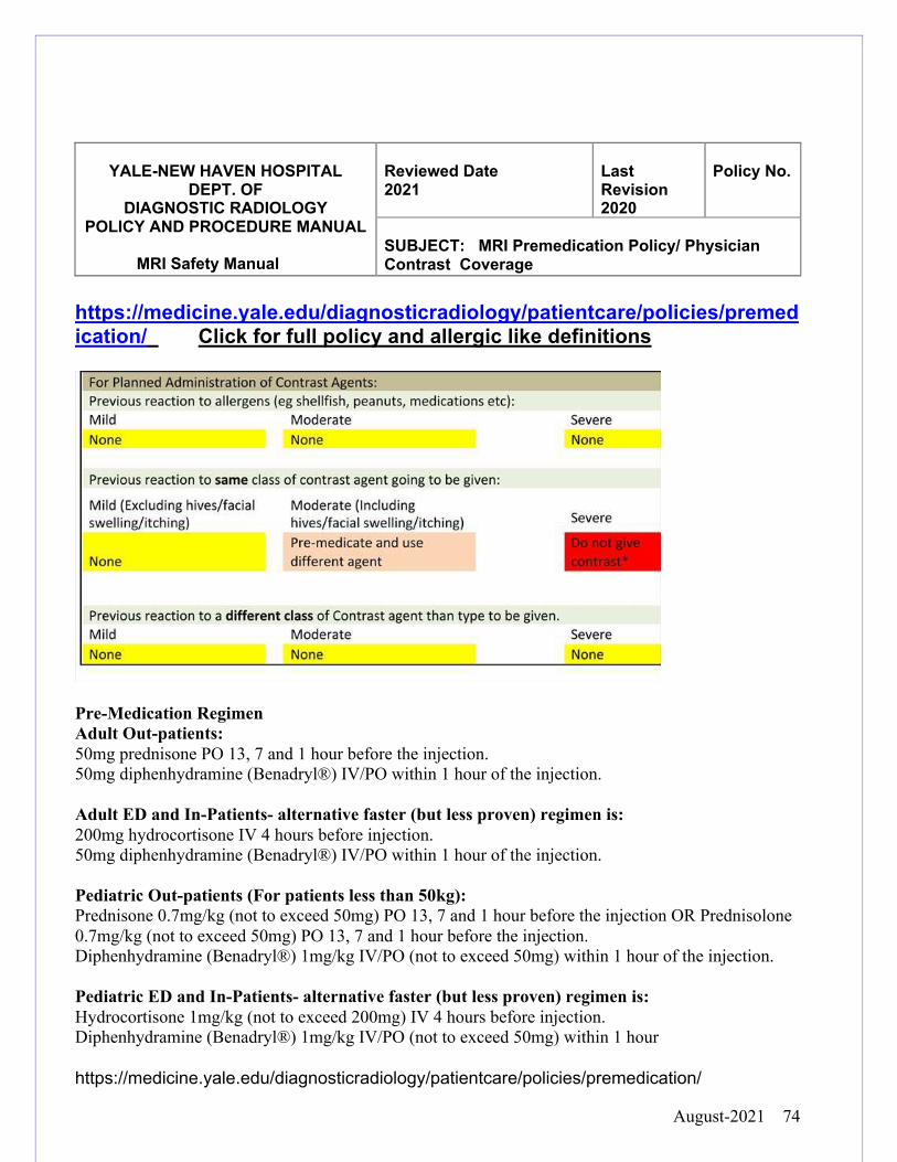
August-2021 74
YALE-NEW HAVEN HOSPITAL
DEPT. OF DIAGNOSTIC RADIOLOGY
POLICY AND PROCEDURE MANUAL
MRI Safety Manual
Reviewed Date 2021
Last Revision 2020
Policy No.
SUBJECT: MRI Premedication Policy/ Physician Contrast Coverage
https://medicine.yale.edu/diagnosticradiology/patientcare/policies/premedication/ Click for full policy and allergic like definitions
Pre-Medication Regimen Adult Out-patients: 50mg prednisone PO 13, 7 and 1 hour before the injection. 50mg diphenhydramine (Benadryl®) IV/PO within 1 hour of the injection. Adult ED and In-Patients- alternative faster (but less proven) regimen is: 200mg hydrocortisone IV 4 hours before injection. 50mg diphenhydramine (Benadryl®) IV/PO within 1 hour of the injection. Pediatric Out-patients (For patients less than 50kg): Prednisone 0.7mg/kg (not to exceed 50mg) PO 13, 7 and 1 hour before the injection OR Prednisolone 0.7mg/kg (not to exceed 50mg) PO 13, 7 and 1 hour before the injection. Diphenhydramine (Benadryl®) 1mg/kg IV/PO (not to exceed 50mg) within 1 hour of the injection. Pediatric ED and In-Patients- alternative faster (but less proven) regimen is: Hydrocortisone 1mg/kg (not to exceed 200mg) IV 4 hours before injection. Diphenhydramine (Benadryl®) 1mg/kg IV/PO (not to exceed 50mg) within 1 hour
https://medicine.yale.edu/diagnosticradiology/patientcare/policies/premedication/
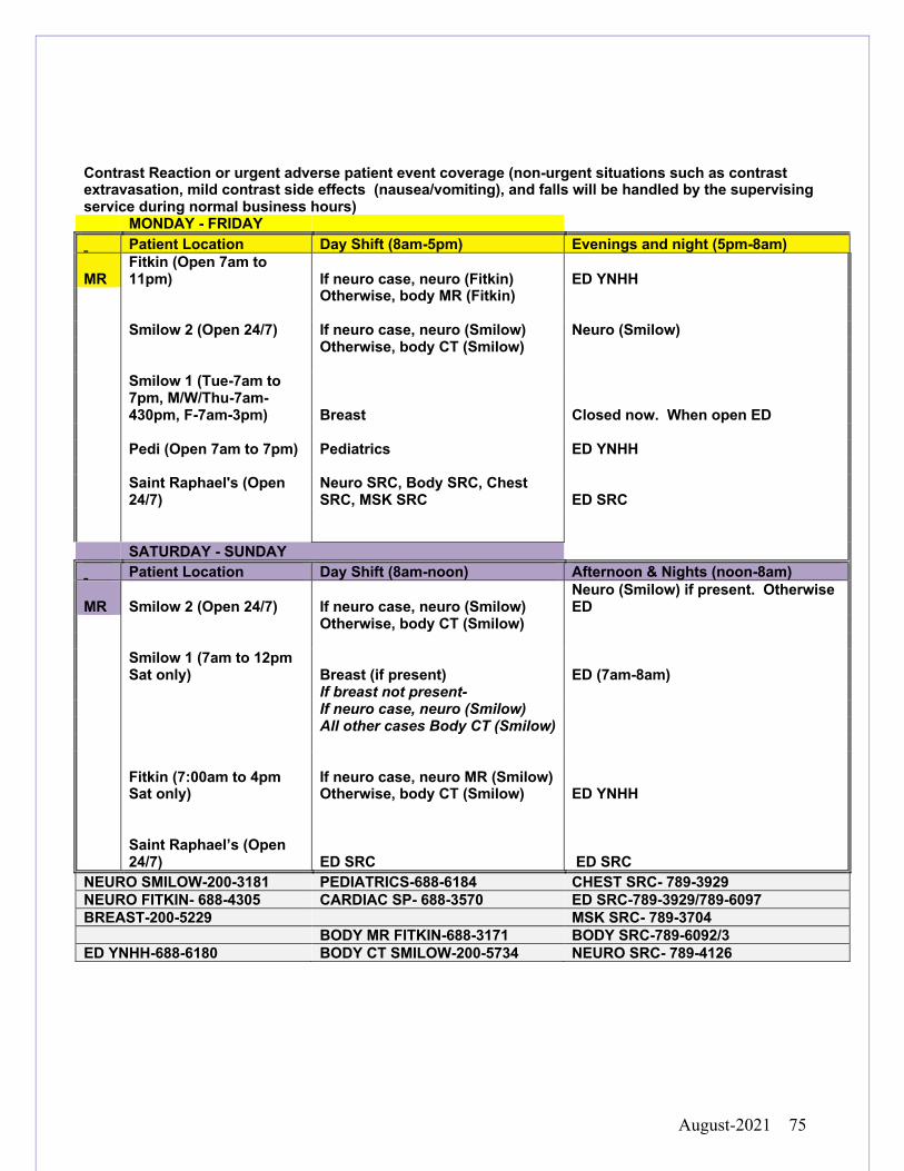
August-2021 75
Contrast Reaction or urgent adverse patient event coverage (non-urgent situations such as contrast extravasation, mild contrast side effects (nausea/vomiting), and falls will be handled by the supervising service during normal business hours) MONDAY - FRIDAY Patient Location Day Shift (8am-5pm) Evenings and night (5pm-8am)
MR Fitkin (Open 7am to 11pm) If neuro case, neuro (Fitkin) ED YNHH
Otherwise, body MR (Fitkin) Smilow 2 (Open 24/7) If neuro case, neuro (Smilow) Neuro (Smilow) Otherwise, body CT (Smilow)
Smilow 1 (Tue-7am to 7pm, M/W/Thu-7am-430pm, F-7am-3pm) Breast Closed now. When open ED
Pedi (Open 7am to 7pm) Pediatrics ED YNHH
Saint Raphael's (Open 24/7)
Neuro SRC, Body SRC, Chest SRC, MSK SRC ED SRC
SATURDAY - SUNDAY Patient Location Day Shift (8am-noon) Afternoon & Nights (noon-8am)
MR Smilow 2 (Open 24/7) If neuro case, neuro (Smilow) Neuro (Smilow) if present. Otherwise ED
Otherwise, body CT (Smilow)
Smilow 1 (7am to 12pm Sat only) Breast (if present) ED (7am-8am)
If breast not present- If neuro case, neuro (Smilow) All other cases Body CT (Smilow)
Fitkin (7:00am to 4pm Sat only)
If neuro case, neuro MR (Smilow) Otherwise, body CT (Smilow) ED YNHH
Saint Raphael’s (Open 24/7) ED SRC ED SRC
NEURO SMILOW-200-3181 PEDIATRICS-688-6184 CHEST SRC- 789-3929 NEURO FITKIN- 688-4305 CARDIAC SP- 688-3570 ED SRC-789-3929/789-6097 BREAST-200-5229 MSK SRC- 789-3704 BODY MR FITKIN-688-3171 BODY SRC-789-6092/3 ED YNHH-688-6180 BODY CT SMILOW-200-5734 NEURO SRC- 789-4126
August-2021 76
ED patients requiring an MRI will receive priority over most inpatient orders. If there are no
available openings on the schedule, the Radiologist will look at the clinical needs of the
inpatients scheduled and decide which patient to reschedule to accommodate the most
emergent case.
There are multiple levels of “Stat” Ordered exams: Life-Threatening and Urgent:
A Stat patient that is considered Life Threatening will be scheduled within 30 minutes of
receipt of the order and the completed safety form. Scanning should begin within 2 hours.
A Stat patient that is considered Urgent will be scheduled within 30 minutes of receipt of the
order and the completed safety form. Scanning should begin within 6 hours.
To schedule an ED MRI two important steps must be done before the patient will be given a
scheduled time:
1. The order must be placed in EPIC
2. The inpatient screening process must be followed with completion of the MRI safety form.
The next available time slot will be given to the ED requested patient and a call will be placed to the ED to alert them of the time. Patient transportation appointment will be scheduled in advance of scan time
YALE-NEW HAVEN HOSPITAL
DEPT. OF DIAGNOSTIC RADIOLOGY
POLICY AND PROCEDURE MANUAL
MRI Safety Manual
Reviewed Date 2021
Last Revision 2018
Policy No.
SUBJECT: MRI EMERGENCY DEPARTMENT and STAT REQUESTED EXAMS
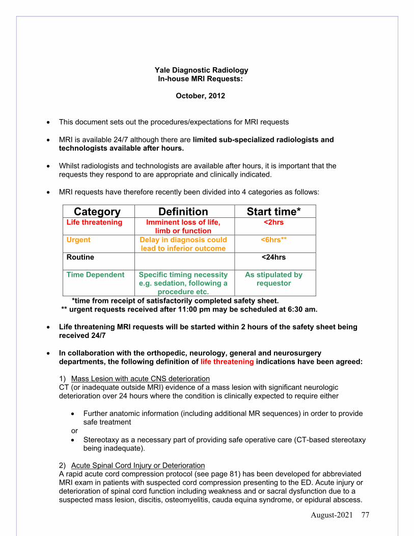
August-2021 77
Yale Diagnostic Radiology In-house MRI Requests:
October, 2012
• This document sets out the procedures/expectations for MRI requests • MRI is available 24/7 although there are limited sub-specialized radiologists and
technologists available after hours. • Whilst radiologists and technologists are available after hours, it is important that the
requests they respond to are appropriate and clinically indicated.
• MRI requests have therefore recently been divided into 4 categories as follows:
Category Definition Start time* Life threatening Imminent loss of life,
limb or function <2hrs
Urgent
Delay in diagnosis could lead to inferior outcome
<6hrs**
Routine
<24hrs
Time Dependent
Specific timing necessity e.g. sedation, following a
procedure etc.
As stipulated by requestor
*time from receipt of satisfactorily completed safety sheet. ** urgent requests received after 11:00 pm may be scheduled at 6:30 am. • Life threatening MRI requests will be started within 2 hours of the safety sheet being
received 24/7
• In collaboration with the orthopedic, neurology, general and neurosurgery departments, the following definition of life threatening indications have been agreed:
1) Mass Lesion with acute CNS deterioration CT (or inadequate outside MRI) evidence of a mass lesion with significant neurologic deterioration over 24 hours where the condition is clinically expected to require either
• Further anatomic information (including additional MR sequences) in order to provide safe treatment
or • Stereotaxy as a necessary part of providing safe operative care (CT-based stereotaxy
being inadequate). 2) Acute Spinal Cord Injury or Deterioration A rapid acute cord compression protocol (see page 81) has been developed for abbreviated MRI exam in patients with suspected cord compression presenting to the ED. Acute injury or deterioration of spinal cord function including weakness and or sacral dysfunction due to a suspected mass lesion, discitis, osteomyelitis, cauda equina syndrome, or epidural abscess.
August-2021 78
• Acute deterioration must have occurred over a short enough time such that rapid
treatment is likely to result in restoration of function. • A likely diagnosis of radiculopathy, sensory loss-only or fixed severe
deficits of >48hrs duration is NOT deemed life threatening. 3) Unstable Spine Correction For planning an unstable spine correction, particularly when an occult soft tissue component may compress neural elements upon deformity correction.
• Neurologic exam may be intact. e.g. bilateral jumped facets to rule out disc herniation.
4) Stroke, if:
1) Diagnosis is uncertain.
2) Time of onset is unclear. (MR perfusion/diffusion in attempt to define ischemic penumbra prior to intravenous lytics/catheter based intervention)
3) Patient has a contrast allergy and needs angiography.
4) Patient is of pediatric age group.
5) Acute Aortic Dissection, if there is a contraindication to iodinated contrast. 6) Acute Appendicitis in Pregnancy, if the general surgery attending consult deems the
patient’s condition to be life-threatening.
August-2021 79
Yale New Haven Hospital
Stat MRI procedure for Acute Stroke Evaluation
Selected acute stroke patients will undergo a stat MRI procedure to further assess cerebral ischemia and possible candidacy for thrombolysis or neuro-intervention. Indications include:
1. Patients with a suspected stroke syndrome (NIHSS > 10)
2. Patients who present with an uncertain time of symptom onset
3. Patients with an uncertain diagnosis
4. Pediatric patients
5. Patients being considered for CTA but with a known contrast dye allergy
6. Patients suspected to be pregnant
1. The acute stroke team (resident/NP/attending) will enter the order (MRI - brain w and w/o contrast), It is the responsibility of the referring clinician to complete the on-line MRI safety sheet. Assistance will be provided by radiology to determine the presence of cochlear implants, orbital metal, pacemakers etc. from previous imaging.
2. The acute stroke team (resident/NP/attending) will then contact the neuroradiology fellow on-call at 203-200-3181 to alert him/her of the acute stroke patient and MRI order.
3. The neuroradiology fellow will alert the MRI tech supervisor who will check for the completed
safety sheet, schedule the case and notify the neuroradiology fellow to protocol the case.
4. Once the case is protocolled the MRI tech supervisor will call the acute stroke team at 203-688-7111 to accompany the patient to the MRI prep hold area. The acute stroke team will remain with the patient at MRI. The only exceptions to this would be if the patient is not found to have an acute stroke, and there are no hemodynamic or respiratory concerns, in which case the patient could be transported back to the ED/floor by standard transport.
• Hyperacute Stroke Protocol includes: Ax DWI, Ax FLAIR and Ax SWI sequences. Ax EPI perfusion will be added if the patient has separate IV access for gadolinium Approved by Stroke Center and Dept. of Radiology: 4.10; 12.10; 05.11; 12.12; 03.13;
August-2021 80
YALE-NEW HAVEN HOSPITAL
DEPT. OF DIAGNOSTIC RADIOLOGY
POLICY AND PROCEDURE MANUAL
MRI Safety Manual
Reviewed Date 2021
Last Revision 2020
Policy No.
SUBJECT: SRC ON CALL PROCESS and OVERNIGHT PROCESS
Process for overnight MR Cases:
1. The clinician places the order in EPIC, MR staff, Nurse or Clinician obtains and completes the MRI safety sheet via Encounters in EPIC.
2. The patient must be escorted to the MR Suite by an MD, PA, or RN, for patients that require monitoring.
Calling in the MRI tech for a case at SRC when a technologist in no longer in house. Technologist on call contact info should NEVER be given to a non-radiologist. Only a radiologist can initiate calling in the on-call MRI technologist.
1. Verify:
(a) This is a truly urgent case that needs to be imaged off hours (b) Ask the ordering physician/team to fill out the MRI safety sheet (c) Protocol the case and call the on-call technologist to let them know you approved
and protocoled the case. (d) For life threatening cases (such as acute cord compression or stroke) the
technologist will come to the hospital once notified of the case. For non-life threatening cases the on call technologist will wait until the safety sheet is completed and cleared before heading to the hospital.
2. Coverage and contact numbers for SRC is detailed below.
SRC is staffed Monday – Friday from 7am – 11pm and Saturday, Sunday 7am – 3:30pm All other times follow on-call details below
Monday – Thursdays: 11pm – 7am
Call Smilow MRI Techs for coverage at 203-200-5144
Fridays 11pm – Saturdays 7am
Page On-call Tech at 203-412-4230 or 203-412-0603
Saturdays 3:30pm – Sundays 7am * then again * Sunday 330pm until 10pm
Page On-call Tech at 203-412-4230 or 203-412-0603
August-2021 81
YALE-NEW HAVEN HOSPITAL DEPT. OF
DIAGNOSTIC RADIOLOGY POLICY AND PROCEDURE MANUAL
MRI Safety Manual
Reviewed Date 2020
Last Revision 2018
Policy No.
SUBJECT: Acute Cord Compression
Acute Cord Compression MRI Protocol The acute cord compression protocol has been designed to allow for rapid imaging of high risk patients presenting with new or worsening symptoms, of under 48 hours duration, suspected to have acute cord compression or cauda equina syndrome. The protocol was developed by Radiology in conjunction with MRI Operations, Spine Surgery and Emergency Medicine, to rapidly diagnose or exclude cord compression. Other neurologic conditions that can potentially mimic cord compression symptoms will not be well assessed by this rapid protocol, and the patient may need to come back for repeat imaging in those cases.
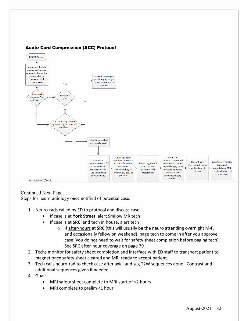
August-2021 82
Continued Next Page… Steps for neuroradiology once notified of potential case:
1. Neuro-rads called by ED to protocol and discuss case: • If case is at York Street, alert Smilow MR tech • If case is at SRC, and tech in house, alert tech
o If after-hours at SRC (this will usually be the neuro attending overnight M-F, and occasionally fellow on weekend), page tech to come in after you approve case (you do not need to wait for safety sheet completion before paging tech). See SRC after-hour coverage on page 79
2. Techs monitor for safety sheet completion and interface with ED staff to transport patient to magnet once safety sheet cleared and MRI ready to accept patient.
3. Tech calls neuro-rad to check case after axial and sag T2W sequences done. Contrast and additional sequences given if needed.
4. Goal: • MRI safety sheet complete to MRI start of <2 hours • MRI complete to prelim <1 hour
August-2021 83
YALE-NEW HAVEN HOSPITAL
DEPT. OF DIAGNOSTIC RADIOLOGY
POLICY AND PROCEDURE MANUAL MRI Safety Manual
Reviewed Date 2021
Last Revision 2018
Policy No.
SUBJECT Magnet Room Cleaning
Cleaning of the MRI suite to include the table, pads, coils, and the inside of the magnet bore is performed by the MRI staff to prevent the transmission of infections. Routine cleaning personnel are not allowed to enter the MRI suite because of the dangerous magnetic field strength that can cause potential catastrophic events harming personnel and the MRI system.
1. Gloves must always be worn when handling contaminated equipment and working with a cleaning disinfectant.
2. Cleaning of the table and pads is performed before and after each patient exam with a hospital approved disinfectant and all cleaning equipment must be MRI safe.
3. Periodic inspection of pads for fraying and tearing is done each month and replaced as necessary.
4. Patient contact inside the magnet bore of the MR unit can transmit infection so cleaning requires an MRI staff member to travel on the table inside the bore to sanitize and disinfect the tunnel walls.
5. MRI magnet room cleaning schedule is available in your area
August-2021 84
YALE-NEW HAVEN HOSPITAL
DEPT. OF DIAGNOSTIC RADIOLOGY POLICY AND PROCEDURE
MANUAL
MRI Safety Manual
Reviewed Date 2021
Last Revision 2018
Policy No.
SUBJECT: LATEX ALLERGIES IN MRI
If a patient has a latex allergy it should be documented in the patient’s chart. Latex allergic
patient’s sensitivity may vary from a reaction only triggered by actual contact with a latex
product to a severe reaction from airborne particles.
The MRI suites at YNHH Main Campus are latex free, accept for the Medrad endo-rectal coil.
(All Invivo Products, O2 Tubing, suction catheters, gloves, Siemens Call ball are latex free)
For more information, please see the Clinical Practice Manual Latex Precautions and Latex Allergies
August-2021 85
YALE-NEW HAVEN HOSPITAL
DEPT. OF DIAGNOSTIC RADIOLOGY
POLICY AND PROCEDURE MANUAL
MRI Safety Manual
Reviewed Date 2020
Last Revision 2018
Policy No.
SUBJECT: PATIENT MONITORING
MONITORING BY NURSING STAFF: CRITICAL CARE PATIENTS
This policy conforms fundamentally with the policy developed jointly by the Society of Critical
Care Medicine and the American Association of Critical Care Nurses. Patients must be
transported with the same level of monitoring required on the sending unit.
It is the responsibility of the health care team on the sending unit to assess the stability of the
patient and assure personnel required for safe transportation.
• All ICU patients are to be transported to diagnostic procedures with a minimum of a
RN and transporter
• All patients requiring cardiac monitoring are to be transported with a minimum of a RN
and transporter
• Patients receiving blood or blood products must be accompanied by an RN
• Patients requiring restraints must be accompanied by an RN or PCA
• Any unstable patient who requires or is at risk for requiring acute intervention beyond
the scope of nursing practice must be accompanied by an MD/LIP
• Non critical care personnel may provide transport for ICU patients deemed
appropriate to travel without an RN or MD support. These patients must be
transported without cardiac monitoring and a MD/LIP order is required indicating the
patient may travel without cardiorespiratory monitoring
August-2021 86
MONITORING BY MRI STAFF FOR THERMAL INJURY
Monitoring of patients is necessary in the MR scanner. The potential for thermal injury from
possibly excessive radio frequency power deposition exists. Sedated, anesthetized and/or
unconscious patients may not be able to express symptoms of such injury. Patients who
require EKG monitoring and who are unconscious, sedated and/or anesthetized should be
examined before scanning and after repositioning to ensure that MR safe EKG leads and
any other electrically conductive material is not in contact with the patient or coiled so as to
induce a current. For more on thermal injuries please refer to page 17.
August-2021 87
The location of the Code Carts should be known by all MRI staff members. These will be checked daily by assigned MR staff for expired medications and functioning equipment.
1. It should be stressed that the magnetic field is Always ON. As in any emergency; it is
the responsibility of appropriately trained and knowledgeable MRI personnel to ensure
the safety of all non MRI personnel as well as that of patients and family.
2. In the event of a code:
YNHH Main Campus:
• Outpatients call 155 for outpatients, and say: Code blue for an adult, Code white for a
pediatric.
• Inpatients, 155
• North Haven Devine St call 911
• St Raphael Medical Campus call 155
• Shoreline Medical Center call 911
• Park Avenue/Trumbull. Call 911 and page overhead for help.
Give the appropriate location:
3. The patient will be removed from the scan room (zone 4) immediately and the scan
room door closed and locked. MR staff members/nursing will transfer patient to
nearest recovery area, get the code cart and start basic life support until the code
YALE-NEW HAVEN HOSPITAL
DEPT. OF DIAGNOSTIC RADIOLOGY
POLICY AND PROCEDURE MANUAL
MRI Safety Manual
Reviewed Date 2021
Last Revision 2018
Policy No.
SUBJECT: CODE PROCEDURE / CODE CART
August-2021 88
team arrives. Full resuscitative measures should not be undertaken in the scanner
room (Zone 4).
4. Other MRI personnel can offer assistance; such as direct the code team to the right
location
5. A radiologist should be notified that a code has been called if possible.
6. While the code is in progress it is imperative that all scanner doors be closed and
monitored by MRI personnel to prevent any accidental entry which could result in
injury.
7. When the code team arrives they are responsible for the patient. MRI personnel will
maintain the safety of all staff in the magnetic environment.
8. After the code it is the responsibility of the MRI staff to call the pharmacy so that the
code cart can be restocked as soon as possible.
Workflow for removing sedated and routine MRI Patients from zone 4
Continued on the Next Page
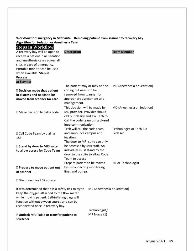
August-2021 89
Workflow for Emergency in MRI Suite – Removing patient from scanner to recovery bay. Algorithm for Sedation or Anesthesia Case StepsinWorkflowA recovery bay will be open to receive a patient in all sedation and anesthesia cases across all sites in case of emergency. Portable monitor can be used when available. Step in Process
Description Team Member
In Scanner � Decision made that patient in distress and needs to be moved from scanner for care
The patient may or may not be coding but needs to be removed from scanner for appropriate assessment and management.
MD (Anesthesia or Sedation)
� Make decision to call a code
This decision will be made by MD provider. Provider should call out clearly and ask Tech to Call the code team using closed loop communication.
MD (Anesthesia or Sedation)
� Call Code Team by dialing 155 � Stand by door to MRI suite to allow access for Code Team
Tech will call the code team and announce campus and location The door to MRI suite can only be accessed by MRI staff. An individual must stand by the door to the suite to allow Code Team to access
Technologist or Tech Aid Tech Aid
� Prepare to move patient out of scanner � Disconnect wall 02 source
Prepare patient to be moved by disconnecting monitoring lines and pumps.
RN or Technologist
It was determined that it is a safety risk to try to keep the oxygen attached to the flow meter while moving patient. Self-inflating bags will function without oxygen source and can be reconnected once in recovery bay.
MD (Anesthesia or Sedation)
� Undock MRI Table or transfer patient to stretcher
Technologist/ MR Nurse (1)
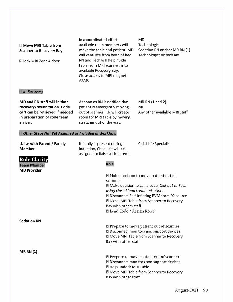
August-2021 90
� Move MRI Table from Scanner to Recovery Bay � Lock MRI Zone 4 door
In a coordinated effort, available team members will move the table and patient. MD will ventilate from head of bed. RN and Tech will help guide table from MRI scanner, into available Recovery Bay. Close access to MRI magnet ASAP.
MD Technologist Sedation RN and/or MR RN (1) Technologist or tech aid
� In Recovery MD and RN staff will initiate recovery/resuscitation. Code cart can be retrieved if needed in preparation of code team arrival.
As soon as RN is notified that patient is emergently moving out of scanner, RN will create room for MRI table by moving stretcher out of the way.
MR RN (1 and 2) MD Any other available MRI staff
� Other Steps Not Yet Assigned or Included in Workflow Liaise with Parent / Family Member
If family is present during induction, Child Life will be assigned to liaise with parent.
Child Life Specialist
RoleClarityTeam Member
Role
MD Provider �Makedecisiontomovepatientoutofscanner�Make decision to call a code. Call-out to Tech using closed loop communication. �Disconnect Self-Inflating BVM from 02 source �Move MRI Table from Scanner to Recovery Bay with others staff �LeadCode/AssignRoles
Sedation RN �Preparetomovepatientoutofscanner�Disconnect monitors and support devices �Move MRI Table from Scanner to Recovery Bay with other staff
MR RN (1) �Preparetomovepatientoutofscanner�Disconnect monitors and support devices �Help undock MRI Table �Move MRI Table from Scanner to Recovery Bay with other staff
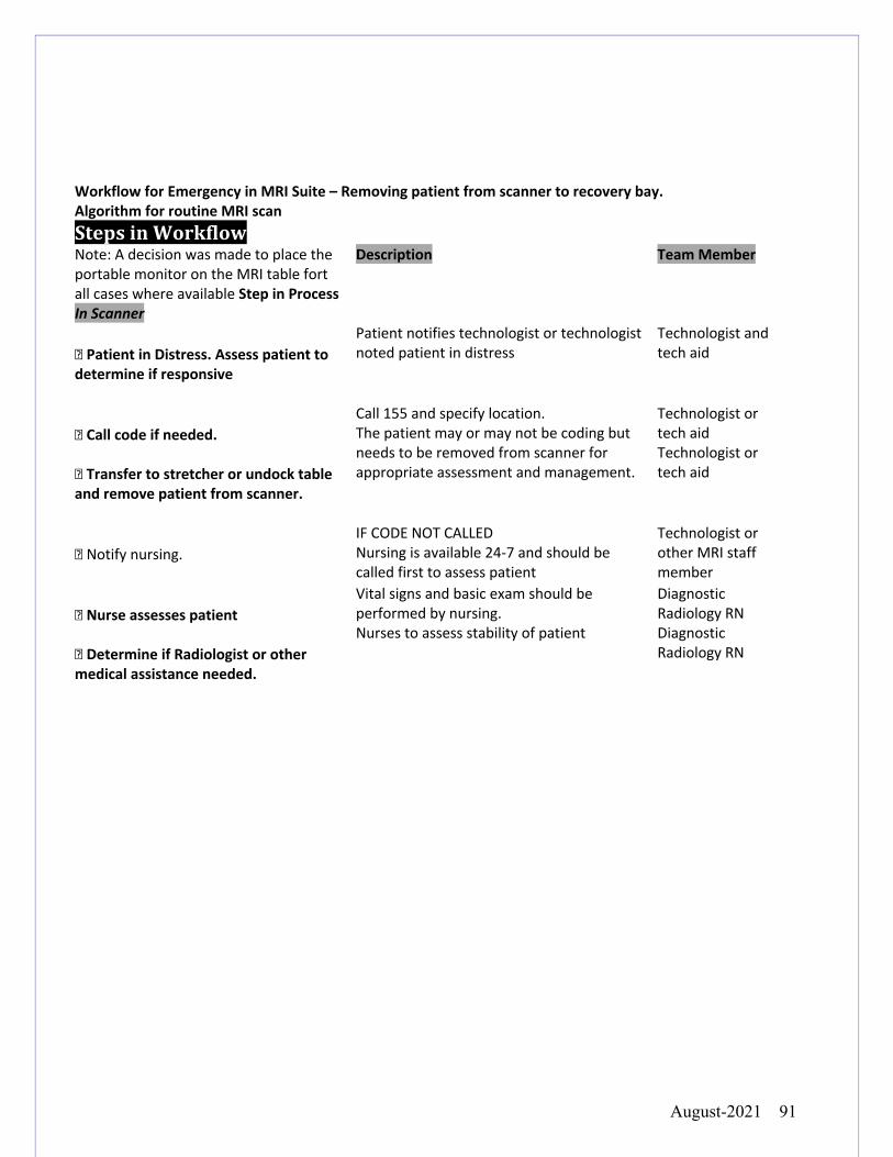
August-2021 91
Workflow for Emergency in MRI Suite – Removing patient from scanner to recovery bay. Algorithm for routine MRI scan StepsinWorkflowNote: A decision was made to place the portable monitor on the MRI table fort all cases where available Step in Process
Description Team Member
In Scanner � Patient in Distress. Assess patient to determine if responsive
Patient notifies technologist or technologist noted patient in distress
Technologist and tech aid
� Call code if needed. � Transfer to stretcher or undock table and remove patient from scanner.
Call 155 and specify location. The patient may or may not be coding but needs to be removed from scanner for appropriate assessment and management.
Technologist or tech aid Technologist or tech aid
� Notify nursing.
IF CODE NOT CALLED Nursing is available 24-7 and should be called first to assess patient
Technologist or other MRI staff member
� Nurse assesses patient � Determine if Radiologist or other medical assistance needed.
Vital signs and basic exam should be performed by nursing. Nurses to assess stability of patient
Diagnostic Radiology RN Diagnostic Radiology RN
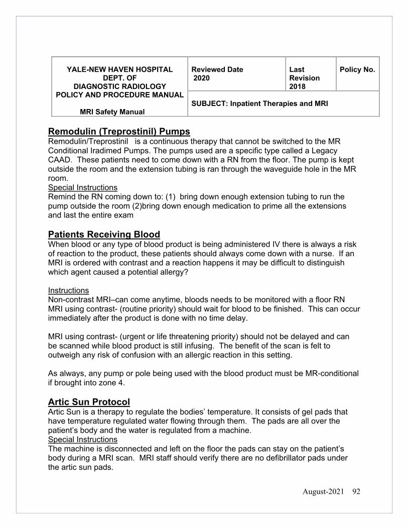
August-2021 92
YALE-NEW HAVEN HOSPITAL DEPT. OF
DIAGNOSTIC RADIOLOGY POLICY AND PROCEDURE MANUAL
MRI Safety Manual
Reviewed Date 2020
Last Revision 2018
Policy No.
SUBJECT: Inpatient Therapies and MRI
Remodulin (Treprostinil) Pumps Remodulin/Treprostinil is a continuous therapy that cannot be switched to the MR Conditional Iradimed Pumps. The pumps used are a specific type called a Legacy CAAD. These patients need to come down with a RN from the floor. The pump is kept outside the room and the extension tubing is ran through the waveguide hole in the MR room. Special Instructions Remind the RN coming down to: (1) bring down enough extension tubing to run the pump outside the room (2)bring down enough medication to prime all the extensions and last the entire exam Patients Receiving Blood When blood or any type of blood product is being administered IV there is always a risk of reaction to the product, these patients should always come down with a nurse. If an MRI is ordered with contrast and a reaction happens it may be difficult to distinguish which agent caused a potential allergy? Instructions Non-contrast MRI–can come anytime, bloods needs to be monitored with a floor RN MRI using contrast- (routine priority) should wait for blood to be finished. This can occur immediately after the product is done with no time delay. MRI using contrast- (urgent or life threatening priority) should not be delayed and can be scanned while blood product is still infusing. The benefit of the scan is felt to outweigh any risk of confusion with an allergic reaction in this setting. As always, any pump or pole being used with the blood product must be MR-conditional if brought into zone 4. Artic Sun Protocol Artic Sun is a therapy to regulate the bodies’ temperature. It consists of gel pads that have temperature regulated water flowing through them. The pads are all over the patient’s body and the water is regulated from a machine. Special Instructions The machine is disconnected and left on the floor the pads can stay on the patient’s body during a MRI scan. MRI staff should verify there are no defibrillator pads under the artic sun pads.
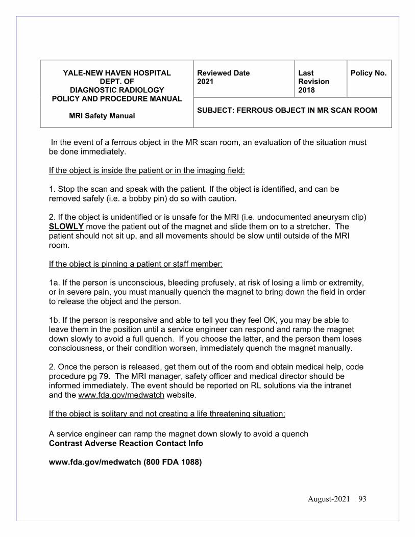
August-2021 93
YALE-NEW HAVEN HOSPITAL DEPT. OF
DIAGNOSTIC RADIOLOGY POLICY AND PROCEDURE MANUAL
MRI Safety Manual
Reviewed Date 2021
Last Revision 2018
Policy No.
SUBJECT: FERROUS OBJECT IN MR SCAN ROOM
In the event of a ferrous object in the MR scan room, an evaluation of the situation must be done immediately. If the object is inside the patient or in the imaging field: 1. Stop the scan and speak with the patient. If the object is identified, and can be removed safely (i.e. a bobby pin) do so with caution. 2. If the object is unidentified or is unsafe for the MRI (i.e. undocumented aneurysm clip) SLOWLY move the patient out of the magnet and slide them on to a stretcher. The patient should not sit up, and all movements should be slow until outside of the MRI room. If the object is pinning a patient or staff member: 1a. If the person is unconscious, bleeding profusely, at risk of losing a limb or extremity, or in severe pain, you must manually quench the magnet to bring down the field in order to release the object and the person. 1b. If the person is responsive and able to tell you they feel OK, you may be able to leave them in the position until a service engineer can respond and ramp the magnet down slowly to avoid a full quench. If you choose the latter, and the person them loses consciousness, or their condition worsen, immediately quench the magnet manually. 2. Once the person is released, get them out of the room and obtain medical help, code procedure pg 79. The MRI manager, safety officer and medical director should be informed immediately. The event should be reported on RL solutions via the intranet and the www.fda.gov/medwatch website. If the object is solitary and not creating a life threatening situation; A service engineer can ramp the magnet down slowly to avoid a quench Contrast Adverse Reaction Contact Info www.fda.gov/medwatch (800 FDA 1088)
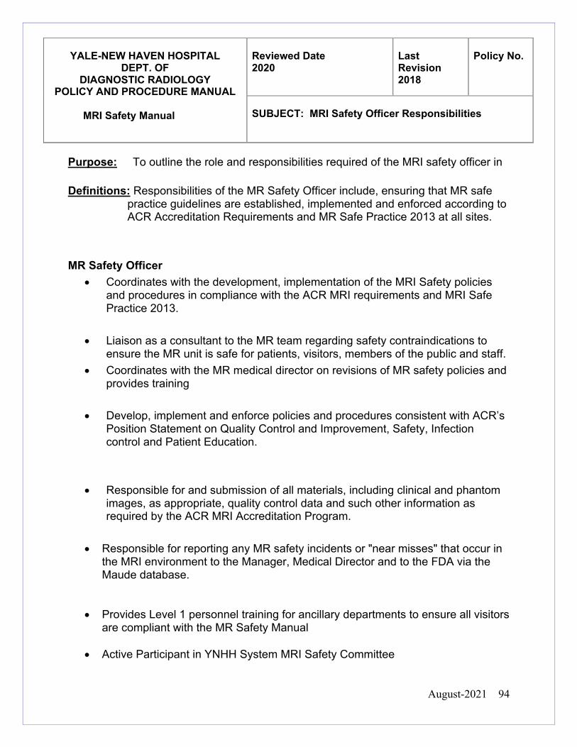
August-2021 94
Purpose: To outline the role and responsibilities required of the MRI safety officer in
Definitions: Responsibilities of the MR Safety Officer include, ensuring that MR safe practice guidelines are established, implemented and enforced according to ACR Accreditation Requirements and MR Safe Practice 2013 at all sites.
MR Safety Officer • Coordinates with the development, implementation of the MRI Safety policies
and procedures in compliance with the ACR MRI requirements and MRI Safe Practice 2013.
• Liaison as a consultant to the MR team regarding safety contraindications to ensure the MR unit is safe for patients, visitors, members of the public and staff.
• Coordinates with the MR medical director on revisions of MR safety policies and provides training
• Develop, implement and enforce policies and procedures consistent with ACR’s Position Statement on Quality Control and Improvement, Safety, Infection control and Patient Education.
• Responsible for and submission of all materials, including clinical and phantom images, as appropriate, quality control data and such other information as required by the ACR MRI Accreditation Program.
• Responsible for reporting any MR safety incidents or "near misses" that occur in the MRI environment to the Manager, Medical Director and to the FDA via the Maude database.
• Provides Level 1 personnel training for ancillary departments to ensure all visitors are compliant with the MR Safety Manual
• Active Participant in YNHH System MRI Safety Committee
YALE-NEW HAVEN HOSPITAL
DEPT. OF DIAGNOSTIC RADIOLOGY
POLICY AND PROCEDURE MANUAL MRI Safety Manual
Reviewed Date 2020
Last Revision 2018
Policy No.
SUBJECT: MRI Safety Officer Responsibilities
August-2021 95
YALE-NEW HAVEN HOSPITAL
DEPT. OF DIAGNOSTIC RADIOLOGY
POLICY AND PROCEDURE MANUAL MRI Safety Manual
Reviewed Date 2020
Last Revision 2018
Policy No.
SUBJECT: MRI Medical Director Responsibilities
Purpose: To outline the role and responsibilities required of the medical director in Definitions: MR medical director whose responsibilities will include ensuring that MR
safe practice guidelines are established and maintained as current and appropriate for the site.
MR Director • Responsible for the development and implementation of MR Safety policies and
procedures in compliance with the most recent ACR White Paper on Magnetic Resonance (MR) Safety.
• Ensures that a physician is present and immediately available when contrast is administered to patients.
• Develop, implement and enforce policies and procedures consistent with ACR’s Position Statement on Quality Control and Improvement, Safety, Infection control, Patient and Staff Education.
• Be responsible for assuring compliance with the recommendations of the medical physicist.
• Be responsible for the oversight and submission of all materials, including clinical and phantom images, as appropriate, quality control data and such other information as required by the MRI Accreditation Program.
• Be responsible for notifying the ACR within 15 days of any changes in imaging equipment (units) or changes in the use of equipment that could affect clinical or phantom images.
• Takes a lead role in YNHH System MRI Safety Committee
• Provides personnel training for ancillary departments to ensure all staff are compliant with the MRI Safety Manual
August-2021 96
Due to the presence of a powerful magnetic field, the MRI environment can be dangerous for patients and staff. Precautions must be taken to assure that ferromagnetic materials/devices do not get close enough to the MRI scanner to pose a danger.
Any loose metallic objects or devices will be placed in a locker or some designated secure space in Zone I or II.
Interrogation with a ferromagnetic detector is an important part of the screening process for all individuals entering the MRI environment.
All non- MR personnel, patients, visitors, ancillary facility members and anyone else MR staff deem necessary, need to be evaluated by thorough “wanding” with ferromagnetic detector in Zone II before given permission to enter Zone III or Zone IV. This group includes but is not limited to nurses and PCA staff accompanying patients, environmental services, cardiology and anesthesia staff".
Employees who regularly work in MR do not need to be wanded. This group includes, but is not limited, to Radiologists, MR nursing, MR technologists and MR technical assistants.
All MR staff members can wand visitors, companions and facility staff
members after appropriate training.
Access to the MRI suite will be denied to any person who is not in compliance
YALE-NEW HAVEN HOSPITAL
DEPT. OF DIAGNOSTIC RADIOLOGY
POLICY AND PROCEDURE MANUAL
MRI Safety Manual
Reviewed Date 2021
Last Revision 2018
Policy No.
SUBJECT: Wanding
August-2021 97
YALE-NEW HAVEN HOSPITAL DEPT. OF
DIAGNOSTIC RADIOLOGY POLICY AND PROCEDURE MANUAL
MRI Safety Manual
Reviewed Date 2020
Last Revision 11/2018
Policy No.
SUBJECT: Exemptions of MR Safety Policies
MRI safety policies are based on FDA standards, manufacturer’s recommendations,
current research and experience, they exist to create a safe standard of care and have
been approved by the YNHH MRI Safety Committee. Exceptions to these policies may
be made at the discretion of the supervising radiologist, such as in the rare instance of
an acute, life-threatening condition, and after consultation with the referring health care
professional. Potential exemptions include, but are not limited to, scanning a patient
with a metallic foreign body of unknown composition, unknown active or passive
implant, or waiving informed consent for a scan that would typically need it. Any
exemptions should be taken seriously and the rationale for the exception must be
documented by the supervising ATTENDING RADIOLOGIST in EPIC via the comments
section for the exam protocol or via a written clinical note in the patients’ medical record
EPIC. A non-radiologist clinician cannot override any MRI safety policy unless there is
co-documentation by the attending radiologist as noted above. MRI technologists will
not scan any patient in these cases until documentation in completed by the supervising
attending radiologist.
August-2021 98
Some programmable shunts have pressure settings that may need to be known pre-
MRI and verified post-MRI. Identify the shunt and its FDA approved process before
imaging.
Programmable Shunts that need to be checked
An outpatient with a programmable shunt THAT NEEDS TO BE CHECKED must have
an appointment set up before scanning. Not all programmable shunts need to be
checked. The shunt settings should be checked within 24 hours by the patient’s “shunt
manager” (i.e. clinician, device rep).
An inpatient with a programmable shunt that needs to be checked must have a
Neurosurgery consult before scanning. (i.e. Neurosurgery needs to be comfortable
checking the shunt post MRI before we can image) PLEASE CONTACT
NEUROSURGERY AS EARLY AS POSSIBLE TO COORDINATE (203 412 1030).
If the type of shunt is unknown, or we have incomplete records, gather as much
information as possible and ask a Neuroradiologist (203 200 3181) to (1) confirm
whether the shunt is programmable or non-programmable using shunt X-rays and (2)
document in EPIC notes. Neurosurgery consult is available to the Neuroradiologist as
YALE-NEW HAVEN HOSPITAL
DEPT. OF DIAGNOSTIC RADIOLOGY
POLICY AND PROCEDURE MANUAL
MRI Safety Manual
Reviewed Date
2021
Last
Revision 11/2018
Policy
No.
SUBJECT: Programmable Shunts
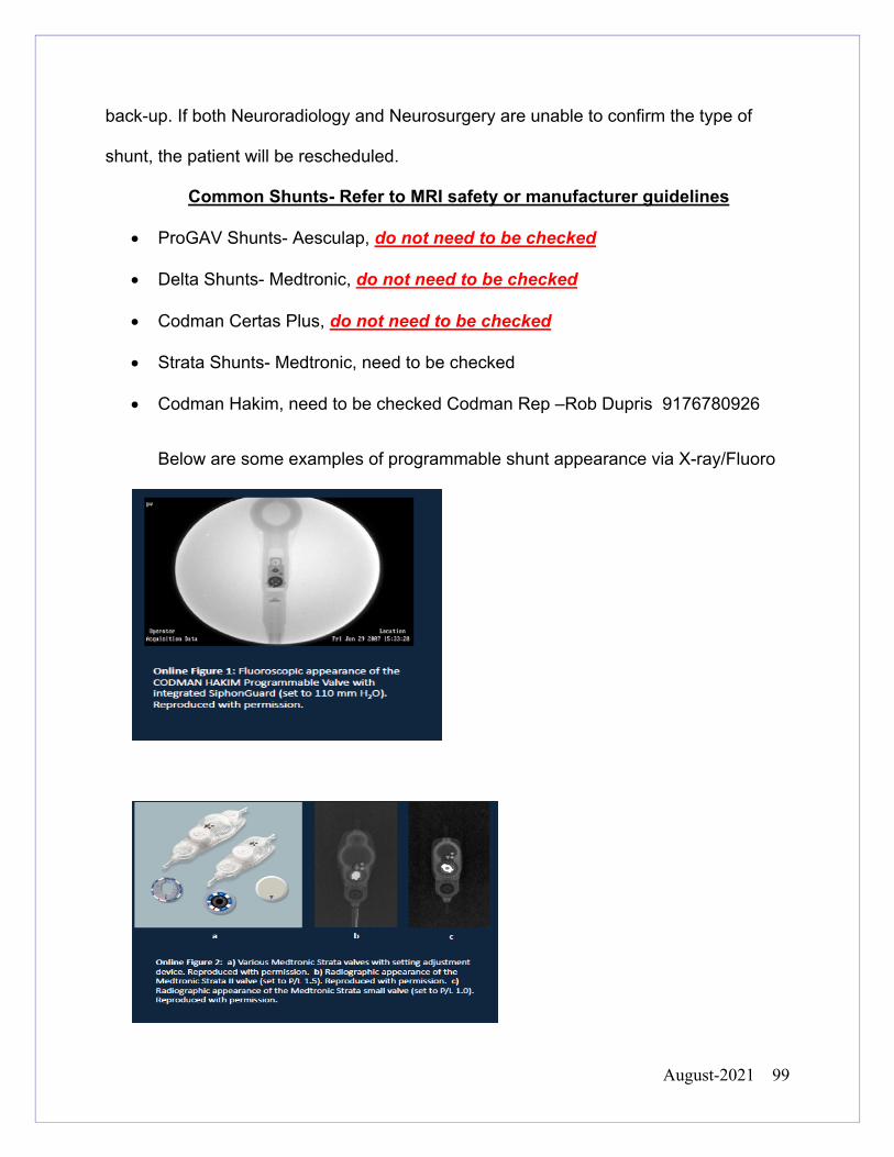
August-2021 99
back-up. If both Neuroradiology and Neurosurgery are unable to confirm the type of
shunt, the patient will be rescheduled.
Common Shunts- Refer to MRI safety or manufacturer guidelines
• ProGAV Shunts- Aesculap, do not need to be checked
• Delta Shunts- Medtronic, do not need to be checked
• Codman Certas Plus, do not need to be checked
• Strata Shunts- Medtronic, need to be checked
• Codman Hakim, need to be checked Codman Rep –Rob Dupris 9176780926
Below are some examples of programmable shunt appearance via X-ray/Fluoro
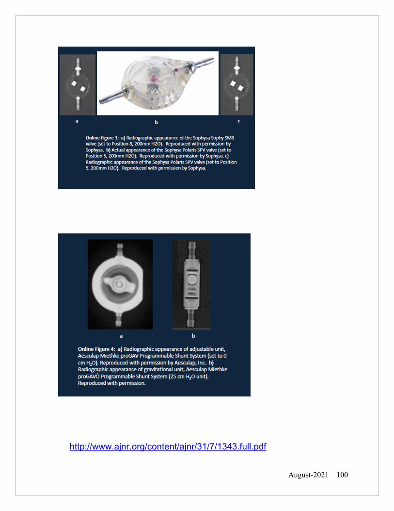
August-2021 100
http://www.ajnr.org/content/ajnr/31/7/1343.full.pdf
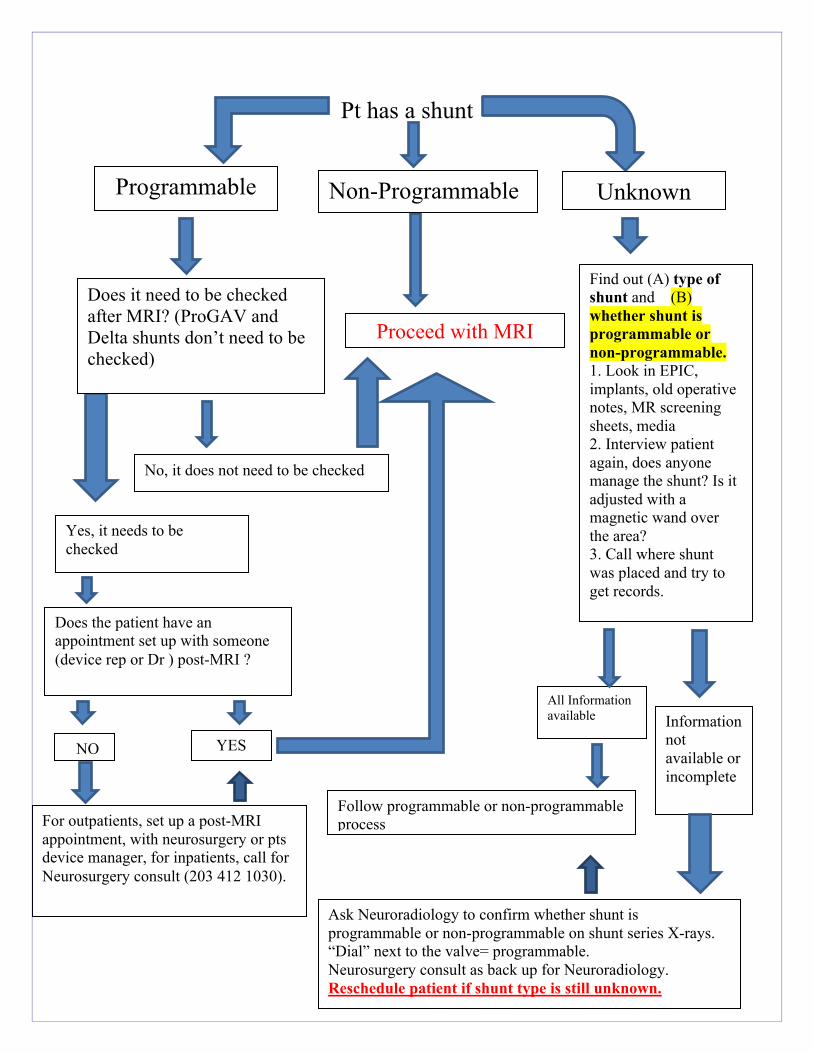
August-2021 101
Pt has a shunt
Ask Neuroradiology to confirm whether shunt is
programmable or non-programmable on shunt series X-rays. “Dial” next to the valve= programmable. Neurosurgery consult as back up for Neuroradiology. Reschedule patient if shunt type is still unknown.
For outpatients, set up a post-MRI appointment, with neurosurgery or pts device manager, for inpatients, call for Neurosurgery consult (203 412 1030).
Does it need to be checked after MRI? (ProGAV and Delta shunts don’t need to be checked)
Proceed with MRI
Yes, it needs to be checked
No, it does not need to be checked
Does the patient have an appointment set up with someone (device rep or Dr ) post-MRI ?
All Information available Information
not available or incomplete
Follow programmable or non-programmable process
YES
Find out (A) type of shunt and (B) whether shunt is programmable or non-programmable. 1. Look in EPIC, implants, old operative notes, MR screening sheets, media 2. Interview patient again, does anyone manage the shunt? Is it adjusted with a magnetic wand over the area? 3. Call where shunt was placed and try to get records.
NO
Programmable Non-Programmable Unknown
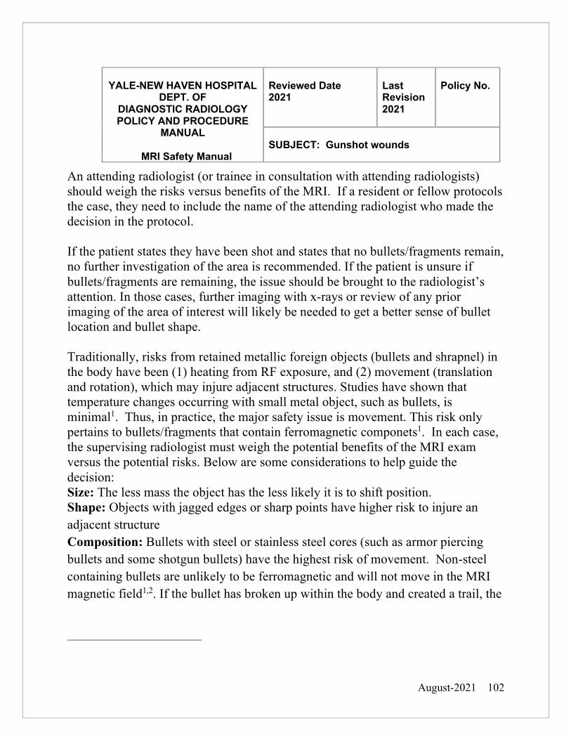
August-2021 102
An attending radiologist (or trainee in consultation with attending radiologists) should weigh the risks versus benefits of the MRI. If a resident or fellow protocols the case, they need to include the name of the attending radiologist who made the decision in the protocol. If the patient states they have been shot and states that no bullets/fragments remain, no further investigation of the area is recommended. If the patient is unsure if bullets/fragments are remaining, the issue should be brought to the radiologist’s attention. In those cases, further imaging with x-rays or review of any prior imaging of the area of interest will likely be needed to get a better sense of bullet location and bullet shape. Traditionally, risks from retained metallic foreign objects (bullets and shrapnel) in the body have been (1) heating from RF exposure, and (2) movement (translation and rotation), which may injure adjacent structures. Studies have shown that temperature changes occurring with small metal object, such as bullets, is minimal1. Thus, in practice, the major safety issue is movement. This risk only pertains to bullets/fragments that contain ferromagnetic componets1. In each case, the supervising radiologist must weigh the potential benefits of the MRI exam versus the potential risks. Below are some considerations to help guide the decision: Size: The less mass the object has the less likely it is to shift position. Shape: Objects with jagged edges or sharp points have higher risk to injure an adjacent structure Composition: Bullets with steel or stainless steel cores (such as armor piercing bullets and some shotgun bullets) have the highest risk of movement. Non-steel containing bullets are unlikely to be ferromagnetic and will not move in the MRI magnetic field1,2. If the bullet has broken up within the body and created a trail, the
YALE-NEW HAVEN HOSPITAL
DEPT. OF DIAGNOSTIC RADIOLOGY POLICY AND PROCEDURE
MANUAL
MRI Safety Manual
Reviewed Date 2021
Last Revision 2021
Policy No.
SUBJECT: Gunshot wounds
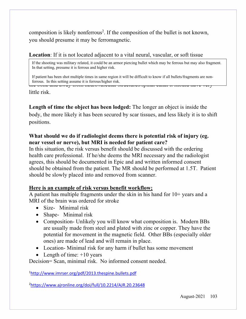
August-2021 103
composition is likely nonferrous2. If the composition of the bullet is not known, you should presume it may be ferromagnetic. Location: If it is not located adjacent to a vital neural, vascular, or soft tissue anatomic structure, movement of the object is unlikely to result in harm, regardless of the composition. For example, a small metallic foreign body in the subcutaneous tissues or bone poses minimal risk to the patient. A bullet embedded fully within the bone and away from neurovascular structures/spinal canal it should have very little risk. Length of time the object has been lodged: The longer an object is inside the body, the more likely it has been secured by scar tissues, and less likely it is to shift positions. What should we do if radiologist deems there is potential risk of injury (eg. near vessel or nerve), but MRI is needed for patient care? In this situation, the risk versus benefit should be discussed with the ordering health care professional. If he/she deems the MRI necessary and the radiologist agrees, this should be documented in Epic and and written informed consent should be obtained from the patient. The MR should be performed at 1.5T. Patient should be slowly placed into and removed from scanner. Here is an example of risk versus benefit workflow; A patient has multiple fragments under the skin in his hand for 10+ years and a MRI of the brain was ordered for stroke
• Size- Minimal risk • Shape- Minimal risk • Composition- Unlikely you will know what composition is. Modern BBs
are usually made from steel and plated with zinc or copper. They have the potential for movement in the magnetic field. Other BBs (especially older ones) are made of lead and will remain in place.
• Location- Minimal risk for any harm if bullet has some movement • Length of time: +10 years
Decision= Scan, minimal risk. No informed consent needed. 1http://www.imrser.org/pdf/2013.thespine.bullets.pdf 2https://www.ajronline.org/doi/full/10.2214/AJR.20.23648
If the shooting was military related, it could be an armor piercing bullet which may be ferrous but may also fragment. In that setting, presume it is ferrous and higher risk. If patient has been shot multiple times in same region it will be difficult to know if all bullets/fragments are non-ferrous. In this setting assume it is ferrous/higher risk.
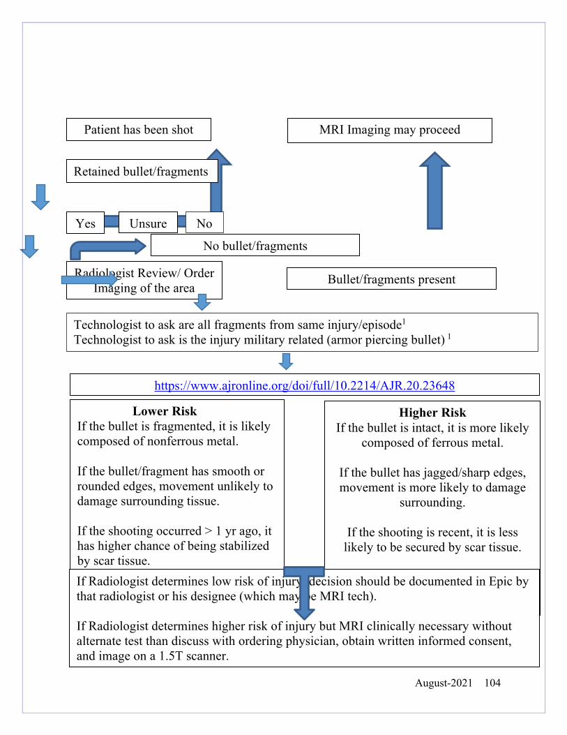
August-2021 104
Patient has been shot
MRI Imaging may proceed
Radiologist Review/ Order Imaging of the area
Bullet/fragments present
No bullet/fragments
Retained bullet/fragments fragfragments left?
Yes No Unsure
https://www.ajronline.org/doi/full/10.2214/AJR.20.23648
Lower Risk If the bullet is fragmented, it is likely composed of nonferrous metal. If the bullet/fragment has smooth or rounded edges, movement unlikely to damage surrounding tissue.
If the shooting occurred > 1 yr ago, it has higher chance of being stabilized by scar tissue. If bullet located within the bone, it is unlikely to move (even if ferrous).
Higher Risk If the bullet is intact, it is more likely
composed of ferrous metal.
If the bullet has jagged/sharp edges, movement is more likely to damage
surrounding.
If the shooting is recent, it is less likely to be secured by scar tissue.
A military related shooting If Radiologist determines low risk of injury, decision should be documented in Epic by
that radiologist or his designee (which may be MRI tech). If Radiologist determines higher risk of injury but MRI clinically necessary without alternate test than discuss with ordering physician, obtain written informed consent, and image on a 1.5T scanner.
Technologist to ask are all fragments from same injury/episode1
Technologist to ask is the injury military related (armor piercing bullet) 1
August-2021 105
Intracranial Electrodes and MRI (Post Grid Protocol) The root cause of epileptic seizures can be difficult to find. A routine EEG using electrodes on the scalp surface may not locate the origin of a patient’s seizures. . In these cases, neurosurgeons may need to do more direct monitoring using intracranial electrodes such as subdural grid, strips and/or depth electrodes. The electrodes are placed in the operative room, and the patient undergoes post operative imaging (MR/CT). They are continuously monitored for a length of time (typically less than 14 days) before the removal of the electrodes. Some companies’ (e.g. Ad-Tech) electrodes do not provide ASTM MRI labelling, but they have an excellent MR safety record with decades of clinical use and research testing. YNHH follows tailored guidelines that have been created based on our years of clinical experience, and published data on this topic. Our MR guidelines are: 1.5T Transmit/Receive Head Coil The protruding leads need to be aligned straight and separated as much as the patients dressing allow. The sequences performed will be kept to what is clinically necessary. Head Imaging Only Davis L. M., Spencer D. D., Spencer S. S., Bronen R. A. (1999). MR imaging of implanted depth and subdural electrodes: is it safe? Epilepsy Res. 35, 95–98. 10.1016/S0920-1211(99)00007-8 Carmichael, D. W., Thornton, J. S., Rodionov, R. , Thornton, R. , McEvoy, A. , Allen, P. J. and Lemieux, L. (2008), Safety of localizing epilepsy monitoring intracranial electroencephalograph electrodes using MRI: Radiofrequency‐induced heating. J. Magn. Reson. Imaging, 28: 1233-1244. doi:10.1002/jmri.21583
YALE-NEW HAVEN HOSPITAL
DEPT. OF DIAGNOSTIC RADIOLOGY POLICY AND PROCEDURE
MANUAL
MRI Safety Manual
Reviewed Date 2021
Last Revision 2019
Policy No.
SUBJECT: POST GRID
August-2021 106
Claustrophobia is defined as extreme or irrational fear of confined places. Many MRI patients feel a varying degree of claustrophobia and/or emotional distress. Having an MRI can have a stressful effect on a patient, in fact a patient might not know they suffer from claustrophobia until their first MRI experience. Some techniques to combat claustrophobia and/or emotional distress are: Before the MR exam:
• Prepare and educate the patient about their MR procedure (e.g., MR machine dimensions, noise and potential vibrations, table movements throughout the exam, coil positions and shapes, intercom system, constant presence of the MRI technologist).
• Allow an appropriately screened companion to remain with the patient during the MR examination.
• Position the patient feet first (if exam appropriate)
During the MR exam:
• Maintain verbal, visual, and/or physical contact with the patient during the MR procedure.
• Audio distractions: appropriate stereo system to provide music to the patient. • Visual distractions: video monitor or goggles, mirrors or prism glasses to redirect
the patient’s line of sight, a blindfold so that the patient is not aware of the surroundings.
• Comforting environment: bright lights inside of the MR system, fan inside of the MR system.
• Use a sedative or other similar medication. • Perform the scan as rapidly as possible and do the most important sequences
first.
YALE-NEW HAVEN HOSPITAL
DEPT. OF DIAGNOSTIC RADIOLOGY
POLICY AND PROCEDURE MANUAL
MRI Safety Manual
Reviewed Date
2021
Last
Revision 2019
Policy
No.
SUBJECT: Claustrophobia and Emotional Distress
August-2021 107
Below is our policy for Tattoos, Jewelry, Body Piercings and Hair Extensions Tattoos: Tattoo ink can contain products such as iron oxide that have potential to react with the RF field creating e-fields and in turn burns. Although this is highly unlikely, safety measures should still be put in place. Before the Exam:
• Tattoos should be discussed with the patient. During the Exam:
• Maintain verbal, and visual contact with the patient during the MR procedure. • If the patient is uncomfortable at any point during the exam stop.
Jewelry/Dermal piercings and Magnetic Eyelashes Before the MR Exam:
• All jewelry that can be removed, should be removed particularly pieces that are in the area of RF exposure.
• If the jewelry can’t be removed easily, position it away from the skin as much as possible.
• Prepare and educate the patient about their MR procedure and potential vibrations felt around the jewelry from the magnet (even nonferrous metal can potentially vibrate during gradient excitation.)
• A tester magnet or a ferromagnetic wand can be used to determine if it is ferromagnetic.
• If it is ferromagnetic, a wrap such as coban or tape can be placed around the item to help prevent it from dislodging.
During the Exam:
• Maintain verbal, and visual contact with the patient during the MR procedure. • If the patient is uncomfortable at any point during the exam stop.
Hair Extensions:
YALE-NEW HAVEN HOSPITAL DEPT. OF
DIAGNOSTIC RADIOLOGY POLICY AND PROCEDURE MANUAL
MRI Safety Manual
Reviewed Date
2021
Last
Revision 2019
Policy
No.
SUBJECT: Tattoos Jewelry Body Piercings and Hair
Extensions
August-2021 108
Occasionally the seams of the hairpiece extension include small amounts of metal that may produce an artifact. Others are semi-permanent and attached with hairpins. Hairpins are typically elongated in shape, very small and highly ferrous. Not only can these pins become projectiles, they can get stuck in the small crevasses of a mechanical MR table, essentially putting the table out of service or creating an artifact that appears on all images until removed. Before the MR exam:
• ALL hairpins need to be removed before proceeding with the MRI. • Ask the patient to remove all hair pieces. Scan the patients head with a
ferromagnetic detector to confirm no hair pins are present.
August-2021 109
Appendix A
YNHH Position on Static Field Gradients
In 2019 the ACR Committee of MR Safety addressed the confusion around the Static Field Gradient, and ASTM MR Conditional labeling. When a device or implant receives MR Conditional labeling, one of the values assigned is the static field gradient (SFG), i.e. the magnetic field change over distance. Interpretations of this value has caused confusion. Example of MR Conditional label: (Static Gradient Field of 720g/cm or less) Vendors provide SFG maps for each magnet. Many of the maps show SFG values higher than a specific device label. For example, you could have an implant that has a SFG rating of 720 g/cm or less and the maximum SFG on your MRI magnet is 1100g/cm. When reviewing SFG maps for determining safety of scanning an MR conditional implant, two things should be addressed for the magnet being used and the implant in question;
(1) Clinically accessible area (2) Patient accessible area
Clinically Accessible Area: The highest SFG areas are not clinically accessible. They are located behind the magnet housing (the plastic bore cover). Additional high SFG regions in front of the plastic covering are in an area utilized by staff not by patients. Patient Accessible Area: Patient accessible areas are rarely higher than the standard ASTM labeling2. In some instances, there is a high SFG at the very periphery of the bore.
2
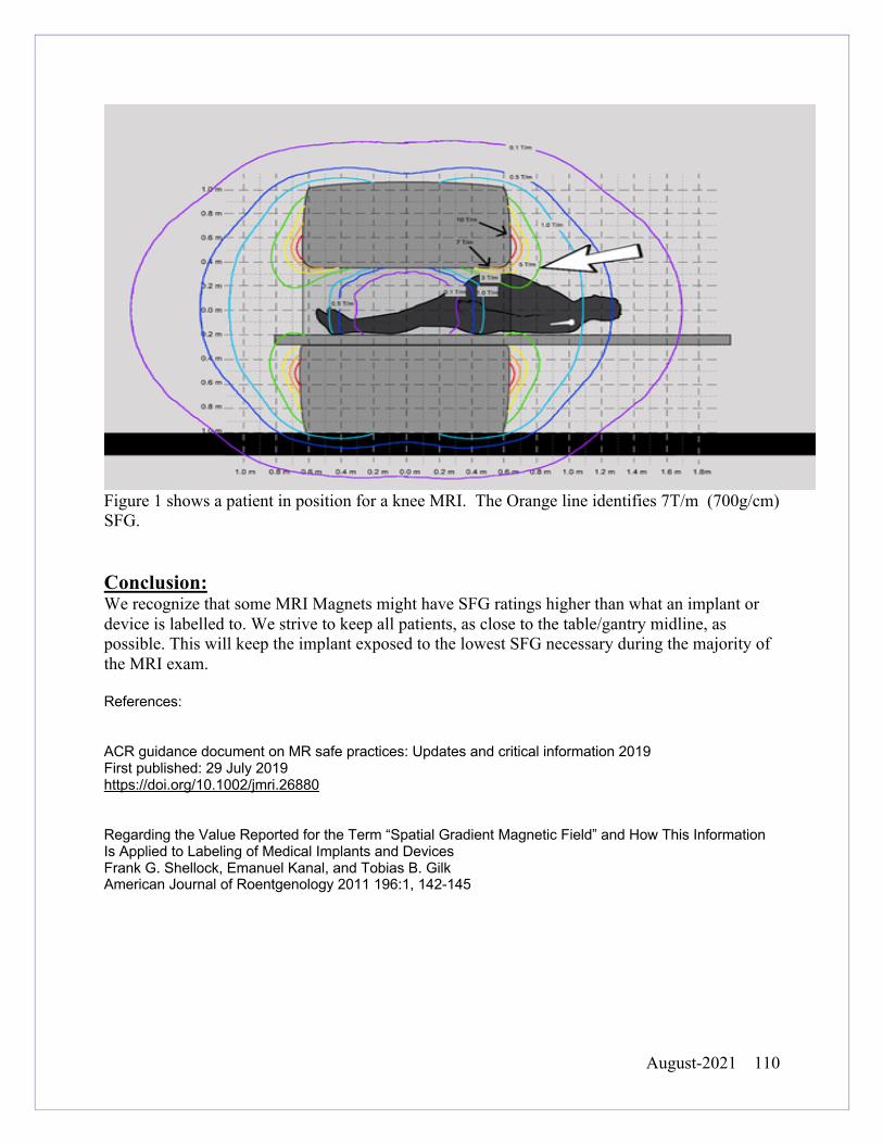
August-2021 110
Figure 1 shows a patient in position for a knee MRI. The Orange line identifies 7T/m (700g/cm) SFG. Conclusion: We recognize that some MRI Magnets might have SFG ratings higher than what an implant or device is labelled to. We strive to keep all patients, as close to the table/gantry midline, as possible. This will keep the implant exposed to the lowest SFG necessary during the majority of the MRI exam. References: ACR guidance document on MR safe practices: Updates and critical information 2019 First published: 29 July 2019 https://doi.org/10.1002/jmri.26880 Regarding the Value Reported for the Term “Spatial Gradient Magnetic Field” and How This Information Is Applied to Labeling of Medical Implants and Devices Frank G. Shellock, Emanuel Kanal, and Tobias B. Gilk American Journal of Roentgenology 2011 196:1, 142-145
August-2021 111
Appendix B Multi Hance (Bracco)
• 1-800-257-5181 Dotarem (Guebert)
• Fill out contrast reaction form
Eovist (Bayer)
• 1-888-84bayer.
Appendix C MRI Safety Websites http://www.mrisafety.com Includes “the List” updated yearly list of MR tested devices http://www.imrser.org MR Safety Papers, guidelines and information http://cmemeded.com/mrisafety MR Safety Courses http://www.acr.org/Quality-Safety/Radiology-Safety/MR-Safety ACR website section on MRI Safety
http://enterprise.astm.org ASTM standards for testing
Appendix D
• https://medicine.yale.edu/diagnosticradiology/patientcare/policies/premedication/
Premedication Policy link
http://translation.ynhh.org/SitePages/Home.aspx
Translated documents link
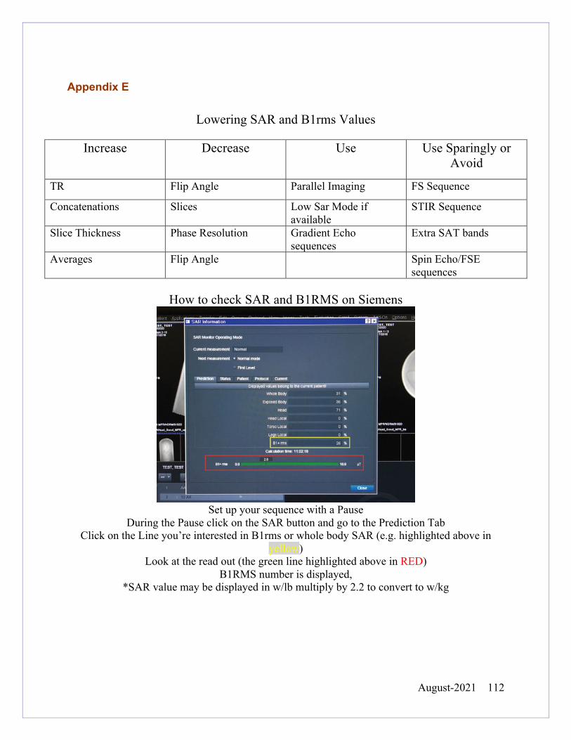
August-2021 112
Appendix E
Lowering SAR and B1rms Values
Increase Decrease Use Use Sparingly or Avoid
TR Flip Angle Parallel Imaging FS Sequence
Concatenations Slices Low Sar Mode if available
STIR Sequence
Slice Thickness Phase Resolution Gradient Echo sequences
Extra SAT bands
Averages Flip Angle Spin Echo/FSE sequences
How to check SAR and B1RMS on Siemens
Set up your sequence with a Pause
During the Pause click on the SAR button and go to the Prediction Tab Click on the Line you’re interested in B1rms or whole body SAR (e.g. highlighted above in
yellow) Look at the read out (the green line highlighted above in RED)
B1RMS number is displayed, *SAR value may be displayed in w/lb multiply by 2.2 to convert to w/kg
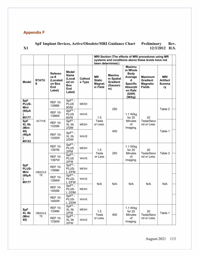
August-2021 113
Appendix F
SpF Implant Devices, Active/Obsolete/MRI Guidance Chart Preliminary Rev.
X1 12/3/2012 H.S.
MRI Section (The effects of MRI procedures using MR systems and conditions above these levels have not been determined.)
Model STATUS
Reference # (Located on Box End Label)
Model Name (Located on Box End Label)
Cathode Type
MR Static Magnetic Field
Maximum Spatial Gradient (Gauss/cm)
Maximum Whole
Body Average
d Specific Absorption Rate (SAR) (W/kg)
Maximum Gradient Magnetic Fields
MRI Artifact Summa
ry
SpF PLUS- Mini (60μA) #0177
ACTIVE
REF 10-1398M
SpF® - PLUS 60/M
MESH
1.5 Tesla
or Less.
250
1.1 W/kg for 25
Minutes of
Imaging.
20 Tesla/Second or Less.
Table 2
REF 10-1398W
SpF® - PLUS 60/W
WAVE
SpF XL IIb (Mini 40) (40μA) #0153
REF 10-1335M
SpF® -XL IIb 2/DM
MESH
450 Table 1
REF 10-1335W
SpF® -XL IIb 2/DW
WAVE
SpF PLUS- Mini (60μA) #0177
OBSOLETE
REF 10-1397M
SpF® - PLUS 2/FM
MESH 1.5 Tesla
or Less 250
1.1 W/kg for 25
Minutes of
Imaging.
20 Tesla/Second or Less.
Table 2
REF 10-1397W
SpF® - PLUS 2/FW
WAVE
REF 10-1399M
SpF® - PLUS-L 2/FM
MESH
N/A N/A N/A N/A N/A
REF 10-1399W
SpF® - PLUS-L 2/FW
WAVE
REF 10-1400M
SpF® - PLUS-L 2/DM
MESH
REF 10-1400W
SpF® - PLUS-L 2/DW
WAVE
SpF XL IIb (Mini 40)
OBSOLETE
REF 10-1336M
SpF® -XL IIb 2/FM
MESH 1.5 Tesla
or Less. 450
1.1 W/kg for 25
Minutes of
Imaging.
20 Tesla/Second or Less.
Table 1
REF 10-1336W
SpF® -XL IIb 2/FW
WAVE
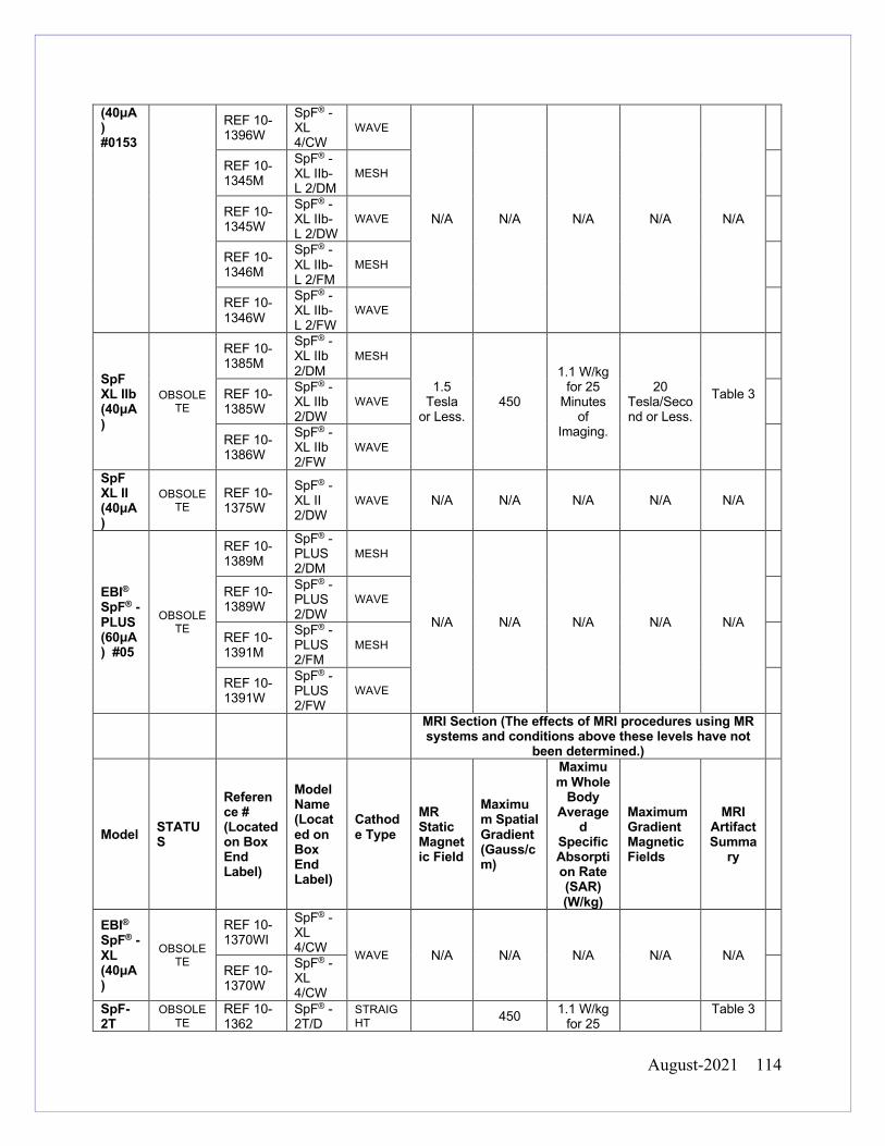
August-2021 114
(40μA) #0153
REF 10-1396W
SpF® - XL 4/CW
WAVE
N/A N/A N/A N/A N/A
REF 10-1345M
SpF® -XL IIb-L 2/DM
MESH
REF 10-1345W
SpF® -XL IIb-L 2/DW
WAVE
REF 10-1346M
SpF® -XL IIb-L 2/FM
MESH
REF 10-1346W
SpF® -XL IIb-L 2/FW
WAVE
SpF XL IIb (40μA)
OBSOLETE
REF 10-1385M
SpF® -XL IIb 2/DM
MESH
1.5 Tesla
or Less. 450
1.1 W/kg for 25
Minutes of
Imaging.
20 Tesla/Second or Less.
Table 3
REF 10-1385W
SpF® -XL IIb 2/DW
WAVE
REF 10-1386W
SpF® -XL IIb 2/FW
WAVE
SpF XL II (40μA)
OBSOLETE
REF 10-1375W
SpF® -XL II 2/DW
WAVE N/A N/A N/A N/A N/A
EBI® SpF® - PLUS (60μA) #05
OBSOLETE
REF 10-1389M
SpF® - PLUS 2/DM
MESH
N/A N/A N/A N/A N/A
REF 10-1389W
SpF® - PLUS 2/DW
WAVE
REF 10-1391M
SpF® - PLUS 2/FM
MESH
REF 10-1391W
SpF® - PLUS 2/FW
WAVE
MRI Section (The effects of MRI procedures using MR systems and conditions above these levels have not
been determined.)
Model STATUS
Reference # (Located on Box End Label)
Model Name (Located on Box End Label)
Cathode Type
MR Static Magnetic Field
Maximum Spatial Gradient (Gauss/cm)
Maximum Whole
Body Average
d Specific Absorption Rate (SAR) (W/kg)
Maximum Gradient Magnetic Fields
MRI Artifact Summa
ry
EBI® SpF® - XL (40μA)
OBSOLETE
REF 10-1370WI
SpF® - XL 4/CW WAVE N/A N/A N/A N/A N/A
REF 10-1370W
SpF® - XL 4/CW
SpF-2T
OBSOLETE
REF 10-1362
SpF® - 2T/D
STRAIGHT 450 1.1 W/kg
for 25 Table 3
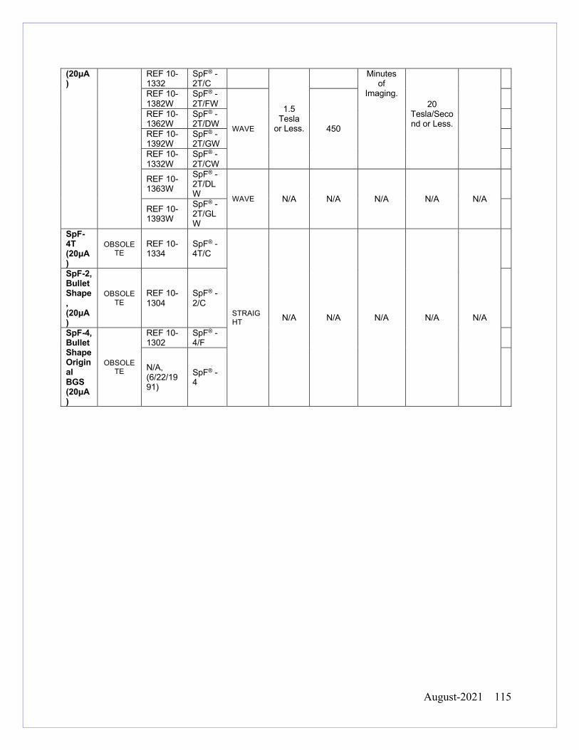
August-2021 115
(20μA)
REF 10-1332
SpF® - 2T/C
1.5 Tesla
or Less.
Minutes of
Imaging. 20
Tesla/Second or Less.
REF 10-1382W
SpF® - 2T/FW
WAVE 450
REF 10-1362W
SpF® - 2T/DW
REF 10-1392W
SpF® - 2T/GW
REF 10-1332W
SpF® - 2T/CW
REF 10-1363W
SpF® - 2T/DLW WAVE N/A N/A N/A
N/A
N/A
REF 10-1393W
SpF® - 2T/GLW
SpF-4T (20μA)
OBSOLETE
REF 10-1334
SpF® - 4T/C
STRAIGHT N/A N/A N/A N/A N/A
SpF-2, Bullet Shape, (20μA)
OBSOLETE
REF 10-1304
SpF® - 2/C
SpF-4, Bullet Shape Original BGS (20μA)
OBSOLETE
REF 10-1302
SpF® - 4/F
N/A, (6/22/1991)
SpF® - 4
August-2021 116
Appendix G
Passive Vascular Implant Background
How do implants get tested?
For a passive implant to acquire a MR Conditional rating, a series of ASTM tests have
to be performed. Two of those tests will be discussed below.
The F2052-15 test measures the deflection angle of an implant to see if gravitational
forces or the B0 forces are more influential on the device. The test does not take into
account any designed or naturally occurring counterforces once a device is implanted in
the human body. Thousands of passive vascular devices mentioned above (coils,
stents, and filters) have been tested and brought to market in the USA have passed this
test at 1.5 and 3 Tesla. There are some devices that were brought to market before
ASTM standards were created, and others before 3T scanners were in use. Based on
additional testing or clinical research, most have had their labeling changed to include
3T. The YNHHS MRI Safety Committee feels the risk of harm is very low from MRI for
a patient from any passive vascular device.
The F2182-11a test assesses the relationship between RF induced heating and the
implant. This is the current standard, but is not very precise and likely is over-
conservative as it does not take into account the cooling effects of blood perfusion,
August-2021 117
which is known to have a major impact on heat reduction of passive vascular devices.
The periphery of the RF transmission field is where maximum heating occurs. During
testing, devices are placed in the area where heating would be maximized to show
worst case scenarios (usually the edges of the phantom), and the results may not reflect
the situations in patients when passive vascular devices are not on the periphery, but
more centered in the body/ field and therefore exposed to lower RF transmission
energies.
How can we predict heating risk?
The standard method to predict heating in a passive implant is to monitor SAR (specific
absorption rate) values. SAR is estimated by adding up all the RF pulses in a
sequence, dividing by TR and then taking that number and dividing by the patient’s
weight (watts of power/kg). Burns are thought to be created within an area of the body
with high resistance to “electron traffic”. RF is introduced into the body and then
dissipated by normal thermoregulatory systems. If there is an area where it cannot
dissipate heat efficiently, an e-field is created (“energy pile-up”), and this is thought to
produce a burn. However, the relationship between RF and e-field creation is not
precise. While RF is a component of the SAR calculation, using SAR to predict burns
caused by e-fields is indirect and difficult to foresee.
It is also important to note SAR levels received in the clinical setting are not accurate.
Different manufacturers calculate SAR with different methods and exponents, and the
SAR levels can even vary between software levels on the same MRI scanner. SAR
August-2021 118
reported at the clinical work station has been shown to be many times higher than
actual values. The limitations of SAR values in clinical MRI are widely known in the MR
community. A newer value called B1rms, which is not patient dependent, is being used
for active implant testing. This value is more accurate, but it’s not the standard ASTM
test for passive implant testing.
In clinical MRI there are two SAR settings “normal mode” and “first level”. The large majority of passive vascular devices have a SAR value that can be satisfied by one of these two modes. There are a handful of devices that have a ASTM labeling with a SAR rating lower than the normal operating mode setting of 2w/kg. Given limitations of testing and SAR estimates, our policy is to always keep the SAR values as low as possible for diagnostic clinical images Reference MR Safety Officer Training Course MR Safety Facebook Group- multiple discussions Dr Donald Mcrobbie SAR B1rms calculation https://drdonaldmcrobbie.com/2018/03/20/sar-and-b1rms-what-are-they/ Coils: Updated stainless steel embolization coil guidelines Cook medical. https://www.cookmedical.com/support/general-product-information/ Slesnick, T.C., Schreier, J., Soriano, B.D. et al. Pediatr Cardiol (2016) 37: 62. https://doi.org/10.1007/s00246-015-1240-3 Standards: ASTM F2182-11a Standard Test Method for Measurement of Radio Frequency Induced Heating On or Near Passive Implants During Magnetic Resonance Imaging, ASTM International, West Conshohocken, PA, 2011, https://doi.org/10.1520/F2182-11A ASTM F2052-15 Standard Test Method for Measurement of Magnetically Induced Displacement Force on Medical Devices in the Magnetic Resonance Environment, ASTM International, West Conshohocken, PA, 2015, https://doi.org/10.1520/F2052-15 Woods, T. O. (2007), Standards for medical devices in MRI: Present and future. J. Magn. Reson. Imaging, 26: 1186-1189. doi:10.1002/jmri.21140 SAR: Baker KB, Tkach JA, Nyenhuis JA, et al. Evaluation of specific absorption rate as a dosimeter of MRI-related implant heating. J Magn Reson Imaging 2004;20:315–320. B1+RMS, The root-mean-square value of the MRI Effective Component of the RF Magnetic (B1) Field http://www.mrisafety.com/SafetyInfov.asp?SafetyInfoID=362 Stents: Coils, Filters, Stents, and Grafts http://www.mrisafety.com/SafetyInfov.asp?SafetyInfoID=171Hiramoto JS, et al. The effect of magnetic resonance imaging on stainless-steel Z-stent-based abdominal aortic prosthesis. J Vasc Surg 2007;45:472-474 T. Resch, M. Malina, B. Lindblad, J. Malina, J. Brunkwall, K. IvancevThe impact of stent design on proximal stent-graft fixation in the abdominal aorta: an experimental study Eur J Vasc Endovasc Surg, 20 (2000), pp. 190-195
August-2021 119
Appendix H
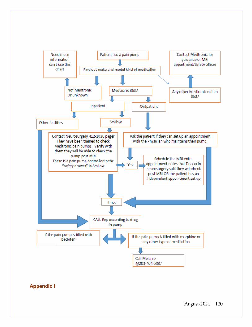
August-2021 120
Appendix I
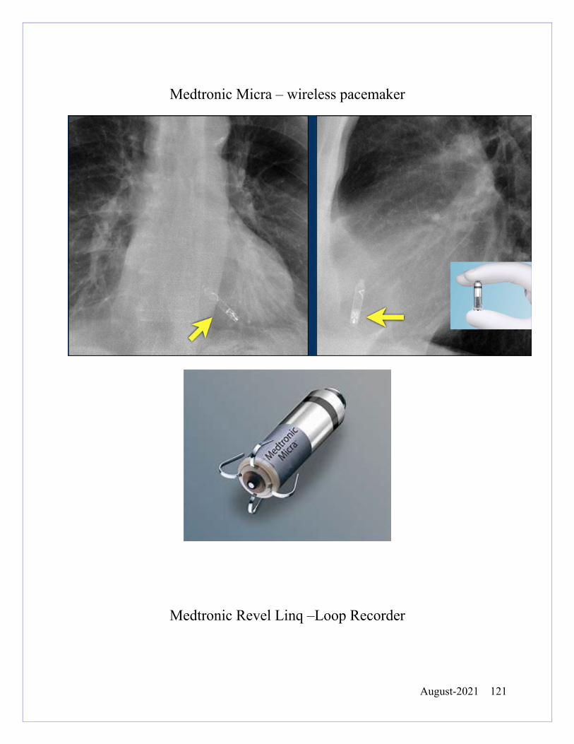
August-2021 121
Medtronic Micra – wireless pacemaker
Medtronic Revel Linq –Loop Recorder
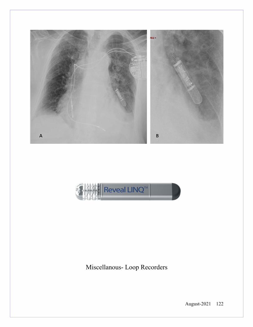
August-2021 122
Miscellanous- Loop Recorders
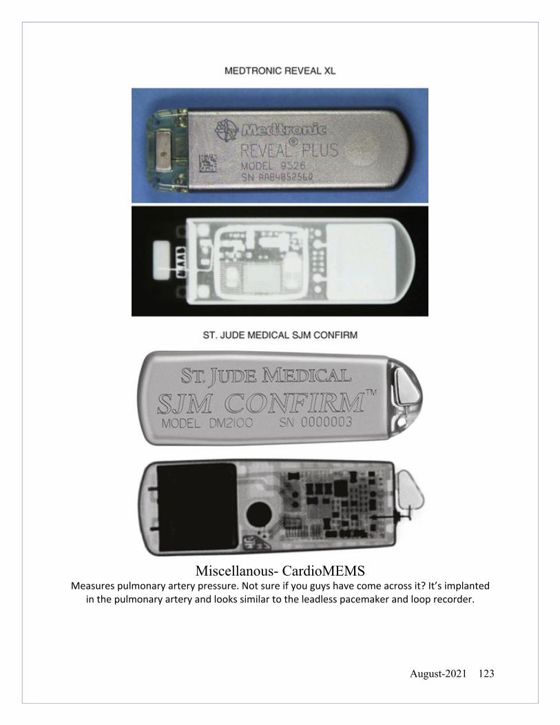
August-2021 123
Miscellanous- CardioMEMS
Measures pulmonary artery pressure. Not sure if you guys have come across it? It’s implanted in the pulmonary artery and looks similar to the leadless pacemaker and loop recorder.
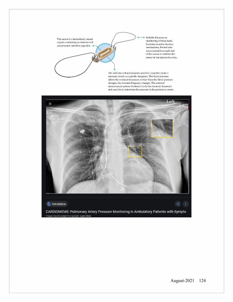
August-2021 124