Embed Size (px)
Citation preview
2010 Spring Clinical 2010 Spring Clinical MRI Education SeminarMRI Education Seminar
Applications of MRI in Applications of MRI in Neurological Diagnostic ImagingNeurological Diagnostic Imaging
Darren P. ODarren P. O’’Neill, MDNeill, MDIndiana University NeuroradiologyIndiana University Neuroradiology
ObjectivesObjectives
••
Review fundamental clinical cases that Review fundamental clinical cases that illustrate the wide array and utility of illustrate the wide array and utility of available neuroradiology MR imaging available neuroradiology MR imaging techniques / applicationstechniques / applications
••
Gain insight into the rationale behind MR Gain insight into the rationale behind MR imaging protocolsimaging protocols
••
Develop a greater understanding of Develop a greater understanding of potential points of patient care impact potential points of patient care impact
OutlineOutline••
Review fundamental clinical cases that illustrate the wide arrayReview fundamental clinical cases that illustrate the wide array
and and utility of available neuroradiology MR imaging techniquesutility of available neuroradiology MR imaging techniques::
••
Gradient echo / SWIGradient echo / SWI••
DiffusionDiffusion
••
FLAIRFLAIR••
IRIR--SPGR / MPSPGR / MP--RAGERAGE
••
MRA / MRVMRA / MRV••
SpectroscopySpectroscopy
••
PerfusionPerfusion
••
Review potential points of patient care impact:Review potential points of patient care impact:––
Additional patient history?Additional patient history?
––
Would an additional study or different protocol be better?Would an additional study or different protocol be better?
––
Would intravenous contrast be helpful?Would intravenous contrast be helpful?
––
Are the referring physician and/or neuroradiologist aware of Are the referring physician and/or neuroradiologist aware of critical findings?critical findings?
Brain Brain ––
ScreenScreen
••
IndicationsIndications––
Screen, Altered mental status, Dementia, Psychiatric disorder, Screen, Altered mental status, Dementia, Psychiatric disorder, HeadachesHeadaches
••
SequencesSequences––
3 PL LOC3 PL LOC––
Sag T1 SESag T1 SE––
Ax T2 FLAIRAx T2 FLAIR––
Sag T2 FLAIRSag T2 FLAIR––
Ax T1 SEAx T1 SE––
Ax T2 TSE FSAx T2 TSE FS––
Ax DWI EPIAx DWI EPI––
Cor T2 TSECor T2 TSE
••
CommentsComments––
Axial scans should be parallel to the ACAxial scans should be parallel to the AC--PC linePC line

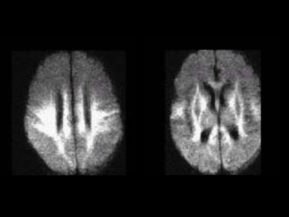
25 year25 year--old pregnant female with mental old pregnant female with mental status changestatus change
Potential Points of ImpactPotential Points of Impact
••
Patient history?Patient history?––
Do we know more than Do we know more than ““mental status changemental status change””??
––
Onset of symptoms?Onset of symptoms?––
Hypertension? Pregnancy? Steroids? Hypertension? Pregnancy? Steroids?
••
Sequences to consider anticipating?Sequences to consider anticipating?––
MRI brain without contrast => evaluate for acute MRI brain without contrast => evaluate for acute ischemiaischemia
––
Avoid contrast with pregnancy!Avoid contrast with pregnancy!
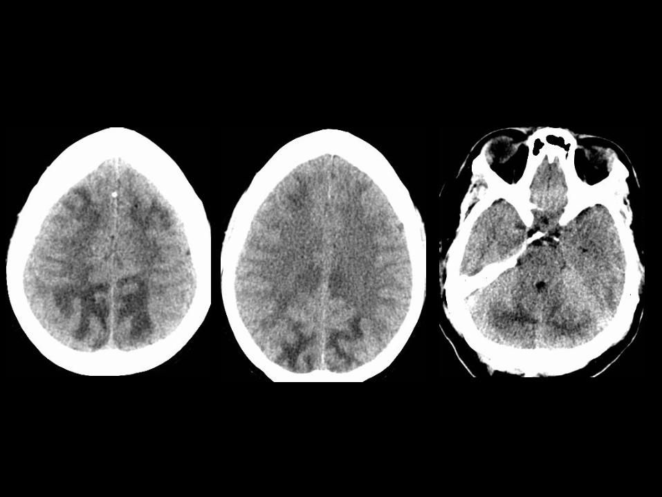
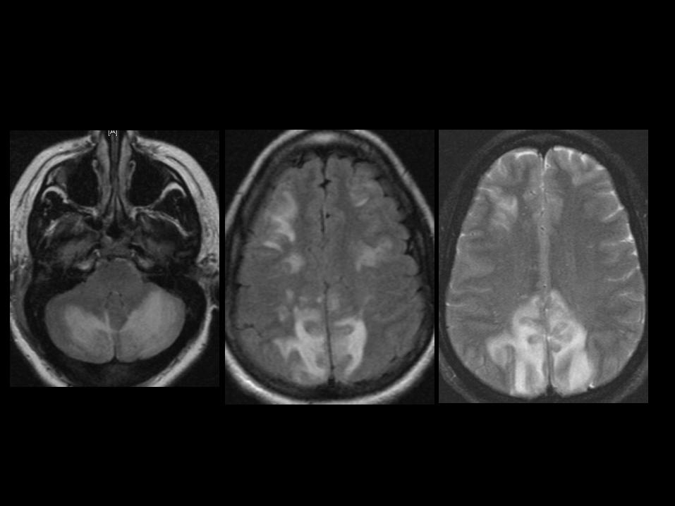
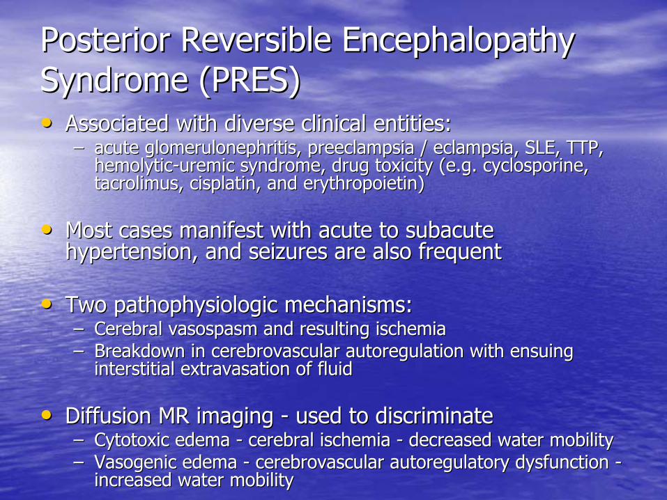
Posterior Reversible Encephalopathy Posterior Reversible Encephalopathy Syndrome (PRES)Syndrome (PRES)••
Associated with diverse clinical entities:Associated with diverse clinical entities:––
acute glomerulonephritis, preeclampsia / eclampsia, SLE, TTP, acute glomerulonephritis, preeclampsia / eclampsia, SLE, TTP, hemolytichemolytic--uremic syndrome, drug toxicity (e.g. cyclosporine, uremic syndrome, drug toxicity (e.g. cyclosporine, tacrolimus, cisplatin, and erythropoietin)tacrolimus, cisplatin, and erythropoietin)
••
Most cases manifest with acute to subacute Most cases manifest with acute to subacute hypertension, and seizures are also frequenthypertension, and seizures are also frequent
••
Two pathophysiologic mechanisms:Two pathophysiologic mechanisms:––
Cerebral vasospasm and resulting ischemiaCerebral vasospasm and resulting ischemia––
Breakdown in cerebrovascular autoregulation with ensuing Breakdown in cerebrovascular autoregulation with ensuing interstitial extravasation of fluid interstitial extravasation of fluid
••
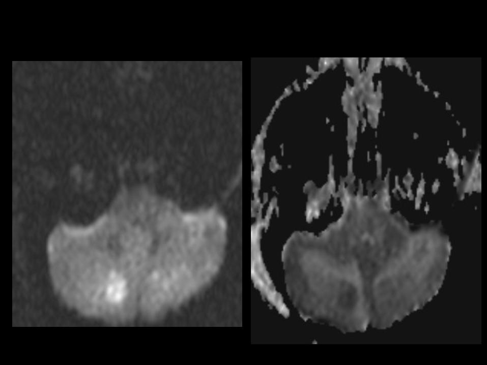
Diffusion MR imaging Diffusion MR imaging --
used to discriminate used to discriminate ––
Cytotoxic edema Cytotoxic edema --
cerebral ischemia cerebral ischemia --
decreased water mobilitydecreased water mobility––
Vasogenic edema Vasogenic edema --
cerebrovascular autoregulatory dysfunction cerebrovascular autoregulatory dysfunction --
increased water mobilityincreased water mobility

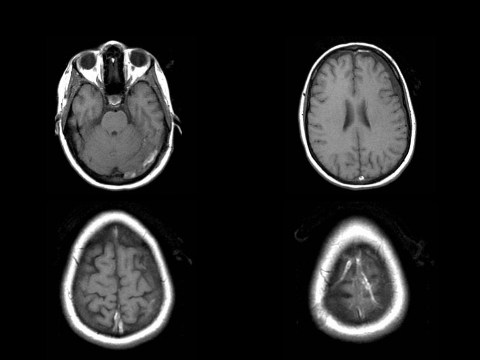
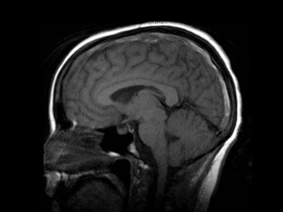
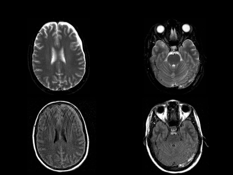
MiddleMiddle--aged female with new onset aged female with new onset parasthesiasparasthesias
Potential Points of ImpactPotential Points of Impact
••
Patient history?Patient history?––
Do we know more than Do we know more than ““parasthesiasparasthesias””??
––
Onset of symptoms? Prior history?Onset of symptoms? Prior history?––
Neurologic deficits?Neurologic deficits?
••
Sequences to consider anticipating?Sequences to consider anticipating?––
Sagittal FLAIR imaging (eg. multiple sclerosis)Sagittal FLAIR imaging (eg. multiple sclerosis)
––
PostPost--contrast imagescontrast images
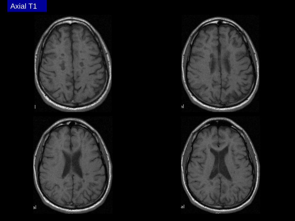
Axial T1
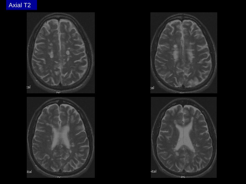
Axial T2

Axial FLAIR
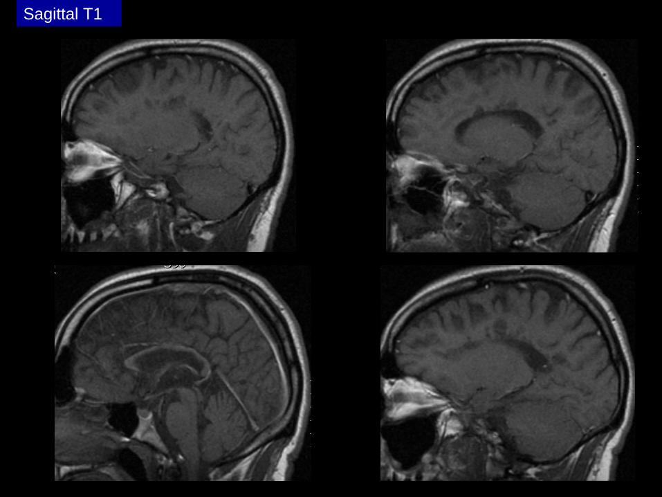
Sagittal T1
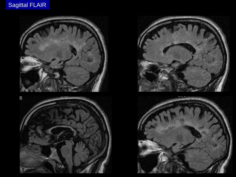
Sagittal FLAIR
Multiple SclerosisMultiple Sclerosis
••
Demyelinating condition of the central nervous Demyelinating condition of the central nervous system system --
generally considered to be autoimmunegenerally considered to be autoimmune
••
White matter tracts are affected, including those White matter tracts are affected, including those of the cerebral hemispheres, infratentorium, and of the cerebral hemispheres, infratentorium, and spinal cord spinal cord
••
Clinical diagnosis supported by radiologic Clinical diagnosis supported by radiologic findingsfindings

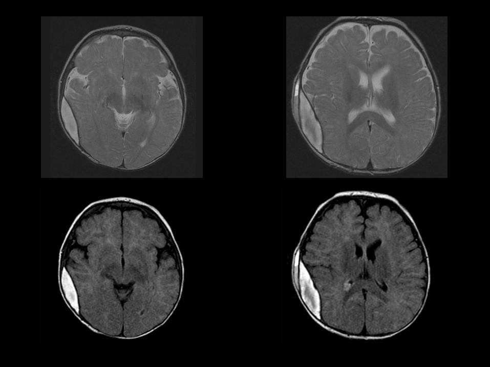
3 month3 month--old male with obtundationold male with obtundation
Potential Points of ImpactPotential Points of Impact
••
Patient history?Patient history?––
Do we know more than Do we know more than ““obtundationobtundation””??
––
Onset of symptoms?Onset of symptoms?––
History of cardiopulmonary arrest?History of cardiopulmonary arrest?
••
Are the referring physician and/or Are the referring physician and/or neuroradiologist aware?neuroradiologist aware?
••
Other studies to consider anticipating?Other studies to consider anticipating?––
MRI => confirm suspected acute ischemiaMRI => confirm suspected acute ischemia
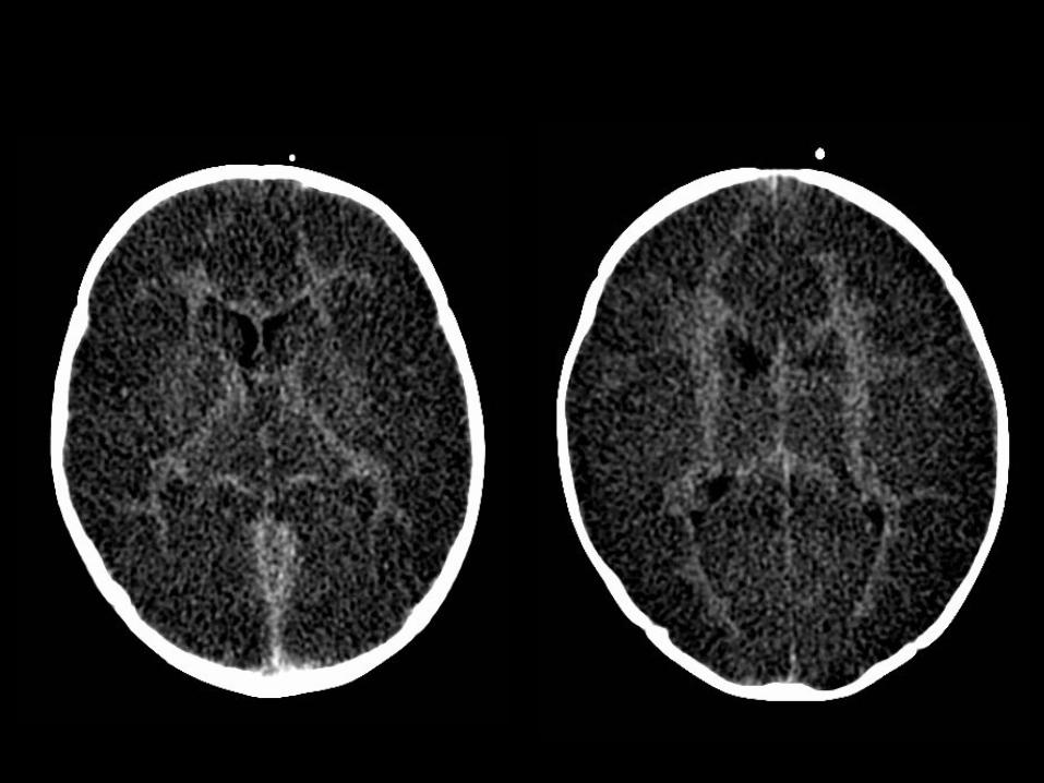
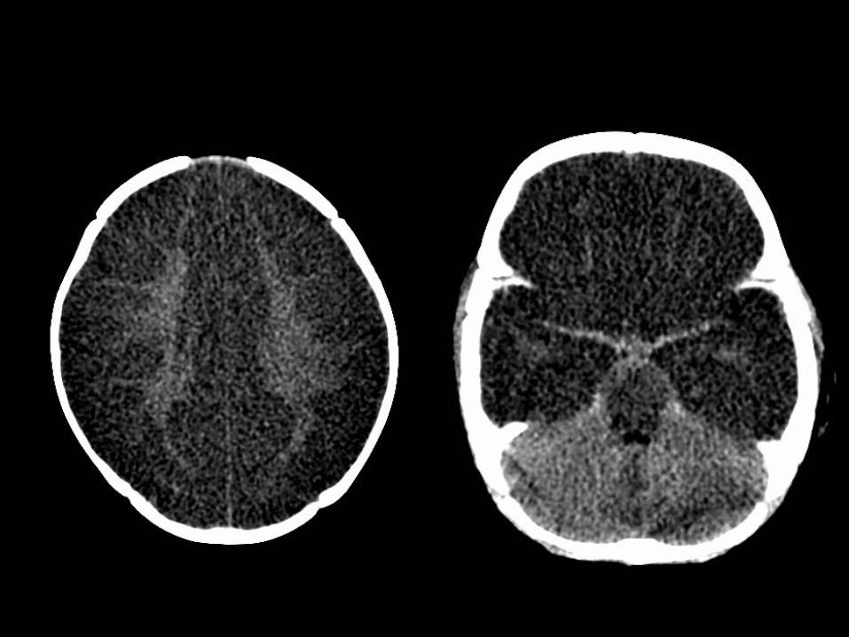
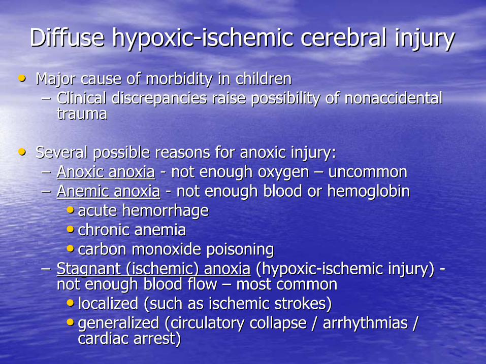
Diffuse hypoxicDiffuse hypoxic--ischemic cerebral injuryischemic cerebral injury
••
Major cause of morbidity in childrenMajor cause of morbidity in children––
Clinical discrepancies raise possibility of nonaccidental Clinical discrepancies raise possibility of nonaccidental traumatrauma
••
Several possible reasons for anoxic injury: Several possible reasons for anoxic injury: ––
Anoxic anoxiaAnoxic anoxia
--
not enough oxygen not enough oxygen ––
uncommonuncommon
––
Anemic anoxiaAnemic anoxia
--
not enough blood or hemoglobin not enough blood or hemoglobin ••
acute hemorrhage acute hemorrhage
••
chronic anemia chronic anemia ••
carbon monoxide poisoningcarbon monoxide poisoning
––
Stagnant (ischemic) anoxiaStagnant (ischemic) anoxia
(hypoxic(hypoxic--ischemic injury) ischemic injury) -- not enough blood flow not enough blood flow ––
most commonmost common
••
localized (such as ischemic strokes) localized (such as ischemic strokes) ••
generalized (circulatory collapse / arrhythmias / generalized (circulatory collapse / arrhythmias / cardiac arrest)cardiac arrest)

HeadacheHeadache
Potential Points of ImpactPotential Points of Impact
••
Patient history?Patient history?––
Do we know more than Do we know more than ““headacheheadache””??
––
Onset of symptoms? Recent trauma?Onset of symptoms? Recent trauma?––
Neurologic deficits?Neurologic deficits?
••
Are the referring physician and/or Are the referring physician and/or neuroradiologist aware?neuroradiologist aware?
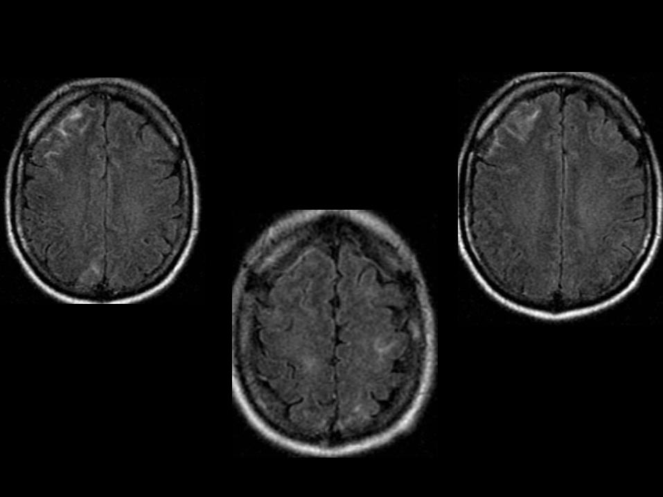
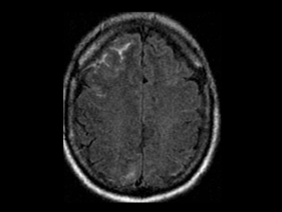
Subarachnoid hemorrhageSubarachnoid hemorrhage••
Differential diagnosis of hyperintense Differential diagnosis of hyperintense FLAIR signal within subarachnoid space:FLAIR signal within subarachnoid space:––
Subarachnoid hemorrhageSubarachnoid hemorrhage
––
Meningitis / pusMeningitis / pus
––
CarcinomatosisCarcinomatosis
––
Supplemental oxygenSupplemental oxygen••
Caution: Most sedated / general anesthesia Caution: Most sedated / general anesthesia patients will have this findingpatients will have this finding
Brain Brain ––
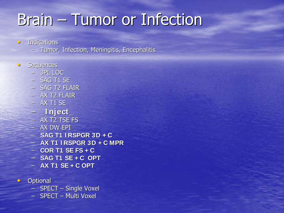
Tumor or InfectionTumor or Infection••
IndicationsIndications––
Tumor, Infection, Meningitis, EncephalitisTumor, Infection, Meningitis, Encephalitis
••
SequencesSequences––
3PL LOC3PL LOC––
SAG T1 SESAG T1 SE––
SAG T2 FLAIRSAG T2 FLAIR––
AX T2 FLAIRAX T2 FLAIR––
AX T1 SEAX T1 SE––
_Inject__Inject_––
AX T2 TSE FSAX T2 TSE FS––
AX DW EPIAX DW EPI––
SAG T1 IRSPGR 3D +CSAG T1 IRSPGR 3D +C––
AX T1 IRSPGR 3D +C MPRAX T1 IRSPGR 3D +C MPR––
COR T1 SE FS +CCOR T1 SE FS +C––
SAG T1 SE +C OPTSAG T1 SE +C OPT––
AX T1 SE +C OPTAX T1 SE +C OPT
••
OptionalOptional––
SPECT SPECT ––
Single VoxelSingle Voxel––
SPECT SPECT ––
Multi VoxelMulti Voxel
Mechanisms of contrast enhancementMechanisms of contrast enhancement
••
Combination of two primary processes in Combination of two primary processes in the central nervous system:the central nervous system:
(1) intravascular (vascular) enhancement(1) intravascular (vascular) enhancement
(2) interstitial (extravascular) enhancement (2) interstitial (extravascular) enhancement
Intravascular enhancementIntravascular enhancement
••
Proportional to increases in blood flow or blood volumeProportional to increases in blood flow or blood volume
••
Related to different pathologic / physiologic processes:Related to different pathologic / physiologic processes:
––
NeovascularityNeovascularity
––
Vasodilation or hyperemiaVasodilation or hyperemia
––
Shortened transit time or shunting Shortened transit time or shunting
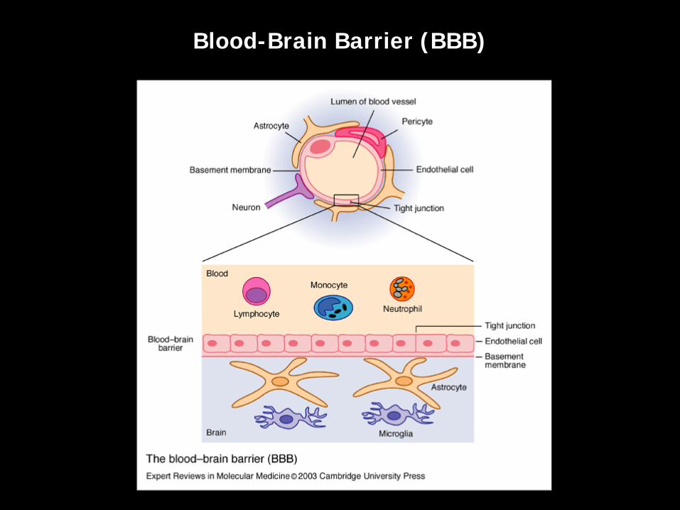
Interstitial enhancementInterstitial enhancement
••
Alterations in permeability of Alterations in permeability of bloodblood--brainbrain--barrier (BBB)barrier (BBB)––
SemiSemi--permeable capillary membranes within brain, spinal cord, and permeable capillary membranes within brain, spinal cord, and proximal nerves proximal nerves --
protect from plasma proteins and inflammatory protect from plasma proteins and inflammatory cellscells
––
Result of endothelial cell specialization Result of endothelial cell specialization ––
requires close relationship requires close relationship of the foot process of the perivascular astrocytes of the foot process of the perivascular astrocytes
––
Neural capillaries have a continuous basement membrane, narrow Neural capillaries have a continuous basement membrane, narrow intercellular gaps, junctional complexes, and a paucity of pinocintercellular gaps, junctional complexes, and a paucity of pinocytotic ytotic vesiclesvesicles
––
Blocks lipophobic compounds and creates a unique interstitial flBlocks lipophobic compounds and creates a unique interstitial fluid uid environment for neural tissues environment for neural tissues
Blood-Brain Barrier (BBB)
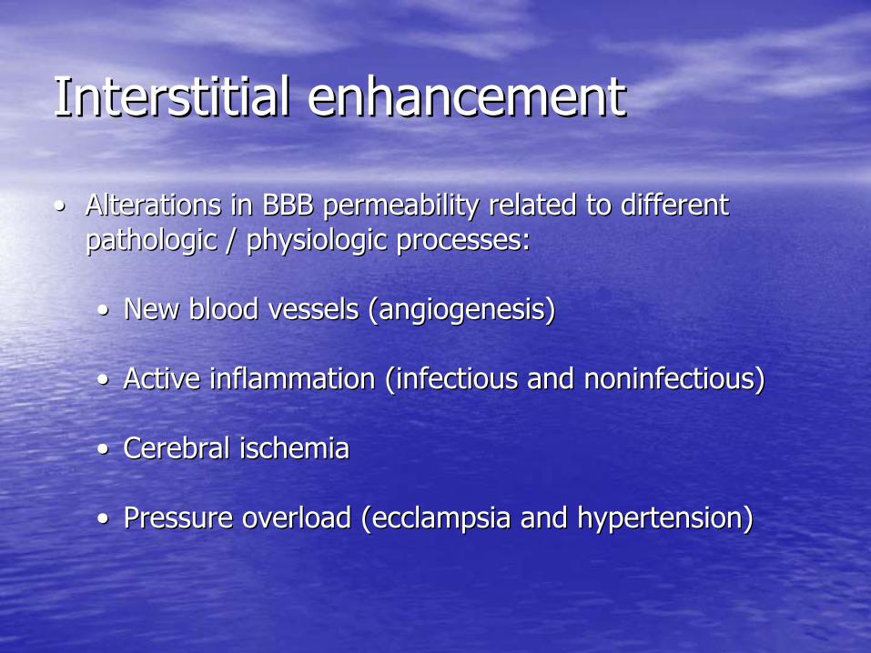
Interstitial enhancementInterstitial enhancement
••
Alterations in BBB permeability related to different Alterations in BBB permeability related to different pathologic / physiologic processes:pathologic / physiologic processes:
••
New blood vessels (angiogenesis)New blood vessels (angiogenesis)
••
Active inflammation (infectious and noninfectious)Active inflammation (infectious and noninfectious)
••
Cerebral ischemiaCerebral ischemia
••
Pressure overload (ecclampsia and hypertension) Pressure overload (ecclampsia and hypertension)
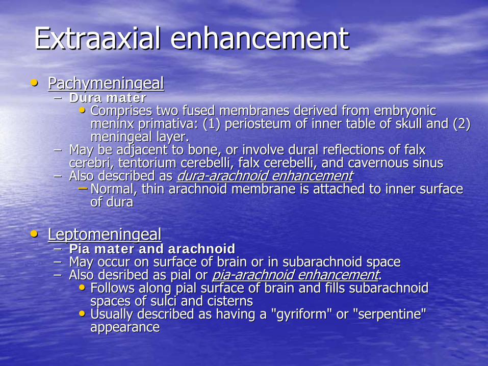
Extraaxial enhancementExtraaxial enhancement••
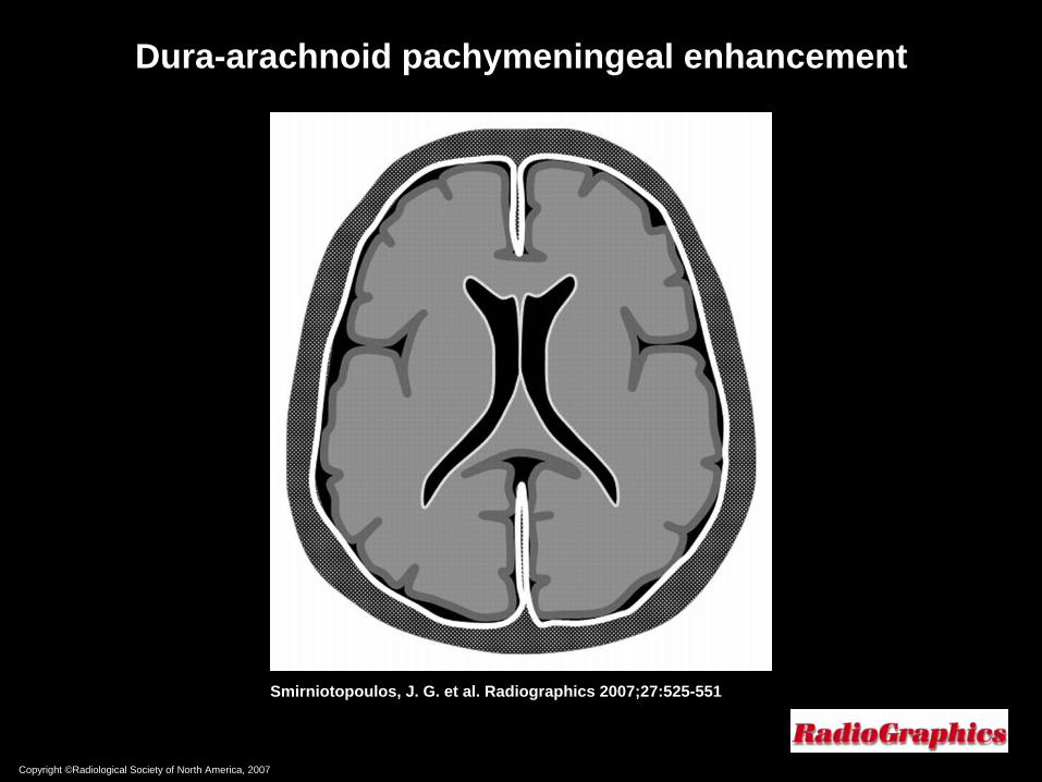
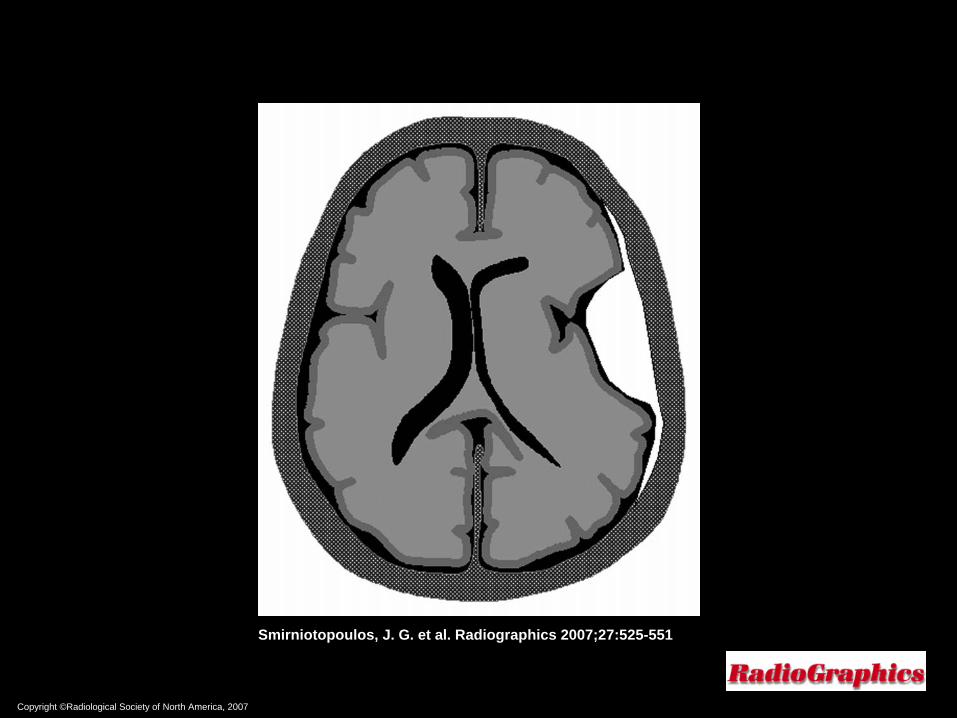
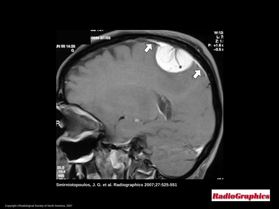
PachymeningealPachymeningeal––
Dura materDura mater••
Comprises two fused membranes derived from embryonic Comprises two fused membranes derived from embryonic meninx primativa: (1) periosteum of inner table of skull and (2)meninx primativa: (1) periosteum of inner table of skull and (2)
meningeal layer. meningeal layer.
––
May be adjacent to bone, or involve dural reflections of falx May be adjacent to bone, or involve dural reflections of falx cerebri, tentorium cerebelli, falx cerebelli, and cavernous sinucerebri, tentorium cerebelli, falx cerebelli, and cavernous sinuss
––
Also described as Also described as duradura--arachnoid enhancementarachnoid enhancement––
Normal, thin arachnoid membrane is attached to inner surface Normal, thin arachnoid membrane is attached to inner surface of duraof dura
••
LeptomeningealLeptomeningeal––
Pia mater and arachnoidPia mater and arachnoid––
May occur on surface of brain or in subarachnoid space May occur on surface of brain or in subarachnoid space ––
Also desribed as pial or Also desribed as pial or piapia--arachnoid enhancementarachnoid enhancement. . ••
Follows along pial surface of brain and fills subarachnoid Follows along pial surface of brain and fills subarachnoid spaces of sulci and cisterns spaces of sulci and cisterns
••
Usually described as having a "gyriform" or "serpentine" Usually described as having a "gyriform" or "serpentine" appearance appearance
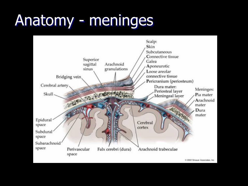
Anatomy Anatomy --
meningesmeninges
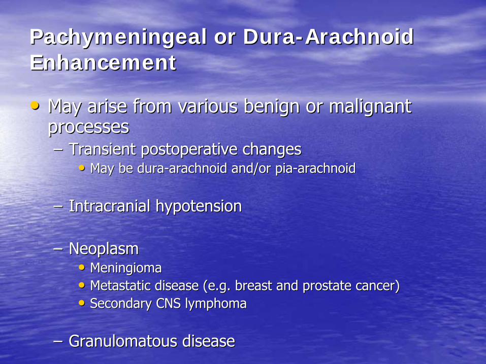
Pachymeningeal or DuraPachymeningeal or Dura--Arachnoid Arachnoid EnhancementEnhancement
••
May arise from various benign or malignant May arise from various benign or malignant processesprocesses––
Transient postoperative changesTransient postoperative changes••
May be duraMay be dura--arachnoid and/or piaarachnoid and/or pia--arachnoidarachnoid
––
Intracranial hypotensionIntracranial hypotension
––
NeoplasmNeoplasm••
MeningiomaMeningioma
••
Metastatic disease (e.g. breast and prostate cancer)Metastatic disease (e.g. breast and prostate cancer)••
Secondary CNS lymphomaSecondary CNS lymphoma
––
Granulomatous diseaseGranulomatous disease
Copyright ©Radiological Society of North America, 2007
Smirniotopoulos, J. G. et al. Radiographics 2007;27:525-551
Dura-arachnoid pachymeningeal enhancement

Copyright ©Radiological Society of North America, 2007
Smirniotopoulos, J. G. et al. Radiographics 2007;27:525-551
Copyright ©Radiological Society of North America, 2007
Smirniotopoulos, J. G. et al. Radiographics 2007;27:525-551
Copyright ©Radiological Society of North America, 2007
Smirniotopoulos, J. G. et al. Radiographics 2007;27:525-551
Copyright ©Radiological Society of North America, 2007
Smirniotopoulos, J. G. et al. Radiographics 2007;27:525-551
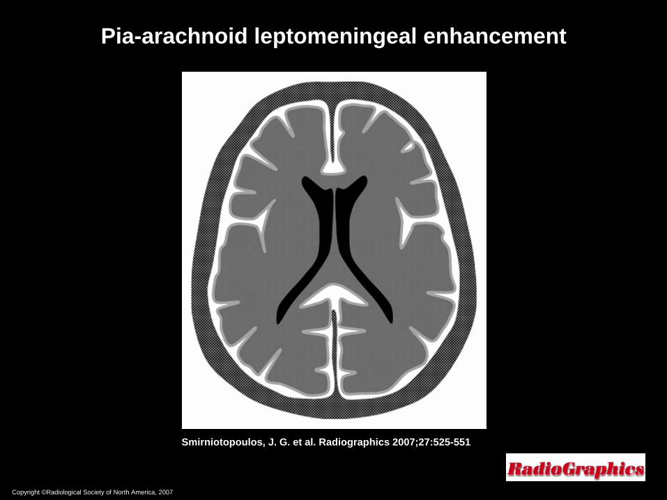
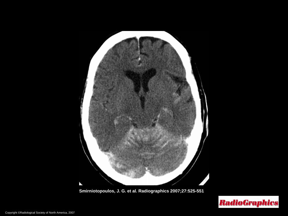
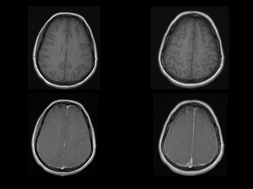
Pia-arachnoid leptomeningeal enhancement
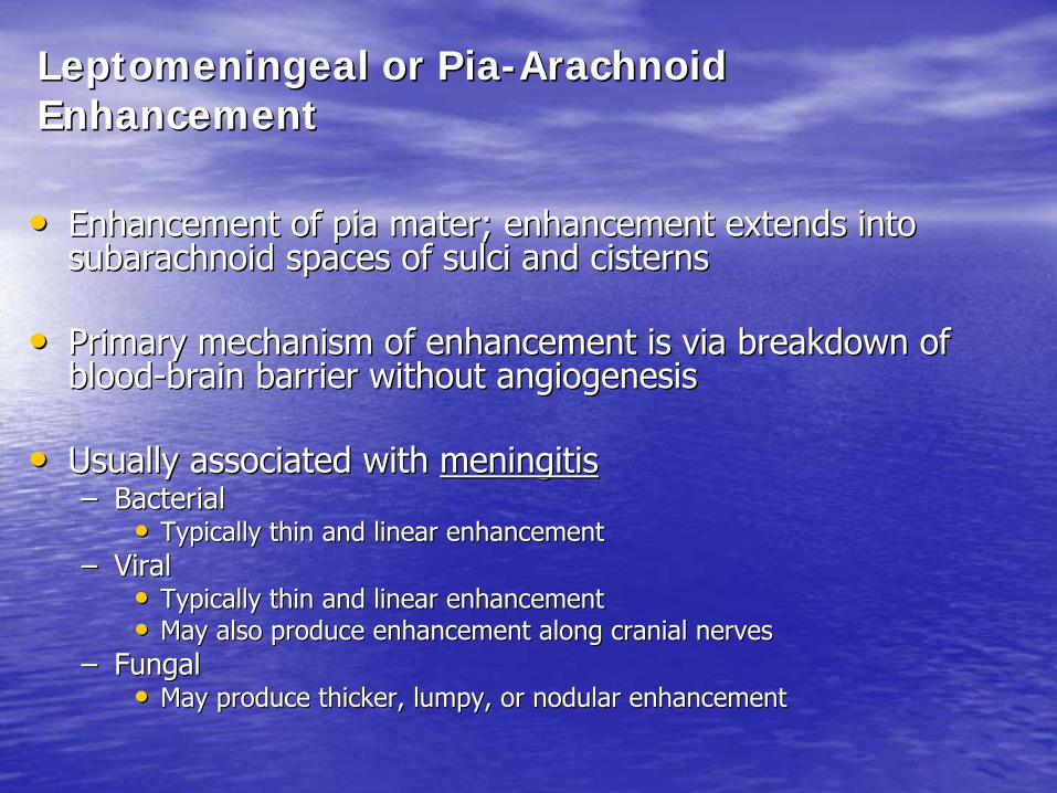
Leptomeningeal or PiaLeptomeningeal or Pia--Arachnoid Arachnoid EnhancementEnhancement
••
Enhancement of pia mater; enhancement extends into Enhancement of pia mater; enhancement extends into subarachnoid spaces of sulci and cisternssubarachnoid spaces of sulci and cisterns
••
Primary mechanism of enhancement is via breakdown of Primary mechanism of enhancement is via breakdown of bloodblood--brain barrier without angiogenesis brain barrier without angiogenesis
••
Usually associated with Usually associated with meningitismeningitis––
Bacterial Bacterial ••
Typically thin and linear enhancementTypically thin and linear enhancement
––
ViralViral••
Typically thin and linear enhancementTypically thin and linear enhancement
••
May also produce enhancement along cranial nervesMay also produce enhancement along cranial nerves
––
FungalFungal••
May produce thicker, lumpy, or nodular enhancementMay produce thicker, lumpy, or nodular enhancement
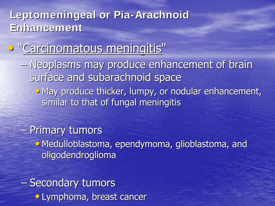
Leptomeningeal or PiaLeptomeningeal or Pia--Arachnoid Arachnoid EnhancementEnhancement
••
““Carcinomatous meningitisCarcinomatous meningitis" " ––
Neoplasms may produce enhancement of brain Neoplasms may produce enhancement of brain surface and subarachnoid spacesurface and subarachnoid space••
May produce thicker, lumpy, or nodular enhancement, May produce thicker, lumpy, or nodular enhancement, similar to that of fungal meningitissimilar to that of fungal meningitis
––
Primary tumorsPrimary tumors••
Medulloblastoma, ependymoma, glioblastoma, and Medulloblastoma, ependymoma, glioblastoma, and oligodendrogliomaoligodendroglioma
––
Secondary tumorsSecondary tumors••
Lymphoma, breast cancer Lymphoma, breast cancer
Copyright ©Radiological Society of North America, 2007
Smirniotopoulos, J. G. et al. Radiographics 2007;27:525-551

Copyright ©Radiological Society of North America, 2007
Smirniotopoulos, J. G. et al. Radiographics 2007;27:525-551
Copyright ©Radiological Society of North America, 2007
Smirniotopoulos, J. G. et al. Radiographics 2007;27:525-551
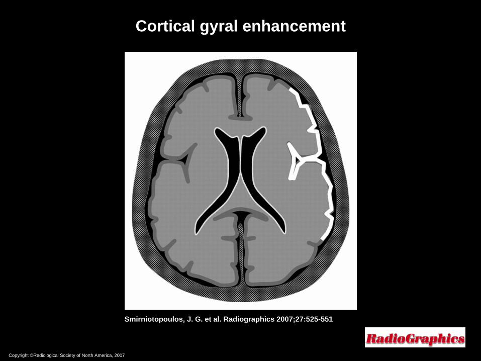
Cortical gyral enhancement
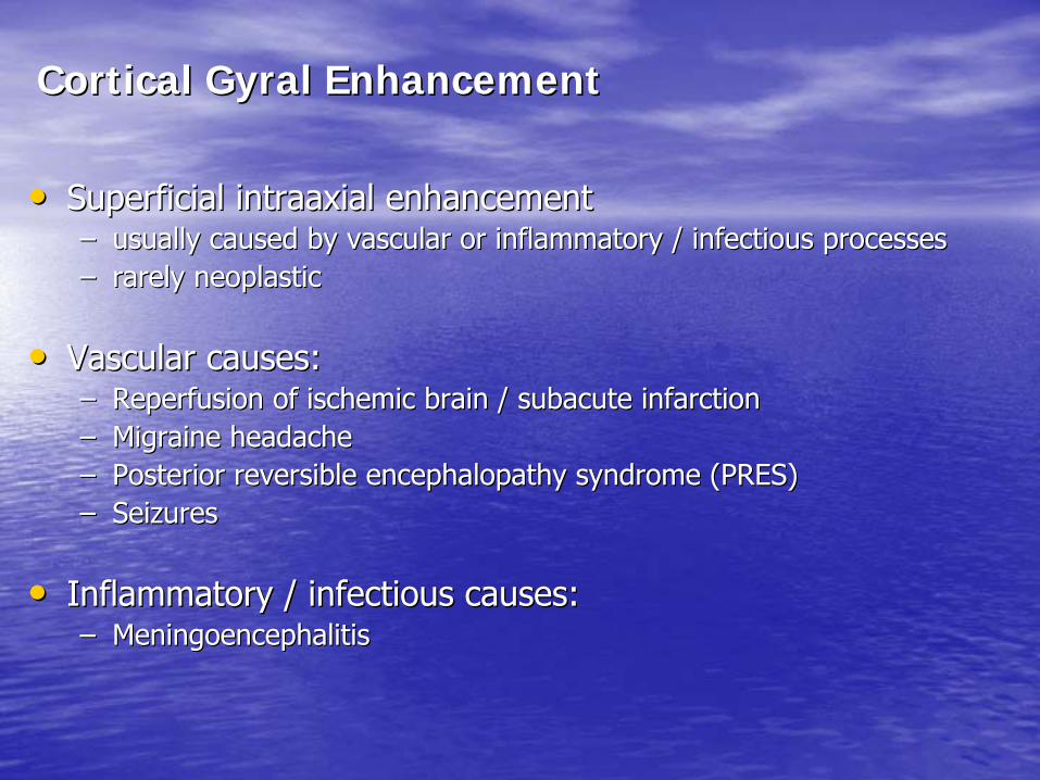
Cortical Gyral EnhancementCortical Gyral Enhancement
••
Superficial intraaxial enhancementSuperficial intraaxial enhancement––
usually caused by vascular or inflammatory / infectious processeusually caused by vascular or inflammatory / infectious processess––
rarely neoplasticrarely neoplastic
••
Vascular causes:Vascular causes:––
Reperfusion of ischemic brain / subacute infarctionReperfusion of ischemic brain / subacute infarction––
Migraine headacheMigraine headache––
Posterior reversible encephalopathy syndrome (PRES)Posterior reversible encephalopathy syndrome (PRES)––
SeizuresSeizures
••
Inflammatory / infectious causes:Inflammatory / infectious causes:––
Meningoencephalitis Meningoencephalitis
Copyright ©Radiological Society of North America, 2007
Smirniotopoulos, J. G. et al. Radiographics 2007;27:525-551
Copyright ©Radiological Society of North America, 2007
Smirniotopoulos, J. G. et al. Radiographics 2007;27:525-551
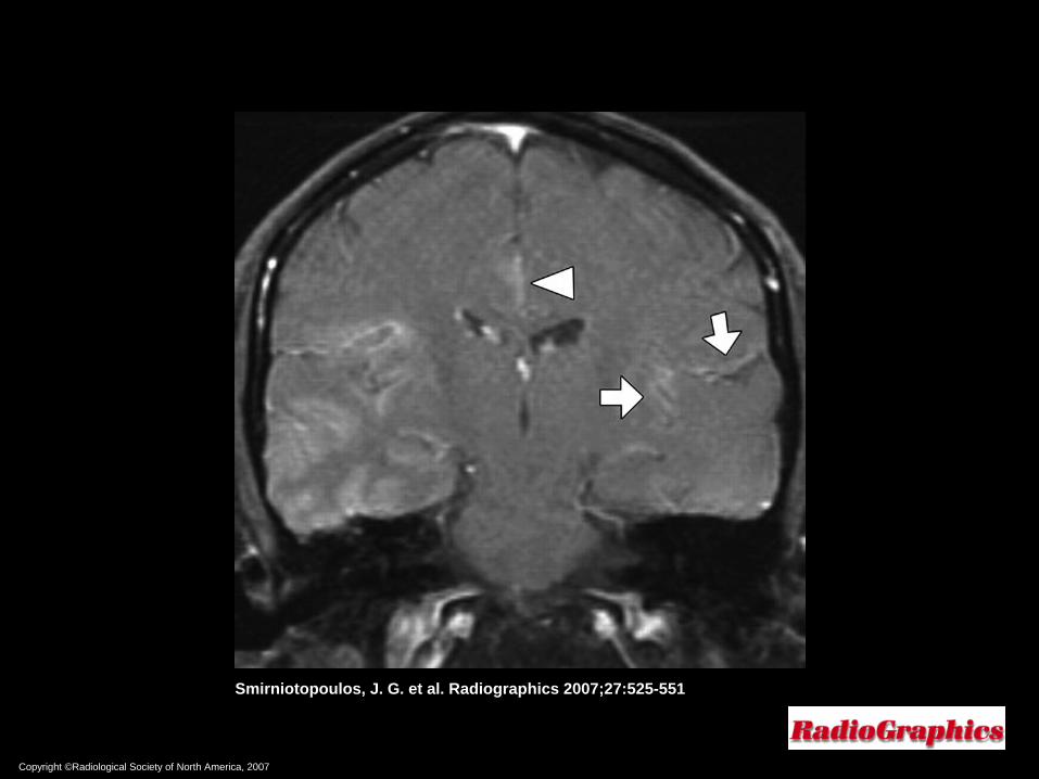
Subcortical nodular enhancement

Nodular Cortical and Subcortical Nodular Cortical and Subcortical EnhancementEnhancement
••
Typical for hematogenous dissemination of metastatic disease andTypical for hematogenous dissemination of metastatic disease and
clot emboliclot emboli
••
Metastatic disease usually travels into brain through arteries aMetastatic disease usually travels into brain through arteries and nd less commonly via venous system less commonly via venous system --
majority are supratentorial majority are supratentorial
••
Metastatic lesions are typically subcortical, occurring in or neMetastatic lesions are typically subcortical, occurring in or near the ar the gray mattergray matter––white matter (corticomedullary) junctionwhite matter (corticomedullary) junction
••
Angiogenesis allows metastases to grow larger but also produces Angiogenesis allows metastases to grow larger but also produces BBB abnormality, which results in contrast enhancement and BBB abnormality, which results in contrast enhancement and considerable perilesional vasogenic edemaconsiderable perilesional vasogenic edema
••
Because of location, cortical and subcortical metastases, even aBecause of location, cortical and subcortical metastases, even as s small lesions, are likely to cause noticeable neurologic symptomsmall lesions, are likely to cause noticeable neurologic symptoms, s, including seizuresincluding seizures
Copyright ©Radiological Society of North America, 2007
Smirniotopoulos, J. G. et al. Radiographics 2007;27:525-551
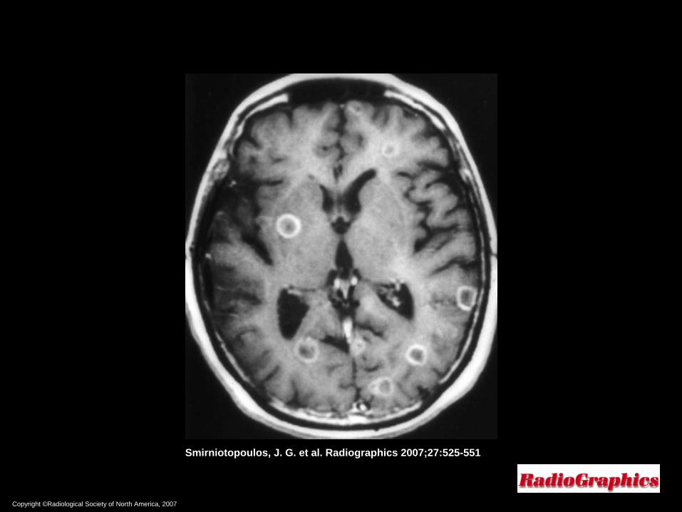
Deep Ring EnhancementDeep Ring Enhancement
••
Deep white matter ringDeep white matter ring--enhancing lesions, especially enhancing lesions, especially those with mass effect and surrounding vasogenic those with mass effect and surrounding vasogenic edema, are most often either primary neoplasms (eg, edema, are most often either primary neoplasms (eg, glioblastoma multiforme) or abscesses glioblastoma multiforme) or abscesses
••
Metastatic deposits are often solid nodular lesions that Metastatic deposits are often solid nodular lesions that may become ringmay become ring--enhancing because of necrosis (eg, enhancing because of necrosis (eg, after chemotherapy or irradiation)after chemotherapy or irradiation)
••
Consider infectious etiology (brain abscess) in patients Consider infectious etiology (brain abscess) in patients with subacute bacterial endocarditis, indwelling with subacute bacterial endocarditis, indwelling catheters, or other implanted devices (eg. cardiac catheters, or other implanted devices (eg. cardiac valves)valves)
Copyright ©Radiological Society of North America, 2007
Smirniotopoulos, J. G. et al. Radiographics 2007;27:525-551
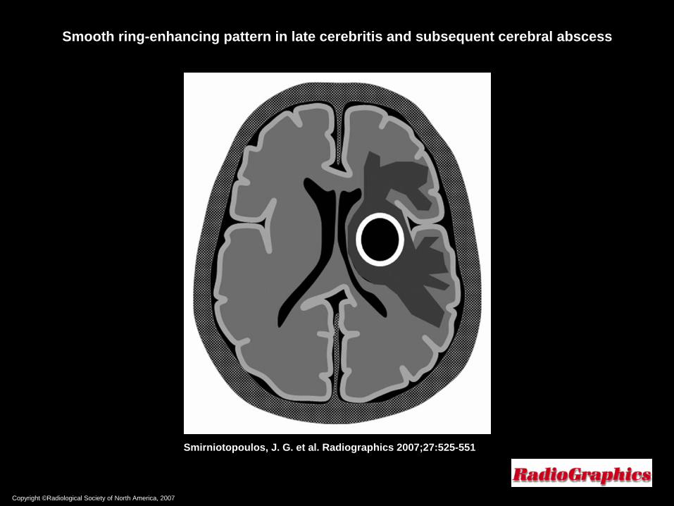
Smooth ring-enhancing pattern in late cerebritis and subsequent cerebral abscess
Copyright ©Radiological Society of North America, 2007
Smirniotopoulos, J. G. et al. Radiographics 2007;27:525-551
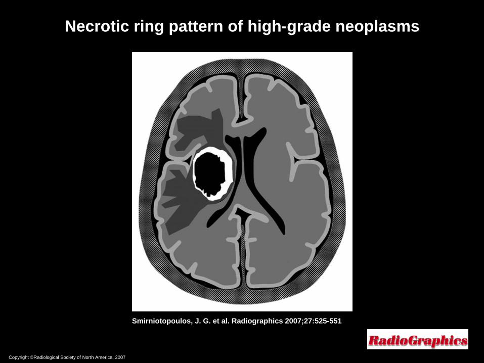
Necrotic ring pattern of high-grade neoplasms
Copyright ©Radiological Society of North America, 2007
Smirniotopoulos, J. G. et al. Radiographics 2007;27:525-551

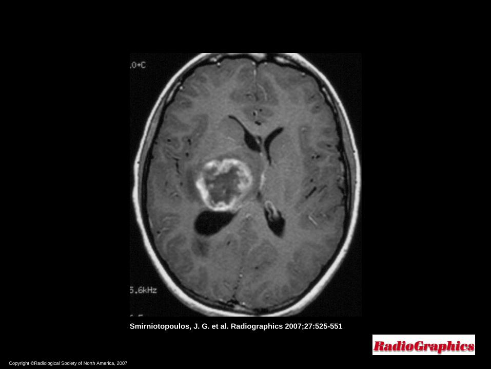
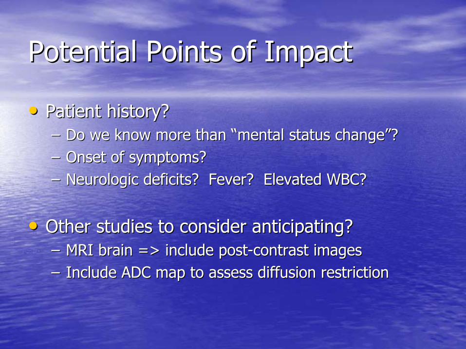
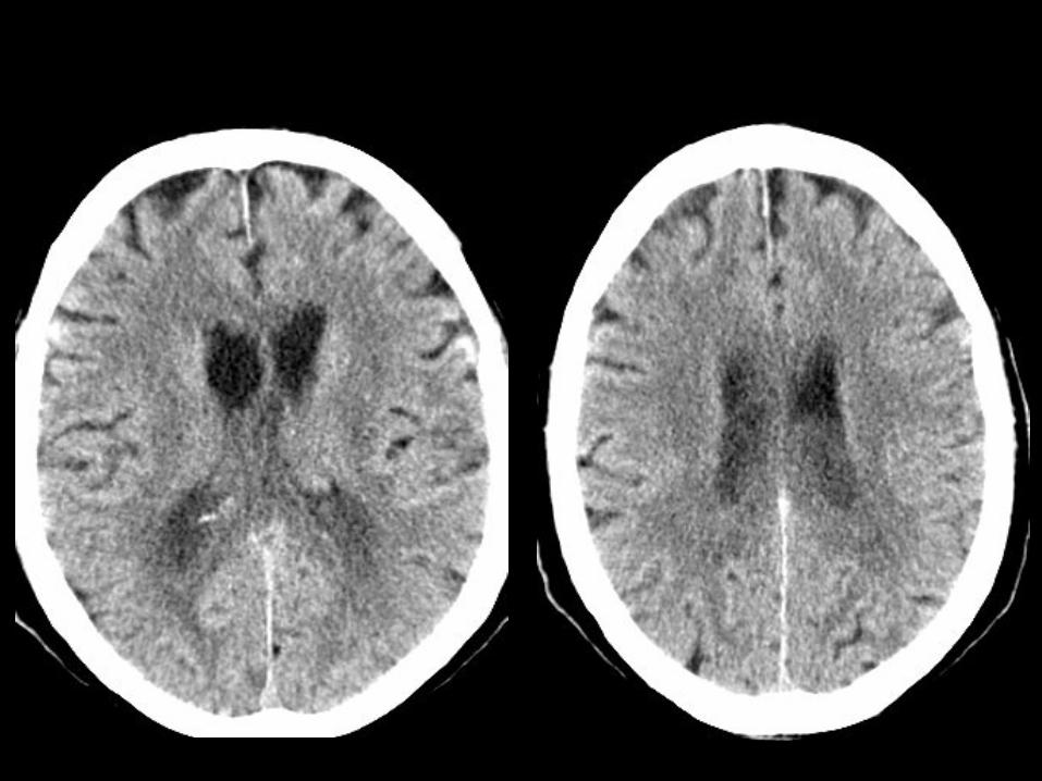
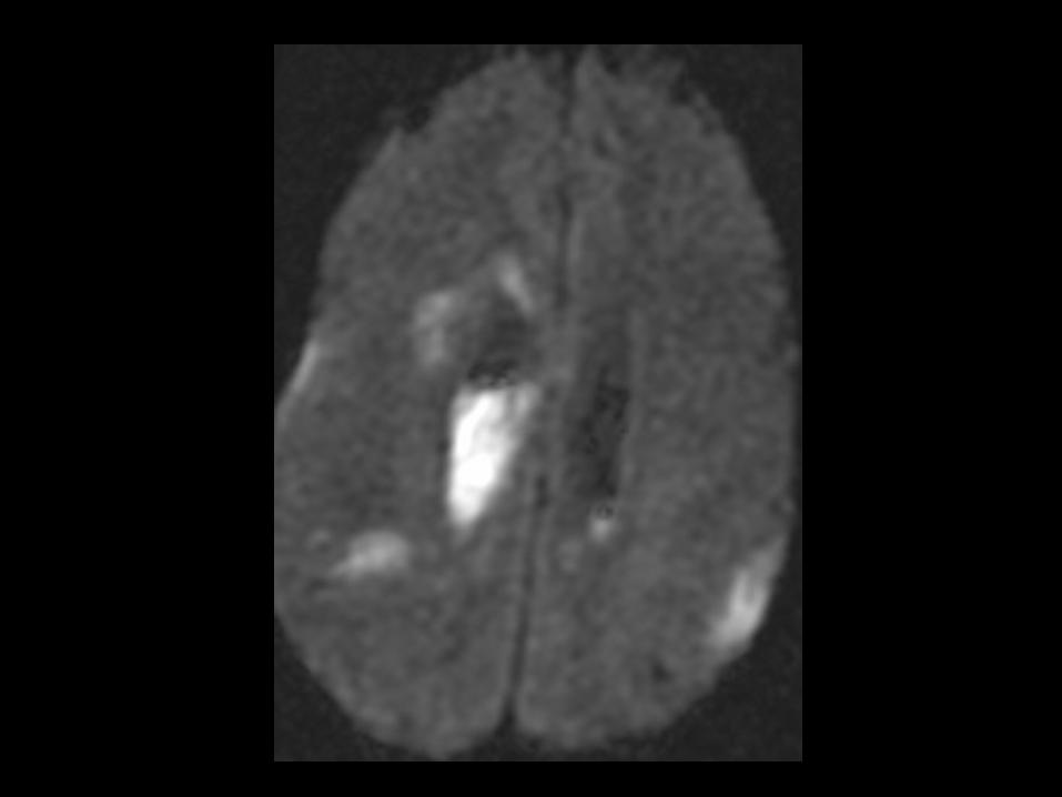
37 year37 year--old male with mental status changeold male with mental status change
Potential Points of ImpactPotential Points of Impact
••
Patient history?Patient history?––
Do we know more than Do we know more than ““mental status changemental status change””??
––
Onset of symptoms?Onset of symptoms?––
Neurologic deficits? Fever? Elevated WBC?Neurologic deficits? Fever? Elevated WBC?
••
Other studies to consider anticipating?Other studies to consider anticipating?––
MRI brain => include postMRI brain => include post--contrast imagescontrast images
––
Include ADC map to assess diffusion restrictionInclude ADC map to assess diffusion restriction
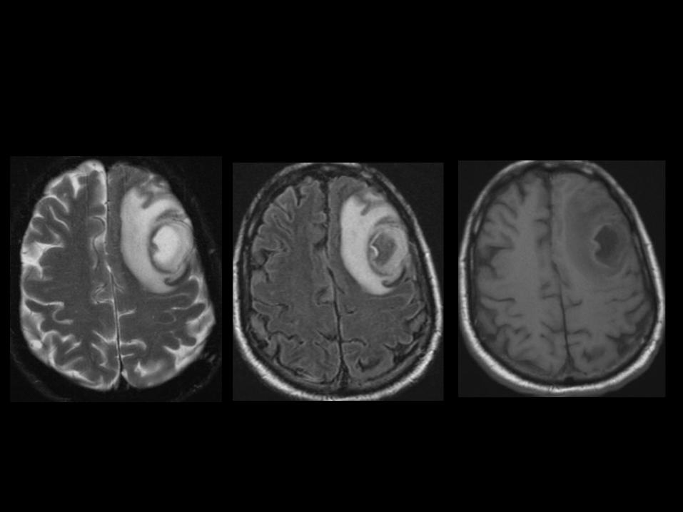
Cerebral abscessCerebral abscess
••
Key characteristics: central diffusion restriction; rim Key characteristics: central diffusion restriction; rim enhancement; vasogenic edemaenhancement; vasogenic edema
••
Differential diagnosis:Differential diagnosis:––
NonNon--HodgkinHodgkin’’s lymphomas lymphoma••
Can have rim enhancement and restricted diffusion, but usually iCan have rim enhancement and restricted diffusion, but usually isoso--
to low T2 signalto low T2 signal
––
GBMGBM••
Usually more heterogeneous, without large fluid center or diffusUsually more heterogeneous, without large fluid center or diffusion ion restrictionrestriction
––
Tumefactive MS (multiple sclerosis)Tumefactive MS (multiple sclerosis)––
MetastasisMetastasis––
Resolving hematomaResolving hematoma––
InfarctInfarct

65 year65 year--old female with altered mental old female with altered mental statusstatus

Potential Points of ImpactPotential Points of Impact
••
Patient history?Patient history?––
Do we know more than Do we know more than ““altered mental statusaltered mental status””??
––
Onset of symptoms?Onset of symptoms?––
Neurologic deficits? Fever? Elevated WBC?Neurologic deficits? Fever? Elevated WBC?
••
Other studies to consider anticipating?Other studies to consider anticipating?––
MRI brain => include postMRI brain => include post--contrast images, contrast images, particularly if infectious process is suspectedparticularly if infectious process is suspected

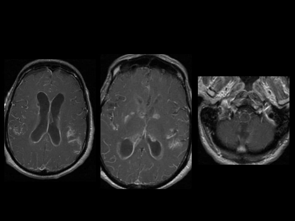
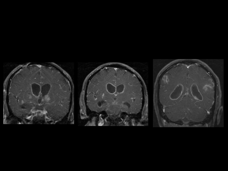
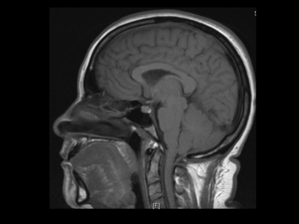
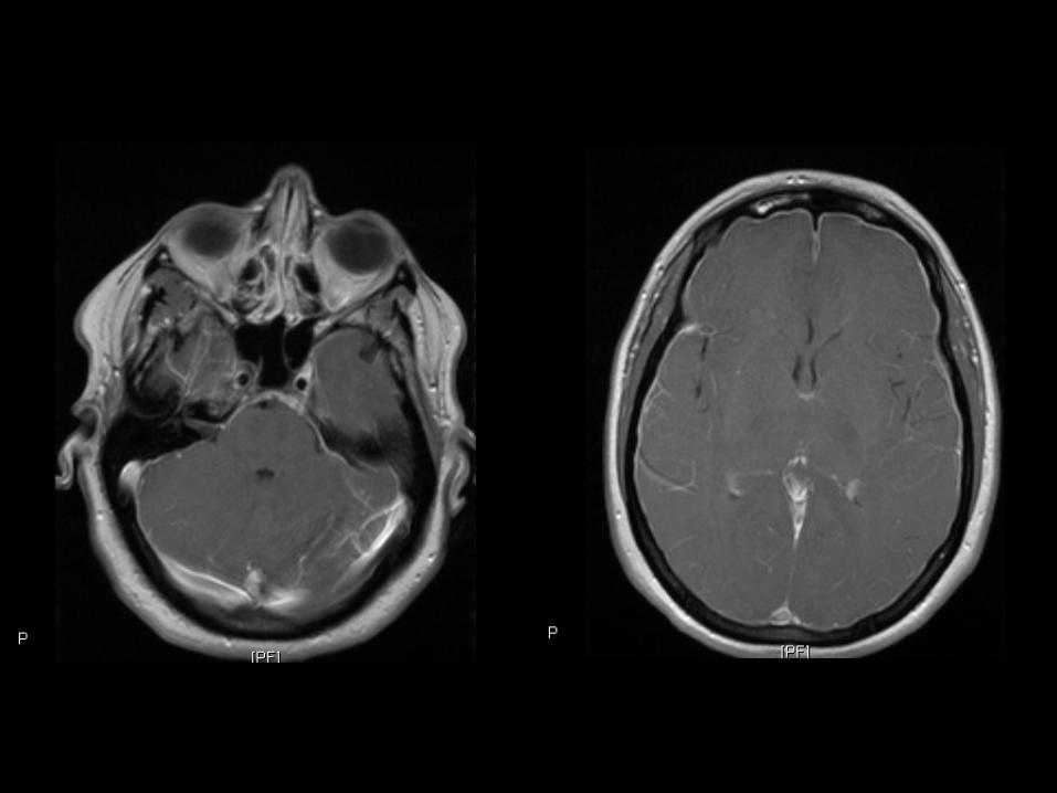
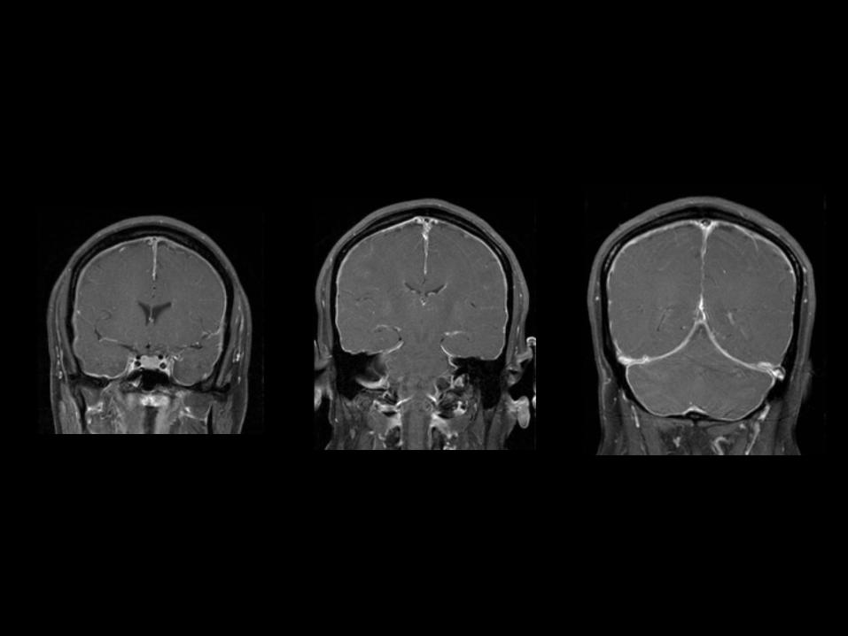
Meningitis / VentriculitisMeningitis / Ventriculitis
••
Debris in ventricles with ependymal Debris in ventricles with ependymal enhancement enhancement
ventriculitisventriculitis
••
Can have ependymal enhancement with NHL, Can have ependymal enhancement with NHL, spread of GBM, etc. but usually more spread of GBM, etc. but usually more nodular/focal and w/o debrisnodular/focal and w/o debris
••
Often associated with abscess rupture or Often associated with abscess rupture or indwelling shunt catheterindwelling shunt catheter
••
4040--80% mortality but often indolent80% mortality but often indolent

45 year45 year--old female who presented with old female who presented with severe headache; recent lumbar puncture severe headache; recent lumbar puncture
performed for suspected meningitisperformed for suspected meningitis
Potential Points of ImpactPotential Points of Impact
••
Patient history?Patient history?––
Do we know more than Do we know more than ““severe headachesevere headache””??
––
Any neurologic symptoms?Any neurologic symptoms?––
Recent surgery?Recent surgery?
••
Other studies to consider anticipating?Other studies to consider anticipating?––
MRI brain WITH contrast MRI brain WITH contrast ––
concern for concern for
meningitismeningitis
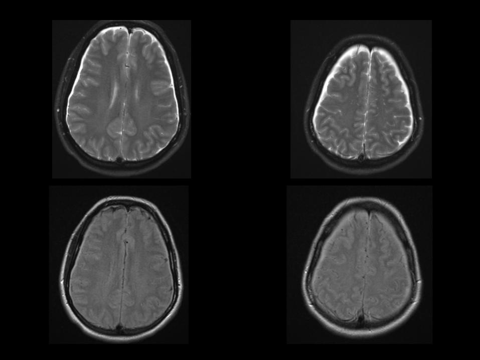

Intracranial HypotensionIntracranial Hypotension••
Result of low CSF volume caused by:Result of low CSF volume caused by:
––
Head traumaHead trauma
––
Tear in spinal nerve root sheath, perineural cyst, or Tear in spinal nerve root sheath, perineural cyst, or spinal arachnoid diverticulumspinal arachnoid diverticulum
––
Iatrogenic causesIatrogenic causes••
Lumbar punctureLumbar puncture
••
Overdraining ventricular or spinal shuntsOverdraining ventricular or spinal shunts
––
SpontaneousSpontaneous••
Results from rupture of spinal arachnoid Results from rupture of spinal arachnoid membrane, which allows CSF passage into membrane, which allows CSF passage into subdural or epidural spacesubdural or epidural space
Brain Brain ––
Trauma / HemorrhageTrauma / Hemorrhage
••
IndicationsIndications––
Trauma, HemorrhageTrauma, Hemorrhage
••
SequencesSequences––
3 PL LOC3 PL LOC––
Sag T1 SESag T1 SE––
Sag T2 FLAIRSag T2 FLAIR––
Ax T2 FLAIRAx T2 FLAIR––
Ax T1 SEAx T1 SE––
Ax T2 TSE FSAx T2 TSE FS––
Ax DWI EPIAx DWI EPI––
Cor T2 TSECor T2 TSE––
Ax GRE or SWIAx GRE or SWI
•
Comments–
Axial GRE should have TE > 25 ms

77--month old male with scalp swellingmonth old male with scalp swelling
Potential Points of ImpactPotential Points of Impact
••
Patient history?Patient history?––
Do we know more than Do we know more than ““scalp swellingscalp swelling””??––
History of trauma or fall?History of trauma or fall?
••
Are the referring physician and/or neuroradiologist Are the referring physician and/or neuroradiologist aware?aware?––
Must exclude nonMust exclude non--accidental traumaaccidental trauma
••
Other studies to consider anticipating?Other studies to consider anticipating?––
CT => characterize suspected fracture and evaluate for potentialCT => characterize suspected fracture and evaluate for potential
associated intracranial hemorrhageassociated intracranial hemorrhage••
Soft tissue and bone algorithmsSoft tissue and bone algorithms
––
MRI => assess for additional parenchymal injuries and MRI => assess for additional parenchymal injuries and potentially assist with injury / hemorrhage datingpotentially assist with injury / hemorrhage dating
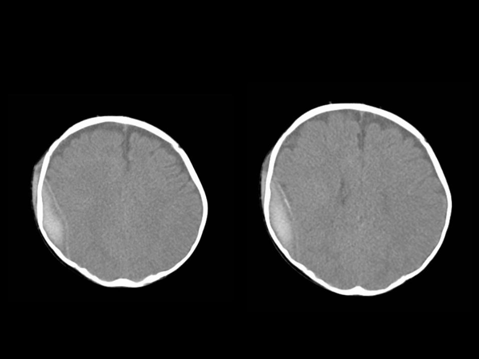
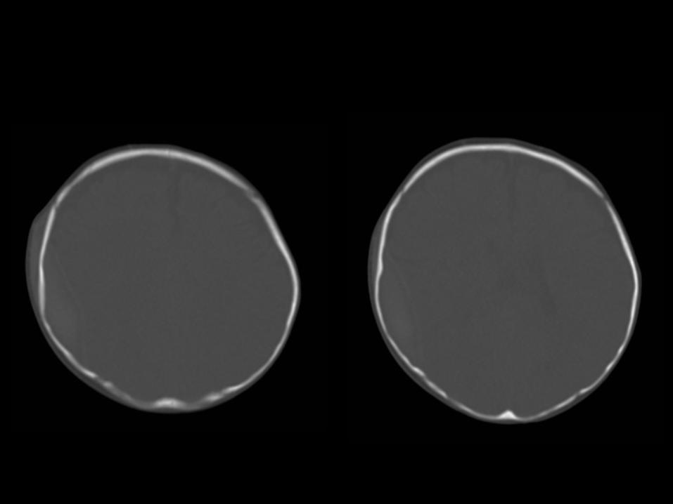


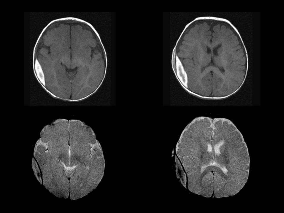
NonNon--accidental traumaaccidental trauma••
CT CT --
recommended in initial evaluation of nonrecommended in initial evaluation of non--accidental traumaaccidental trauma––
High sensitivity in detecting acute intracranial bleed, fractureHigh sensitivity in detecting acute intracranial bleed, fractures, s, cerebral edema and hypoxiccerebral edema and hypoxic--ischemic injury ischemic injury
––
Attenuation of subdural / epidural hematoma varies by chronicityAttenuation of subdural / epidural hematoma varies by chronicity::••
Acute Acute --
hyperdense hyperdense ••
Subacute Subacute ––
isodenseisodense••
Chronic Chronic ––
hypodense hypodense
••
MRI MRI --
essential second investigationessential second investigation––
Best performed 5Best performed 5--10 days after insult10 days after insult––
Reliably differentiate between acute and chronic subdural hematoReliably differentiate between acute and chronic subdural hematoma ma ––
Most sensitive modality for detecting early ischemic changes Most sensitive modality for detecting early ischemic changes ––
Clearly delineates anatomic locations difficult to image with CTClearly delineates anatomic locations difficult to image with CT••
posterior fossa, anterior part of middle cranial fossa, close toposterior fossa, anterior part of middle cranial fossa, close to
inner table of skullinner table of skull

80 year80 year--old male with dementia that has old male with dementia that has progressed over the past 4 years progressed over the past 4 years
Potential Points of ImpactPotential Points of Impact
••
Patient history?Patient history?––
Do we know more than Do we know more than ““dementiadementia””??
––
Previous CVA symptoms? Risk factors?Previous CVA symptoms? Risk factors?––
Neurologic deficits?Neurologic deficits?
••
Other studies to consider anticipating?Other studies to consider anticipating?––
GRE imaging GRE imaging ––
evaluate for previous evaluate for previous
hemorrhage associated with infarctshemorrhage associated with infarcts
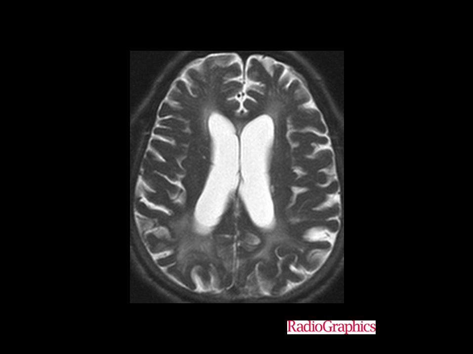
Figure 3b. Sensitivity of GRE imaging for hemosiderin in an 80-year-old man with dementia that has progressed over the past 4 years
Chao C P et al. Radiographics 2006;26:1517-1531
©2006 by Radiological Society of North America

Figure 3a. Sensitivity of GRE imaging for hemosiderin in an 80-year-old man with dementia that has progressed over the past 4 years
Chao C P et al. Radiographics 2006;26:1517-1531
©2006 by Radiological Society of North America
Cerebral Amyloid AngiopathyCerebral Amyloid Angiopathy••
Cerebrovascular disorder characterized by Cerebrovascular disorder characterized by deposition of deposition of ββ--amyloid protein in the media and amyloid protein in the media and adventitia of small and mediumadventitia of small and medium--sized vesselssized vessels
••
Both sporadic and hereditary forms may occurBoth sporadic and hereditary forms may occur
••
Manifests radiologically as part or all of a Manifests radiologically as part or all of a constellation of findings including acute or constellation of findings including acute or chronic ICHs in a distinctive corticalchronic ICHs in a distinctive cortical--subcortical subcortical distribution, leukoencephalopathy, and atrophy distribution, leukoencephalopathy, and atrophy

SusceptibilitySusceptibility--Weighted ImagingWeighted Imaging
••
3D high3D high--resolution velocityresolution velocity--compensated compensated gradientgradient--echo sequence that exploits echo sequence that exploits magnetic properties of tissuesmagnetic properties of tissues
••
Refers to use of resultant magnitude and Refers to use of resultant magnitude and phase images, or a combination thereofphase images, or a combination thereof
••
Can provide detailed assessment of Can provide detailed assessment of venous architecturevenous architecture
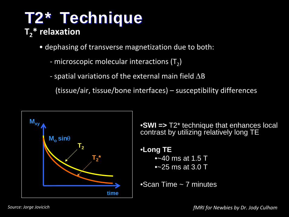
T2* TechniqueT2* Technique
Source: Jorge Jovicich
time
Mxy
Mo sinT2
T2 *
T2
* relaxation
• dephasing of transverse magnetization due to both:
‐
microscopic molecular interactions (T2
)
‐
spatial variations of the external main field B
(tissue/air, tissue/bone interfaces) –
susceptibility differences
fMRI for Newbies by Dr. Jody Culham
•SWI => T2* technique that enhances local contrast by utilizing relatively long TE
•Long TE•~40 ms at 1.5 T•~25 ms at 3.0 T
•Scan Time ~ 7 minutes
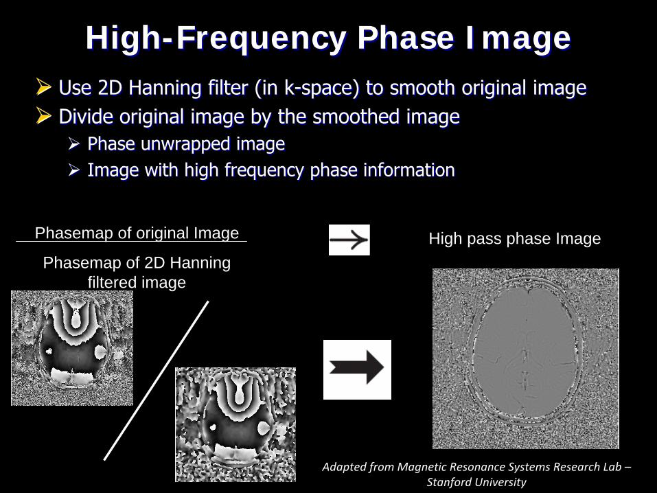
Use 2D Hanning filter (in kUse 2D Hanning filter (in k--space) to smooth original imagespace) to smooth original image
Divide original image by the smoothed image Divide original image by the smoothed image
Phase unwrapped imagePhase unwrapped image
Image with high frequency phase informationImage with high frequency phase information
Phasemap of original Image
Phasemap of 2D Hanning filtered image
High pass phase Image
HighHigh--Frequency Phase ImageFrequency Phase Image
Adapted from Magnetic Resonance Systems Research Lab –
Stanford University
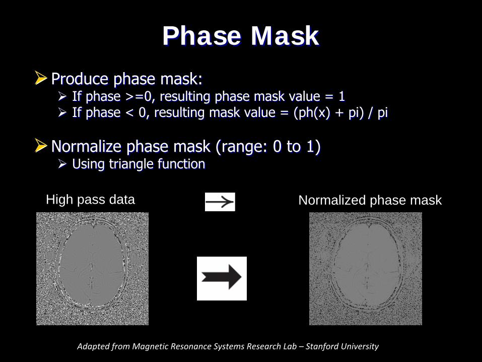
Produce phase mask:Produce phase mask:
If phase >=0, resulting phase mask value = 1 If phase >=0, resulting phase mask value = 1
If phase < 0, resulting mask value = (ph(x) + pi) / piIf phase < 0, resulting mask value = (ph(x) + pi) / pi
Normalize phase mask (range: 0 to 1)Normalize phase mask (range: 0 to 1)
Using triangle functionUsing triangle function
Normalized phase maskHigh pass data
Phase MaskPhase Mask
Adapted from Magnetic Resonance Systems Research Lab – Stanford University
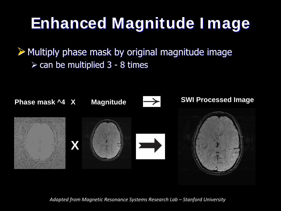
Multiply phase mask by original magnitude imageMultiply phase mask by original magnitude image can be multiplied 3 can be multiplied 3 --
8 times8 times
X
Phase mask ^4 X Magnitude SWI Processed Image
Enhanced Magnitude ImageEnhanced Magnitude Image
Adapted from Magnetic Resonance Systems Research Lab – Stanford University
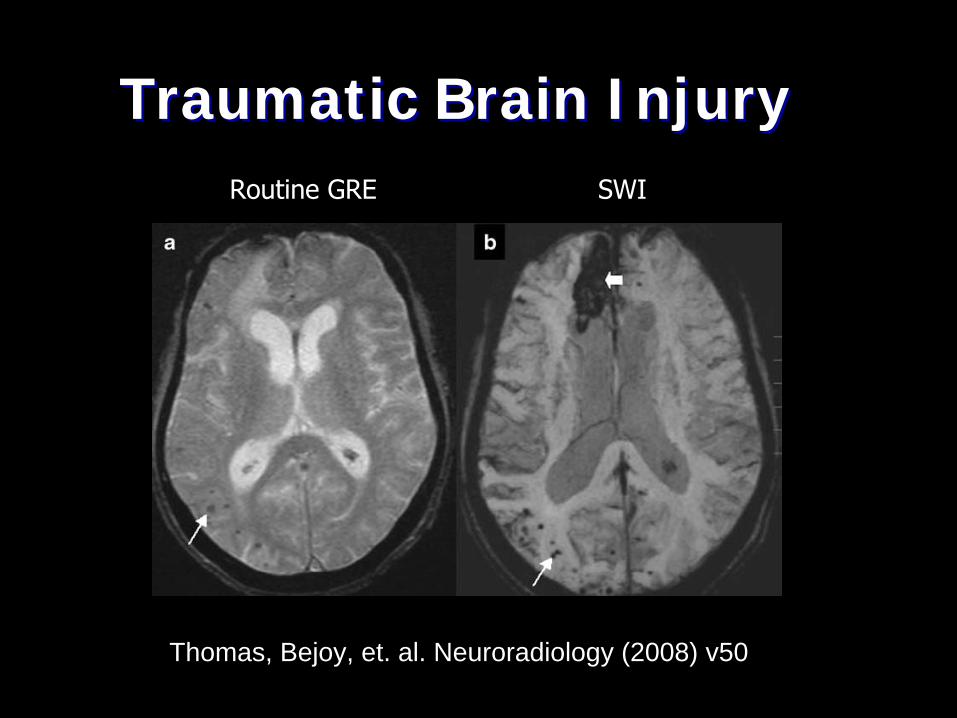
Traumatic Brain InjuryTraumatic Brain Injury
Thomas, Bejoy, et. al. Neuroradiology (2008) v50
Routine GRE SWI
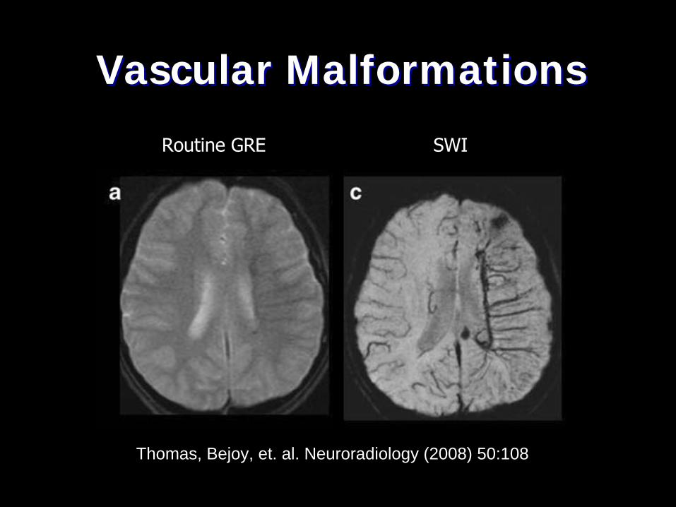
Vascular MalformationsVascular Malformations
Thomas, Bejoy, et. al. Neuroradiology (2008) 50:108
Routine GRE SWI
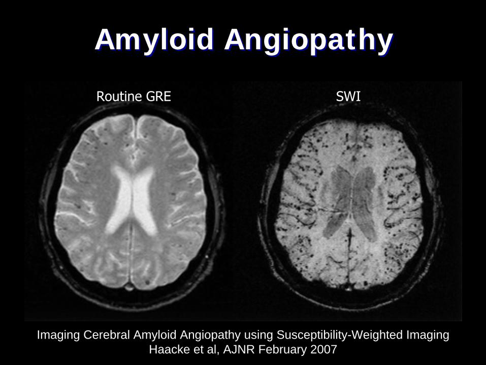
Amyloid AngiopathyAmyloid Angiopathy
Imaging Cerebral Amyloid Angiopathy using Susceptibility-Weighted Imaging Haacke et al, AJNR February 2007
Routine GRE SWI

Brain Brain ––
Seizure / DysplasiaSeizure / Dysplasia••
IndicationsIndications––
Seizure, Dysplasia, Mesial temporal sclerosisSeizure, Dysplasia, Mesial temporal sclerosis
••
SequencesSequences––
3 PL LOC3 PL LOC––
Sag T1 SESag T1 SE––
Ax T2 FLAIRAx T2 FLAIR––
Ax T1 SEAx T1 SE––
Ax T2 TSE FSAx T2 TSE FS––
Ax DWI EPIAx DWI EPI––
Cor T2 TSE Cor T2 TSE (angled perpendicular to temporal lobes)––
Cor FLAIR Cor FLAIR (angled perpendicular to temporal lobes)––
Ax IRAx IR--SPGR / MPSPGR / MP--RAGE RAGE or or Cor IRCor IR--SPGR / MPSPGR / MP--RAGERAGE
•
Comments–
Coronal sequences should be thin section perpendicular to the long axis of the hippocampus

2 year2 year--old female with seizures old female with seizures
Potential Points of ImpactPotential Points of Impact
••
Patient history?Patient history?––
Do we know more than Do we know more than ““seizuresseizures””??
––
Onset of symptoms?Onset of symptoms?––
Prior studies / previous surgery / trauma?Prior studies / previous surgery / trauma?
••
Pulse sequences to consider anticipating?Pulse sequences to consider anticipating?––
Axial IRAxial IR--SPGR / MPSPGR / MP--RAGERAGE
––
Coronal T2 and FLAIR through hippocampiCoronal T2 and FLAIR through hippocampi
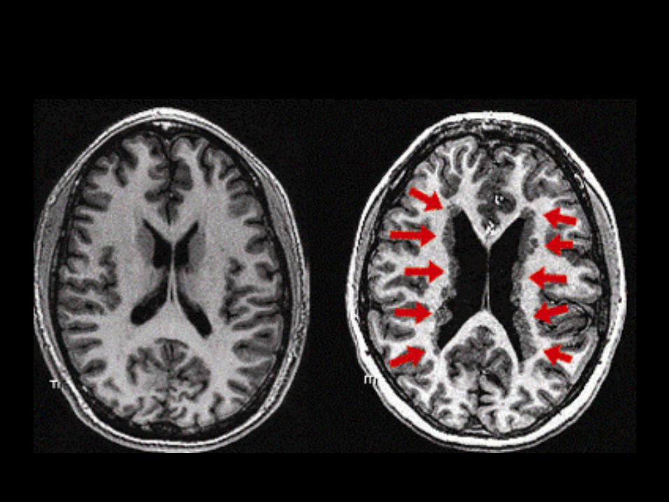
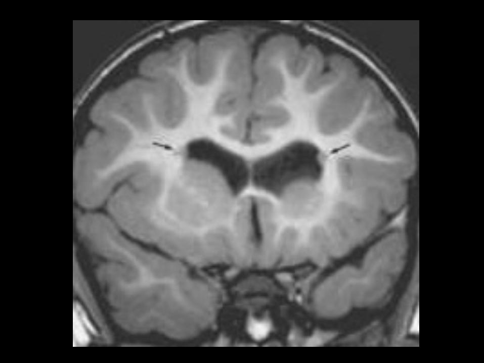
Periventricular Nodular HeterotopiaPeriventricular Nodular Heterotopia
••
In a normal brain, much of the gray matter (consisting In a normal brain, much of the gray matter (consisting mostly of nerve cells) appears on the brain surface, mostly of nerve cells) appears on the brain surface, while white matter (consisting mostly of nerve fibers while white matter (consisting mostly of nerve fibers interconnecting areas of gray matter) runs deeperinterconnecting areas of gray matter) runs deeper
••
In PNH, a migrational abnormality occurs during In PNH, a migrational abnormality occurs during development development --
portions of gray matter sit deep in the portions of gray matter sit deep in the
brain core, within the white matter, having failed to brain core, within the white matter, having failed to migrate out to the surfacemigrate out to the surface––
May serve as elliptogenic fociMay serve as elliptogenic foci
Brain Brain ––
Advanced ProtocolsAdvanced Protocols
••
Dural venous sinus thrombosis Dural venous sinus thrombosis ––
Cor 2D TOF SPGRCor 2D TOF SPGR
––
Sag 2D TOF SPGR (slight oblique angle)Sag 2D TOF SPGR (slight oblique angle)
••
Stroke, TIA, Vertebrobasilar infarct, AneurysmStroke, TIA, Vertebrobasilar infarct, Aneurysm––
Ax 3D TOF SPGRAx 3D TOF SPGR
––
Ax PerfusionAx Perfusion
••
Tumor, Metabolic abnormalityTumor, Metabolic abnormality–
Single voxel spectroscopy (short and long echo; eg. TE 35 and 144) on all new mass lesions
–
Multi voxel spectroscopy -
suspected gliomas–
Perfusion•
Gd –
20 ml @ 3-5 ml/s

30 year30 year--old female with mental status old female with mental status changeschanges
Potential Points of ImpactPotential Points of Impact
••
Patient history?Patient history?––
Do we know more than Do we know more than ““mental status changesmental status changes””??
––
Any neurologic symptoms?Any neurologic symptoms?––
Recent surgery? Dehydration?Recent surgery? Dehydration?
••
Are the referring physician and/or Are the referring physician and/or neuroradiologist aware?neuroradiologist aware?
••
Other studies to consider anticipating?Other studies to consider anticipating?––
CTV head + contrast; reconstructions / MIP imagesCTV head + contrast; reconstructions / MIP images
––
MRI brain MRI brain ––
evaluation for ischemia / hemorrhageevaluation for ischemia / hemorrhage
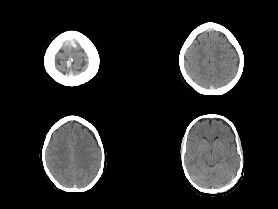
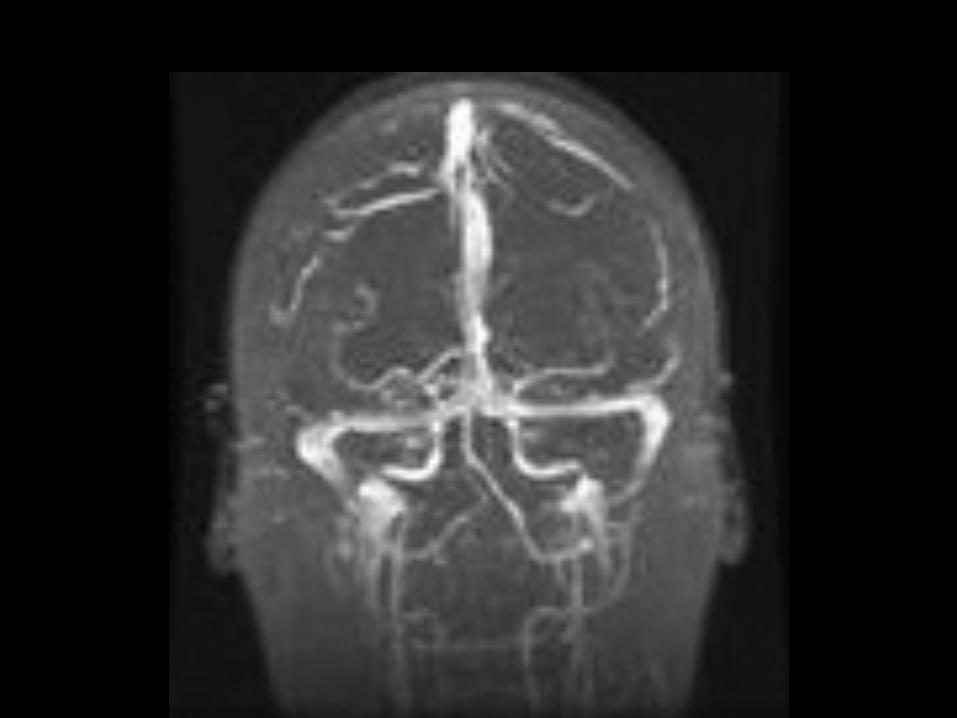
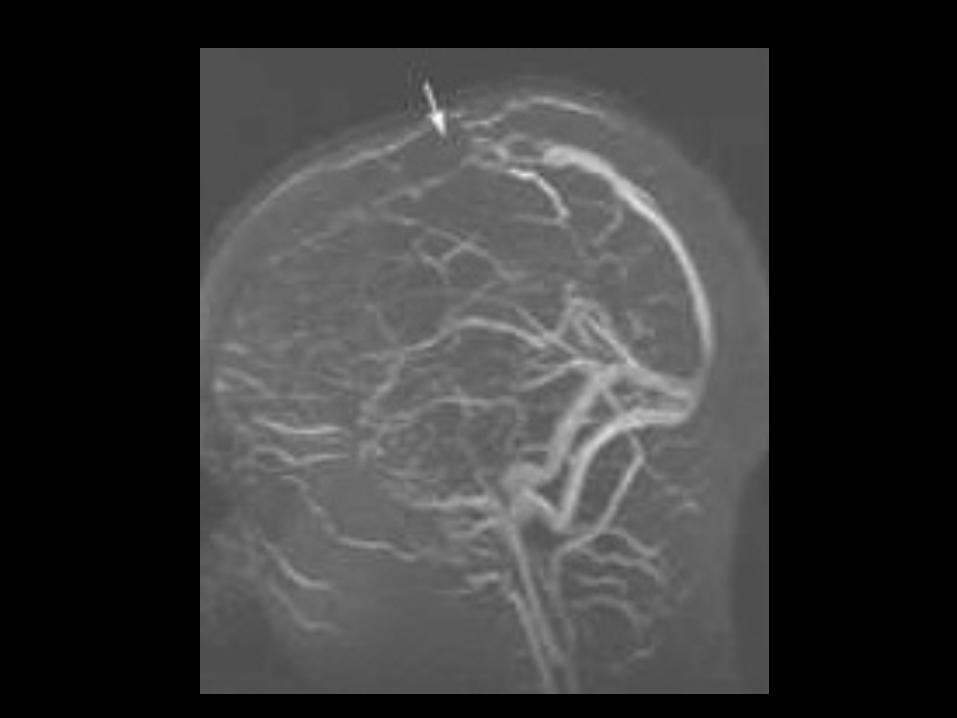
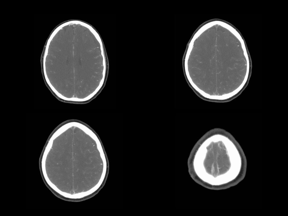
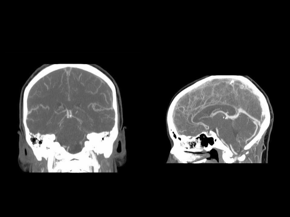
Dural Venous ThrombosisDural Venous Thrombosis
••
MRIMRI––
Main sign is lack of expected signal flow void Main sign is lack of expected signal flow void on standard T1on standard T1--
and T2and T2--weighted images weighted images
––
Challenging diagnosis in acute stageChallenging diagnosis in acute stage••
Hypointense signal of acute thrombus mimics Hypointense signal of acute thrombus mimics normal flow void on T2normal flow void on T2--weighted images weighted images
––
Absence of flow void on T1Absence of flow void on T1--weighted images weighted images must be carefully sought because thrombus must be carefully sought because thrombus may be isointense / mildly hyperintense to may be isointense / mildly hyperintense to brain tissuebrain tissue

69 year69 year--old female with chronic headacheold female with chronic headache
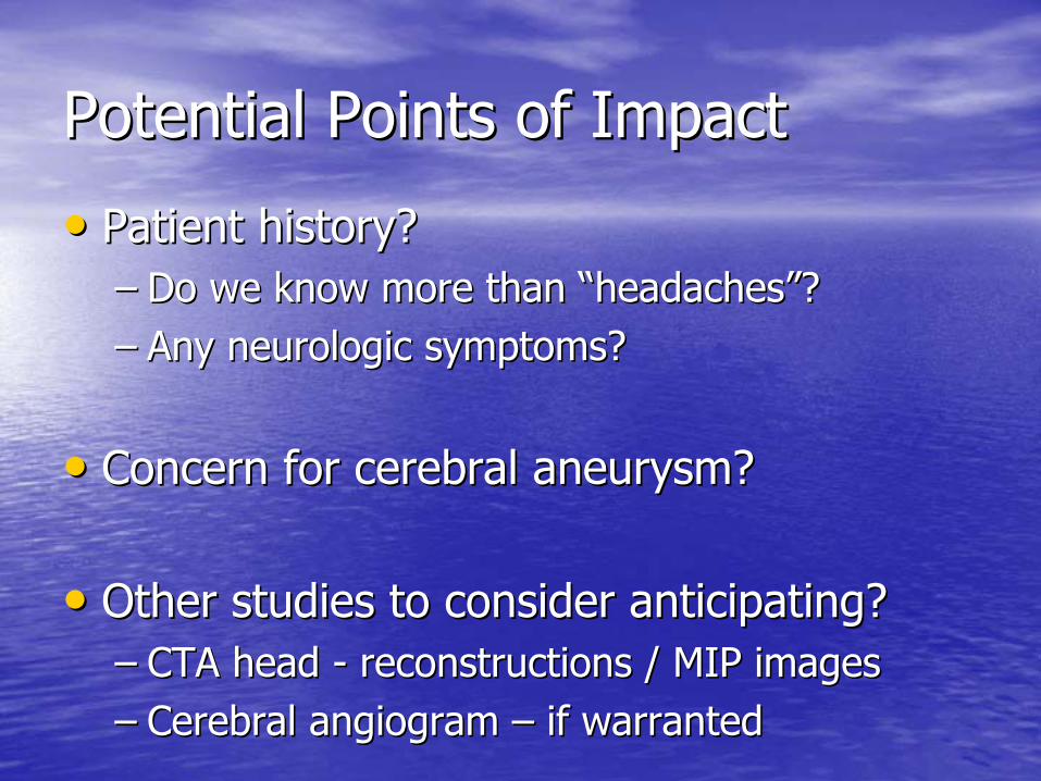
Potential Points of ImpactPotential Points of Impact
••
Patient history?Patient history?––
Do we know more than Do we know more than ““headachesheadaches””??
––
Any neurologic symptoms?Any neurologic symptoms?
••
Concern for cerebral aneurysm?Concern for cerebral aneurysm?
••
Other studies to consider anticipating?Other studies to consider anticipating?––
CTA head CTA head --
reconstructions / MIP imagesreconstructions / MIP images
––
Cerebral angiogram Cerebral angiogram ––
if warrantedif warranted
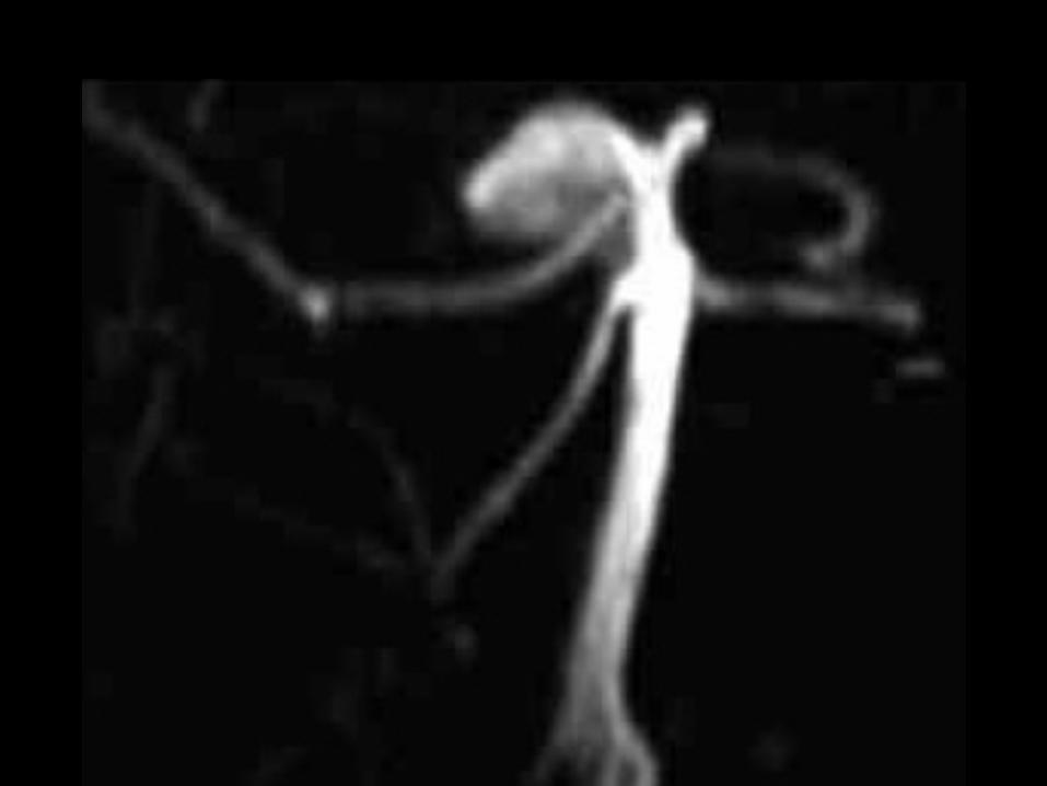
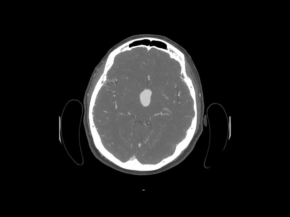
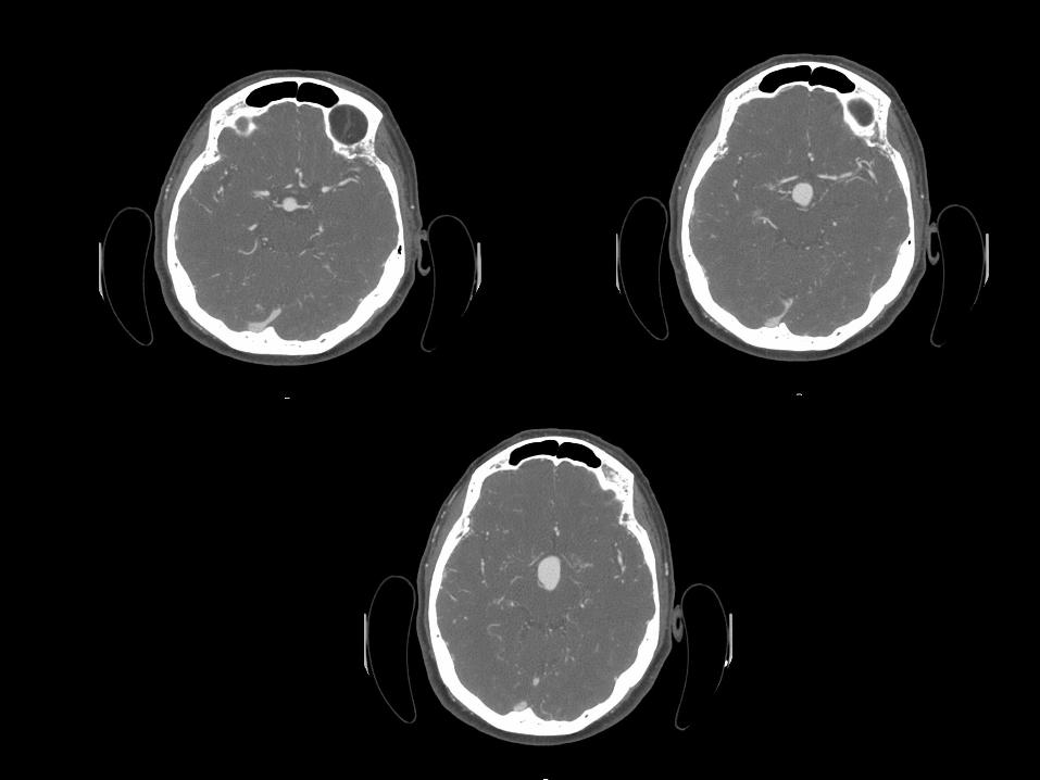
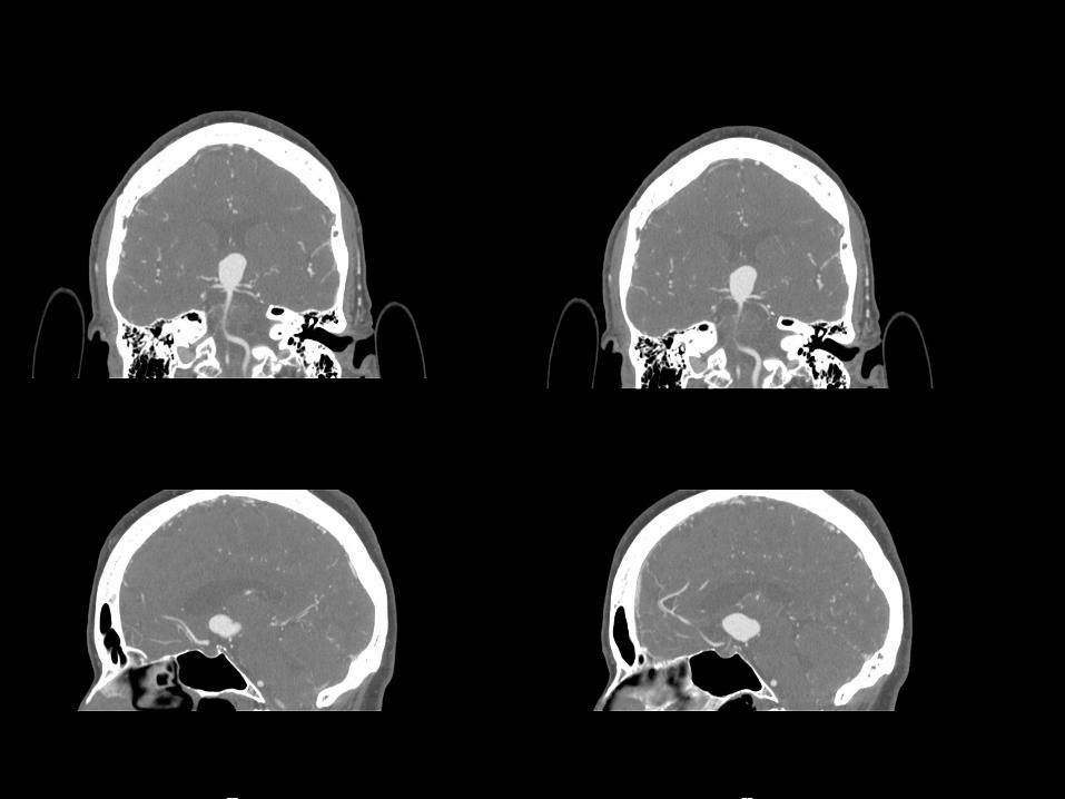
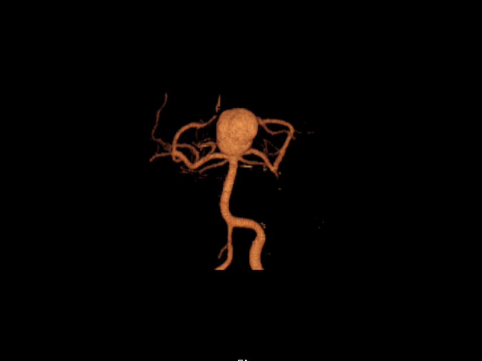
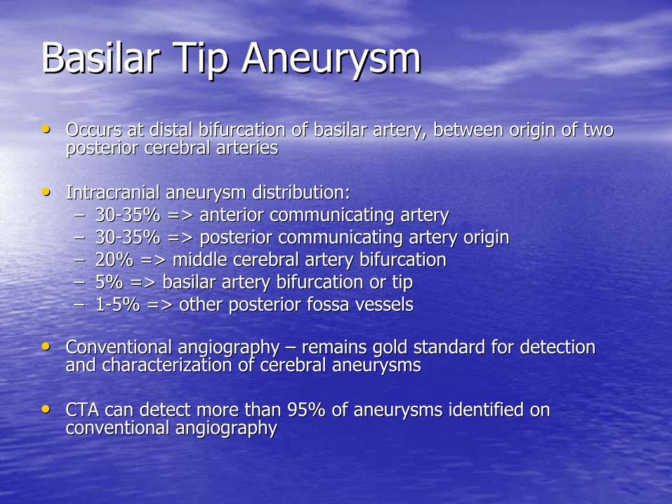
Basilar Tip AneurysmBasilar Tip Aneurysm
••
Occurs at distal bifurcation of basilar artery, between origin oOccurs at distal bifurcation of basilar artery, between origin of two f two posterior cerebral arteriesposterior cerebral arteries
••
Intracranial aneurysm distribution: Intracranial aneurysm distribution: ––
3030--35% => anterior communicating artery 35% => anterior communicating artery ––
3030--35% => posterior communicating artery origin 35% => posterior communicating artery origin ––
20% => middle cerebral artery bifurcation 20% => middle cerebral artery bifurcation ––
5% => basilar artery bifurcation or tip 5% => basilar artery bifurcation or tip ––
11--5% => other posterior fossa vessels5% => other posterior fossa vessels
••
Conventional angiography Conventional angiography ––
remains gold standard for detection remains gold standard for detection and characterization of cerebral aneurysmsand characterization of cerebral aneurysms
••
CTA can detect more than 95% of aneurysms identified on CTA can detect more than 95% of aneurysms identified on conventional angiographyconventional angiography
50 year50 year--old male with brain tumor found on old male with brain tumor found on an outside hospital MRI study an outside hospital MRI study

Potential Points of ImpactPotential Points of Impact
••
Patient history?Patient history?––
Do we know more than Do we know more than ““tumortumor””??
––
Previous surgery?Previous surgery?––
Neurologic deficits?Neurologic deficits?
––
Outside images available for radiologist review?Outside images available for radiologist review?
••
Other studies to consider anticipating?Other studies to consider anticipating?––
MR spectroscopyMR spectroscopy
––
MR perfusionMR perfusion––
PostPost--contrast IRcontrast IR--SPGR (for radiation therapy)SPGR (for radiation therapy)
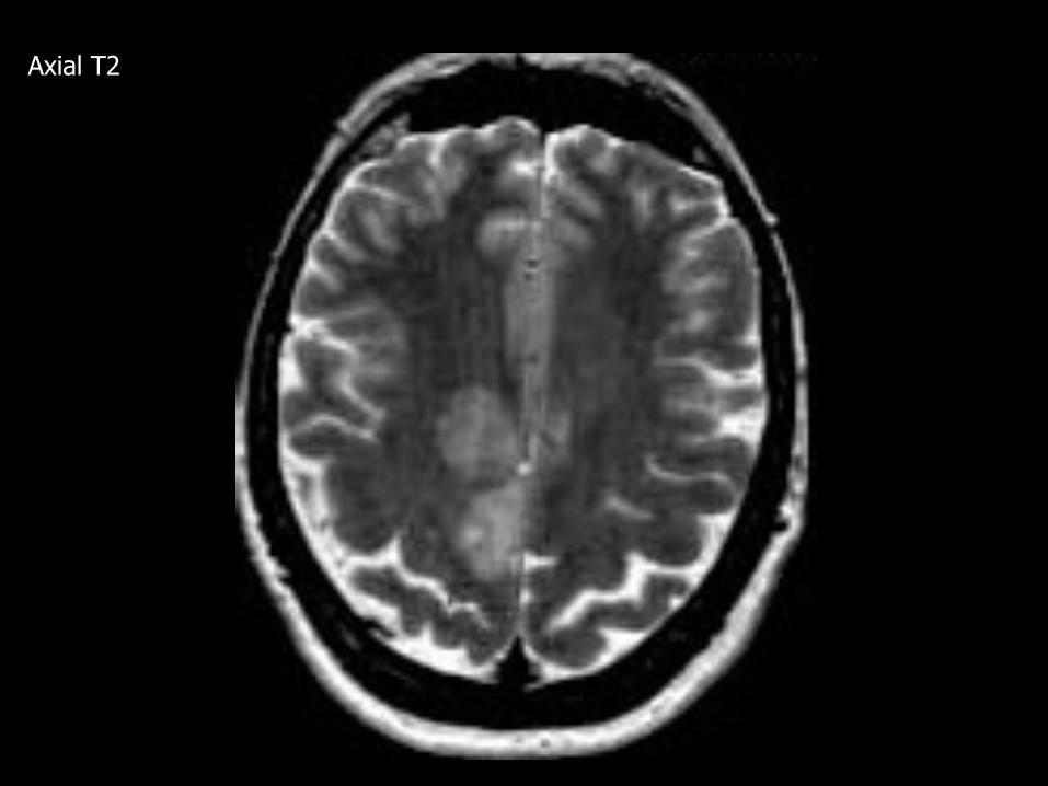
Axial T2
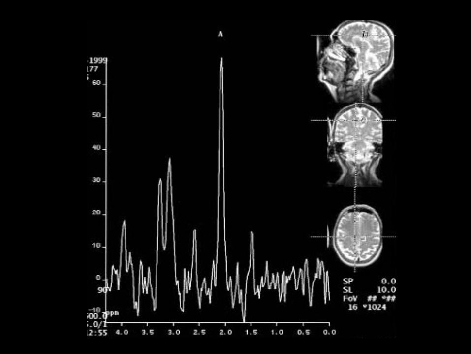
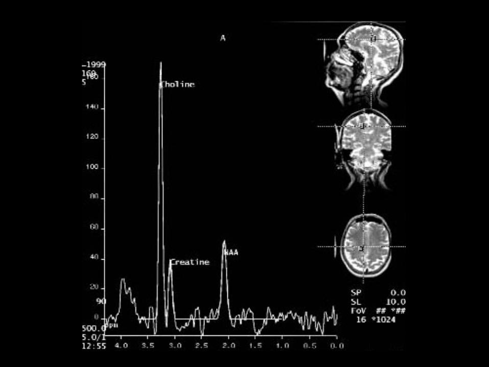

MR SpectroscopyMR Spectroscopy
••
Useful in tumor evaluation and surgical / biopsy planningUseful in tumor evaluation and surgical / biopsy planning
••
Although water and fat contribute virtually all of the Although water and fat contribute virtually all of the signal in proton MR imaging, it is possible to suppress signal in proton MR imaging, it is possible to suppress these signals and assess the signal from other these signals and assess the signal from other metabolites including choline, creatine, and NAA metabolites including choline, creatine, and NAA
••
Metabolic mapping of spectra allows rapid assessment of Metabolic mapping of spectra allows rapid assessment of spectral peaks and choline map also demonstrates the spectral peaks and choline map also demonstrates the most malignant site to biopsymost malignant site to biopsy
••
Elevated Elevated cholinecholine
probably represents the cell membrane probably represents the cell membrane breakdown secondary to the tumor, while breakdown secondary to the tumor, while NAANAA
is a is a
metabolite of normal neuronal tissue and metabolite of normal neuronal tissue and creatinecreatine reflects energy storesreflects energy stores

MiddleMiddle--aged female with a brain tumoraged female with a brain tumor
Potential Points of ImpactPotential Points of Impact
••
Patient history?Patient history?––
Do we know more than Do we know more than ““tumortumor””??
––
Previous surgery and/or biopsy results?Previous surgery and/or biopsy results?––
Neurologic deficits?Neurologic deficits?
––
Outside images available for radiologist review?Outside images available for radiologist review?
••
Other studies to consider anticipating?Other studies to consider anticipating?––
MR spectroscopyMR spectroscopy
––
MR perfusionMR perfusion––
PostPost--contrast IRcontrast IR--SPGR (for radiation therapy)SPGR (for radiation therapy)
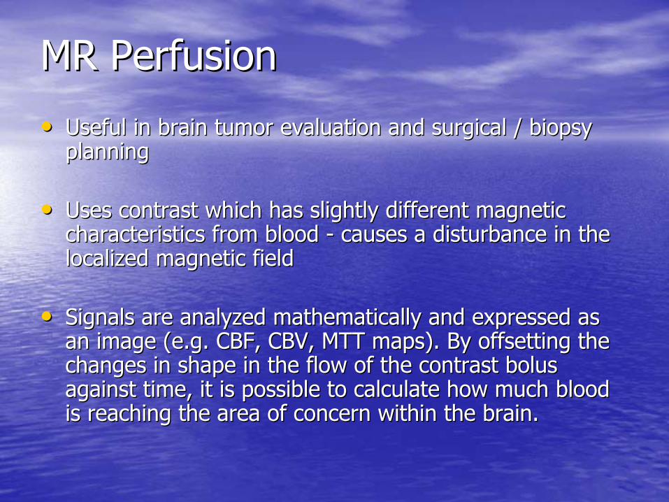
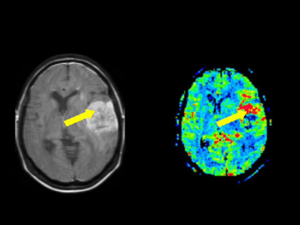
MR PerfusionMR Perfusion
••
Useful in brain tumor evaluation and surgical / biopsy Useful in brain tumor evaluation and surgical / biopsy planningplanning
••
Uses contrast which has slightly different magnetic Uses contrast which has slightly different magnetic characteristics from blood characteristics from blood --
causes a disturbance in the causes a disturbance in the
localized magnetic fieldlocalized magnetic field
••
Signals are analyzed mathematically and expressed as Signals are analyzed mathematically and expressed as an image (e.g. CBF, CBV, MTT maps). By offsetting the an image (e.g. CBF, CBV, MTT maps). By offsetting the changes in shape in the flow of the contrast bolus changes in shape in the flow of the contrast bolus against time, it is possible to calculate how much blood against time, it is possible to calculate how much blood is reaching the area of concern within the brain. is reaching the area of concern within the brain.

THANK YOU!THANK YOU!
HAVE A GREAT DAY!HAVE A GREAT DAY!


![The diagnostic value of MRI multi-parameter combination ... · hancement (internal enhancement pattern) [1-3] of breast lesions on dynamic contrast-enhanced MRI (DCE-MRI) indicates](https://img.pdfslide.us/doc/110x75/5fa73044450d904265457571/the-diagnostic-value-of-mri-multi-parameter-combination-hancement-internal.jpg)