Embed Size (px)
Citation preview

MRes Medical Imaging Sciences – Research Project Titles 2010/11
1. Direct incorporation of [11C]CO2 into ureas, Prof Tony Gee 2. Simulation Tool for Novel Contrast Mechanisms, Prof Rene Botnar
2011/12
1. Hypoxia imaging agents for cardiovascular systems Rafaael Torres, Rebekka Heuting 2. Towards multimodal imaging of Matrix Metalloproteinase activity, Rafaael Torres 3. Simulating Delayed Enhancement Cardiac MRI sequences and analyzing contrast improvement Prof.
Tobias Schaeffter 4. Optimisation of signal response of Spin Echo, Gradient Echo and Inversion Recovery in the presence of
contrast agents Prof. Rene Botnar 5. PCA of Matabolic Brain Networks in Dystonia, Dr Joel Dunn 6. Resting state fMRI in Dementia
2012/13
1. Computational dynamic PET simulations of the developing human brain Supervisor: Dr. Charalampos Tsoumpas (email: [email protected]) Location: Office Suite 2, The Rayne Institute, 4th Floor, Lambeth Wing, St. Thomas’ Hospital Positron Emission Tomography (PET) imaging can be of great use for diagnosis of several developmental diseases. However, PET scanning requires the administration of radioactive material, the radiotracer, into the patient. As a consequence imaging children with PET may increase risk effects caused by ionising radiation such as leukaemia and other forms of cancer. However, in practice the significant majority of PET acquisitions are performed on adults, thus there are not greatly optimised acquisition and reconstruction techniques for imaging children.* The aim of this project is the generation of realistic PET images that would have been acquired if very low radiotracer amounts were administered in children of different age. This project involves literature review on the physics and acquisition of PET [1]. Following good understanding of fundamental issues involved with the paediatric PET imaging, the student will prepare brain PET acquisitions for a family of hybrid computational phantoms [2]. A fast analytic simulation technique will be used to generate dynamic brain PET data [3]. The image quality will be assessed on images and the impact of low-injected dose will be studied. This research project opens up the possibility of reducing radioactive dose in children during paediatric brain PET imaging. The student would be co/author if a publication results from their work. Project contents
Literature review on PET physics and paediatric imaging techniques
Data processing using Matlab and / or imaging related software tools
Competency in computer use is required but you will not be developing new tools / programs so advanced knowledge is not essential.
* More information can be obtained from the Image Gently campaign: http://www.pedrad.org/associations/5364/ig/
References [1] Physics in Nuclear Medicine, by SR Cherry, JA Sorenson, ME Phelps, (Saunders, 3rd Edition, 2003) [2] Bolch et al (2010) Hybrid Computational Phantoms for Medical Dose Reconstruction, Radiat Environ Biophys 49(2): 155–168. doi:10.1007/s00411-009-0260-x [3] C Tsoumpas et al 2011 Fast generation of 4D PET-MR data from real dynamic MR acquisitions Phys Med Biol 56 6597 doi:10.1088/0031-9155/56/20/005
2. Maths and computing background: Project Supervisor: Shaihan Malik [email protected] Magnetic Resonance Imaging (MRI) is increasingly performed to assess neonatal brain damage. With better care leading to a reduction in major focal lesions, diffuse white matter injury and delayed myelination are now the most common abnormalities observed on qualitative MRI in the preterm population. Quantitative techniques are required to assess objectively brain development and injury. Quantitative MRI is however a challenge: the signal in an MR image is affected by a large number of interacting parameters, some of which are intrinsic properties of the tissue being imaged. This richness makes MRI an excellent tool for generating images with a multitude of different types of soft tissue contrast, but this same variability must be accounted for when attempting quantitative measurement. A recently developed quantitative MR imaging approach, mcDESPOT1, has been used to map myelin water content2 in infants from 15 weeks after birth. mcDESPOT works by obtaining multiple images with different contrast settings; knowledge of the expected relationships between tissue properties and the received signals is then used to extract tissue information using a multi-component fit. It is possible that mcDESPOT adds complimentary information to that obtained from qualitative MR imaging and other quantitative approaches, such as diffusion tensor imaging, but it has never been tested in neonates soon after birth. The aim of this study is to develop an image analysis pipeline for neonatal MR image data, in order to generate T1/T2 and myelin water fraction maps. Once developed, this capability will be used to measure these parameters together for the first time in this patient cohort, and if successful there is a strong likelihood the results will be published. The student will be expected to write code for image analysis in Matlab, and so a computational/numerate background is preferred. They can expect to learn about the fundamentals of MRI and if interested, they might also take part in fine-tuning image acquisition parameters. 1.Deoni S et al. Gleaning multicomponent T1 and T2 information from steady-state imaging data. Magn Reson Med 2008;60:1372-1387 2.Deoni S et al. Mapping infant brain myelination with magnetic resonance imaging. J Neurosci 2011; 31: 784-91
3. Pretargeting in radionuclide imaging and therapy: review of present status and evslustion of novel approaches
Supervisor: Prof. Phil Blower
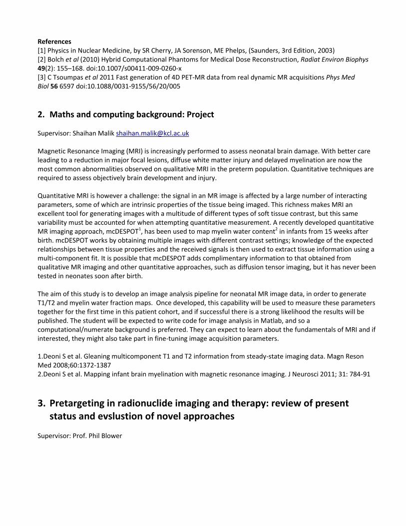
4. Laplace inversion of multiexponential data – applications to the measurement of kinetic parameters from imaging experiments
Supervisors: Dr Thomas Eykyn, Dr Richard Southworth, Dr Joel Dunn, Miss Erika Mariotti Many imaging modalities give rise to exponential or multiexponential data as a result of multicompartment kinetics. This includes, but is not limited to, (i) PET radionuclide uptake and washout kinetics, (ii) diffusion MRI of water and other metabolites in cells or in vivo due to different intra vs extra cellular or restricted diffusion and (iii) hyperpolarised 13C metabolic imaging for measuring kinetics of enzyme reactions. An example is shown below for conversion of hyperpolarised 13C pyruvate to lactate in a suspension of cancer cells. This example shows biexponential rates and in this case the reaction kinetics gives information about the activity of lactate dehydrogenase, a key enzyme in energy metabolism. Deriving the amplitudes and rates from multiexponential data yields a wealth of biophysical parameters that can be used as biomarkers of disease and treatment response. The aims of this project will be to explore the applications of Laplace inversion of multiexponential data using maximum entropy and non-linear least squares fitting to derive kinetic parameters using a model-free approach. Objectives:
1. Familiarisation with a range of imaging modalities
and data acquired from hyperpolarised 13C MR
imaging, high resolution NMR, diffusion MRI and
FDG PET.
2. Computer simulation using MATLAB and MemExp
to test robustness of Laplace inversion for deriving exponential rates and exploring the influence of
heterogeneous kinetics and experimental noise.
3. Application of the technique to real data, PET and NMR, and experimental measurements.
4. Team working, critical thinking, presentation and communication skills, scientific writing.
5. Possible publication, conference.
This project would suit a student with a strong mathematical background, and experience with computer simulations.
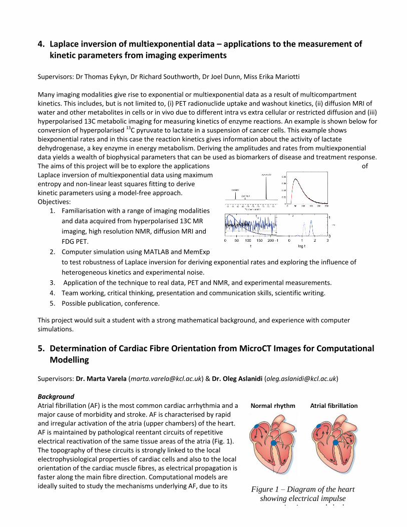
5. Determination of Cardiac Fibre Orientation from MicroCT Images for Computational Modelling
Supervisors: Dr. Marta Varela ([email protected]) & Dr. Oleg Aslanidi ([email protected]) Background Atrial fibrillation (AF) is the most common cardiac arrhythmia and a major cause of morbidity and stroke. AF is characterised by rapid and irregular activation of the atria (upper chambers) of the heart. AF is maintained by pathological reentant circuits of repetitive electrical reactivation of the same tissue areas of the atria (Fig. 1). The topography of these circuits is strongly linked to the local electrophysiological properties of cardiac cells and also to the local orientation of the cardiac muscle fibres, as electrical propagation is faster along the main fibre direction. Computational models are ideally suited to study the mechanisms underlying AF, due to its
Figure 1 – Diagram of the heart
showing electrical impulse
propagation in normal rhythm
(left) and atrial fibrillation
(right).

complex multi-factorial nature. Whole-atria 3D models are currently being developed in our group to understand the genesis and progression of AF1. These models require detailed information about atrial anatomy and cardiac fibre orientation. High-resolution (70 -vivo micro-computed tomography (microCT) images of the atria have been acquired by our group2. Tissue was stained with iodine prior to acquisition to obtain a high contrast between cardiac muscle fibres and connective tissue. The details of cardiac fibre orientation can be inferred from these images (Fig. 2) using dedicated image processing algorithms, such as structure tensor analysis2,3.
Figure 2 - Anterior view of the atria. A) Segmented atrial anatomy. B) Orientation of cardiac fibres in the same geometry.
Aims
Obtain quantitative measures of fibre orientation across the atria using microCT data and Matlab;
Compare fibre orientations computed from microCT data with those from histological cross-sections (this gold-standard technique has recently been applied to the atria3);
Generate a unique description of fibre orientation across different areas in the atria, particularly those known to be involved in the genesis of AF (such as sleeves of the pulmonary veins in the left atrium2).
The information obtained will be incorporated into anatomically realistic 3D computational models of AF1, which will be applied to explore relationships between cardiac anatomy and pathophysiology of the disease. There is a strong possibility of publication of the results on the successful completion of the project. References 1. Aslanidi OV, et al. 3D virtual human atria: A computational platform for studying clinical atrial fibrillation. Progress in Biophysics & Molecular Biology 107 (2011), 156-168. 2. Aslanidi OV, et al. Application of Micro-computed tomography with iodine staining to cardiac imaging, segmentation and computational model development. IEEE Transactions on Medical Imaging (2012), doi: 10.1109/TMI.2012.2209183. 3. Zhao J, et al. An image-based model of atrial muscular architecture: effects of structural anisotropy on electrical activation. Circulation Arrhythmia & Electrophysiology 5 (2012), 361-370.
6. Non-Newtonian Effects in the Flow in Vascular Networks Supervisor: Taha Sochi
Non-Newtonian fluids are characterized by non-proportionality between stress and rate of strain. To simplify the flow models and their computational implementation in the flow simulation codes, fluids are generally assumed Newtonian and hence these effects are ignored. However, in many normal and pathological situations, these effects are important and therefore they should be included in the flow models. The proposed project is based on including these effects in the existing models and codes which are derived from two principal mathematical formulations: one-dimensional Navier-Stokes, and Poiseuille, with certain emphasis on the first. There are two main approaches for representing non-Newtonian effects: incorporating these effects in the fluid viscosity, and incorporating them in the flow profile through the alpha-factor in the 1D model. The second approach is based on defining a vessel-dependent alpha field.

The theoretical and computational infrastructure for this project is currently available in terms of mathematical models and computer codes. The student is required to introduce some enhancements on the existing codes with limited extensions to incorporate non- Newtonian effects. The project requires familiarity with the physical principles and mathematical techniques of fluid dynamics with good computational skills preferably in C++ language. The project also requires modest literature investigation which should be easy to do. Expertise for this project is available in the KCL Imaging Science Department, and therefore the outcome of the project should be predictable. The student should acquire good knowledge and expertise with regard to the theory and techniques of flow simulation which have many biomedical applications. Other benefits include exposure to literature, training on writing and publishing scientific papers, as well as acquisition and enhancement of computational and mathematical skills. On successful accomplishment of this project, the results should be publishable in refereed journals and the student should benefit directly by being the main author, or at least a co-author, of a scientific paper which improves his/her CV and increases the chance of having a career in research.
7. Cryomicrotomic Cardiovascular Image Extraction Supervisor: Taha Sochi This ongoing project is based on developing mathematical models, computational algorithms, and computer codes for image extraction and reconstruction of cardiovascular networks from cryomicrotomes. The current project is also interested in developing algorithms and codes to deal with the extracted networks for the purpose of testing, optimization, discretization, enhancement, efficiency, etc. Beside their use in imaging and visualization, e.g. for the purpose of inspection and diagnosis as part of the general imaging effort, the extracted networks are also used in the flow simulation where the fidelity of these networks has a great impact on the simulation results. Since this is an existing project with well established theory, algorithms and codes, the student is required to join the ongoing effort mainly to improve theoretical foundations and computer techniques, and to extend and optimize the existing codes. The student is expected to get sufficient materials and credit for his own project as part of this collective effort. He/she is also expected to join other members of this project in any related publications in the refereed journals. The main requirements for this project are strong interest in image extraction with general familiarity of the subject, and good mathematical and computational skills especially in Matlab. Knowledge of C++ and fluency in Linux scripting is an advantage. Reliability and coordination skills are also required. The student should acquire adequate knowledge and experience related to the theory and techniques of image extraction and network manipulation. Other benefits include exposure to literature, training on writing and publishing scientific papers, as well as acquisition and enhancement of mathematical and computational skills.
8. Development of Novel 18F Labelled Mitochondria Targeting Tracers for Dual Modality Cancer Imaging with PET and Fluorescence Imaging Techniques
Supervisor: Dr Ran Yan
Background:
Rhodamine 123 is a cationic fluorescent dye widely used for labelling respiring mitochondria. It has shown increased uptake and prolonged retention on a large variety of human carcinoma cell lines such as breast, colon, prostate and pancreas cancer, whereas it was rapidly washed out from the normal epithelial cells (Figure 1a).1 Rhodamine 123 together with three other members of rhodamine family has been conjugated with galactosamine serum albumin (GmSA), a D-galactose receptor targeting reagent, for in vivo spectral fluorescence imaging of ovarian cancer xenografts. Excellent tumor to background contrast was achieved with sustained fluorescence from all four imaging reagents (Figure 1 b to e).2 These characteristics of Rhodamine 123 make it an attractive lead compound for the development of a new generation of 18F labelled dual modality cancer imaging reagents.

1a
1b 1c
1d 1e
O NH2H2N
O
OMe
Cl
Figure 1. 1a) Rhodamine 123 stained human breast cancer (MCF-7) cells; 1b-e) in vivo ovarian cancer xenograft fluorescence imaging with rhodamine-GmSA conjugates
Objectives:
In this project, you will synthesize cold reference compounds bearing different rhodamine moiety and fluorine substituted functionalities and measure their fluorescent properties and logPs. Subsequently, the lead compounds will be submitted to in vitro screening on carcinoma cell lines with flow cytometry and fluorescence microscopy. Promising lead compounds that have high uptake and prolonged retention on carcinoma cell lines will be identified for 18F labelling.
References:
1. I. C. Summerhayes, K. K. Nadakavukaren, E. L. Shepherd, and L. B. Chen; PNAS, 1982, 79 (17), 5292-5296;
2. M. R. Longmire, M. Ogawa, Y. Hama, N. Kosaka, C. A. Regino, P. L. Choyke, H. Kobayashi; Bioconjugate Chem., 2008, 19, 1735–1742.
2013/14
1. Image-based study of the role of cardiac tissue fibrosis in atrial fibrillation Supervisors: Dr. Steven Niederer ([email protected]) & Dr. Oleg Aslanidi ([email protected])
Background. Atrial fibrillation (AF) is the most common arrhythmia: it is characterized by uncoordinated electrical activations of the atria with consequent deterioration of the cardiac mechanical function. AF increases risks of other cardiovascular disease, stroke and death1. Atrial tissue fibrosis (Fig. 1) has been identified as both a cause and an effect of AF,2 and it can persist following reversion to sinus rhythm (SR). The presence of fibrosis is associated with an increased risk of AF recurrence even after its clinical termination3. Besides, fibrosis alters the electrical conduction anisotropy and fibre orientation in the atria,4,5 which can directly impact the speed and pathways of activation (Fig. 2), and hence provide a substrate for AF. The quantification of fibre reorientation during AF is hindered by the absence of a reference state that describes the distribution fibres in the healthy atria.
Aims. This project aims to address the lack of atrial fibre information by developing a computer model of fibre orientation based on a recent 3D high
resolution (50 m) reconstruction of the atria.6 The generated model of atria fibres will be used to explore the impact of fibrosis4,5 on the speed and sequence of atrial activation, as well as the possible breakdowns of the normal electrical activation patterns and initiation of AF, using biophysically detailed 3D simulations.7
Figure 1. Distribution of
fibrotic tissue (green) during
AF and SN.

Figure 2. 3D reconstruction of the atria. A) Orientation of atrial fibres. B) Activation sequence determined by the fibre directions.
Skills. This project will involve analysing ex-vivo imaging data, image processing, basic programming and computer simulations. There is a possibility of publication of the results upon the completion of the project.
References 1. Camm, J. A., et al. Guidelines for the management of atrial fibrillation. Europace 12, 1360-1420 (2010). 2. Allessie, M., Ausma, J. & Schotten, U. Electrical, contractile and structural remodeling during atrial fibrillation.
Cardiovascular Research 54, 230-246 (2002). 3. Wijffels, M. C., Kirchhof, C. J., Dorland, R. & Allessie, M. A. Atrial fibrillation begets atrial fibrillation. A study in awake
chronically instrumented goats. Circulation 92, 1954-1968 (1995). 4. Burstein, B. & Nattel, S. Atrial fibrosis: Mechanisms and clinical relevance in atrial fibrillation. Journal of the American
College of Cardiology 51, 802-809 (2008). 5. Li, D., Fareh, S., Leung, T. K. & Nattel, S. Promotion of atrial fibrillation by heart failure in dogs: Atrial remodeling of a
different sort. Circulation 100, 87-95 (1999). 6. Zhao, J., et al. An image-based model of atrial muscular architecture: effects of structural anisotropy on electrical
activation. Circulation Arrhythmia & Electrophysiology 5, 361-370 (2012). 7. Aslanidi, O.V., et al. 3D virtual human atria: A computational platform for studying clinical atrial fibrillation. Progress
in Biophysics & Molecular Biology 107, 156-168 (2011).
2. Using Magnetic Resonance Spectroscopy Imaging to Distinguish Metastatic From
Non-metastatic Tumours In Vivo.
Supervisors: Dr Thomas Eykyn and Dr Gilbert Fruhwirth
Project Synopsis
This project aims at setting up non-invasive pre-clinical magnetic resonance spectroscopy imaging
(MRSI) in mice. The goal of the project is to test the hypothesis that the metabolomic changes observed
between highly metastatic and non-metastatic breast cancer cell lines in vitro can be extrapolated to and
quantified in real tumours in vivo by MRSI.
Background
We have already generated a model system of highly metastatic and non-metastatic breast cancer cell
lines and characterized their metabolomic changes by nuclear magnetic resonance spectroscopy (NMR)
in vitro (Figure 1). This cellular system revealed that in a breast adenocarcinoma cell line the over-
expression of the pro-metastatic protein CXCR4 led to a marked reduction of total choline levels (Figure

1C). Furthermore, over-expression of the even more migratory active truncation mutant D34-CXCR4
resulted in additional significant decrease of the total lipid content (Figure 1D).
Figure 1. NMR identifies metabolic changes between cell lines with different metastatic potentials.
(A) Different metastatic potentials are conferred by over-expression of wild-type or mutant CXCR4. (B) Typical high
resolution magic angle spinning NMR spectra of the different cell lines. (C) Total lipid content is decreased upon
over-expression of CXCR4. (D) The even more invasive truncation mutant D34 CXCR4 shows a decreased lipid
content in addition to the reduced choline level.
Objectives
We propose the use of these already existing breast cancer cell lines to generate tumour tissues for
MRSI measurements. These cell lines are routinely used in our laboratory and consistent tumour growth
and metastasis has been observed before.
1) Once tumours are established, we will first collect MRSI data from the freshly harvested intact
tumours ex vivo and subsequently homogenize and extract these tumour tissues for NMR analysis to
compare the two methods. We expect MRSI to be established in due course as all required
equipment is already available.
2) Subsequent to the ex vivo set-up and concomitant cross-validation, we will pre-clinically MRSI image
mice bearing two tumours, one established from the metastatic and one from the non-metastatic cell
line in order to distinguish them within the same animal in vivo.
3) As an optional objective depending on project progress, we propose to repeat 2) in the presence or
absence of cancer relevant drugs that are known to affect either tumour growth/viability (e.g. growth
factor receptor inhibitor/etoposide) or the metastatic behaviour of the cancer cells (e.g. CXCR4
inhibitor, Rho kinase inhibitor).
Student Benefits
The student will obtain hands-on experience in cell culture, immuno-cytochemistry, magnetic resonance
spectroscopy (1H, 31P), and pre-clinical MRSI imaging as well as large-scale data analysis (imaging and
metabolomics). The student will further be trained and prepared for future animal research work by
acquiring as many hands-on skills as possible (dictated by UK legislation). Furthermore, the student will

be embedded in a research-active environment and, pending successful results, will contribute with
her/his report to the writing of a scientific publication.
In conclusion, the student will gain multi-disciplinary hands-on research experience in cancer cell biology,
biochemistry, metabolomic imaging, animal research and scientific writing within a truly multi-disciplinary
environment with the prospect of acquiring many skills required for a future research career.
Reference
1) Vermeer LS, Fruhwirth GO, Pandya P, Ng T, Mason AJ (2012), Journal of Proteome Research 11:
2996-3003.
3. Generation and characterization of a platform for the evaluation of novel PEGylated
multi-modal imaging agents.
Supervisors: Dr Gilbert Fruhwirth and Dr Rafael Torres M de Rosales
Project Synopsis
This project aims to generate cancer cell lines that stably express a PEG-binding cell surface molecule.
Following generation and characterization, those cancer cells will be grafted onto mice to establish
tumours, which will then be used to evaluate the in vivo properties of polyethylene glycol (PEG)-modified
nanoparticles by PET/CT imaging.
Background
Nanoparticles hold great promise as multi-modal imaging agents, for instance for PET/MR imaging. We
have developed novel techniques to synthesis such particles and modify them for the purpose of
radionuclide imaging (SPECT or PET) (1). PEG modification is a frequently used method to coat
nanoparticles to make them bio-compatible and also to increase their circulation time in the blood and
thereby facilitate specific targeting processes (e.g. tumour targeting). Recently, an antibody molecule was
developed that has PEG-binding capabilities (2). This molecule was developed further so it can be
expressed on the surface of cells post retroviral infection with the antibody recognition domain being
available for PEG-binding.
Figure 1. (A) Schematic representation of the PEGylated nanoparticles that will be used in this project (left) and
biodistribution by PET-CT in a normal mouse (right); B) Schematic showing the PEG-binding reporter is expressed

on the cell surface with its binding domain receptive for extracellular PEGylated imaging probes. Tumours
established from those cells in mice will help discern the properties of different PEGylated nanoparticles in vivo.
Objectives
1) Infection of cancer cells with retrovirus particles carrying the DNA of the PEG-binding protein and
subsequent selection to achieve stable cell clones.
2) Generation of differently PEGylated nanoparticles ready for subsequent radiolabelling
3) Characterization of stable cell lines by immuno-blotting and immuno-cytochemistry (using fluorescent
PEG).
4) Establishment of tumour xenografts for subsequent radionuclide/CT imaging to compare differently
PEGylated and radiolabelled nanoparticles (comparison of circulation times by dynamic PET,
comparison of specific tumour uptake versus non-specific particle uptake/binding).
Student Benefits
The student will obtain hands-on experience in cell culture, retroviral transduction technology, immuno-
cytochemistry, nanoparticle synthesis, and radionuclide imaging as well as image data analysis. The
student will further be trained and prepared for future animal research work by acquiring as many hands-
on skills as possible (dictated by UK legislation). Furthermore, the student will be embedded in a
research-active environment and, pending successful results, will contribute with her/his report to the
writing of a scientific publication.
In conclusion, the student will gain multi-disciplinary experience in cancer cell biology, nanoparticle
chemistry, pre-clinical radionuclide imaging, and scientific writing within a truly multi-disciplinary
environment with the prospect of acquiring many skills required for a future research career.
Reference
1) Sandiford et.al. ACS Nano, January 2013, 7, 500
2) Chuang et al. J Nucl Med June 2010 51:933
4. Direct extraction of cardiac and respiratory signals from MR image data
Supervisors: Dr Andy King / Christian Baumgartner
Figure 1: Example of navigator extraction from cardiac cine images

Physiological motion in the thorax due to respiration and cardiac contraction remains to respiration and cardiac
contraction remains a limiting factor in numerous medical applications. In MR image acquisitions it leads to
blurring of the scans which in turn may negatively affect diagnosis. In MR-guided cardiac treatments such as
balloon dilation of pulmonary valves [1] or MR-guided high-intensity focused ultrasound (HIFU) ablations [2] it
leads to misalignment of the anatomy and guidance information acquired pre-treatment.
External gating signals are not always readily available, are limited in their accuracy and may complicate the image
acquisitions. Recently a group of techniques collectively known as manifold learning have been proposed which
lend themselves ideally to extract such signals directly from the image data. Some initial work on ultrasound and
MR gating using this approach has been performed by Wachinger et al. [3]. However, this study was restricted to
one such technique. In this project the student will compare and contrast the performance of different techniques
for the extraction of physiological motion navigators directly from imaging data. The student will be expected to:
Formulate a method for evaluating the accuracy of different motion measurements.
Identify datasets on which the evaluations will be performed. Possibly involvement in the acquisition of
additional data on the MR scanner will be required.
Develop MATLAB scripts in collaboration with the project supervisors to process the data to extract the
navigators.
Collect and statistically evaluate the results, and formulate a conclusion.
The student will work with the supervisors to achieve the goals of the project. All MATLAB software will be
developed in collaboration with the project supervisors, so basic competency in computer use is required together
with a desire to learn more.
[1] Tzifa et al., Magnetic resonance-guided cardiac interventions using magnetic resonance-compatible devices: a
preclinical study and first-in-man congenital interventions, Circulation 3(6):585-592, 2010.
[2] Tempany et al., MR imaging-guided focused ultrasound surgery of uterine leiomyomas: a feasibility study,
Radiology, 226(3):897-905, 2003
[3] Wachinger et al., Manifold learning for image-based breathing gating in ultrasound and MRI, Medical image
analysis, 16(4):806-818, 2012
2013/14 Research Projects carried out by MRes students
1. Methods to improve the specific activity of [F-18] tetrafluoroborate for use in thyroid imaging
Supervisors: Prof Phil Blower / Dr Alexander Koshnevisan
2. Imaging of Complement Protein Post Heart Transplantation
Supervisors: Dr Ehsan Sharif-Paghaleh / Dr Greg Mullen

3. Evaluation of bone marrow using quantitative advanced magnetic resonance imaging (MRI)
Supervisor: Prof Vicky Goh
4. FDG PET Imaging in Paediatric Dystonia: comparisons of brain glucose metabolism between patients with Cerebral Palsy and Neurodegeneration with Brain Iron Accumulation
Supervisors: Dr Joel Dunn / Dr Daniel Lumsden
5. The use of cardiac MR in the subclinical detection of cardiovascular changes in HIV positive patients
Supervisors: Dr Valentina Puntmann / Prof Eike Nagel
6. Investigation into the effect of aortic injury on the formation of atherosclerotic lesions in an Apolipoprotien E- deficient mouse model.
Supervisors: Prof René Botnar / Dr Alkystis Phinikaridou
7. Histology Analysis of Myocyte Structure and Expression of Cardiac Hypertrophy in Patients with Aortic Stenosis
Supervisors: Dr Valentina Puntmann / Prof Manuel Mayr
8. Are native T1 values to characterize myocardial tissue equivalent between various sequences: comparison of reproducibility and discriminatory accuracy for MOLLI and 3’5-MOLLI
Supervisors: Dr Valentina Puntmann / Prof Eike Nagel
9. Cardiac MR texture analysis to predict outcomes of patients undergoing ICD implantation following MI
Supervisors: Dr Greg Mullen / Dr Amedeo Chiribiri
10. MR Characterisation of anal cancer
Supervisors: Prof Vicky Goh / Dr Sofia Gourtsoyianni