Embed Size (px)
Citation preview

0
MRes in Biomedical Sciences and Translational Medicine
Programme Handbook 2017 - 2018
Master of Research Strands:
Biology of Cancer
Biomedical Imaging and Biosensing
Biostatistics (with Health Informatics)
Cancer Medicine
Cellular and Molecular Physiology
Drug Safety
Medical Sciences
Molecular and Clinical Gastroenterology
Molecular and Clinical Pharmacology
Nanomedicine
Neuroscience
Stem Cells, Tissues and Disease
Women’s, Children’s and Perinatal Health

1

2
Student Handbook 2017-18
Contents
Page Number
(in Booklet) Section 1 Introduction
3
Section 2 Programme Organisation and Student Support
11
Section 3 Programme Overview & Timetable
18
Section 4 Assessment
24
Section 5 Research Projects
29
Section 6 Techniques and Frontiers in Biomedical Sciences
53
Section 7 Transferable Skills
73
Section 8 Attendance Monitoring & Absence Reporting
98
Section 9 Student Feedback and Representation
100
Section 10 Appendix
100

3
Section 1 Introduction 1.1 A welcome to new students I am pleased to welcome you as a new student into the Institute of Translational Medicine and hope that you will find your time studying on the MRes in Biomedical Sciences and Translational Medicine programme an enriching experience. The course combines hands-on laboratory research work with lectures and tutorials to give you direct knowledge and experience of cutting-edge biomedical and clinical research. You will also receive training in transferable skills, which will broaden your existing skill set and help prepare you for your subsequent careers. Feedback from previous MRes students indicates that you will need to work hard, but that the course is both enjoyable and rewarding. This handbook contains essential information about all aspects of the MRes programme, so it is important that you read and understand it. Additional information and announcements about the programme will also be issued during the course, either electronically by email or through VITAL (the University’s online teaching resource), or via posters on the MRes notice boards. It is therefore very important that you check your e-mail, relevant VITAL pages and the notice board every day, as there may be important messages relating to the course or changes in schedule, etc. One of the first things you will want to know is who to contact for help, information and advice. The MRes programme is run by a team consisting of the Programme Director, the Programme Administrators and the Strand Convenors. They are happy to help you if you are having any difficulties, whether academic, administrative or personal in nature, including disability-related issues. Any problems should initially be discussed with your Strand Convenor, who will either deal with the issue directly or will refer the matter to the Programme Director or Administrator as appropriate. If the issue has not been resolved within two weeks, you should contact the Programme Director, who will then deal with the matter. If the Programme Director fails to resolve the issue within two weeks of being alerted, you should then refer the matter to the Director of Postgraduate Research for the Institute of Translational Medicine, Prof Andrea Varro. She will be happy to discuss any unresolved problems with you provided that the appropriate line of communication described above has been followed and exhausted. Contact details for each member of the team can be found in Section 2.1 of this handbook, along with a brief description of their area of responsibility, to guide you to the most suitable person to deal with your question. I hope that you find the MRes in Biomedical Sciences and Translational Medicine both useful and enjoyable. Good luck with your studies.
Programme Director: Dr Alec Simpson
Email: [email protected] Telephone: 0151 794 5510

4
1.2 Induction Timetable for MRes Students The first two weeks of the programme will provide you with essential introductory information and practical training that will be of benefit to you throughout the programme. It is compulsory to attend these activities on time. You should also check you know how to find the activities that will take place outside of the Sherrington Buildings.

5
Monday 18 September 2017
Tuesday 19 September 2017
Wednesday 20 September 2017
Thursday 21 September 2017
Friday 22 September 2017
(Surnames A – K) 10:00 - 11:00
Group 1 - Registration PC Centre
Sherrington Building
Completing registration using “Liverpool Life”
Overseas students
Paperwork completion - bring passport, BRP,
original certificates/transcript
09:30 - 11:30
Laser Radiation Training Dr Pete Cole
NWCR, 200 London Road
09:00 - 17:00 Strand convenors to meet
with students (All students / All strands)
(pre-arranged with Strand
Convenors)
Women’s strand to meet at the
University Meeting Room, First Floor, Women’s
Hospital
09:00 - 17:00 Strand convenors to meet
with students (All students / All strands)
(pre-arranged with Strand
Convenors)
(Surnames L – Z) 11:00 - 12:00
Group 2 - Registration PC Centre
Sherrington Building
Completing registration using “Liverpool Life”
Overseas students
Paperwork completion - bring passport, BRP,
original certificates/transcript
11:30 - 12:30
Health & Safety Dr Geoff Williams
NWCR, 200 London Road
12:30 - 13:30 13:00 - 13:30
Overview of ITM Dr Alec Simpson
NWCR, 200 London Road
13:00 - 14:00 Research Ethics Dr Alec Simpson
NWCR, 200 London Road
13:30 - 14:30 Academic Integrity Talk
Dr Carlos Rubbi NWCR, 200 London Road
13:30 - 14:30 Overview of MRes Course
Dr Alec Simpson NWCR, 200 London Road
14:00 - 16:00 Basic Radiation Training
Dr Pete Cole NWCR, 200 London Road
14:30 - 15:30 Dr Jeff Barclay
Science skills (literature and database searching) NWCR, 200 London Road
14:30 - 15:00 The Guild Rep Talk
Rachel Coleman NWCR, 200 London Road
16:00 - 17:00 Library Introduction Talk
Ken Linkman (not compulsory for UoL
Graduates and Intercalating students)
15:30 – 16:30 Basic Science Skills
Dr Alec Simpson NWCR, London Road
15:00 -15:30 ITM PGR Society Talk
Tom Leather Ana Illera
NWCR, 200 London Road
16:30 - 17:00 Q&A Session
Dr Alec Simpson NWCR, 200 London Road
15:30 - 16:00 Students meet with Strand
Convenors NWCR, 200 London Road
16:00 - 17:30 Welcome drinks & nibbles NWCR, 200 London Road
6
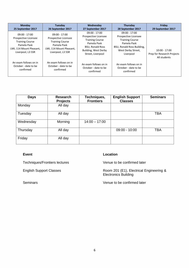
Days Research Projects
Techniques, Frontiers
English Support Classes
Seminars
Monday
All day
Tuesday
All day TBA
Wednesday
Morning 14:00 – 17:00
Thursday
All day 09:00 - 10:00 TBA
Friday
All day
Event Location Techniques/Frontiers lectures Venue to be confirmed later English Support Classes Room 201 (E1), Electrical Engineering &
Electronics Building Seminars Venue to be confirmed later
Monday 25 September 2017
Tuesday 26 September 2017
Wednesday 27 September 2017
Thursday 28 September 2017
Friday 29 September 2017
09:00 - 17:00 Prospective Licensee
Training Course Pamela Pask
LMI, 114 Mount Pleasant, Liverpool, L3 5SR
An exam follows on in October - date to be
confirmed
09:00 - 17:00 Prospective Licensee
Training Course Pamela Pask
LMI, 114 Mount Pleasant, Liverpool, L3 5SR
An exam follows on in October - date to be
confirmed
09:00 - 17:00 Prospective Licensee
Training Course Pamela Pask
BSU, Ronald Ross Building, West Derby
Street, Liverpool
An exam follows on in October - date to be
confirmed
09:00 - 17:00 Prospective Licensee
Training Course Pamela Pask
BSU, Ronald Ross Building, West Derby Street,
Liverpool
An exam follows on in October - date to be
confirmed
10:00 - 17:00 Prep for Research Projects
All students

7
MRes Coursework Deadline Dates 2017-18
Project/Date Project type Copies students submit Copies in ITM MRES Office
Markers
Short Review 1 (Techniques and Frontiers Module) 13 November 2017 17 November 2017 18 December 2017
Make Choice Title allocated Submit Report
Submit 1 to Turnitin
E-copy on data base
1st Marker Lecturer giving the topic 2nd Marker Strand Convenor
Research Project 1 (Research Projects) 12 January 2018 Oral/Poster
Presentation 15 January 2018 Submit Report Submit 1 to their supervisor
Submit 1 to internal assessor (allocated by strand convenor)
Submit 1 to Turnitin
E-copy on data base 1st Marker Rotation One Supervisor 2nd Marker Academic Staff, chosen by Strand Convenor
Referees Report – Journal Club (Techniques and Frontiers Module) 26 February 2018 Submit Report Submit 1 to Turnitin
E-copy on data base 1st Marker
Lecturer giving the journal club, decided by strand convenor 2nd Marker Strand Convenor
Research Project 2 (Research Projects) 13 April 2018
Oral/Poster Presentation E-copy on data base
16 April 2018 Submit Report Submit 1 to their supervisor Submit 1 to internal assessor
(allocated by strand convenor)
Submit 1 to Turnitin
E-copy on data base 1st Marker Rotation Two Supervisor 2nd Marker Academic Staff, chosen by Strand Convenor
Short Review 2 (Techniques and Frontiers Module) 21 May 2018
Submit Report Submit 1 to Turnitin
E-copy on data base 1st Marker Lecturer giving the topic 2nd Marker Strand Convenor
Research Project 3 (Research Projects) 6 July 2018 Oral/Poster
Presentation E-copy on data base 9 July 2018
Submit Report Submit 1 to their supervisor Submit 1 to internal assessor
(allocated by strand convenor)
Submit 1 to Turnitin
E-copy on data base 1st Marker Rotation Three Supervisor 2nd Marker Academic Staff, chosen by Strand Convenor
Grant Application (Transferable Skills Module) 9 July 2018 Workshop Lecture Theatre 1, Sherrington
Building Alec Simpson
6 August 2018 Submit Report Submit 1 to Turnitin
E-copy on data base
1st Marker Strand convenor 2nd Marker Strand Convenor from another strand chosen by strand convenor

8
Please note: Students need to be resident in Liverpool until 13 August 2018 except for the
timetabled holidays indicated below. Permission to be absent from University outside of these holidays can only be granted by the Programme Director following a written request.
Timetabled holidays: 19 December 2017 – 7 January 2018 31 March 2018 – 8 April 2018 14 August 2018 – 31 August 2018 (Inclusive)
Viva dates 3 – 7 September 2018
Careers Workshop TBC LT1, Sherrington
Seminar Room 1, Sherrington 09:00 – 13:00
IP Commercialisation Workshop (Transferable Skills Module) TBC (July 2018)
20 July 2018 TBC
Portfolio (Transferable Skills Module) 13 August 2018 Project hand-in Submit 1 to ITM MRES Office Pass to Alec Simpson to
mark 1st Marker –Alec Simpson

9
Later Events: Some induction activities are scheduled to take place after the first 2 weeks. Details of these are given below: Event: Safety Seminar Date: TBC Time: Location: Event: English Language Classes for International Students Date: 5 October 2017 to 23 November 2017 Time: 09:00 – 10:00 Location: Room 201 (E1), Electrical Engineering & Electronics Building Building number 235, grid reference E7
https://www.liverpool.ac.uk/files/docs/maps/liverpool-university-campus-map.pdf
https://www.liverpool.ac.uk/english-language-centre/in-sessional-support/academic-classes-international-research-students/
Event: Demonstrator Training Workshop Date: TBC Time: Location: Event: Biostatistics Workshop Date: 17 and 22 November 2017 Time: Location: TBC Event: IP and Commercialisation Workshop Date: TBC Time: Location:

10
1.3 Using the Handbook This handbook is to be used in conjunction with other information you may be given about different aspects of the MRes Programme, and compulsory courses organized by the University of Liverpool. It provides essential information required for the Degree Programme; you should read it carefully and keep it in a safe place. It also presents information on how the student charter is implemented in this course. It includes details of: • the broader aims and objectives of the MRes
• the modules available
• the means by which the course will be assessed overall
• the assessment criteria that will be used.
• the aims and objectives of each individual module or similar unit of study and what you
should be able to achieve by the end of it
• the teaching and learning methods that will be used and the means by which more general skills (such as working in teams and making oral presentations) will be developed and assessed.
• the facilities and support services provided by the University that may be useful to you.
• the staff responsible for organising the programme and its strands or who undertake other duties of relevance and their contact details
• the means by which your views on individual modules or units on courses of study overall and on other aspects of your experience will be sought – both individually and collectively – and how information on the responses to those views will be fed back to you.
• how you will be provided with systematic information on your individual progress, on your areas of strength and weakness, and on the means by which you can improve your performance.

11
Section 2 Programme Organisation and Student Support The MRes in Biomedical Sciences and Translational Medicine programme is run by a team consisting of Strand Convenors, Programme Director and Programme Administrators. They are happy to help you if you are having any difficulties, whether academic, administrative or personal in nature, including disability-related issues. In addition, the University provides many useful services to help you adjust to life on campus and to help with various difficulties you may face. Any problems should initially be discussed with your Strand Convenor, who will either deal with the issue directly or will refer the matter to the Programme Director or Administrator, as appropriate. If the issue has not been resolved within two weeks, you should contact the Programme Director, who will then deal with the matter. If the Programme Director fails to resolve the issue within two weeks of being alerted, you should then refer the matter to the Director of Postgraduate Research for the Institute of Translational Medicine, Prof Andrea Varro. She will be happy to discuss any unresolved problems with you provided that the appropriate line of communication described above has been followed and exhausted. In addition, we are offering a voluntary mentoring system to all MRes students. There are four mentors throughout the Institute (2 females and 2 males). Their names and affiliation are below. You can choose to go to any of them if you have any personal problems that you are reluctant to discuss with your strand convenor or the course organiser. If your problems continue don’t hesitate to contact Professor Varro. Please note, that any course related issues should be discussed through the mechanisms as described above starting with the strand convenor.
Dr Carrie Duckworth Gastroenterology [email protected]
Dr Janet Risk
Molecular & Clinical Cancer
Dr Lakis Liloglou Molecular & Clinical Cancer [email protected]
Dr Lee Haynes
Cellular & Molecular Physiology
Contact details for each member of the team can be found below, along with a brief description of their area of responsibility, to guide you to the most suitable person to deal with your question.

12
2.1 The MRes Team Strand Convenors Responsible for the organisation of the various MRes strands, convenors will help with any academic and administrative problems relating to their specific MRes strand. Strand convenors will be able to help with most issues and should normally be the first person you contact when problems arise. If they are unable to help, they will refer you to the Programme Director/Administrator or other staff as appropriate. Contact details for the various strand convenors are given below: Biology of Cancer Dr Eithne Costello-Goldring 0151 706 4178 [email protected] Dr Carlos Rubbi 0151 794 8842 [email protected]
Biomedical Imaging and Biosensing Professor Harish Poptani 0151 794 5444 [email protected]
Biostatistics (with Health Informatics) Dr Gabriella Czanner 0151 794 9132 [email protected]
Cancer Medicine Dr Jason Parsons 0151 794 8848 [email protected]
Cellular & Molecular Physiology Dr Jeff Barclay 0151 794 5307 [email protected]
Drug Safety Dr Takao Sakai 0151 794 5459 [email protected]
Medical Sciences Dr Carrie Duckworth 0151 794 6811 [email protected]
Molecular and Clinical Gastroenterology Professor Chris Probert 0151 795 4011 [email protected]
Molecular and Clinical Pharmacology Dr Takao Sakai 0151 794 5459 [email protected]
Nanomedicine Professor Andrew Owen 0151 794 8211 [email protected]
Neuroscience Dr Simon Keller 0151 529 5943 [email protected]
Stem Cells, Tissues and Disease Dr Antonius Plagge 0151 795 4987 [email protected]
Women’s, Children’s and Perinatal Health Dr Dharani Hapangama 0151 795 9559 [email protected]

13
Programme Director – Dr Alec Simpson Responsible for the overall organisation of the MRes programme, he will help with general academic and administrative problems relating to the MRes course. He will also help with any specific issues that are not able to be resolved by strand convenors. Alec’s office, room 136 can be accessed from the 2nd floor of the Nuffield Wing of the Sherrington Building (Building 312 on the campus map). He can be contacted by email, [email protected] or telephone on (0151) 794 5510. Issues that cannot be resolved by the Programme Director will be referred to the Institute Director of Postgraduate Studies (Professor Andrea Varro). Programme Administrator – Joanne Isherwood Responsible for the general administration of the MRes programme, she will help with non-academic problems related to the course, including attendance issues, deadlines, schedule alterations, etc. Joanne can be found in the ITM Postgraduate Office in Room LG40 on the ground floor of the Sherrington Building on Ashton Street. She can be contacted by e-mail at [email protected] or by telephone on (0151) 794 9901. Postgraduate Students Team The PGR team is based in Room LG40, Sherrington Building, they will help you with general administrative issues and non-academic related problems. The team can be contacted by e-mail at [email protected]. 2.2 Academic Staff involved in the MRes programme In addition to those listed above involved in organizing the MRes, a large number of staff contribute to the delivery of the course and supervision of research projects. Most staff are based in the Institute of Translational Medicine, although staff from other Institutes also contribute to the programme. 2.3 Safety Institute of Translational Medicine has a dedicated Safety Officer. All students must attend a safety talk given by Geoff Williams in the first week and will receive written guidelines from him on safety within the Institute. You are also required to attend University training sessions dealing with general safety and radiation protection, as detailed in the induction timetable in Section 1.2. Additional safety information will be given by Departmental safety officers within the Departments in which you conduct your Research Projects. Risk assessment forms must be completed for your Research Projects and copies of these provided to the appropriate safety officers in your department. It is the duty of every employee and every student of the University to take reasonable care for the health and safety of themselves and of other persons who may be affected by their acts or omissions; and to act in accordance with the University Safety Policy and with the Health and Safety arrangements made by the University and its departments.

14
Working with human subjects and/or human material Supervisors have a responsibility to ensure that all work involving human subjects is covered by appropriate Ethics Comittee Permission. They should also ensure that students conducting research projects involving human subjects and/or material understand the permission given for their work, and in writing their dissertation, they make a clear statement of the Ethics Comittee Approval for the work. Working with animals Supervisors have a responsibility to ensure that the appropriate Home Office Authority (both personal and project licence) are in place before working with experimental animal is started. They should also ensure that students conducting research projects involving experimental animals understand the permission given for their work, and in writing their dissertation, they make a clear statement of the Home Office Approval for the work. You need to read carefully and obey all the instructions regarding safety that have been given to you before commencing experimental work in the laboratory. Normal working hours are 09.00 - 17:30 Monday – Friday. Work outside these hours, including weekends, is only permitted if either your supervisor or a suitably qualified person approved by your supervisor is present. Further information, including current safety codes of practice and guidance, can be found at www.liv.ac.uk/safety. 2.4 Mail and Messages Mail coming into the Institute addressed to students will be left in the Postgraduate Student Office, Sherrington Building. An email alert will be sent to you if any post arrives. Urgent messages received for students, wherever possible, are relayed either by telephone or email. There are also MRes notice boards in two locations: outside the Seminar Room in Physiology, near to the main entrance; and outside the Postgraduate Student Office, Sherrington Building. Information and announcements about the programme will also be issued during the course, either electronically by email or through VITAL (the University’s online teaching resource), or via posters on the MRes notice boards. It is therefore very important that you check your e-mail, relevant VITAL pages and the notice board every day for important information and schedule changes etc. 2.5 Common Rooms Tea and coffee facilities and chilled water are available in the Physiology Common Room, where you will also find a microwave and vending machines. There are similar facilities in the 3rd floor Nuffield Wing (3.02N and 3.03N) and in Pharmacology. There is also Meeting Room 3.04N available for group interactions and group working, or the Physiology Committee room located next door to the Physiology Seminar Room, which can be booked via the Programme Administrator.

15
2.6 Computer, Library and Other Academic Services There are three computers in the Physiology common room connected to University Network (Physiology Cybercafé) for all students to use. There is also a computer suite on the ground floor of the Sherrington Building, near the main entrance, which has 50 computers available for general use (the room is occasionally booked; bookings are shown on a diary on the door). All students registered for the MRes will also have access to all the University services. These are detailed in the postgraduate handbook and on the University website: http://www.liv.ac.uk/gradschool/pgrhandbook/index.htm. Students are expected to make full use of these services. The Harold Cohen Library, with 295 PCs and seating for 500 readers, contains the main collections in Dentistry, Engineering, Science, Medicine, Veterinary Science and Mathematics. Facilities for both group and quiet study are available. There is also a branch library at the Veterinary Teaching Hospital on the Wirral. Your student smart card will give you access to the libraries and enable you to self-issue and return books. There are introductory talks and tours available for new students and staff will help you find your way around and show you how to use the online catalogue. Printed and web guides to the various libraries and services are available and staff at the Information Support Desks or Computing Helpdesks will be happy to help if you have a problem or a question. Further information can be found at www.liv.ac.uk/library. Computing Services The Computing Services Department provides central computing and information technology services to assist the University in carrying out its learning, teaching, research and administration. There are a number of PC Teaching Centres which are primarily used for teaching but, when not booked for classes, they are available for individual student use. These are located in centres across the campus and at the Leahurst Veterinary Centre. There are also a number of Learning Centres on the campus and in some Halls of Residence that are not bookable by tutors for classes and are therefore available for individual student use. The Teaching and Learning Centres each contain PCs linked together by a network. To use the PCs, you first need to self-register by following the information on the screen of a PC Teaching or Learning Centre, or at www.liv.ac.uk/register on any computer connected to the internet. The PC Teaching Service is based on Microsoft Windows and provides access to a wide range of services, including electronic mail, the internet, VITAL (the University’s Virtual Learning Environment), word processing, spreadsheets and databases. There are a number of locations within the precinct where students may use either WiFi (wireless) or a wired connection to connect their own laptop computer (or other mobile device) to the University's network.

16
The main CSD Helpdesk is located in the Brownlow Hill Building (Building no. 224; Ref F7; www.liv.ac.uk/maps/), with satellites available in the two main libraries. The Helpdesk provides a full range of support services including problem solving, software sales and registration queries. More information about the support offered by the Helpdesk can be found at www.liv.ac.uk/csd/helpdesk/. 2.7 Liverpool Life This is one of the most important facilities you will need to use. Liverpool Life is your portal to all of your essential personal and academic information. Liverpool Life can be accessed by entering the URL Liverpool-life.liv.ac.uk into your browser or by following the ‘Liverpool Life’ link on the Digital University (student.liv.ac.uk). You will need your student ID (displayed on your student smart card) and PIN. You should familiarise yourself with Liverpool Life as a matter of priority. Further information about Liverpool Life, including user guides, can be found at www.liv.ac.uk/student-administration/liverpool-life/. 2.8 Students with disabilities The University Disability Support Team co-ordinates and maintains the support required to help you succeed on your course. Practical support The team can help you to:
• inform academic departments about your support requirements
• arrange appropriate support in using the libraries and other academic support services
• organise study assistants
• find financial support for services. With consent, and when appropriate, the team can liaise on a continual basis with your academic department and prepare documentation to confirm your support arrangements. Advice The Disability Support Team also provides advice on:
• support requirements
• how to apply for Disabled Students Allowance and other sources of funding
• who to contact for support and advice.

17
You can find further information and details of who to contact regarding disability issues from the following website: http://www.liv.ac.uk/studentsupport/disability/index.htm The Institute contact for disability matters is Professor Andrea Varro. 2.9 University Guide and Handbook The University of Liverpool have produced a guide for studying and living in Liverpool. We strongly recommend that you read through ‘Your Liverpool’ guide during your stay in Liverpool. It provides important information on University supports services and general advice on living in Liverpool. The ‘Your Registration’ handbook also contains import information regarding registration and key contacts. https://www.liverpool.ac.uk/student-administration/student-administration-centre/student-handbooks/ https://www.liverpool.ac.uk/media/livacuk/welcome/Your,Registration.PDF

18
Section 3 Programme Overview & Timetable 3.1 Aims & Learning Outcomes Aims The MRes in Biomedical Sciences and Translational Medicine provides a supportive learning environment to enable students to:
1. Develop intellectual, practical, learning and research skills.
2. Acquire, within an interdisciplinary environment, an understanding of methods in biomedical sciences and translational medicine relevant to modern research at the forefront of the subject.
3. Develop the capacity for individual work and teamwork.
4. Develop academic competence at the highest level attainable leading to the forefront
of current knowledge in biomedical sciences.
5. Take the first postgraduate steps required for future roles leading biomedical research in clinical, industrial and public sectors.
Learning outcomes On completion of the programme students will be able to: Demonstrate systematic understanding and knowledge and a critical awareness of:
a. Cutting edge topics in biomedical sciences and translational medicine.
b. How scientific understanding advances, and how current awareness of physiological, biomedical, biotechnological and socio-economic issues can be maintained and incorporated into ongoing projects.
c. Cutting edge techniques applicable to research in biomedical sciences and translational medicine.
d. Interdisciplinary approaches and techniques that can be deployed to work towards a defined goal in biomedical sciences and translational medicine research.
e. The relevance of biomedical research to business opportunities and the commercial sector.
f. The importance and complexity of ethics in scientific research
g. How research is communicated to scientific and lay communities
19
Demonstrate application of knowledge to:
a. Evaluate critically current research in biomedical sciences and translational medicine.
b. Plan, manage and execute research projects in a rigorous scientific manner within the prescribed timeframe and, where appropriate, formulate scientific hypotheses.
c. Use laboratory equipment correctly and safely to generate new data.
d. Analyse experimental data, interpret their validity and apply statistical analyses.
e. Effectively record experimental procedures and laboratory protocols.
f. Prepare scientific reports in an appropriate format.
g. Prepare grant applications to obtain research funding
h. Prepare business plans to exploit research findings commercially
i. Debate complex ethical issues in biomedical research
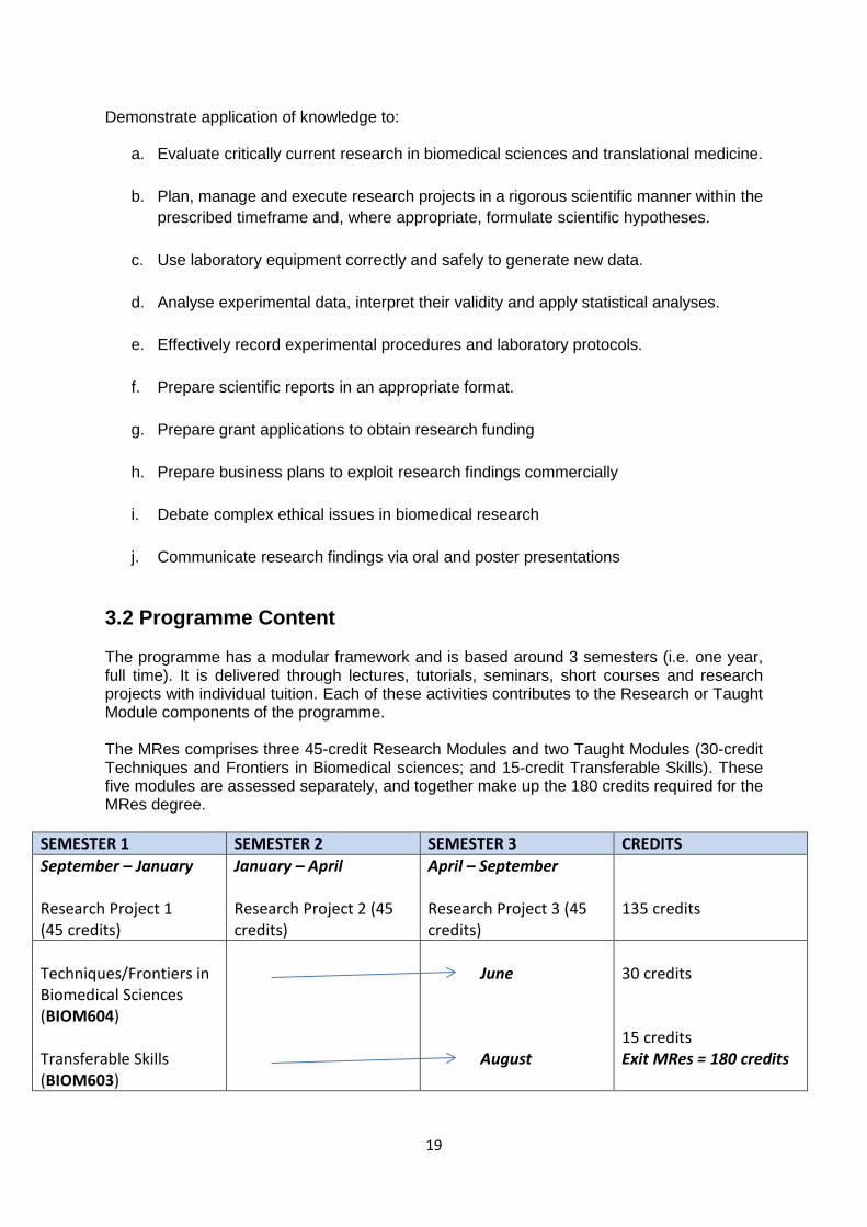
j. Communicate research findings via oral and poster presentations 3.2 Programme Content The programme has a modular framework and is based around 3 semesters (i.e. one year, full time). It is delivered through lectures, tutorials, seminars, short courses and research projects with individual tuition. Each of these activities contributes to the Research or Taught Module components of the programme. The MRes comprises three 45-credit Research Modules and two Taught Modules (30-credit Techniques and Frontiers in Biomedical sciences; and 15-credit Transferable Skills). These five modules are assessed separately, and together make up the 180 credits required for the MRes degree.
SEMESTER 1 SEMESTER 2 SEMESTER 3 CREDITS September – January Research Project 1 (45 credits)
January – April Research Project 2 (45 credits)
April – September Research Project 3 (45 credits)
135 credits
Techniques/Frontiers in Biomedical Sciences (BIOM604) Transferable Skills (BIOM603)
June August
30 credits 15 credits Exit MRes = 180 credits

20
An overview of each module is given below.
A) Research Project modules Students will undertake three research projects, comprising 10 weeks of lab work followed by 2 weeks in which to write a project report and prepare a presentation. In most strands the research project is chosen by the student after discussion with the appropriate strand convenor, who is responsible for matching student interests with available projects and supervisors. In the Cellular & Molecular Physiology strand, the first two research projects are allocated to students and are chosen to augment their existing technique and knowledge base. For example those who have previously followed courses in molecular biology might do a project involving electrophysiology and vice versa. The third project is chosen by Cellular & Molecular Physiology strand students after discussion with staff involved in the program. During the course of the project, all students will be encouraged to suggest experiments, design experimental protocols, as well as being taught subject specific techniques and advanced knowledge in transferable skills. Each of the three research projects taken should include a different research technique to enhance experimental training skills that need to be clearly stated at the end of each 10 week project. Students will have regular (usually daily) contact with the supervisors or other laboratory members for advice and guidance (see later section 3.6). Time will be allowed to undertake the necessary literature searches during the 10-week experimental period, and a further 2 weeks is to be spent out of the lab in order to write a project report and make an oral or a poster presentation to the Institute at the end of each project. Students will be assessed on their project report, their presentation and their general performance in the lab.
B) Techniques and Frontiers in Biomedical sciences module Techniques in Biomedical Sciences This part of the module consists of a series of lectures on a wide range of modern research techniques, including transgenic approaches, use of fluorescence and electron microscopy, gene expression analysis, proteomic approaches, etc. These lectures are complemented by tutorials designed to develop the fundamental skills required for laboratory research, including data handling, generation of figures, scientific writing and preparation of poster and oral presentations. Frontiers in Biomedical Sciences In this part of the module, the emphasis is on how state-of-the-art research techniques are used to advance knowledge in specific biomedical research areas and on modern methods/approaches employed in the diagnosis of disease and the treatment of patients. Lectures on these topics are complemented by tutorials and journal clubs designed to develop analytical and critical thinking skills.

21
The Techniques and Frontiers module is enhanced by Institute Seminars and Biomedical Review Lectures within the Institute of Translational Medicine. Eminent scientists from throughout the UK contribute to this by presenting their research on a variety of topics. It is important that MRes students attend both Seminars and Biomedical Review Lectures to broaden their knowledge and range of learning experiences. Seminars are organized by the individual Departments within the Institute and are advertised regularly via email. Attendance at certain Departmental Seminars may be recommended by strand convenors to enhance awareness of research that is particularly relevant to individual MRes strands. Finally, strand-specific activities are an important part of the Techniques and Frontiers module, as they facilitate awareness of the science associated with particular research strands. Students will be assessed on this module via one short review based on a Techniques lecture, one short review based on a Frontiers lecture, and a referees report based on a Journal Club.
C) Transferable Skills module Training in this module is on-going throughout the year and is delivered by staff involved with the program and the University via central provisions. It includes training in research techniques and the development of personal and professional transferable skills. Topics include research philosophy, principles and ethics, managing research progress, data analysis and presentation, health and safety, scientific and technical writing, patent law, exploitation of research, team work skills, time and resource management, communication skills, self-assessment skills and leadership skills. In addition, there is a weekly “English Support” session run by The English Language Unit to improve communication skills for students with English as a second language. Important and innovative parts of the transferable skills module include the “IP and Commercialisation” and “Writing a PhD Studentship” workshops and debates for public understanding of science (science & society). At the end of the module you will need to prepare a Portfolio of Assessment commentary as part of the transferable skills training. Finally, you will attend a Demonstrator Training session as part of the transferable skills training. Further information on the 3 assessed components of the module is given below (detailed information can be found later in this handbook): The IP and Commercialisation Workshop will raise awareness of the issues associated with, and necessary for, successful commercialisation of academic research. Intellectual property is a key component for business success. You will learn about the types and importance of intellectual property and how to search for patent information on the Web. The routes available for creating value from basic research will be described. There will be a comparison of the licensing and spin-out routes, together with detail of the business planning process and the role of the technology transfer office. You will be working as a group to prepare a written business plan for potential commercialisation of research. In addition, you will be required to present your idea as a ‘pitch’ to a panel of judges in a similar way to the popular television programme “Dragons’ Den”.

22
The “Grant Application” workshop will enable students to create a coherent and feasible research proposal for a scientific project to continue studying for a PhD and to present this in a form suitable for external scrutiny. The Portfolio of Assessment Commentary will enable students to evaluate and reflect on the aims and objectives of the individual programme components, as well as their own achievements. Students will be required to assemble a professional-looking portfolio that documents their progression through the programme in a form suitable for external scrutiny. Students will be assessed on the business plan and presentation given in the IP and Commercialisation component, on the Grant application, and on the Portfolio of Assessment Commentary. Students MUST attend a viva with the MRes external examiner in order to be awarded an MRes degree. The specific date for your viva will be set nearer the time but you must be available in Liverpool from 3 – 7 September 2018, when vivas will be held. English Support Classes are compulsory for students with English as a second language. Please visit the English Language Centre page here for more information about classes for international research students: https://www.liverpool.ac.uk/english-language-centre/in-sessional-support/academic-classes-international-research-students/ More general English language support can be found here: https://www.liverpool.ac.uk/english-language-centre/in-sessional-support/ 3.3 Schedule of Techniques, Frontiers and Transferable Skills sessions Time: 14:00 – 15:00 Wednesdays Venue: North West Cancer Research Centre, 200 London Road Precise details of lectures will be sent separately to students and staff via email. 3.4 Expectations of MRes project supervisors:
1. Supervisors are responsible for ensuring that appropriate risk assessments are in place and that all work involving human subjects or animals is covered by the appropriate Ethics Comittee or Home Office documentation.
2. Supervisors are expected to have regular meetings with MRes students during the research projects to discuss progress and plan experiments - at least 1 hour per week is recommended for this. If assistance with day-to-day lab work is delegated to post-docs or graduate students, this should be made clear to the MRes student.
3. Normal working hours are 09:00 – 17:30 Monday-Friday. In cases where it is
necessary for students to work outside these hours, supervisors must ensure that a suitably experienced supervisor is present (students are not permitted to perform lab work alone). In such cases, the out of hours work book must be signed on entering and leaving the building stating the name of the person who is supervising the student.

23
4. If students are absent from lab work without authorisation or explanation, supervisors should inform both the MRes programme administrator and the student’s Strand Convenor.
5. In addition to their research project, students have various other commitments that they must attend (Wednesday afternoon lectures, training courses, seminars, strand specific activities, etc). Supervisors should timetable lab work around these sessions to enable students to attend these compulsory activities.
6. Supervisors should be prepared to give advice on the content and organisation of their students’ assignments (project reports, talks, posters, studentship application) and to listen to practice talks. However, supervisors should not give feedback on written drafts of reports.
3.5 Expectations of MRes students:
1. Be familiar with the content of the MRes Programme Handbook.
2. Be professional in your dealings with your supervisor, and other research staff you encounter.
3. Attend punctually for both lectures and lab work.
4. Ensure that you inform your supervisor and strand convenor as soon as possible if you
are absent and subsequently complete the appropriate absence form.
5. Ensure that you check your email regularly, and respond promptly to messages from your supervisor and other MRes staff.
6. Submit all assignments promptly by the deadlines stated in the Handbook.

24
Section 4 Assessment 4.1 Overview of Assessment The accreditation for a Research Masters Degree is regulated by The University of Liverpool Ordinances and Regulations. Detailed rules about assessment can be found within the University Framework for Postgraduate Modular Provision and appropriate sections of the Code of Practice on Assessment, which can be accessed via the University website: https://www.liv.ac.uk/tqsd/code-of-practice-on-assessment/ The award of MRes requires that a minimum of 180 credits be obtained. In order to be eligible for the award of an MRes, candidates must achieve a minimum mark of 50% in each of the modules that comprise the programme (three Research project modules, the Techniques and Frontiers in Biomedical Sciences module, and the Transferable Skills module). Where the average of the total marks in all modules is 50% or above, a mark in the range 40-49% may be deemed compensatable only in the Transferable Skills module. In addition, a minimum of 70% recorded attendance at the compulsory sessions in the Techniques/Frontiers and Transferable Skills modules is required to pass the MRes degree. Candidates who fail to satisfy the examiners in a module assessment shall be permitted to re-present the failed work on one further occasion only, at a time specified by the examiners. For the purposes of calculating the overall average mark and determining classification, marks for modules passed by reassessment will be capped at 50%. 4.1.1 Late submission of work
Please note that the standard University penalty for late submission of written work applies; 5% of the total marks available for the assessment will be deducted from the final mark for each day after the submission date up to maximum of five working days. Work received after this time will receive a mark of zero.
4.1.2 Marking of submitted work
We aim to assess all written work and provide feedback to students within 3-4 working weeks after submission.
4.1.3 Extenuating Circumstances
When awarding degrees, the Board of Examiners will take into consideration any extenuating circumstances that may have adversely affected a candidate’s performance providing these have been notified in writing to the Programme Director. Where illness is involved a medical certificate should be supplied. Rules and regulations can be downloaded from the University website: http://www.liv.ac.uk/tqsd/pol_strat_cop/cop_assess/appendix_M_cop_assess.pdf.

25
Please note that the appropriate application form needs to be filled out to be eligible for consideration. A copy of the extenuating circumstances form can be found at the back of this handbook. Documentation must be supplied within a reasonable timeframe, forms submitted long after the event will not be considered. The form and supporting evidence must be submitted as soon as possible (normally within five days) after the events under consideration occur, and no later than one week before the meeting of the Board of Examiners at which the results of the assessments concerned will be considered. If you are unable to submit the form within the normal five days please contact the MRes Programme Administrator.
4.1.4. External Examiner
The External Examiner(s) will oversee course assessment procedures and assess annually the quality and relevance of the subjects taught. The External Examiner(s) will conduct a viva voce examination on the research elements of all candidates. Attendance at the viva is a prerequisite for obtaining the MRes degree.
4.1.5 Award of Merit or Distinction
A Merit or Distinction grade will be awarded in accordance with the CODE OF PRACTICE ON ASSESSMENT APPENDIX C University Framework for Postgraduate Modular Provision, which can be accessed from the following website https://www.liverpool.ac.uk/media/livacuk/tqsd/code-of-practice-on-assessment/appendix_C_2015-16_cop_assess.pdf
4.1.6 Progression to PhD Progression to PhD of MRes graduates is subject to obtaining 65% or above in the Research Projects and is also subject to the discretion of the supervisor.
4.2 Role of the External Examiners The MRes external examiners have the important task of checking and validating the marks and degree recommendations of the Board of Examiners for the MRes. This process is carried out at several levels by Strand examiners, who oversee the research elements, and by the Lead examiner, who oversees the taught module components and has overall responsibility for the programme. • The Lead external examiner views all marks and examples of in-course assessment work
from all taught modules. • The Lead external examiner also assesses all student portfolios. • Strand external examiners are provided with the marks and all research project reports
for each student in their strand. • Strand examiners viva all students for around 20-30 minutes. The focus of the viva is the
student’s research projects and its assessment in the portfolio. The Strand Convenor is also present throughout the viva process.

26
• External examiners perform a significant role at the final Examiners Board meeting. The
examiner might comment at this meeting on student performance and on the course in general.
• All external examiner write an annual report on the course and on the assessment
process. There will be several Strand external examiners representing research expertise in the main strands of the MRes degree programme. The vivas will take place in September (3 – 7 September 2018) and attendance at the viva is a prerequisite for obtaining the degree. You must be available to attend a viva in Liverpool in this week. 4.3 Academic Integrity Academic integrity is concerned with the moral and ethical code that applies to the standards by which the academic community operates. Students who embrace academic integrity understand that they must produce their own work, acknowledging explicitly any material that has been included from other sources or legitimate collaboration, and to present their own findings, conclusions or data based on appropriate and ethical practice. There are conventions of academic practice, such as established referencing and citation protocols, which both display and ensure academic integrity. Failure to adhere to these conventions can result in poor academic practice or, if there is a clear intention to deceive examiners and assessors, to unfair or dishonest academic practice. Cases of suspected breaches of academic integrity will be dealt with in accordance with the University of Liverpool’s Code of Practice on Academic Integrity. Cases will be considered by the Strand Convenor and Programme Director, taking advice from the Plagiarism Assessment Officer and Director of Postgraduate Studies. Penalties applied will depend on the severity of the offence in keeping with the guidelines in the Code of Practice. A talk on how to avoid plagiarism in bioscience and use of the Turnitin system for plagiarism/collusion detection will be given in the 1st semester. Definitions of various breaches of academic integrity taken from the Code of Practice are given below for your attention. All students must read this section and be aware and observe these rules in regard of all written work submitted for assessment. An academic integrity form should be signed to cover all written work submitted for the MRes programme (a copy of this form can be found at the back of this handbook). Poor academic practice Poor academic practice occurs when there has been failure, due to lack of academic ability or understanding, to observe the expected standards associated with academic integrity when undertaking academic work. Poor academic practice covers a range from minor errors such as missing quotation marks or mistakes in referencing to plagiarism, copying from others or embellishment, fabrication or falsification of data. This category also captures first offences in which dishonesty can be presumed but intent to deceive cannot be established because there has been no prior warning.

27
Unfair and dishonest practice Unfair and dishonest practice occurs when a student intends to gain an advantage over other students by wilfully seeking to deceive assessors and/or examiners. Such acts are often but not always premeditated and would include offences subsequent to a prior written warning of academic malpractice. Minor Errors Minor errors arise when a student has attempted to adopt academically acceptable practices but has failed to do so accurately or fully. Examples would be forgetting to insert quotation marks, minor mistakes in referencing or citation, gaps in the bibliography or reference list, non-compliance with some aspects of presentation guidelines. Collusion Collusion occurs when, unless with official approval (e.g. in the case of group projects), two or more students consciously collaborate in the preparation and production of work which is ultimately submitted by each in an identical or substantially similar form and/or is represented by each to be the product of his or her individual efforts. Collusion also occurs where there is unauthorised co-operation between a student and another person in the preparation and production of work which is presented as the student’s own. Coercive collusion would be considered a serious breach of academic integrity. Copying Copying occurs when a student consciously presents as their own work material copied directly from a fellow student or other person without their knowledge. It includes the passing off of another’s intellectual property, not in the public domain, as one’s own. It differs from collusion in that the originator of the copied work is not aware of or party to the copying. Copying of work from published sources would be dealt with as plagiarism. Submission of commissioned or procured coursework The dishonest practice occurs when a student presents as their own work coursework assessment tasks (or parts thereof) which have been intentionally procured (by financial or other inducement means) for this purpose. The definition includes the practice of requesting another party to prepare all or part of a course assignment (with or without payment) on the student’s behalf. Fabrication Throughout this policy the term “fabrication” is used to cover one or more of the following: Embellishment or Falsification of Data occurs when a proportion of the total data is altered, enhanced or exaggerated in order to emphasise data which has been obtained by legitimate means. Fabrication of Data occurs when a student creates and presents an extensive amount or significant piece of data in order to conceal a paucity of legitimate data; or wholly fabricates a set of data in the absence of legitimate data.
28
Plagiarism Plagiarism occurs when a student misrepresents, as his/her own work, the work, written or otherwise, of any other person (including another student) or of any institution. Examples of forms of plagiarism include: • the verbatim (word for word) copying of another’s work without appropriate and correctly presented acknowledgement and citation of the source; • the close paraphrasing of another’s work by simply changing a few words or altering the order of presentation, without appropriate and correctly presented acknowledgement and citation of the source; • failure to reference appropriately or to adequately identify the source of material used; • unacknowledged quotation of phrases from another’s work; • the deliberate and detailed presentation of another’s concept as one’s own. • You should not present work or part thereof for assessment that has previously been
submitted for assessment in another University of Liverpool module;
• You should not incorporate into any assignment material that has been submitted by me or any other person in support of a successful application for a degree of this or any other University or degree awarding body.
4.4 Academic Integrity form A Declaration of Academic Integrity form should be signed to cover all written work submitted for the MRes programme (a copy of this form can be found at the back of this handbook). 4.5 Procedure for requesting a deadline extension In cases where you are unable to meet assessment deadlines, for example due to illness, you must request an extension to the deadline from your Strand Convenor, who will decide whether or not to grant your request in consultation with the Programme Director.
29
Section 5 Research Projects Students will undertake three research projects, comprising 10 weeks of lab work followed by 2 weeks in which to write a project report and prepare an oral or poster presentation. During the course of the project, all students will be encouraged to suggest experiments, design experimental protocols, as well as being taught subject specific techniques and advanced knowledge transferable skills. The research projects will include at least three different research techniques to enhance experimental training skills. Students will have regular (usually daily) contact with supervisors and other laboratory members for advice and guidance during the 10 weeks in the lab. Time will be allowed to undertake the necessary literature searches during the 10-week experimental period, and a further 2 weeks is to be spent out of the lab in order to write a project report and make an oral or a poster presentation to the Institute at the end of each project. Supervisors will provide advice on which results should be included in the project report, the presentation of Figures, the interpretation of results and the overall structuring of the project report. However, supervisors are not permitted to comment on written draft reports. Supervisors are also expected to provide advice on the preparation of the oral and poster presentations. Students will be assessed on their project report, their presentation and their general performance in the lab. Further information on these assessments is provided later in this Section. 5.1 Laboratory safety and working hours You need to read carefully and obey all the instructions regarding safety that have been given to you before commencing experimental work in the laboratory. You are normally be expected to work in the lab between 09:00 – 17:00 Monday to Fridays, although flexibility is required depending on the type of experiments undertaken after discussion with supervisor. A supervisor who is frequently away from the laboratory is expected to allocate a post doc or a experienced PhD student to help with your day to day supervision. Work outside these hours, including at weekends, is only permitted if either your supervisor or a suitably qualified person approved by your supervisor is present. The out of hours work book must be signed on entering and leaving the building stating the name of the person who is supervising you.You are not expected or advised to work in the lab longer than the 10 weeks allocated for your project, in order to allow you enough time to complete your writing and prepare your talk or poster by the end of your project placement. If for any reason you need to be absent (e.g. other meetings, courses, illness, etc) you should inform your supervisor as soon as possible, at the latest by 09:30 on the day that you will be away from the lab, by calling or emailing them. You must provide information on when and why you will be absent, and ask him/her to make arrangements for any ongoing experiments that you cannot complete that day. You must also call or email your strand convenor to formally report your absence.

30
Working with human subjects and/or human material Supervisors have a responsibility to ensure that all work involving human subjects is covered by appropriate Ethics Comittee Permission. They should also ensure that students conducting research projects involving human subjects and/or material understand the permission given for their work, and in writing their dissertation, they make a clear statement of the Ethics Comittee Approval for the work. Working with animals Supervisors have a responsibility to ensure that the appropriate Home Office Authority (both personal and project licence) are in place before working with experimental animal is started. They should also ensure that students conducting research projects involving experimental animals understand the permission given for their work, and in writing their dissertation, they make a clear statement of the Home Office Approval for the work. Laboratory Books Laboratory books are the property of Liverpool University and are to be handed to the supervisor at the end of the project placement along with a completion of project work form. Completion of project work A Completion Form must be filled out and signed by your project supervisor(s) at the conclusion of the research projects, to confirm that all material has been safely accounted for and that any useful data has been passed on to your supervisor. 5.2 Preparation of Research Project Reports These notes are intended to help you in the preparation of the report describing your project. You will also be given a lecture on how to prepare a good project report as part of the Techniques in Biomedical Sciences lecture series. Your project should be prepared in the format of a research paper recently published in the Biochemical Journal. Please consult a recent issue of this journal to check on the appropriate style to adopt. The report should be produced on a computer using appropriate word processing, bibiographic and graphic software. It is expected that your report will be produced to “publication quality”, which means that you should pay close attention to spelling, punctuation and grammar, as well as scientific content. You should also take care with the quality of figures, clarity of legends, and citation of references. Figures and tables should be embedded in the text, in the style of papers published in the Biochemical Journal. An example of a project report is given at the end of this Section for you to refer to. Please note this example is there to give you an idea of the overall content, quality and depth of a report. The style should be as with the current format of Biochemical Journal. Your report should be typed single spaced, in Calibri font size 12 and must be 4000 ± 400 words in length. This word limit covers all text sections of the report, including the Abstract and legends to Figures/Tables, but does not apply to text contained within Figures/Tables or to the References section (i.e., the list of citations at the end does not count toward the 4000 words). Marks will be deducted proportionally for exceeding the upper limit of 4400 words. For example, 4600 words = 200 words above the limit = 5% deducted; 4800 words = 400 words above the limit = 10% deducted.

31
However, the mark will not be reduced below the pass mark of 50% for the assignment. Marks will not be deducted for being below the lower limit of the word count (i.e. 3600 words), but students should be aware that short reports are highly likely to be awarded lower marks due to a lack of coverage and discussion of key areas. Please attach a front cover sheet to your report stating your name, your student number, the title of the report, the name of your supervisor, the name of your internal assessor (2nd marker) if known, and the word count of your report. A template cover sheet is supplied at the end of this Section for this purpose. You should submit two copies of your final project report; one to your supervisor and one to your internal assessor. You also need to submit an electronic copy of the final version via Turnitin. These files need to be able to be uploaded onto Turnitin with all Figures included so where necessary your files will need to be compressed. Deadlines for submission of the project reports and dates of the oral/poster presentations are given in Section 3.3. The report must conform to the following style:
1. Abstract This is a concise summary of the work. It should deal with the reasons why the work was performed, the methods used, the results obtained, and the major conclusions reached. This section must not exceed 250 words.
2. Introduction This should describe the background to the relevant scientific literature
and the work performed. The rationale for the work and the hypothesis to be tested should be explained, and the major aims should be specified.
3. Experimental The description of methods should be adequate for a competent worker
in the area to follow and repeat your experiments. You should however be concise; again recent papers in your field of study should provide a guide for you.
4. Results This section should consist of text which describes the experimental data
obtained and where appropriate describes the rationale that links one experiment to the next. The text should be cross-referenced to the relevant figure or table. Is it not necessary to reproduce the same material in tables and figures. This section must not take the form of a diary of your experimental observations in the laboratory, nor need every single experimental observation be recorded. Instead, you must take responsibility for collating the data, and whatever statistical analysis are appropriate, and presenting your findings in a way that makes it possible for the reader to understand your major conclusions. Each figure should have an explanatory legend that enables the reader to understand how the experiment was performed. Figures and tables should be inserted into the main body of the text as close as possible to the relevant section.
5. Discussion This section should focus on the interpretation of your results, and set them
in the context of current knowledge in the field. It should not be necessary to repeat your description of the experimental data, but you will want to summarise your main findings and explain how they are meaningful.
6. References Again, this should follow the the Biochemical Journal style. It is strongly
recommended that you use reference organising software (such as EndNote) to construct your references, which will ensure that you use the correct Biochemical Journal style. References in the text should be cited as a number in the order in which they appear. The reference list should be correspondingly numbered, and references listed in order of their citation in the text.

32
7. Footnotes Abbreviations and acronyms used in the text must be defined immediately after the first use of the abbreviation. In addition, a complete list of all abbreviations used should also be cited in a single Footnote section. The abbreviations of some important biochemical compounds, e.g. ATP, NADH, DNA, and amino acids in proteins, need not be defined.
5.3 Preparation of Oral and Poster Presentations You will be required to make a presentation for each of your Research projects. This will comprise 2 poster presentations and 1 oral presentation. The dates of these presentation sessions are given in Section 3.3, but the scheduling of oral/poster presentations for individual strands will be announced nearer the time. You will be given instructions on how to prepare good oral and poster presentations as part of the Science Skills series in the 1st semester. You will also be given advice and assistance from your supervisor in preparing your presentations. The cost of printing your poster will be provided by your supervisor. 5.4 Assessment of Research Projects For each research project, students will be assessed on their project report, their presentation and on their general performance in the lab. Assessment of the project report will be conducted by the supervisor (1st marker) and an internal assessor (2nd marker). Continual assessment of performance in the lab is provided by the supervisor alone. Assessment of the presentation is by 2 independent markers. Each research project contributes 45 credits out of a total of 180 credits for the MRes. A brief explanation of how marks are awarded for these individual assessments and how they are combined to give the final mark is given below: Supervisor and Internal Assessor Mark 1: Awarded for the project report; this will reflect the scientific quality of the dissertation, its clarity and thoroughness, and quality of presentation; the internal assessor will also base his/her mark on a short informal mini-viva (project discussion). Supervisor Mark 2: This will be a continual assessment mark awarded for assessment of the student’s conduct during the project taking into account organization, initiative, effort and performance in the lab. Two Internal Assessors Mark 3: Awarded for oral/ poster presentation.

33
Supervisor and Internal Assessor
Mark 1 - Project report 40 marks
Supervisor
Mark 2 - Continual Assessment 10 marks
Internal Assessors Mark 3 - Oral/poster Presentation
10 marks
Total = 60 marks
Details of the assessment criteria used and the assessment forms that will be used by markers are given on the on the pages that follow.

34
5.4.1 Assessment Form for research project reports

35

36
5.4.2 Marking Criteria for assessment of written assignments
Distinction Level 100% - 90%
89% - 80% 79% - 70%
Outstanding. No (or virtually no – use scaling) better result conceivable at Masters level. Entirely correct and complete, with extensive evidence of critical thinking. Evidence of extensive research of relevant literature. Extremely logical structure, faultlessly written and presented. Clear evidence of highly original thought and cogent scientific argument. Excellent. Clear evidence of achievement on a scale reserved for exceptionally high quality work at Masters level. Essentially correct and complete, with significant evidence of critical thinking and excellent use of relevant literature. Highly logical structure, extremely well written and presented, displaying a significant amount (use scaling) of original thought and cogent scientific argument. Very Good. High quality work that demonstrates comprehensive understanding and shows some evidence of critical thinking. Evidence of very good research of relevant literature, with all key literature identified/discussed. Very well written and presented, with a very logical structure. Some evidence of original thought and cogent scientific argument
Merit Level
69% - 60%
Good. Good quality work that demonstrates generally sound scientific understanding. Evidence of good research of relevant literature, but some relevant literature not identified/discussed. Generally well written and presented, with a logical structure. Evidence of original/critical thinking may be limited
Pass Level 59% - 50% Satisfactory. Learning objectives achieved, but may contain deficiencies in one or more aspects of knowledge of the relevant literature, scientific understanding, structure and presentation.
Fail Level 49% - 40% Unsatisfactory. Learning objectives not achieved. Essentially an incomplete report with significant flaws or omissions and major deficiencies in one or more aspects of knowledge of the relevant literature, scientific understanding, structure and presentation.
39% - 0%
Poor. Severely deficient in content, understanding and application and containing many serious errors and omissions.

37
5.4.3 Marking Criteria for continual assessment of student performance
Distinction Level 100% - 90%
89% - 80% 79% - 70%
Outstanding. Student capable of working independently at the highest level on all aspects of project. As good as can be expected at Masters level. No room for improvement in project understanding, design, execution and motivation. Excellent. Student able to design and execute project work independently at a very high level with the minimum of assistance. Little room for improvement in project understanding, design, execution and motivation. Very Good. Student able to generate and analyse data at a high level of proficiency with the minimum of assistance. Demonstrates very good motivation, understanding of the project and critical scientific judgement.
Merit Level 69% - 60%
Good. Student able to generate and analyse data at a good level of proficiency with some assistance. Demonstrates generally good motivation, understanding of the project and some evidence of critical scientific judgement.
Pass Level 59% - 50%
Satisfactory. Learning objectives achieved, but student needs significant assistance to generate and analyse data. Some deficiencies in motivation, understanding of the project and critical scientific judgement may be apparent.
Fail Level 49% - 40%
Unsatisfactory. Learning objectives not achieved. Student requires constant help, produces work of poor quality and is unable to analyse data effectively. Significant deficiencies in motivation, understanding of the project and critical scientific judgement are apparent.
39% - 0%
Poor. Student displays inability to understand or carry out project work and low or no motivation (use scaling)
38
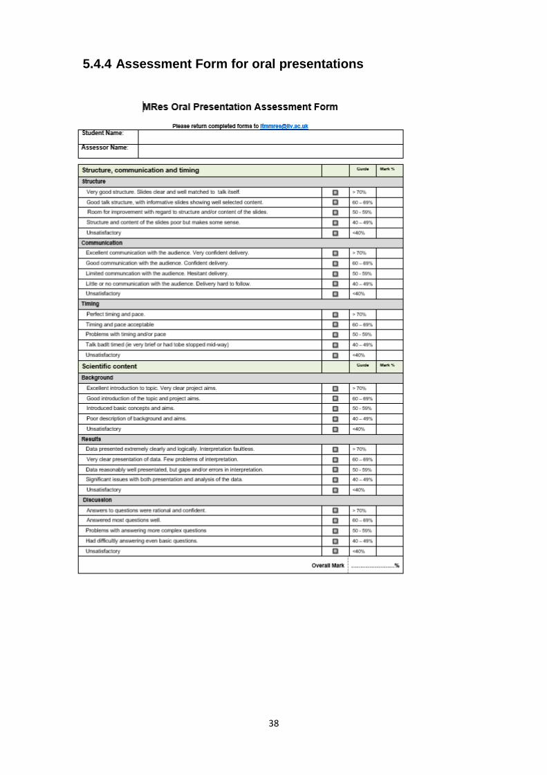
5.4.4 Assessment Form for oral presentations

39

40
5.4.5 Assessment Form for poster presentations

41

42
5.5 Submitting your Research Project Report You must adhere to the following instructions:
1. Use the example in section 5.6 to create a front sheet for each of your project reports
2. You must also submit an electronic copy via Turnitin which must be the final version of the project by 10:00 on the submission date.

43
5.6 Front sheet for all Research Project Report submissions (example)
Research Project Report [Insert number]
TITLE OF YOUR RESEARCH PROJECT REPORT
Joe Bloggs Student I.D. 200700000
Supervisor: [Insert name] Internal Assessor: [Insert name]
Strand: [Insert name of your strand]
Word Count: [Insert word count]
By submitting this work I confirm that I have read, understood, and adhered to the University’s Academic Integrity Policy (Appendix L in the University Code of Practice) and that I have read, understood and signed a Declaration of Academic Integrity.

44
5.7 Example of a Research Project Report Counting Calories in Yeast: Investigating Mechanisms of Lifespan Extension Associated with Dietary Restriction Name Department of xxx, Institute of Translational Medicine, University of Liverpool, Crown St, Liverpool, L69 3BX, UK. Dietary restriction (DR) extends the lifespan of several animal models, though the exact mechanisms underlying this remain unclear. The decline of heat shock protein (HSP) chaperone activity is closely tied to aging. Small HSPs 12 and 26 are expressed in response to DR in Saccharomyces cerevisiae and HSP12 is essential for lifespan extension associated with DR. Up-regulated in response to stress, HSPs of the α–crystallin family such as HSP26 act as intracellular chaperones. HSP12 aids membrane stability. To elucidate the role of these HSPs, two genome-wide screens for genetic interactions (GIs) were recently performed: synthetic genetic array (SGA) and quantitative fitness analysis (QFA). These identify GIs based on synthetic sickness whereby viable deletion mutants are combined resulting in reduced fitness. This implicates both gene products in crucial cellular functions. This study validated a subset of the putative interactions from QFA and SGA data with a 20% success rate, highlighting the need for validation of high-throughput (HTP) GI screens. Reproducible GIs of hsp12 were linked to genes of the stress response while autophagy was enriched in GIs with hsp26. Further analysis of double-mutant strains in a second genetic background highlights the potential lack of strain-specific GIs. INTRODUCTION Dietary Restriction (DR)1 is the single robust physiological intervention known to extend lifespan in a variety of organisms including: Saccharomyces cerevisiae, Drosophila Melanogaster, Caenorhabditis elegans and rodents [1–4]. Also known as calorie restriction, the beneficial effects of DR include protection against cancer and age-related disease [5], delayed senescence and extension of good health [6]. Whether these effects are seen in non-human primates is debated [7,8]. Few studies have analysed the effect of DR on humans, not least because adherence to DR is challenging. However, available data suggests DR with suitable nutrient intake results in a reduction of risk 1 Abbreviations: Dietary restriction (DR), Target of rapamycin (TOR), Replicative lifespan (RLS), Synthetic lethality (SL), Synthetic sickness interaction (SSI), Genetic interaction (GI), Synthetic genetic array (SGA), Quantitative fitness analysis (QFA), Open reading
factors for cancer, cardiovascular disease and diabetes [9,10].
The exact mechanisms mediating longevity remain unclear. Sirtuins, a family of NAD+-dependent deacetlylases, regulate glucose and lipid metabolism in response to energy status and stress resistance [4]. The yeast homologue, silent information regulator (Sir2), mediates longevity via increased silencing of DNA, ultimately avoiding extrachromosomal rDNA circle formation [11]. However, DR mediated longevity has been induced in sir2Δ strains [12]. Mechanisms independent of sirtuins include nutrient sensing pathways: insulin/insulin-like growth factor 1, AMP kinase and the target of rapamycin (TOR)
frame (ORF), Heat shock protein (HSP), Yeast peptone dextrose (YPD), Synthetic complete (SC), Dimethylsulfoxide (DMSO), High through-put (HTP), High osmolarity glycerol (HOG),
45
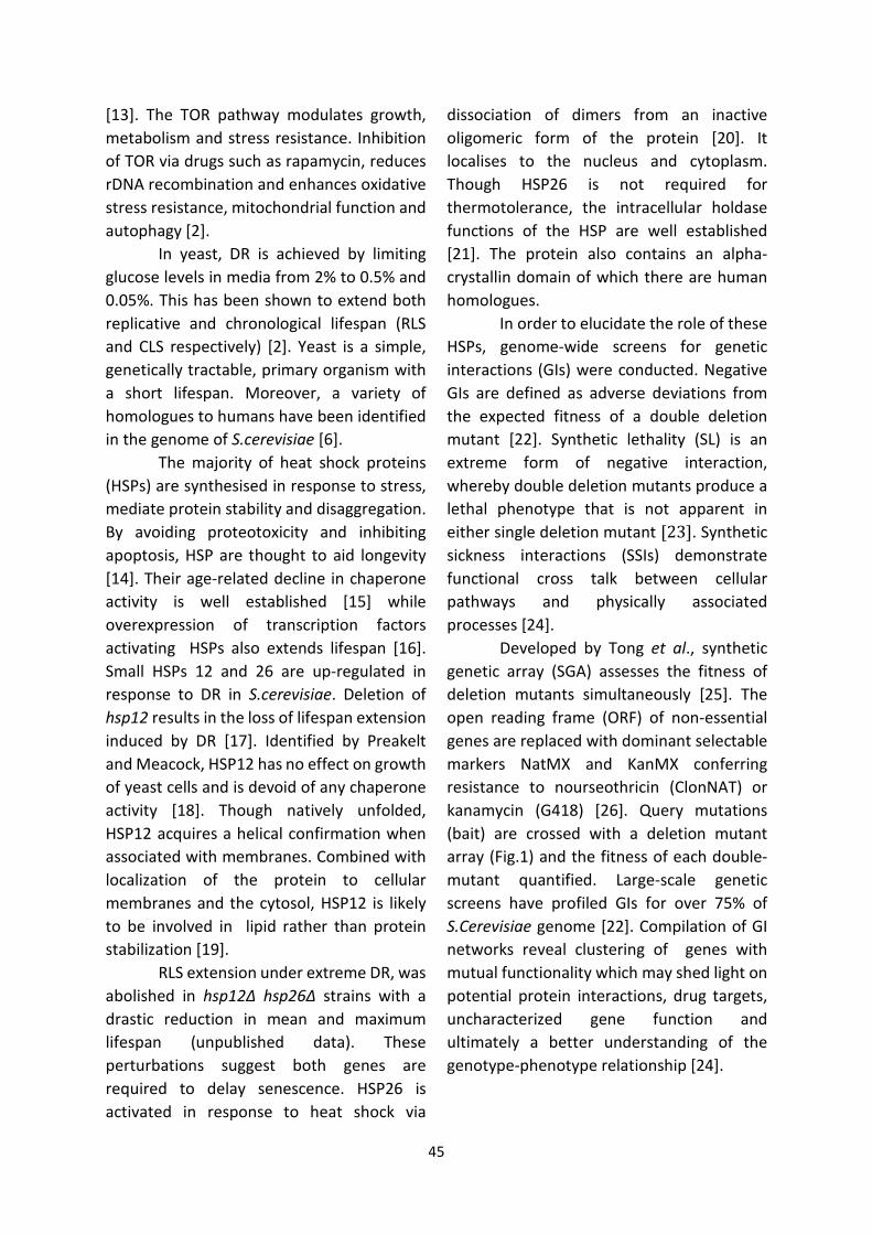
[13]. The TOR pathway modulates growth, metabolism and stress resistance. Inhibition of TOR via drugs such as rapamycin, reduces rDNA recombination and enhances oxidative stress resistance, mitochondrial function and autophagy [2].
In yeast, DR is achieved by limiting glucose levels in media from 2% to 0.5% and 0.05%. This has been shown to extend both replicative and chronological lifespan (RLS and CLS respectively) [2]. Yeast is a simple, genetically tractable, primary organism with a short lifespan. Moreover, a variety of homologues to humans have been identified in the genome of S.cerevisiae [6].
The majority of heat shock proteins (HSPs) are synthesised in response to stress, mediate protein stability and disaggregation. By avoiding proteotoxicity and inhibiting apoptosis, HSP are thought to aid longevity [14]. Their age-related decline in chaperone activity is well established [15] while overexpression of transcription factors activating HSPs also extends lifespan [16]. Small HSPs 12 and 26 are up-regulated in response to DR in S.cerevisiae. Deletion of hsp12 results in the loss of lifespan extension induced by DR [17]. Identified by Preakelt and Meacock, HSP12 has no effect on growth of yeast cells and is devoid of any chaperone activity [18]. Though natively unfolded, HSP12 acquires a helical confirmation when associated with membranes. Combined with localization of the protein to cellular membranes and the cytosol, HSP12 is likely to be involved in lipid rather than protein stabilization [19].
RLS extension under extreme DR, was abolished in hsp12Δ hsp26Δ strains with a drastic reduction in mean and maximum lifespan (unpublished data). These perturbations suggest both genes are required to delay senescence. HSP26 is activated in response to heat shock via
dissociation of dimers from an inactive oligomeric form of the protein [20]. It localises to the nucleus and cytoplasm. Though HSP26 is not required for thermotolerance, the intracellular holdase functions of the HSP are well established [21]. The protein also contains an alpha-crystallin domain of which there are human homologues.
In order to elucidate the role of these HSPs, genome-wide screens for genetic interactions (GIs) were conducted. Negative GIs are defined as adverse deviations from the expected fitness of a double deletion mutant [22]. Synthetic lethality (SL) is an extreme form of negative interaction, whereby double deletion mutants produce a lethal phenotype that is not apparent in either single deletion mutant [23]. Synthetic sickness interactions (SSIs) demonstrate functional cross talk between cellular pathways and physically associated processes [24].
Developed by Tong et al., synthetic genetic array (SGA) assesses the fitness of deletion mutants simultaneously [25]. The open reading frame (ORF) of non-essential genes are replaced with dominant selectable markers NatMX and KanMX conferring resistance to nourseothricin (ClonNAT) or kanamycin (G418) [26]. Query mutations (bait) are crossed with a deletion mutant array (Fig.1) and the fitness of each double-mutant quantified. Large-scale genetic screens have profiled GIs for over 75% of S.Cerevisiae genome [22]. Compilation of GI networks reveal clustering of genes with mutual functionality which may shed light on potential protein interactions, drug targets, uncharacterized gene function and ultimately a better understanding of the genotype-phenotype relationship [24].

46
Quantitative fitness analysis (QFA) facilitates measurement of growth phenotypes to a higher resolution by taking
hsp12Δ
ΔΔ Δ
:Nxxx :K
:N, xxx :Khsp12Wild type
Sporulation
MATa haploid progeny selection
Double mutant selection
MATα MATa
a/α
Mating
ClonNAT and G418 media
-HIS me d ia
ClonNAT and G418 media
Sporulationmedia
Fig.1 SGA Procedure: The query mutant hsp12:N containing the NatMX resistance cassette (filled red circle) is systematically crossed with a library of deletion mutants (filled blue circle) containing KanMX cassettes (xxx:K). These dominant markers provide resistance to nourseothricin (ClonNAT) and kanamycin (G418) respectively. Haploids containing each deletion mutant are mated. Sporulation of the diploid is induced on media containing reduced nitrogen. The progeny of MATa are selected on –HIS media, due to the inclusion of the mating-type specific promoter (MFA1pr-HIS3). Double-mutants are isolated and assembled into an array via robotic manipulation (Based on Tong and Boone, 2007). multiple photographs of plates during the incubation phase [27]. While fewer strains are grown on each plate, time allocated for growth is greater in QFA than SGA screens [28]. This makes growth phenotypes more apparent.
These screens were the first to incorporate hsp12 and hsp26 as bait which have isolated a myriad of putative genetic interactions. However, both screens were performed using isogenic strains. Various genetic backgrounds differ in sensitivity to mutational perturbation and little is known about their influence on GI network topology [29]. This study aims to verify putative interactions from both the SGA and QFA screens via manual replication through spot test assays. Recreation of the double-mutants in a different background strain may provide insight into strain specific interactions. Ultimately the analysis of these interactions may shed light on the roles of these proteins in lifespan extension accompanying DR. EXPERIMENTAL PROCEDURES All chemicals were of laboratory grade and obtained from Sigma-Aldrich (Poole, UK) unless otherwise stated. Reagents used for growth media were also provided by Formedium (Norwich, UK). PCR primers were from Sigma-Aldrich (Poole, UK) and PCR reagents supplied by Bioline (London, UK). Materials for gel electrophoresis were obtained from GE-Healthcare.
Strains and Growth media – The strains used in this study included a library of DLY7325 deletion mutants (MATa lyp1::HPH::LEU2 can1∆::STE2pr-Sp_his5 his3∆ leu2∆ ura3∆ met15∆ (plus hsp12::NATMX or hsp26::NATMX plus xxx::KANMX as appropriate) provided by Peter Banks (Newcastle University). These were generated by mating an DLY7325 starter strain (MATα plus hsp26/12::N) onto the BY4741 kanMX deletion library (MAT a his3∆ leu2∆ ura3∆ met15∆ plus xxx::K). The AEY1017 strain (MATα ade2-1 ura3-1 his3-11,15 leu2-3, 112 trp1-1 can 1-100 TELVIIL::URA3), donated by Dr Downs (University of Sussex), is a W303 derivative
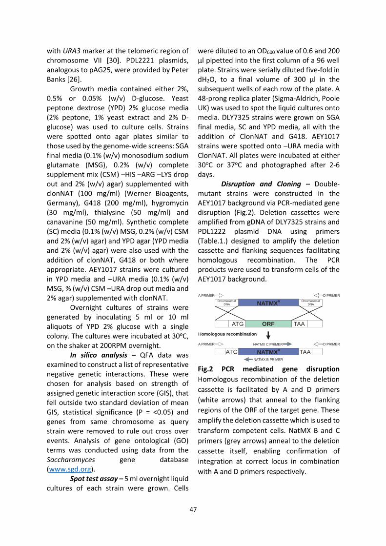
47
with URA3 marker at the telomeric region of chromosome VII [30]. PDL2221 plasmids, analogous to pAG25, were provided by Peter Banks [26].
Growth media contained either 2%, 0.5% or 0.05% (w/v) D-glucose. Yeast peptone dextrose (YPD) 2% glucose media (2% peptone, 1% yeast extract and 2% D-glucose) was used to culture cells. Strains were spotted onto agar plates similar to those used by the genome-wide screens: SGA final media (0.1% (w/v) monosodium sodium glutamate (MSG), 0.2% (w/v) complete supplement mix (CSM) –HIS –ARG –LYS drop out and 2% (w/v) agar) supplemented with clonNAT (100 mg/ml) (Werner Bioagents, Germany), G418 (200 mg/ml), hygromycin (30 mg/ml), thialysine (50 mg/ml) and canavanine (50 mg/ml). Synthetic complete (SC) media (0.1% (w/v) MSG, 0.2% (w/v) CSM and 2% (w/v) agar) and YPD agar (YPD media and 2% (w/v) agar) were also used with the addition of clonNAT, G418 or both where appropriate. AEY1017 strains were cultured in YPD media and –URA media (0.1% (w/v) MSG, % (w/v) CSM –URA drop out media and 2% agar) supplemented with clonNAT.
Overnight cultures of strains were generated by inoculating 5 ml or 10 ml aliquots of YPD 2% glucose with a single colony. The cultures were incubated at 30oC, on the shaker at 200RPM overnight.
In silico analysis – QFA data was examined to construct a list of representative negative genetic interactions. These were chosen for analysis based on strength of assigned genetic interaction score (GIS), that fell outside two standard deviation of mean GIS, statistical significance (P = <0.05) and genes from same chromosome as query strain were removed to rule out cross over events. Analysis of gene ontological (GO) terms was conducted using data from the Saccharomyces gene database (www.sgd.org).
Spot test assay – 5 ml overnight liquid cultures of each strain were grown. Cells
were diluted to an OD600 value of 0.6 and 200 µl pipetted into the first column of a 96 well plate. Strains were serially diluted five-fold in dH2O, to a final volume of 300 µl in the subsequent wells of each row of the plate. A 48-prong replica plater (Sigma-Aldrich, Poole UK) was used to spot the liquid cultures onto media. DLY7325 strains were grown on SGA final media, SC and YPD media, all with the addition of ClonNAT and G418. AEY1017 strains were spotted onto –URA media with ClonNAT. All plates were incubated at either 30oC or 37oC and photographed after 2-6 days.
Disruption and Cloning – Double-mutant strains were constructed in the AEY1017 background via PCR-mediated gene disruption (Fig.2). Deletion cassettes were amplified from gDNA of DLY7325 strains and PDL1222 plasmid DNA using primers (Table.1.) designed to amplify the deletion cassette and flanking sequences facilitating homologous recombination. The PCR products were used to transform cells of the AEY1017 background.
NATMXR
ORF
NATMXR
NATMX B PRIMER
NATMX C PRIMER
D PRIMERA PRIMER
ATG TAA
ATG TAA
Homologous recombination
Chromosomal DNA
Chromosomal DNA
D PRIMERA PRIMER
Fig.2 PCR mediated gene disruption Homologous recombination of the deletion cassette is facilitated by A and D primers (white arrows) that anneal to the flanking regions of the ORF of the target gene. These amplify the deletion cassette which is used to transform competent cells. NatMX B and C primers (grey arrows) anneal to the deletion cassette itself, enabling confirmation of integration at correct locus in combination with A and D primers respectively.

48
Table.1 Primer sequences and expected product sizes
Primers Sequence 5' to 3' Wildtype Product (bp)
Deletion Cassette Product (bp)
Nat B GGCAGGGCATGCTCATGTAGA N/a N/a hsp12 A GTATACGCAAGCATTAATACAACCC
1023 2040 hsp12 D GCTTGGATTTGAGACGGATTTC hsp26 A CCTGTCAAGGTGCATTGTTGG 1555 2257 hsp26 D GTCGAGGTTTAACGCTTATTACC atg13 A GAGGCGAGAAGTAGAAACAAGG
2686 1826 atg13 D CAGCGGGTGACAAATAAGC atg2 A GGTGCGGCGAATGGAAATAATC 5106 1684 atg2 D GGCTTTCCCACCATTCCAC atg23 A CTATTGGAGAACCGGCTAAG 1938 1933 atg23 D CAACGGCTCCTGTGGGTAC atg7 A GGAAGAACAAGCCACCACATG 2491 1955 atg7 D CCAATTTCTTAGCCGTCAGC emi5 A GTTCCGCCGGTTAATGAACG 1009 1768 emi5 D GGACCGCTATGAAGAAGGACTG git1 A CTTTGATGAGGGACATCTGCTGG 1858 1921 git1 D GATGCAGCTTACTCCCAAGAGTG his3 A GGAGTCACTGCCAGGTATCG 1227 1921 his3 D CTTACGGAATACCACTTGCCAC kex2 A GGACGTGTTCTTTCTCTCG 2809 1721 kex2 D GCGGGTGCAAACAATGCAAAG nbp2 A CCCTTTCAAATGCCAGCCTC 1243 1889 nbp2 D GGAAGAGTTGGACATAGAGGAAG spe1 A CTGAAATAGCGGAACCTGC 1742 1698 spe1 D CCACCACTGTACGCCATTG
gDNA extraction – 10 ml liquid cultures were inoculated with single colonies and grown overnight. gDNA was extracted from the cultures with a Yeast gDNA Extraction Kit (Thermo Scientific, UK) and stored at -20oC.
PCR and Gel electrophoresis – 50 µl PCR reactions were generated in the following manner: 22 µl dH20, 1 µl each of forward and reverse primers (100 µM), 1 µl of plasmid DNA or purified gDNA from deletion strain and 25 µl 2X My Taq® (Bioline, London UK). In order to aid amplification of HSP26:N, 5% dimethylsulfoxide (DMSO) was included in the reaction as previously described [26]. Reactions were run with an annealing temperature of 94oC and 1 min extension time at 55oC per cycle. PCR products were resolved on 1% agarose gel and photographed.
Transformation of AEY1017 strain – Following the method previously described by Schiestl and Geitz [31], AEY1017 cells were transformed with successfully amplified deletion cassettes. Solutions consisting of: 240 µl 50% (w/v) polyethylene glycol, 36 µl 1M lithium acetate, 50 µl salmon testes single stranded DNA and 34 µl of the PCR product were incubated at 30oC for 30 min and 42oC for the following 30 min. The cells were pelleted and resuspended in 1 ml dH20. 4 ml YPD 2% glucose were added to each solution and cells grown overnight.
Transformants were selected on YPD 2% glucose media containing 100 mg/ml clonNAT. Double-mutants were grown on YPD media containing 100 mg/ml clonNAT and 200 mg/ml G418. Successful integration of cassettes were determined via gDNA extraction from secondary transformants and PCR amplification of the cassette.

49
RESULTS Initial analysis of QFA data – As negative GIs are more likely to reveal functional cross talk between pathways than positive interactions
[32], we first compiled a list of statistically significant, negative GIs from the QFA dataset (Table.S1 and Table.S2). These genes displayed deleterious growth defects
Representative negative genetic interactions identified by SGA (Blue) and QFA screens. (Grey) Double-mutant strains not present in the DLY7325 library prevented further analysis.
(Underlined) Genes related to autophagy. in combination with the query mutant, under a range of stressful conditions. A subset of this list, comprising of the top double-mutants displaying growth defects under a range of conditions (core GIs) and those specific to certain stressors (stress-specific GIs), were selected. These were combined with genes represented in the SGA screen (Table.2). GO term analysis revealed an enrichment of genes associated with autophagy, in putative GIs with hsp26. Due to the SL phenotypes of core interactions, some double-mutant strains were not present in the library and so only a subset could be examined.
Reproduction of genetic interactions – In order to compare double-mutant fitness with that of the query mutant, spot test assays were performed. This technique facilitates comparison of growth phenotypes to a medium through-put scale [33]. All strains of the DLY7325 background contained the null mutation his3Δ1. Therefore double-mutant strains with his3Δ were included as
controls for query mutant fitness. Fig.2 illustrates SSIs in combination with hsp12. Cells were spotted onto minimal media including SGA final media and SC media. Plates were subject to stressful conditions similar to SGA and QFA. All
Table 2. Genetic interactions from QFA and SGA screens: Genes interacting with hsp12 Genes interacting with hsp26
ash1 fmt1 ire1 pol2 slm1 ald4 cbt1 hap4 qri7 tim50 ala1 fre1 jjj1 rad9 tfc7 asc1 cox8 hap4 rps14a tre2
cdc34 fus2 kns1 ras1 tir4 atg13 csm1 inp51 rps16b vac17 cka2 gal11 mbf1 rfa1 tma16 atg2 cyk3 kex2 sdh2 crn1 gir2 mdn1 rpc11 top1 atg23 dbr1 lhs1 sfc1 csm1 gre2 mnt4 rpo21 tub4 atg7 dep1 oms1 sok1 dnl4 hat1 nbp2 rpp2b bch2 elc1 ost4 spe1 dot1 hsf1 pbn1 rps19a bdf2 emi5 paa1 srb8 dut1 ibd2 pdr5 rrp42 bst1 fkh2 pat1 ssa1
ecm34 inp52 pno1 ser33 bud17 git1 pph21 ssa2

43
2% Glucose (30 ̊C)
Cell Density
hsp12 cdc34
hsp12 dut1 hsp12 fus2
hsp12 mbf1
Δ Δ
Δ ΔΔ Δ
Δ Δ
hsp1
hsp12
2 crn1Δ Δ
Δ Δ gal11
hsp12 his3 hsp12 ibd2
hsp12 jjj1 hsp12 mnt4
hsp12 pno1 hsp12 ras1 hsp12 slm1
Δ Δ
Δ Δ
Δ Δ
Δ ΔΔ Δ
Δ Δ
Δ Δ
hsp12Δ Δ nbp2
hsp12 pdr5
2 rpp2b
2 top1
Δ Δ
Δ Δ
Δ Δ
hsp12 rfa1
hsp12 rps19a
hsp12 tub4
Δ Δ
Δ Δ
Δ Δ
hsp1
hsp1
Fig.2 First round of spot test assays: Serial dilutions of strains were spotted to SGA final media containing 2% glucose and incubated at 30oC. hsp12Δ his3Δ strain was included as a control for query mutant fitness. (Red) Double-mutant strains display synthetic sickness.
hsp12 his3 hsp26 his3
hsp12 emi5 hsp26 emi5hsp12 gal11 hsp26 gal11 hsp12 pdr5
Δ Δ
Δ ΔΔ Δ
Δ Δ
Δ ΔΔ ΔΔ Δ
hsp26 pdr5 Δ Δ
2% Glucose(37 ̊C)
hsp12 his3 hsp26 his3 hsp12 kex2
hsp26 lhs1
hsp26 nbp2
Δ ΔΔ ΔΔ Δ
Δ Δ
Δ Δ
hsp26 kex2Δ Δhsp12 lhs1
hsp12 nbp2
Δ Δ
Δ Δ
2% Glucose (30 ̊C)Cell Density
Fig.3 Second round of spot test assays: Serial dilutions of strains were spotted onto SGA final media containing 2% glucose and incubated at either 30oC or 37oC. his3Δ double-mutants were included as controls for query mutant viability. Strains displaying growth defects when combined with the query mutant hsp12Δ (red) or with hsp26Δ (blue). These phenotypes are not due to in-viability of the single mutant as strains are viable in combination with the opposite query mutant. strains failed to grow on 0.8M NaCl plates and those subject to an additional stress of 37oC. All SSIs are listed Fig.S1.
A second round of spot tests were performed to assess the viability of both
single-mutants. This included all strains that had shown synthetic sickness in the first stage of analysis. Fig.3 highlights SL in hsp12Δ gal11Δ and hsp26Δ gal11Δ. This could be due to a shared GI between query mutants or

44
in-viability of gal11Δ strains. For this reason, strains exhibiting SSIs in both query mutant combinations were discounted. Instead, phenotypes specific to each query mutant combination were included. Fig.S2 and Fig.S3 list all negative GIs isolated in the second stage of validation.
This generated a list of reproducible genetic interactions specific to hsp12 or hsp26 (Table.3 and Table.4). Analysis of GO term enrichment, in genes partnered with hsp12, illustrated the prevalence of genes related to the nucleus and membrane of the endoplasmic reticulum. Genes associated with the high osmolarity glycerol (HOG) pathway and DNA stress response were also apparent. GIs with hsp26 were enriched in nuclear and plasma membrane related genes. Most phenotypes reflected initial QFA analysis. However bdf2, csm1, and lhs1 (Table.3) are not interactions with hsp12
identified by the QFA screen (Table.S1). This is also the case for pdr5 and rpp2b (Table.4) for hsp26 interactions (Table.S2). These genes were identified in GIs with the opposite query mutant. This study reproduced GIs with roughly a 20% success rate.
Replicating interactions using rich media – In a further spot test assay, we analysed growth phenotypes on nutrient rich media (Table.S4). This facilitated faster growth of strains and analysis of phenotypes under conditions that proved difficult with minimal media (such as salt and 37oC). The majority of the SSIs on YPD are evident on plates with additional stress conditions such as heat.
Analysis of fitness in AEY1017 genetic background – In order to extract general principles concerning budding yeast,
Table.3 Validated genetic interactions with hsp12 and associated GO terms: Genes highlighted blue interact with hsp26 in QFA data.
Gene GO: Biological Process GO: Cellular Component GO: Molecular Function
bdf2 negative regulation of heterochromatin assembly nucleus core promoter binding, histone acetyl-lysine binding,
histone binding, TFIID-class transcription factor binding
csm1 homologous chromosome segregation, meiotic
chromosome segregation, protein localization to nucleolar rDNA repeats, rDNA condensation
monopolin complex, nuclear envelope,
nucleolus Unknown
gir2 cytoplasmic translation, polysome Unknown
gre2 ergosterol metabolic process, filamentous growth,
osmotic stress response cytoplasm, nucleus
3-methylbutanol:NAD(P) oxidoreductase activity, methylglyoxal reductase (NADPH-dependent) activity
ire1 endoplasmic reticulum unfolded protein response, fungal-
type cell wall organization, inositol metabolic process, protein homooligomerization, protein phosphorylation
integral component of endoplasmic reticulum
membrane, nucleus
endoribonuclease activity, protein homodimerization activity, protein serine/threonine kinase activity,
unfolded protein binding
lhs1 posttranslational protein targeting to membrane,
translocation, response to unfolded protein endoplasmic reticulum
lumen adenyl-nucleotide exchange factor activity, ATP
binding, unfolded protein binding
nbp2 hyperosmotic response, inactivation of MAPK activity involved in cell wall organization, regulation of protein
kinase activity, response to heat, osmotic stress response cytoplasm Unknown
top1
chromatin assembly or disassembly, chromatin silencing at rDNA, DNA strand elongation involved in DNA replication,
DNA topological change, mitotic chromosome condensation, regulation of mitotic recombination
nucleolus, nucleus, replication fork
protection complex DNA topoisomerase type I activity
tre2 protein targeting to vacuole involved in ubiquitin-
dependent protein catabolic process via the multivesicular body sorting pathway
Unknown Unknown
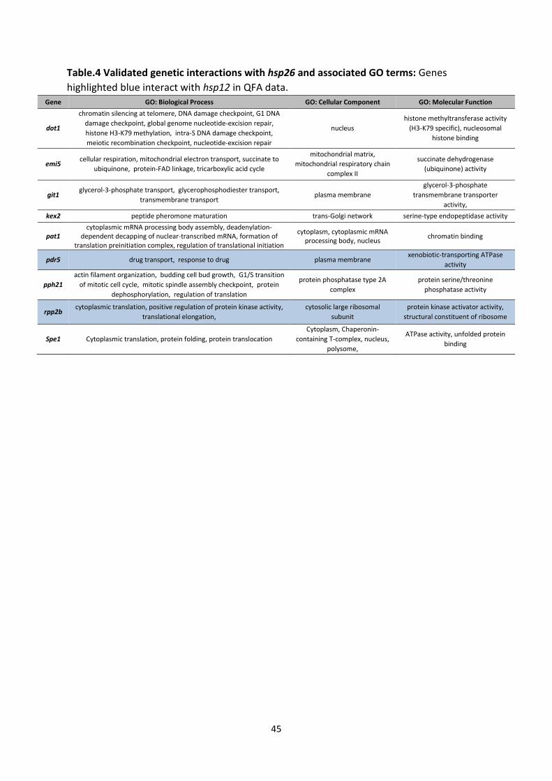
45
Table.4 Validated genetic interactions with hsp26 and associated GO terms: Genes highlighted blue interact with hsp12 in QFA data.
Gene GO: Biological Process GO: Cellular Component GO: Molecular Function
dot1
chromatin silencing at telomere, DNA damage checkpoint, G1 DNA damage checkpoint, global genome nucleotide-excision repair, histone H3-K79 methylation, intra-S DNA damage checkpoint, meiotic recombination checkpoint, nucleotide-excision repair
nucleus histone methyltransferase activity
(H3-K79 specific), nucleosomal histone binding
emi5 cellular respiration, mitochondrial electron transport, succinate to
ubiquinone, protein-FAD linkage, tricarboxylic acid cycle
mitochondrial matrix, mitochondrial respiratory chain
complex II
succinate dehydrogenase (ubiquinone) activity
git1 glycerol-3-phosphate transport, glycerophosphodiester transport,
transmembrane transport plasma membrane
glycerol-3-phosphate transmembrane transporter
activity, kex2 peptide pheromone maturation trans-Golgi network serine-type endopeptidase activity
pat1 cytoplasmic mRNA processing body assembly, deadenylation-
dependent decapping of nuclear-transcribed mRNA, formation of translation preinitiation complex, regulation of translational initiation
cytoplasm, cytoplasmic mRNA processing body, nucleus chromatin binding
pdr5 drug transport, response to drug plasma membrane xenobiotic-transporting ATPase
activity
pph21 actin filament organization, budding cell bud growth, G1/S transition
of mitotic cell cycle, mitotic spindle assembly checkpoint, protein dephosphorylation, regulation of translation
protein phosphatase type 2A complex
protein serine/threonine phosphatase activity
rpp2b cytoplasmic translation, positive regulation of protein kinase activity,
translational elongation, cytosolic large ribosomal
subunit protein kinase activator activity,
structural constituent of ribosome
Spe1 Cytoplasmic translation, protein folding, protein translocation Cytoplasm, Chaperonin-
containing T-complex, nucleus, polysome,
ATPase activity, unfolded protein binding

46
NatMX hs
p12
NatMX hs
p26
A and
D hs
p26
A and
D hs
p12
A and
D hs
p12
A and
D hs
p26
200015001000
Primers
B.
200015001000
MW atg2Δ
:K
atg7Δ
:K
atg13Δ:K
emi5Δ
:K
atg23
Δ:K
A and
D atg7
A and
D emi5
git1Δ
:K
kex2
Δ:K
nbp2Δ
:K
Primers
A and
D atg2
A and
D atg13
A and
D atg23
A and
D git1
A and D
kex2
A and D
nbp2
A and
D sp
e1
200015001000
MW
D.
atg7
Δ:K
emi5
Δ:K
AEY1017hsp26Δ:N atg2Δ:K
1 2 3
A and D atg2Primers
hsp12Δ
20001500
1000
MW hsp12Δhsp26Δ hsp26Δ
A and
D hsp12
A (
and N
at B
hsp12
) A an
d D
hsp26
A (
and N
at B
hsp26
) A a
nd D
hsp12
A (
and N
at B
hsp12
) A a
nd D
hsp26
A (
and N
at B
hsp26
)
hsp26Δ hsp12Δ WildtypePDL1222MW
spe1
Δ:KC.
Primers
A.
A and D atg7 A and D emi5
1 2 3 1 2 3
AEY1017hsp26Δ:N atg7Δ:K
AEY1017hsp26Δ:N emi5Δ:K
atg2
Δ:K
Fig.4 Deletion mutant construction in AEY1017 genetic background: (A) Amplification of NatMX cassettes at the hsp12 and hsp26 locus from gDNA and PDL222 plasmids using corresponding A and D primers generating bands of predicted sizes. Wild-type genes were included as negative controls. (B) PCR confirmation of NatMX cassette integration from transformed AEY1017 strains. hsp12:N successfully amplified using A and D primers (2040bp) (lane 1) and confirmation through inclusion of NatMX B primer (Lane2). Wild-type hsp12:N amplified from hsp26 gDNA (1023bp) (lane3). No band present in lane 4 as expected. Though A and D primers failed to amplify hsp26:N (lane5), the inclusion of a NatMX specific primer confirms integration of the deletion cassette at the hsp26 locus (lane6). Wild-type hsp26 (1555bp) successfully amplified (lane 7). Artefacts present in lane 8. (C) Amplification of KanMX cassettes at loci of genes shown have reproducible GIs. Bands of predicted sizes were obtained for atg2:K, atg7:K and emi5:K only. (D) PCR confirmation of KanMX cassette integration from transformants compared to positive controls amplified from DLY strains. Only hsp26:K emi5:N clone 1 produces band of predicted size. Bands running at size of wild-type emi5 from hsp26:K emi5:N clones 2 and 3.

47
0.05% Glucose(30 ̊C)0.05% Glucose(37 ̊C)
Cell Density
hsp26 hsp26 atg2 hsp26 atg7
hsp26 hsp26 atg2hsp12 atg7 hsp26 emi5
(clone 1)(clone 1)
(clone 2)(clone 2)(clone 2)
ΔΔ ΔΔ Δ
ΔΔ ΔΔ ΔΔ Δ
hsp26 clone 1)Δ Δ emi5 (
(-URA +ClonNAT Media) (YPD +ClonNAT Media)
Fig.5 Spot test of AEY1017 strains: Serial dilutions of strains were spotted onto -URA and YPD 0.05% glucose media with ClonNAT. Plates were incubated at 30oC and 37oC. Hsp26 single mutants were included as controls for query mutant fitness. (Blue) Synthetic sickness displayed by successfully transformed hsp26Δ emi5Δ (clone1). comparisons between genetic backgrounds must be made [34]. To assess the strain specific nature of the validated GIs, we constructed double deletion mutants using the AEY1017 wild-type strain. Query mutant deletions were first constructed by amplifying hsp12Δ:N and hsp26Δ:N from plasmid and gDNA (Fig.4A). As the flanking regions of deletion cassettes amplified from DLY7325 gDNA shared superior homology to the target gene loci, these cassettes were used to transform AEY1017 parent strain. Successful integration of hsp12Δ:N and hsp26Δ:N was established (Fig.4B).
Deletion cassettes atg2:K, atg7:K and emi5:K were amplified (Fig.4C) and used to transform AEY1017 hsp26Δ strain. To establish successful integration at the correct loci, the deletion cassettes were recovered from transformant gDNA (Fig.4D). Successful transformation of hsp26Δ emi5Δ clone 1 was confirmed.
The transformants displaying growth on G418 plates were included in a spot test assay (Fig.5). Synthetic sickness was observed in hsp26 emi5 (clone 1) on -URA
0.05% glucose plates. This phenotype was also seen in YPD media at 37oC. The SSI is apparent in comparison to hsp26Δ and to the unsuccessful transformant hsp26Δ emi5Δ clone 2.
DISCUSSION SL and SSIs provide an unbiased insight into the function of a query gene product in vivo [35]. In this study we reproduce a subset of GIs from novel QFA and SGA screens whereby hsp12 and hsp26 were included as bait. These interactions were further replicated in a second wild-type strain to rule out genetic background specific GIs. Negative interactions may denote components of a multi-subunit machine but are more likely to occur between pathways [32]. By examining the functional connotations of GIs isolated by QFA and SGA, insight into the roles of HSP12 and HSP26 can be made.
Genetic interactions of hsp26 – Small HSPs such as HSP26 antagonise protein disaggregation by trapping unfolded proteins into co-complexes. HSP104/HSP70/HSP40 chaperone system reactivates the substrate

48
in an energy-dependent manner [36]. HSP26 also acts in synergy with HSP42 to inhibit prionogenesis [37]. GIs reflecting this were isolated previously via phenotypic enhancement assays [38]. Though these were not reproduced by our QFA screen, this study genetically linked hsp26 with ssa1 (encoding HSP70 in yeast). To validate this interaction, the double-mutant must be constructed. This study did not isolate any genetic partners of hsp26 identified in previous HTP screens [22,28,39]. A strong genetic link between autophagy and hsp26 was established. Described as the catabolism of defective cellular components, autophagy is induced by DR [40] and nutrient sensing pathways [41]. Inhibition causes premature aging of tissues. QFA data from this study identifies links with: ATG23, a peripheral membrane protein; subunits from the ATG1-13 complex involved in its retrieval from the preautophagosomal structure [42] and Spe1, essential for the synthesis of polyamines which have been shown to induce autophagy [43]. Therefore autophagy may play a role in DR mediated lifespan extension.
Characterizing the role of HSP26 in autophagy may shed light on physical cross talk between pathways. Autophagy can be induced via inactivation of TOR kinase through nitrogen starvation and monitored microscopically by immuno-electron microscopy against autophagosomal marker proteins and accumulation of autophagic bodies in the vacuole [44]. Biochemical assays include quantifying degradation of radiolabeled proteins and measuring delivery of components to the lysosome. Finally protein modification methods monitor autophagy via SDS-PAGE and western blotting of LC3 cleavage [42]. ATG2, 3 and 23 are found in retrieval step of autophagy which can be monitored via the transport of
ATG9 after knocking out ATG1 (TAKA) assay [44]. This measures cycling of ATG9-YFP in double-mutants lacking atg1Δ [45]. Using expression of fluorescent autophagosome marker implicates atg7 in nutrient-mediated autophagy [46]. Analysis of autophagy in hsp26Δ strains and those subject to DR may illustrate its role in DR mediated longevity.
Analysis of hsp26Δ emi5Δ suggests a non-specific GI to strain or mating type. However further comparison to single mutant controls is needed.
Genetic interactions of hsp12 – Previous SGA screens have isolated GIs of hsp12 [22,35,47]. However only oms1, encoding a mitochondrial membrane protein, was identified in this study. Construction of this double mutant would facilitate validation.
Costanzo et al. [22] identifies dep1 as a putative GI with hsp12 in contrast to the QFA data in this study. Spot test assays suggest it is a common interaction between both query mutants. However, further comparison to single-mutant controls is necessary.
Activation of hsp12 in response to DNA damage, osmotic stress, DR and heat is well established. This is reflected by the strong genetic link between hsp12 and genes related to the stress response identified in this study. Validated GIs were enriched in genes associated with the HOG pathway, ER membrane and DNA replication stress. Activated by the HOG pathway, HSP12 is induced under late phase ER-stress [48] and drug-induced DNA replication stress highlighted up-regulation of HSP12 [49]. Functional analysis may provide a better understanding of HSP12s role in this process.
Validity of SGA and QFA screens — The GIs identified in this study share little homology with previous experimental data. However this is unsurprising as GIs have been

49
found to be unidirectional, whereby loss of one gene can be compensated by the other but not visa-versa [50]. These are the first HTP screens using hsp12 and hsp26 as bait and a plethora of novel GIs were isolated.
The prevalence of false positive interactions became apparent from validation of GIs. Moreover this study confirmed GIs with limited success. When measuring fitness-based GIs, experimental variability contributes significantly to the generation of false positives. It is important to normalize batch consistency, nutritional competition and location of strains on the plate. This allows uniform access to nutrients and effective inoculation of the plate. Though genome-wide screens assess replicates of stains with robotic manipulation, HTP studies often optimize throughput and sacrifice accuracy [32]. Interpretation of large-scale genetic interaction assays is challenging due to the number of observed interactions and experimental noise [50]. This raises the need to validate the increasing number of HTP GI screens.
Attempts to optimize the spot test analysis of fitness using rich media revealed growth defects only in plates subject to additional stressors such as heat. This reinforced the necessity of stressful SGA media in order to induce growth defects.
Summary – Screens for GIs highlight shared functions between individual gene products. Here, we validate a subset of the negative GIs of hsp12 and hsp26. Construction and analysis of double mutants absent from the DLY7325 library are needed for validation. Further comparison of the AEY1017 hsp26Δ emi5Δ to single-mutant controls may reveal a non-specific genetic interaction. This study highlights the genetic link between hsp26 and autophagy. Further functional analysis may illustrate the role of
HSP12 and 26 and their function under DR conditions. ACKNOWLEDGEMENTS I would like to thank …. REFERENCES 1 Bishop, N. A. and Guarente, L. (2007)
Genetic links between diet and lifespan: shared mechanisms from yeast to humans. Nat. Rev. Genet. 8, 835–844.
2 Kaeberlein, M. (2010) Lessons on longevity from budding yeast. Nature 464, 513–519.
3 Rizza, W., Veronese, N. and Fontana, L. (2014) What are the roles of calorie restriction and diet quality in promoting healthy longevity? Ageing Res. Rev. 13, 38–45.
4 Houtkooper, R. H., Pirinen, E. and Auwerx, J. (2012) Sirtuins as regulators of metabolism and healthspan. Nat. Rev. Mol. Cell Biol. 13, 225–238.
5 Tannenbaum, A. (1944) The Dependence of the Genesis of Induced Skin Tumors on the Caloric Intake during Different Stages of Carcinogenesis. Cancer Res. 4, 673–677.
6 Fontana, L., Partridge, L. and Longo, V. D. (2010) Extending healthy life span--from yeast to humans. Science 328, 321–326.
7 McKiernan, S. H., Colman, R. J., Lopez, M., Beasley, T. M., Aiken, J. M., Anderson, R. M. and Weindruch, R. (2011) Caloric restriction delays aging-induced cellular phenotypes in rhesus monkey skeletal muscle. Exp. Gerontol. 46, 23–29.
8 Mattison, J. A., Roth, G. S., Beasley, T. M., Tilmont, E. M., Handy, A. M., Herbert, R. L., Longo, D. L., Allison, D. B., Young, J. E., Bryant, M., et al. (2012) Impact of caloric restriction on health and survival in rhesus monkeys from the NIA study. Nature 489, 318–321.
9 Holloszy, J. O. and Fontana, L. (2007) Caloric Restriction in Humans. Exp. Gerontol. 42, 709–712.
10 Fontana, L. and Klein, S. (2007) Aging, adiposity, and calorie restriction. JAMA J. Am. Med. Assoc. 297, 986–994.

50
11 Lin, S.-J., Kaeberlein, M., Andalis, A. A., Sturtz, L. A., Defossez, P.-A., Culotta, V. C., Fink, G. R. and Guarente, L. (2002) Calorie restriction extends Saccharomyces cerevisiae lifespan by increasing respiration. Nature 418, 344–348.
12 Kaeberlein, M., Kirkland, K. T., Fields, S. and Kennedy, B. K. (2004) Sir2-Independent Life Span Extension by Calorie Restriction in Yeast. PLoS Biol. 2.
13 Chang, H.-C. and Guarente, L. (2014) SIRT1 and other sirtuins in metabolism. Trends Endocrinol. Metab. 25, 138–145.
14 Tower, J. (2011) Heat shock proteins and Drosophila aging. Exp. Gerontol. 46, 355–362.
15 Kuznik, B. I., Linkova, N. S. and Khavinson, V. K. (2012) Heat shock proteins: Changes related to aging, development of thrombotic complications, and peptide regulation of the genome. Adv. Gerontol. 2, 175–186.
16 Morley, J. F. and Morimoto, R. I. (2004) Regulation of Longevity in Caenorhabditis elegans by Heat Shock Factor and Molecular Chaperones. Mol. Biol. Cell 15, 657–664.
17 Herbert, A. P., Riesen, M., Bloxam, L., Kosmidou, E., Wareing, B. M., Johnson, J. R., Phelan, M. M., Pennington, S. R., Lian, L.-Y. and Morgan, A. (2012) NMR structure of Hsp12, a protein induced by and required for dietary restriction-induced lifespan extension in yeast. PloS One 7, e41975.
18 Praekelt, U. M. and Meacock, P. A. (1990) HSP12, a new small heat shock gene of Saccharomyces cerevisiae: analysis of structure, regulation and function. Mol. Gen. Genet. MGG 223, 97–106.
19 Welker, S., Rudolph, B., Frenzel, E., Hagn, F., Liebisch, G., Schmitz, G., Scheuring, J., Kerth, A., Blume, A., Weinkauf, S., et al. (2010) Hsp12 is an intrinsically unstructured stress protein that folds upon membrane association and modulates membrane function. Mol. Cell 39, 507–520.
20 Haslbeck, M., Walke, S., Stromer, T., Ehrnsperger, M., White, H. E., Chen, S., Saibil, H. R. and Buchner, J. (1999) Hsp26: a temperature-regulated chaperone. EMBO J. 18, 6744–6751.
21 Haslbeck, M., Braun, N., Stromer, T., Richter, B., Model, N., Weinkauf, S. and
Buchner, J. (2004) Hsp42 is the general small heat shock protein in the cytosol of Saccharomyces cerevisiae. EMBO J. 23, 638–649.
22 Costanzo, M., Baryshnikova, A., Bellay, J., Kim, Y., Spear, E. D., Sevier, C. S., Ding, H., Koh, J. L. Y., Toufighi, K., Mostafavi, S., et al. (2010) The Genetic Landscape of a Cell. Science 327, 425–431.
23 Tong, A. H. Y. and Boone, C. (2007) 16 High-Throughput Strain Construction and Systematic Synthetic Lethal Screening in. In Methods in Microbiology (Ian Stansfield and Michael JR Stark, ed.), pp 369–707, Academic Press.
24 Boone, C., Bussey, H. and Andrews, B. J. (2007) Exploring genetic interactions and networks with yeast. Nat. Rev. Genet. 8, 437–449.
25 Tong, A. H. Y., Evangelista, M., Parsons, A. B., Xu, H., Bader, G. D., Pagé, N., Robinson, M., Raghibizadeh, S., Hogue, C. W. V., Bussey, H., et al. (2001) Systematic Genetic Analysis with Ordered Arrays of Yeast Deletion Mutants. Science 294, 2364–2368.
26 Goldstein, A. L. and McCusker, J. H. (1999) Three new dominant drug resistance cassettes for gene disruption in Saccharomyces cerevisiae. Yeast Chichester Engl. 15, 1541–1553.
27 Banks, A. P., Lawless, C. and Lydall, D. A. (2012) A Quantitative Fitness Analysis Workflow. J. Vis. Exp. JoVE.
28 Addinall, S. G., Holstein, E.-M., Lawless, C., Yu, M., Chapman, K., Banks, A. P., Ngo, H.-P., Maringele, L., Taschuk, M., Young, A., et al. (2011) Quantitative fitness analysis shows that NMD proteins and many other protein complexes suppress or enhance distinct telomere cap defects. PLoS Genet. 7, e1001362.
29 Chari, S. and Dworkin, I. (2013) The Conditional Nature of Genetic Interactions: The Consequences of Wild-Type Backgrounds on Mutational Interactions in a Genome-Wide Modifier Screen. PLoS Genet 9, e1003661.
30 Meijsing, S. H. and Ehrenhofer-Murray, A. E. (2001) The silencing complex SAS-I links histone acetylation to the assembly of repressed chromatin by CAF-I and Asf1 in

51
Saccharomyces cerevisiae. Genes Dev. 15, 3169–3182.
31 Schiestl, R. H. and Gietz, R. D. (1989) High efficiency transformation of intact yeast cells using single stranded nucleic acids as a carrier. Curr. Genet. 16, 339–346.
32 Baryshnikova, A., Costanzo, M., Kim, Y., Ding, H., Koh, J., Toufighi, K., Youn, J.-Y., Ou, J., Luis, B.-J. S., Bandyopadhyay, S., et al. (2010) Quantitative analysis of fitness and genetic interactions in yeast on a genome scale. Nat. Methods 7, 1017–1024.
33 Schwer, B. and Shuman, S. (2014) Structure-function analysis of the Yhc1 subunit of yeast U1 snRNP and genetic interactions of Yhc1 with Mud2, Nam8, Mud1, Tgs1, U1 snRNA, SmD3 and Prp28. Nucleic Acids Res. gku097.
34 Brem, R. B. and Kruglyak, L. (2005) The landscape of genetic complexity across 5,700 gene expression traits in yeast. Proc. Natl. Acad. Sci. U. S. A. 102, 1572–1577.
35 Krause, S. A., Cundell, M. J., Poon, P. P., McGhie, J., Johnston, G. C., Price, C. and Gray, J. V. (2012) Functional specialisation of yeast Rho1 GTP exchange factors. J. Cell Sci. 125, 2721–2731.
36 Jackrel, M. E., DeSantis, M. E., Martinez, B. A., Castellano, L. M., Stewart, R. M., Caldwell, K. A., Caldwell, G. A. and Shorter, J. (2014) Potentiated Hsp104 Variants Antagonize Diverse Proteotoxic Misfolding Events. Cell 156, 170–182.
37 Haslbeck, M., Miess, A., Stromer, T., Walter, S. and Buchner, J. (2005) Disassembling Protein Aggregates in the Yeast Cytosol THE COOPERATION OF HSP26 WITH SSA1 AND HSP104. J. Biol. Chem. 280, 23861–23868.
38 Duennwald, M. L., Echeverria, A. and Shorter, J. (2012) Small Heat Shock Proteins Potentiate Amyloid Dissolution by Protein Disaggregases from Yeast and Humans. PLoS Biol 10, e1001346.
39 Moehle, E. A., Ryan, C. J., Krogan, N. J., Kress, T. L. and Guthrie, C. (2012) The Yeast SR-Like Protein Npl3 Links Chromatin Modification to mRNA Processing. PLoS Genet 8, e1003101.
40 Alvers, A. L., Fishwick, L. K., Wood, M. S., Hu, D., Chung, H. S., Dunn, W. A., Jr and Aris,
J. P. (2009) Autophagy and amino acid homeostasis are required for chronological longevity in Saccharomyces cerevisiae. Aging Cell 8, 353–369.
41 Alvers, A. L., Wood, M. S., Hu, D., Kaywell, A. C., Dunn, W. A. and Aris, J. P. (2009) Autophagy is required for extension of yeast chronological life span by rapamycin. Autophagy 5, 847–849.
42 Yorimitsu, T. and Klionsky, D. (2005) Autophagy: molecular machinery for self-eating. Cell Death Differ. 12, 1542–1552.
43 Eisenberg, T., Knauer, H., Schauer, A., Büttner, S., Ruckenstuhl, C., Carmona-Gutierrez, D., Ring, J., Schroeder, S., Magnes, C., Antonacci, L., et al. (2009) Induction of autophagy by spermidine promotes longevity. Nat. Cell Biol. 11, 1305–1314.
44 Klionsky, D. J., Cuervo, A. M. and Seglen, P. O. (2007) Methods for Monitoring Autophagy from Yeast to Human. Autophagy 3, 181–206.
45 Cheong, H., Yorimitsu, T., Reggiori, F., Legakis, J. E., Wang, C.-W. and Klionsky, D. J. (2005) Atg17 Regulates the Magnitude of the Autophagic Response. Mol. Biol. Cell 16, 3438–3453.
46 Mizushima, N., Yamamoto, A., Matsui, M., Yoshimori, T. and Ohsumi, Y. (2004) In Vivo Analysis of Autophagy in Response to Nutrient Starvation Using Transgenic Mice Expressing a Fluorescent Autophagosome Marker. Mol. Biol. Cell 15, 1101–1111.
47 Sharifpoor, S., Dyk, D. van, Costanzo, M., Baryshnikova, A., Friesen, H., Douglas, A. C., Youn, J.-Y., VanderSluis, B., Myers, C. L., Papp, B., et al. (2012) Functional wiring of the yeast kinome revealed by global analysis of genetic network motifs. Genome Res. 22, 791–801.
48 Bicknell, A. A., Tourtellotte, J. and Niwa, M. (2010) Late phase of the endoplasmic reticulum stress response pathway is regulated by Hog1 MAP kinase. J. Biol. Chem. 285, 17545–17555.
49 Tkach, J. M., Yimit, A., Lee, A. Y., Riffle, M., Costanzo, M., Jaschob, D., Hendry, J. A., Ou, J., Moffat, J., Boone, C., et al. (2012) Dissecting DNA damage response pathways by analysing protein localization and abundance changes during DNA replication stress. Nat. Cell Biol. 14, 966–976.

52
50 Kaeberlein, M., Andalis, A. A., Liszt, G. B., Fink, G. R. and Guarente, L. (2004) Saccharomyces cerevisiae SSD1-V confers longevity by a Sir2p-independent mechanism. Genetics 166, 1661–1672.
51 Jaimovich, A., Rinott, R., Schuldiner, M., Margalit, H. and Friedman, N. (2010) Modularity and directionality in genetic interaction maps. Bioinformatics 26, i228–i236.

53
Section 6 Techniques and Frontiers in Biomedical Sciences Techniques in Biomedical Sciences This part of the module consists of a series of lectures on a wide range of modern research techniques, including optical and electron microscopy, use of bioluminescent intracellular probes, gene expression analysis, proteomic approaches, etc. These lectures are complemented by tutorials designed to develop the fundamental skills required for laboratory research, including data handling, generation of Figures, scientific writing and preparation of poster and oral presentations. Frontiers in Biomedical Sciences In this part of the module, the emphasis is on how state-of-the-art research techniques are used to advance knowledge in specific biomedical research areas and on modern methods/approaches employed in the diagnosis of disease and the treatment of patients. Lectures on these topics are complemented by tutorials and journal clubs designed to develop analytical and critical thinking skills. Both the Techniques and Frontiers modules are enhanced by Institute Seminars within the Institute of Translational Medicine. Eminent scientists from throughout the UK contribute to this by presenting their research on a variety of topics. It is important that MRes students attend ITM Seminars to broaden their knowledge and range of learning experiences. Seminars are organized by the individual Departments within the Institute and are advertised regularly via email. Attendance at certain Departmental seminars may be recommended by strand convenors to enhance awareness of research that is particularly relevant to individual MRes strands. Finally, strand-specific activities are an important part of the Techniques and Frontiers modules, as they facilitate awareness of the science associated with particular research strands. Students will be assessed on one short review based on the Techniques module, one short review based on the Frontiers module, and a referees report based on a journal club. 6.1 Preparation of Short Reviews and Referees Reports Short Reviews Students should make every effort to ensure that their work is presented in good English, and is clearly written. The sources used to prepare the review should be listed at the end, in full, and cited at the appropriate point in the text; citations and bibliography should appear in Trends in Cell Biology style. Any material - text or figures - that is taken verbatim from other sources must be fully identified. In the case of text, it is essential to use quotation marks (“....”) to identify such material in order to avoid accusations of plagiarism (which is a serious academic offence). Any schematic diagrams, cartoons etc used as Figures should, wherever possible, be created by yourself using appropriate graphics packages, rather than by copying and pasting from published (and hence copyright protected) literature. The University policy with regard to academic integrity is explained earlier in this handbook.

54
A good review should include: (a) an opening statement that introduces the subject, sets it in the context of published work, and attracts the attention of the reader. (b) In the main part of the review, the major theme, should be developed and critically discussed. Points should be made systematically using paragraph headings if appropriate and avoiding repetition. The text should include citations to the relevant literature, which should be described and discussed in sufficient detail for the review to stand on its own as a piece of scientific writing. (c) A final concluding statement may take the form of an overview, summary or outline of prospects for future work (or all three). Short reviews should include up to 30 citations to peer-reviewed papers. It will often be the case that many more papers have been consulted; the process of selecting the most appropriate literature citations is therefore a matter of judgement and this will be reflected in the quality of the final product. An example of a short review is given at the end of this Section for you to refer to (note, however, that this is NOT in the style of Trends in Cell Biology). Short Reviews should be prepared in the style of articles published in the journal Trends in Cell Biology. They should be typed single spaced, in Calibri font size 12 and must be 2500 ± 250 words in length. This word limit covers all text sections of the report, including the Abstract and legends to Figures/Tables, but does not apply to text contained within Figures/Tables or to the References section (i.e., the list of citations at the end does not count toward the 2500 words). Marks will be deducted proportionally for exceeding the upper limit of 2750 words. For example, 3000 words = 250 words above the limit = 10% deducted. However, the mark will not be reduced below the pass mark of 50% for the assignment. Marks will not be deducted for being below the lower limit of the word count (i.e. 2250 words), but students should be aware that short reports are highly likely to be awarded lower marks due to a lack of coverage and discussion of key areas.
- Submitting your Short Reviews: Submit an electronic copy of the final version (including any Figures) via Turnitin.
Please attach a front cover sheet to your report stating your name, the title of your review, your student number, the name of the lecturer giving the topic, the title of the report, and the word count. A template cover sheet is supplied at the end of this Section for this purpose. Short review 1 Choose up to 3 techniques in order of preference that were discussed in the lecture course “Techniques in Biomedical Sciences” in semester 1. They should not be connected to the main methods used in your research projects, and should not be a lecture given by one of your supervisors or your strand convenor. Inform the PGR office ([email protected]) and the strand convenor of your choice and order of preference, deadline 10:00 Monday 13 November 2017. We plan to tell you which technique you have been allocated to review by Friday 17 November 2017. Prepare a short review style article discussing the principles on which the technique is based, any practical problems in applying the technique, and the importance of the technique in modern Biomedical Sciences. You should ask the lecturer concerned for advice on the content and scope of the review.

55
Short review 2 Choose 3 topics covered in “Frontiers in Biomedical Sciences” in semesters 2 and 3 and place in order of preference. They should not be connected to the main methods used in your research projects, and should not be a lecture given by one of your supervisors or your strand convenor. Inform the MRes Programme Administrator ([email protected]) and the strand convenor of your choice and order of preference. You will then be notified of topic for your allocated review. Dates for making a choice will be released in Semester 2. Prepare a short review style article discussing the importance of the subject, its topicality, and why or how recent progress has been made. Journal Club The idea behind a journal club is to provide insights into how to critically analyse papers and allow an appreciation of recent advances. Our journal club will be organised as follows: You will be allocated a paer within your strand The paper will be chosen by the Strand Convenor. An electronic copy will be made available (or a reference to the paper given) in VITAL. The paper may be chosen to illustrate some important technical aspects or novel biological findings, or to highlight problems with the data/interpretations in the paper. Excessively long papers will be avoided. All participating students will be expected to read the paper and prepare a brief summary, as a series of bullet points, of the major issues. These will be helpful for the discussions of the paper and should be emailed to the chairperson in advance of each journal club. There will be one Journal Club in Semester 1 and another in Semester 2. The Journal Club will be chaired by a member of academic staff. In the lecture series before a Journal Club there will be a Journal Club Lecture where you will receive a demonstration of how to analyse and critique a paper. Referee’s report Your strand convenor will inform you which paper has been be chosen to referee nearer the time. Guidance on how to construct referees’ reports will be given by the Programme Director in the Science Skills session on Scientific Publishing and also in the introductory lecture to the Frontiers module, and these can be used to provide the basis for the written account. Your referees report should be 1000 ± 100 words and the word count should be indicated. Marks will be deducted proportionally for exceeding the upper limit of 1100 words. For example, 1200 words = 100 words above the limit = 10% deducted. However, the mark will not be reduced below the pass mark of 50% for the assignment. Marks will not be deducted for being below the lower limit of the word count (i.e. 900 words), but students should be aware that short reports are highly likely to be awarded lower marks due to a lack of coverage of key areas. Submitting your Referees Report: Submit an electronic copy of the final version (including any Figures) via Turnitin by 10:00 on the deadline date. Please attach a front cover sheet to your report stating your name, your student number, the name of the lecturer giving the topic, the title of the report, and the word count. A template cover sheet is supplied at the end of this Section for this purpose.

56
6.2 Assessment Each short review will contribute 40% of the marks, and the referee’s report 20% of the total marks. The Techniques and Frontiers module contributes 30 credits out of a total of 180 credits for the MRes degree. Both short reviews and referees reports will be double marked by the person who gave the lecture or journal club and a 2nd internal assessor (usually the strand convenor). The mark awarded will be the average of these two marks. Please note that the standard university penalty for late submission of written work applies (see earlier, under overview of assessment). Details of the assessment criteria used and the assessment forms that will be used by markers are given on the on the pages that follow, as are examples of a short review and a referee’s report.

57
6.2.1 Assessment Form for short reviews

58

59
6.2.2 Marking Criteria for assessment of written assignments
Distinction Level 100% - 90% 89% - 80% 79% - 70%
Outstanding. No (or virtually no – use scaling) better result conceivable at Masters level. Entirely correct and complete, with extensive evidence of critical thinking. Evidence of extensive research of relevant literature. Extremely logical structure, faultlessly written and presented. Clear evidence of highly original thought and cogent scientific argument. Excellent. Clear evidence of achievement on a scale reserved for exceptionally high quality work at Masters level. Essentially correct and complete, with significant evidence of critical thinking and excellent use of relevant literature. Highly logical structure, extremely well written and presented, displaying a significant amount (use scaling) of original thought and cogent scientific argument. Very Good. High quality work that demonstrates comprehensive understanding and shows some evidence of critical thinking. Evidence of very good research of relevant literature, with all key literature identified/discussed. Very well written and presented, with a very logical structure. Some evidence of original thought and cogent scientific argument
Merit Level
69% - 60%
Good. Good quality work that demonstrates generally sound scientific understanding. Evidence of good research of relevant literature, but some relevant literature not identified/discussed. Generally well written and presented, with a logical structure. Evidence of original/critical thinking may be limited
Pass Level 59% - 50% Satisfactory. Learning objectives achieved, but may contain deficiencies in one or more aspects of knowledge of the relevant literature, scientific understanding, structure and presentation.
Fail Level 49% - 40% Unsatisfactory. Learning objectives not achieved. Essentially an incomplete report with significant flaws or omissions and major deficiencies in one or more aspects of knowledge of the relevant literature, scientific understanding, structure and presentation.
39% - 0%
Poor. Severely deficient in content, understanding and application and containing many serious errors and omissions.

60
6.2.3 Assessment Form for referees’ reports

61

62
6.3 Front sheet for referee’s report and short review submissions (example)
Short Review [Insert number] or Referee’s Report
TITLE OF YOUR REVIEW OR REPORT
Joe Bloggs Student I.D. 200700000
Lecturer (who gave the Techniques/Frontiers lecture): [Insert name] Strand: [Insert name of your strand]
Word Count: [Insert word count]
By submitting this work I confirm that I have read, understood, and adhered to the University’s Academic Integrity Policy (Appendix L in the University Code of Practice) and that I have read, understood and signed a Declaration of Academic Integrity.

63
6.4 Example of a short review – note this is an example from a previous year and so not in the journal style currently recommended. The role of sirtuins in ageing Name Department of xxx , Institute of Translational Medicine, University of Liverpool, UK. Population ageing is a global phenomenon with socioeconomic consequences, largely due to age-related health problems. It is thus important to dissect the underlying mechanisms of ageing in order to target these problems at the root. Calorie restriction (CR) is a natural intervention with positive effects on ageing in terms of lifespan and healthspan. Sirtuins, a group of seven Class III histone deacetylases, are involved in mediating these effects of CR; SIRTs 1 to 7 are the mammalian sirtuins. In addition, they also regulate physiological processes such as glucose and lipid metabolism in peripheral tissues, and brain activities. As such, sirtuins make attractive pharmacological targets to counteract ageing and age-related pathologies. Introduction Ageing is a phenomenon which affects every living organism, and loosely refers to the progressive functional deterioration of living organisms with time. Population ageing is a global occurrence, and is due to an increase in numbers of individuals aged 65 and over as a result of both increasing life expectancies and decreased rates of fertility. In a report published by the World Health Organisation (WHO) in 2011 [1], this subset of the global population was estimated to triple to 1.5 million in 2050 from 500 million in 2010. Within the ageing population, the subpopulation of the “oldest old”, constituting of 85-and-overs, is predicted to expand much faster with a more than triple increment in numbers from 2010 to 2050 as compared to a two-fold one from 65-and- overs. These figures are alarming as population ageing places much strain on both the economy and society, mostly as a result of age-related health problems. Dementia is a common age-related condition. Within the ageing population, the oldest olds are most susceptible to dementia, usually in the form of Alzheimer’s disease; dementia affects 30% of 85 to 89 year olds as compared to 3% of 65 to 69 year olds. The economic burden of dementia is eminent by healthcare expenditure for it in 2010, which was greater than USD600 billion. Aside from dementia, the ageing population is also more susceptible to infectious diseases as a result of immunosenescence and frailty, and other noncommunicable conditions such as cardiovascular disease, cancer, and diabetes, contributing to more healthcare costs. In addition, there will be an increasing need for families to spend time caring for the ailing elderly. However, decreasing fertility rates meant that there will be potentially less primary support from children to care for their aged parents; this can place more burden on the healthcare system as the elderly are likely to be sent to nursing homes. By and large, it is evident that population ageing presents many problems. The inevitability of global population ageing meant that there is a need to devise strategies to cope with the various age-associated health problems as illustrated above. Directing more finances into healthcare to cope with these problems will only place more economic burden with time, hence more effective long-term solutions have to be sought. An important strategy is to dissect the underlying mechanisms of ageing. By understanding such mechanisms, potential pharmacological intervention can be devised to alleviate or prevent age-related diseases, in turn improving both lifespan and healthspan; healthspan refers to the length of time during which an individual has optimal health. Calorie restriction (CR) was shown to have positive effects on lifespan and healthspan across various experimental organisms (2-4). Further studies demonstrated a role for sirtuins, a group of Class III histone deacetylases, in mediating these effects of CR; hence, sirtuins make potential pharmacological targets. This review will illustrate proposed shared cellular

64
mechanisms which underlie the ageing process, followed by the protective effects of CR and sirtuins against ageing. The feasibility of targeting sirtuins in ageing will also be highlighted. The ageing process Recently, nine general cellular mechanisms or hallmarks were proposed to drive the process of ageing, listed as follows: 1) epigenetic alterations, 2) telomere attrition, 3) genomic instability, 4) loss of proteostasis, 5) mitochondrial dysfunction, 6) deregulated nutrient sensing, 7) cellular senescence, 8) altered intercellular communication, and 9) stem cell exhaustion (5). These nine hallmarks were suggested to co-occur and inter-connect, and were further categorised; the first three hallmarks were suggested to initiate ageing, whilst hallmarks 5 to 7 were thought to be cellular responses to those events, with the last two hallmarks proposed to affect tissue function directly, resulting in ageing phenotypes. Of these hallmarks, defective nutrient sensing has attracted much research interest. Pathways implicated in the ageing process include those mediated by insulin and insulin growth factor 1 (IGF-1), mammalian target of rapamycin (mTOR), AMP- activated protein kinase (AMPK), and sirtuins. Nutrient availability is detected and signalled by insulin, IGF-1, and mTOR, in contrast to signalling of nutrient scarcity by AMPK and sirtuins. Generally, studies on these pathways indicated that signalling of nutrient availability promotes ageing, whilst the opposite has an ageing-suppressing effect through lifespan extension. Effects of calorie restriction on ageing CR is defined by low caloric consumption without malnutrition (2). It was previously shown to protect against ageing by extending lifespan in various experimental organisms such as rodents, non-human primates, Saccharomyces (S.) cerevisiae, Drosophila (D.) melanogaster, and Caenorhabditis (C.) elegans (2, 3). In addition to regulating lifespan, CR was also shown to regulate healthspan in rodents (2). For instance, the onset and progression of cancer, diabetes, kidney disease, and autoimmune conditions were delayed (2). In the brain, CR exerted neuroprotective effects by preventing age-related deterioration in spatial and psychomotor memory tasks, and reducing neuronal loss characteristic of Alzheimer’s and Parkinson’s diseases. Age-associated muscle atrophy was also shown to be attenuated by CR (6). Recently, a 25-year study conducted on rhesus monkeys showed that lifespans of these primates were not affected by CR (4); these findings contradicted an earlier long- term study on rhesus monkeys (3). However, it should be noted that control animals between both studies were not given the same diet; controls in the earlier study were fed a more sucrose-rich diet (28% sucrose) as opposed to those in the later study (4% sucrose). It is possible that the observed effect of CR in the earlier study is attributed to a much higher mortality rate from controls. Nevertheless, animals on CR in both studies displayed delayed disease onset and better health markers than controls, showing the positive effect of CR on health. Protective effects against ageing by CR can be mediated through the aforementioned nutrient-sensing pathways mediated by insulin, IGF-1, mTOR, AMPK, and sirtuins (7). Due to the non-exhaustive nature of this review, only the role of the sirtuins in ageing will be briefly illustrated; induced expression of sirtuins by CR was previously observed in mice and humans (8). The sirtuins In 1984, the first sirtuin was reported in S. cerevisiae (9). In mammals, there are seven sirtuin isoforms, namely SIRT1 to 7 (6). Sirtuins are generally classified as Class III histone deacetylases due to their dependence on the co-factor beta-nicotinamide adenine dinucleotide (NAD+), which signals the nutrient state of the cell (10). In reality, however, they also exert other isoform-specific enzymatic activities such as ADP- ribosylation, demalonylation, and desuccinylation; hence they are more accurately referred to as deacylases (Fig. 1) (6, 11).

65
Furthermore, the sirtuins also show isoform- specific localisation in the cell; SIRTs 1, 6, and 7 localise to the nucleus, SIRTs 3 to 5 and 2 localise to the mitochondria and cytosol respectively. To add to the complexity of sirtuins, they exert their biological activities on a range of targets such as transcription factors (TFs), histones, and tubulin, to bring about different physiological outcomes.
Figure 1. Sirtuins as deacylases. Deacylation of specific substrates requires NAD+ as a co-factor, with nicotinamide produced as a by-product of the enzymatic reaction. Nicotinamide then acts to inhibit further sirtuin activity, hence providing negative feedback. Deacylation activities of specific sirtuins are indicated, with additional demalonylation and desuccinylation activities unique to SIRT5 (indicated in red). Figure produced with information from Houtkooper et al, 2012 (11). Sirtuins and lifespan extension Studies have shown the potential role of several sirtuins in mediating lifespan extension by CR. Of the seven sirtuins, SIRT1 is the most extensively studied and best characterised. Its (and its invertebrate orthologs’) role in lifespan extension by CR was first shown in mice, C. elegans, S. cerevisiae, and D. melanogaster (12-15). However, these initial findings were controversial due to contradictory findings from other later studies, by which overexpression of SIRT1 and its orthologs in mice, C. elegans, and D. melanogaster failed to reproduce lifespan extension (16, 17). Although the most recent studies have reaffirmed the involvement of SIRT1 and its orthologs in longevity (18-21), an important caveat to note about these studies on sirtuins and longevity is the different experimental designs between them which can affect experimental outcomes. Aside from SIRT1, findings have also implicated SIRTs 3 and 6 in ageing. In mice, SIRT6 knockout accelerated ageing and shortened lifespan, with animals dying by the 4th week (22). When SIRT6 was overexpressed, lifespan was extended in male but not female mice, suggesting a gender-specific role of SIRT6 in ageing (23). In the case of SIRT3, polymorphisms in its genetic locus were linked to increased lifespan in humans (24, 25). The roles of the other sirtuins in longevity are not understood. Sirtuins and healthspan Sirtuins regulate glucose and lipid metabolism in the liver, pancreas, muscles, and white adipose tissues (WATs) (6, 11). Gluconeogenesis occurs mainly in the liver, and is stimulated by SIRTs 1 and 2. SIRT1 seems to have a paradoxical role in regulating gluconeogenesis as it may also inhibit the process. SIRTs 3 and 4 promotes and represses gluconeogenesis respectively. Suppression of glycolysis was shown to be mediated by SIRTs 1, 3, and 6. Sirtuins can also regulate glucose metabolism by modulating insulin secretion by pancreatic β cells; insulin stimulates glucose uptake by the liver, WATs, and skeletal muscles. SIRT1 stimulates insulin secretion in response to glucose, whereas SIRT4 inhibits insulin secretion in response to amino acids. Aside from regulation of insulin secretion, sensitisation of tissues to insulin may also be mediated by SIRTs 1 and 3.
66
Lipid metabolism was shown to be regulated by SIRTs 1, 2, 3, 4 and 6. In WATs, SIRT1 suppresses adipogenesis and stimulates lipolysis, whilst SIRT2 exerts the opposite effects. In the liver, fatty acid oxidation is induced by SIRT1; this effect of SIRT1 was shown to protect against hepatic steatosis in mice. SIRTs 3 and 6 may also stimulate fatty oxidation, whereas SIRT4 may inhibit it. Sirtuins also mediate processes in the brain (6). For instance, protein abundances of SIRTs 1 and 5 are upregulated in cerebral tissues of rats on CR, which correlated with enhanced cognition. Evidence also suggests that SIRT1 may have positive effects on hippocampal plasticity, which can promote learning and memory processes. In the hypothalamus, SIRT1 regulates the circadian rhythm and responses to gut hormones, where feeding behaviour and energy expenditure are affected by the latter. The documented physiological effects of sirtuins are much more extensive than that illustrated herein. Regardless, it is evident that sirtuins have important physiological roles in the body. Hence, deregulation of their activities can have negative consequences on health. In fact, sirtuins are implicated in a range of age-related pathologies which include neurodegeneration diseases, metabolic syndrome, cardiovascular disease, Type 2 diabetes mellitus, and cancer (6, 26). Although the effect of sirtuins in lifespan is promising overall, the role of sirtuins in healthspan may be more relevant with regards to counteracting socioeconomic problems of population ageing. Sirtuins as pharmacological targets The important role of sirtuins in regulating both lifespan and healthspan makes them attractive pharmacological targets to counteract ageing and age-related diseases. Resveratrol is a polyphenol in peanuts and berries which mediates a range of health benefits by activating SIRT1 (6, 11, 27). In mice, for instance, resveratrol treatment was demonstrated to augment mitochondrial function, in turn protecting them from diet-induced obesity; exercise performance was also improved. In mice given a high- fat but not a standard diet, resveratrol extended lifespan, suggesting that SIRT1 may have a predominant protective role against stress. Resveratrol was also shown to exert anti-cancer, anti-inflammation, cardioprotective, and neuroprotective effects (28). These findings are consistent with the overall health effects of sirtuins and CR. In addition to resveratrol, other polyphenols such as quercetin were also identified which were able to extend lifespan in C. elegans via its SIRT1 ortholog (6, 27). This further led to the development of small synthetic SIRT1-activating compounds (STACs) with better pharmacokinetic parameters and potency, which also extended lifespan in a range of experimental organisms (27). The specificity and mechanism of SIRT1 activation by STACs, however, was a topic of debate. However, studies have shown that there are sequence and structural requirements on both SIRT1 and their substrates which mediate binding and activation of SIRT1 (27). Since sirtuins can exert isoform-specific, antagonising effects in the body, as briefly illustrated above, it is important to develop other isoform-specific sirtuin activators or inhibitors. Before that can be accomplished, further characterisation of the roles of each sirtuin in health and ageing needs to be done. Although STACs show promising effects against ageing, it has been a challenge to bring them forward to clinical trials. Ageing is not recognised by government regulatory agencies, such as the Food and Drug Administration (FDA) in USA, as a pathology because it is a common phenomenon. In addition, a human trial conducted for ageing will take a longer time than usual trials, incurring much higher costs. To circumvent this problem, clinical trials with STACs can be conducted to assess effects on specific age-related pathologies rather than on ageing per se. It is noteworthy that several STACs are in clinical trials at the moment (27).
67
Conclusion Sirtuins have potential roles in lifespan and healthspan. However, our understanding of sirtuins is still rudimentary, as evident from current findings which only shed light on some, but not all sirtuins. In addition, isoform-specific and tissue-specific effects also need to be considered for effective pharmacological intervention; different sirtuin isoforms can have antagonising biological effects which would require the development of isoform-specific drugs. It is also difficult to introduce potential compounds for ageing in clinical trials, as ageing is not recognised by regulatory agencies as a disease. Until such challenges are met, the only way to counteract the socioeconomic effects of population ageing at present is to promote awareness of healthy living and to continue to provide and improve support for the old. References 1. Suzman, R. and Beard, J. Global Health and Aging [Online]. World Health Organisation. http://www.who.int/ageing/publications/global_health/en/ [26 April 2014] 2. Koubova J and Guarente L. How does calorie restriction work? Genes & Development 17:313-321, 2003 3. Colman RJ, Anderson RM, Johnson SC, Kastman EK, Kosmatka KJ, Beasley TM, Allison DB, Cruzen C, Simmons HA, Kemnitz JW, and Weindruch R. Caloric restriction delays disease onset and mortality in rhesus monkeys. Science 325:201-204, 2009 4. Mattison JA, Roth GS, Beasley TM, Tilmont EM, Handy AM, Herbert RL, Longo DL, Allison DB, Young JE, Bryant M, Bernard D, Ward WF, Qi W, Ingram DK, and de Cabo R. Impact of caloric restriction on health and survival in rhesus monkeys from the NIA study. Nature 489:318-321, 2012 5. Lepez-Otin C, Blasco MA, Partridge L, Serrano M, Kroemer G. The hallmarks of aging. Cell 153:1194-217, 2013 6. Morris BJ. Seven sirtuins for seven deadly diseases of aging. Free Radical Biology and Medicine 56:133-171, 2013 7. Dilova I, Easlon E, and Lin SJ. Calorie restriction and the nutrient sensing signaling pathways. Cellular and Molecular Life Sciences 64:752-767, 2007 8. Guarente L. Calorie restriction and sirtuins revisited. Genes & Development 27:2072-2085, 2013 9. Shore D, Squire M, and Nasmyth KA. Characterization of 2 genes required for the position-effect control of yeast mating-type genes. Embo Journal 3:2817- 2823, 1984 10. Schemies J, Uciechowska U, Sippl W, and Jung M. NAD+-dependent histone deacetylases (sirtuins) as novel therapeutic targets. Medicinal Research Reviews 30:861-889, 2010

68
11. Houtkooper RH, Pirinen E, and Auwerx J. Sirtuins as regulators of metabolism and healthspan. Nature Reviews Molecular Cell Biology 13:225-238, 2012 12. Kaeberlein M, McVey M, and Guarente L. The SIR2/3/4 complex and SIR2 alone promote longevity in Saccharomyces cerevisiae by two different mechanisms. Genes & Development 13:2570-2580, 1999 13. Rogina B and Helfand SL. Sir2 mediates longevity in the fly through a pathway related to calorie restriction. Proceedings of the National Academy of Sciences of the United States of America 101:15998-16003, 2004 14. Tissenbaum HA and Guarente L. Increased dosage of a sir-2 gene extends lifespan in Caenorhabditis elegans. Nature 410:227-230, 2001 15. Li Y, Xu W, McBurney MW, and Longo VD. SirT1 inhibition reduces IGF- I/IRS-2/Ras/ERK1/2 signaling and protects neurons. Cell Metabolism 8:38-48, 2008 16. Burnett C, Valentini S, Cabreiro F, Goss M, Somogyvari M, Piper MD, Hoddinott M, Sutphin GL, Leko V, McElwee JJ, Vazquez-Manrique RP, Orfila A, Ackerman D, Au C, Vinti G, Riesen M, Howard K, Neri C, Bedalov A, Kaeberlein M, Soti C, Partridge L, and Gems D. Absence of effects of Sir2 overexpression on lifespan in C. elegans and Drosophila. Nature 477:482- 485, 2011 17. Herranz D, Munoz-Martin M, Canamero M, Mulero F, Martinez-Pastor B, Fernandez-Capetillo O, and Serrano M. Sirt1 improves healthy ageing and protects from metabolic syndrome-associated cancer. Nature Communications 1, 2010 18. Stumpferl SW, Brand SE, Jiang JC, Korona B, Tiwari A, Dai J, Seo JG, and Jazwinski SM. Natural genetic variation in yeast longevity. Genome Research 22:1963-1973, 2012 19. Banerjee KK, Ayyub C, Ali SZ, Mandot V, Prasad NG, and Kolthur-Seetharam U. dSir2 in the adult fat body, but not in muscles, regulates life span in a diet- dependent manner. Cell Reports 2:1485-1491, 2012 20. Schmeisser K, Mansfeld J, Kuhlow D, Weimer S, Priebe S, Heiland I, Birringer M, Groth M, Segref A, Kanfi Y, Price NL, Schmeisser S, Schuster S, Pfeiffer AF, Guthke R, Platzer M, Hoppe T, Cohen HY, Zarse K, Sinclair DA, and Ristow M. Role of sirtuins in lifespan regulation is linked to methylation of nicotinamide. Nature Chemical Biology 9:693-700, 2013 21. Satoh A, Brace CS, Rensing N, Cliften P, Wozniak DF, Herzog ED, Yamada KA, and Imai S. Sirt1 extends life span and delays aging in mice through the regulation of Nk2 homeobox 1 in the DMH and LH. Cell Metabolism 18:416- 430, 2013 22. Mostoslavsky R, Chua KF, Lombard DB, Pang WW, Fischer MR, Gellon L, Liu P, Mostoslavsky G, Franco S, Murphy MM, Mills KD, Patel P, Hsu JT, Hong AL,Ford E, Cheng HL, Kennedy C, Nunez N, Bronson R, Frendewey D, Auerbach W, Valenzuela D, Karow M, Hottiger MO, Hursting S, Barrett

69
JC, Guarente L,Mulligan R, Demple B, Yancopoulos GD, and Alt FW. Genomic instability and aging-like phenotype in the absence of mammalian SIRT6. Cell 124:315-329, 2006 23. Kanfi Y, Naiman S, Amir G, Peshti V, Zinman G, Nahum L, Ziv B, and Cohen HY. The sirtuin SIRT6 regulates lifespan in male mice. Nature 483:218-21, 2012 24. Albani D, Ateri E, Mazzuco S, Ghilardi A, Rodilossi S, Biella G, Ongaro F, Antuono P, Boldrini P, Di Giorgi E, Frigato A, Durante E, Caberlotto L, Zanardo A, Siculi M, Gallucci M, and Forloni G. Modulation of human longevity by SIRT3 single nucleotide polymorphisms in the prospective study "Treviso Longeva (TRELONG)". Age 36:469-478, 2014 25. Rose G, Dato S, Altomare K, Bellizzi D, Garasto S, Greco V, Passarino G, Feraco E, Mari V, Barbi C, BonaFe M, Franceschi C, Tan Q, Boiko S, Yashin AI, and De Benedictis G. Variability of the SIRT3 gene, human silent information regulator Sir2 homologue, and survivorship in the elderly. Experimental Gerontology 38:1065-1070, 2003 26. Hall JA, Dominy JE, Lee Y, and Puigserver P. The sirtuin family’s role in aging and age-associated pathologies. The Journal of Clinical Investigation 123:973-979, 2013 27. Hubbard BP and Sinclair DA. Small molecule SIRT1 activators for the treatment of aging and age-related diseases. Trends in Pharmacological Sciences. 35:146-154, 2014 28. Bhat KPL, Kosmeder JW, and Pezzuto JM. Biological effects of resveratrol. Antioxidants & Redox Signaling 3:1041-1064, 2001
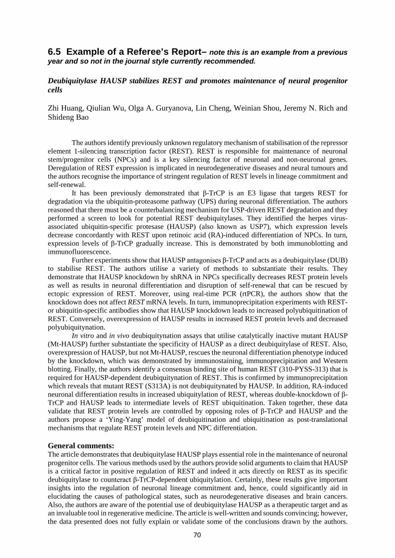
70
6.5 Example of a Referee’s Report– note this is an example from a previous year and so not in the journal style currently recommended. Deubiquitylase HAUSP stabilizes REST and promotes maintenance of neural progenitor cells Zhi Huang, Qiulian Wu, Olga A. Guryanova, Lin Cheng, Weinian Shou, Jeremy N. Rich and Shideng Bao
The authors identify previously unknown regulatory mechanism of stabilisation of the repressor element 1-silencing transcription factor (REST). REST is responsible for maintenance of neuronal stem/progenitor cells (NPCs) and is a key silencing factor of neuronal and non-neuronal genes. Deregulation of REST expression is implicated in neurodegenerative diseases and neural tumours and the authors recognise the importance of stringent regulation of REST levels in lineage commitment and self-renewal.
It has been previously demonstrated that β-TrCP is an E3 ligase that targets REST for degradation via the ubiquitin-proteasome pathway (UPS) during neuronal differentiation. The authors reasoned that there must be a counterbalancing mechanism for USP-driven REST degradation and they performed a screen to look for potential REST deubiquitylases. They identified the herpes virus-associated ubiquitin-specific protesase (HAUSP) (also known as USP7), which expression levels decrease concordantly with REST upon retinoic acid (RA)-induced differentiation of NPCs. In turn, expression levels of β-TrCP gradually increase. This is demonstrated by both immunoblotting and immunofluorescence.
Further experiments show that HAUSP antagonises β-TrCP and acts as a deubiquitylase (DUB) to stabilise REST. The authors utilise a variety of methods to substantiate their results. They demonstrate that HAUSP knockdown by shRNA in NPCs specifically decreases REST protein levels as well as results in neuronal differentiation and disruption of self-renewal that can be rescued by ectopic expression of REST. Moreover, using real-time PCR (rtPCR), the authors show that the knockdown does not affect REST mRNA levels. In turn, immunoprecipitation experiments with REST- or ubiquitin-specific antibodies show that HAUSP knockdown leads to increased polyubiquitination of REST. Conversely, overexpression of HAUSP results in increased REST protein levels and decreased polyubiquitynation.
In vitro and in vivo deubiquitynation assays that utilise catalytically inactive mutant HAUSP (Mt-HAUSP) further substantiate the specificity of HAUSP as a direct deubiquitylase of REST. Also, overexpression of HAUSP, but not Mt-HAUSP, rescues the neuronal differentiation phenotype induced by the knockdown, which was demonstrated by immunostaining, immunoprecipitation and Western blotting. Finally, the authors identify a consensus binding site of human REST (310-PYSS-313) that is required for HAUSP-dependent deubiquitynation of REST. This is confirmed by immunoprecipitation which reveals that mutant REST (S313A) is not deubiquitynated by HAUSP. In addition, RA-induced neuronal differentiation results in increased ubiquitylation of REST, whereas double-knockdown of β-TrCP and HAUSP leads to intermediate levels of REST ubiquitination. Taken together, these data validate that REST protein levels are controlled by opposing roles of β-TrCP and HAUSP and the authors propose a ‘Ying-Yang’ model of deubiquitination and ubiquitination as post-translational mechanisms that regulate REST protein levels and NPC differentiation. General comments: The article demonstrates that deubiquitylase HAUSP plays essential role in the maintenance of neuronal progenitor cells. The various methods used by the authors provide solid arguments to claim that HAUSP is a critical factor in positive regulation of REST and indeed it acts directly on REST as its specific deubiquitylase to counteract β-TrCP-dependent ubiquitylation. Certainly, these results give important insights into the regulation of neuronal lineage commitment and, hence, could significantly aid in elucidating the causes of pathological states, such as neurodegenerative diseases and brain cancers. Also, the authors are aware of the potential use of deubiquitylase HAUSP as a therapeutic target and as an invaluable tool in regenerative medicine. The article is well-written and sounds convincing; however, the data presented does not fully explain or validate some of the conclusions drawn by the authors.

71
Therefore, to substantiate their final conclusions, I recommend the following changes before submission of the paper: Major points:
1) Figure 2 – immunoblotting experiments use CoREST as a negative control to show that HAUSP knockdown specifically reduces REST protein levels. Although CoREST is a primary co-factor of REST, the authors do not show whether HAUSP knockdown affects the levels of other factors in the repressor complex with REST, such as HDAC-1 and -2 (Abrajano et al. PLoS One. (2009) 4(12):e7936.). It would be good to confirm whether these protein levels remain unchanged to validate whether HAUSP specifically affects REST.
2) Figure 4 – similarly to Figure 2, CoREST is used as a negative control for rtPCR to check whether mRNA levels of REST are affected by HAUSP knockdown. The authors should also investigate mRNA levels of other REST co-factors and possibly other transcription factors involved in NPC differentiation.
3) Figure S4d – the authors argue that ‘forced expression of flag-HAUSP (...) increased REST protein levels’. This is not convincing as immunoblotting does not actually show any significant increase in protein levels.
4) Figure 5e – in vitro deubiquitylation assay utilises proteins that were overexpressed in 293T cells and subsequently purified. This means that there is a potential risk of co-purifying other DUBs and/or other factors or scaffold proteins that could affect REST polyubiquitination status. The result might be particularly dubious if WT-HAUSP, but not Mt-HAUSP was contaminated, but this could be checked by mass spectrometry or protein sequencing.
5) Figure 7 – the authors claim to identify a consensus HAUSP binding sequence in REST, but fail to show a direct loss of interaction between HAUSP and mutant REST. Although decreased deubiquitination of REST(S313A) is demonstrated, immunoprecipitation of HAUSP and immunoblotting with mutant REST would be crucial to validate the authors’ conclusions.
6) Figure 8 – the concept of direct modulation of REST protein levels and, hence, lineage commitment and self-renewal, by β-TrCP and HAUSP as presented in the ‘Ying-Yang’ model is very simplified. The authors fail to discuss other potential factors involved in this regulation, only with a brief mention of TRF2. Certainly, REST is a critical factor in regulating NPC differentiation, but other factors are also implicated. For example, RA-induced differentiation results in activation of LIF (Asano et al Stem Cells (2009) 27(11):2744-52). Finally, authors do not show how β-TrCP and HAUSP antagonise each other at the molecular level and whether modulation of REST protein levels is phosphorylation-dependent, as β-TrCP requires a phosphodegron for ubiquitination (Westbrook et al. Nature (2008) 452, 370-374).
Minor points
1) Figure 1 – the authors show protein levels of HAUSP, β-TrCP, REST and TUJ1 by immunoblotting during NPC differentiation and the results are confirmed by immunostaining of all proteins, but β-TrCP. It would be nice to include β-TrCP in this experiment and also to be consistent with the cell line used, as the authors sometimes utilise 15167 and sometimes ENStemA NPCs.
2) Figure 3e – immunoblot shows protein levels of ectopically expressed Flag-REST after HAUSP knockdown; it would be good to see also total REST levels.

72
3) Figure 7b – immunoblot shows incomplete disruption of deubiquitylation of REST(S313A) (lane 4) as compared to control not overexpressing HAUSP (lane 2) – this is not discussed in text.
4) Figure S6: a –REST is almost completely degraded in differentiated cells (lane 2), although a proteasomal inhibitor is used. b – REST ubiquitination in cells overexpressing HAUSP is only slightly reduced (lane 1) as compared to control (lane 2). These results are not explained.
5) Most of the immunoprecipitation experiments do not include input REST levels (Figure 5c, e, f, Figure 6d, Figure 7b, c, d, Figure S4c and Figure S6), so it is hard to assess the relevancy of the results of deubiquitination assays.
6) Figure 1, 3 and 6 – apart from TUJ1, it would be nice to use antibodies against other neuronal markers to substantiate the results.

73
Section 7 Transferable Skills Training in this module is on-going throughout the year and is delivered by staff involved with the program, the central University and external provisions. It includes training in research techniques and the development of personal and professional transferable skills. Assessment of this module is via three assignments: the business plan and presentation given in the IP and Commercialisation component (40% of the marks), the Grant Application (50% of the marks), and the Portfolio of Assessment Commentary (10% of the marks). Altogether, these 3 assignments in the Transferable Skills module contribute 15 credits out of the total 180 credits for the MRes degree. Further information on the 3 assessed components of the module is given on the pages that follow. Grant Applications are assessed by two independent markers. The Portfolio of Assessment Commentary will be evaluated by the course director. The Business Proposal will be assessed by a panel comprising external IP and business experts and the Programme Director. Please note the standard University penalty for late submission of written work applies (see under overview of assessment). 7.1 Intellectual Property and Commercialisation The IP and Commercialisation Workshop will raise awareness of the issues associated with, and necessary for, successful commercialisation of academic research. Intellectual property is a key component for business success. You will learn about the types and importance of intellectual property and how to search for patent information on the Web. The routes available for creating value from basic research will be described. There will be a comparison of the licensing and spin-out routes, together with detail of the business planning process and the role of the technology transfer office. You will be working as a group to prepare a written business plan for potential commercialisation of research. In addition, you will be required to present your idea as a ‘pitch’ to a panel of judges in a similar way to the popular television programme “Dragon’s Den”. Marks will be awarded to each group by the judging panel based on both the business plan and the presentation. 7.2 Writing a Grant Application The “Writing a Grant Application” workshop will enable students to create a coherent and feasible research proposal for a scientific project, and to present this in a form suitable for external scrutiny. Research project supervisors should be also be consulted for advice on preparing the grant application, which should include at least one face-to-face meeting during the write-up period. An overview of the material covered in the workshop is given below. Aims:
- To improve your understanding of the concepts of hypothesis testing and experimental planning.
- To practice the skills required for producing a scientific proposal in writing.

74
Learning outcomes: On completion of this assignment, you will have improved your ability to:
- formulate testable hypotheses - explain the rationale and design of experiments to test specific hypotheses - write a scientific proposal in a structured way.
How is this relevant to me?
- In your later career you may need to think analytically about new experiments, and may need to explain your thinking in writing.
- Almost all scientific activity that occurs in the outside world (and almost all new scientific activity in Universities) is based on written proposals that justify the work to be done.
- In some cases, your future salary (or that of your colleagues) may even depend on the ability to write a convincing proposal describing what you intend to do.
Examples of types of proposal Deciding what experiments are worth doing, how to design them to produce useful data, and then how to explain these processes to other people, is central to all modern scientific activity. The same generic approach is used at many different levels, including PhD. projects proposal you will need write as part of your transferable skill task, and proposals for grants that provide the salary for a post-doctoral researcher or a research fellow. The same general principles also apply to the formulation of proposals for large projects in industry designed to produce new products (eg drug discovery and development), and for big international research projects that might involve the collaboration of many different research groups. The elements of a scientific proposal The general questions that provide the basis for any scientific proposal are:
(a) What is already known about the specific topic ? This component often takes the form of a review of previous studies in the area.
(b) What are the gaps in present knowledge ? A critical analysis of previous work in the field may identify unanswered questions, or there may be conflicting data that need to be explained and reconciled. Developments in one field, including new types of technology, may suggest new applications in another field, or new ways of looking at specific questions in other fields.
(c) What specific hypothesis will provide the basis for the proposed experiments ? The concept of hypothesis-testing lies at the foundation of much of modern science. A section below deals with this in more detail.
(d) What experimental plan will be followed ? For a proposal to be convincing, it needs to be clear what types of experiment are to be done and how; what problems are anticipated, and what strategies are available to overcome these problems ? There are often several different types of experiment that can be done to address a specific question, so it is useful to ask what is the rationale for a particular approach. Can multiple lines of evidence be brought to a support a particular conclusion ? If so what are they ?
(e) What outcomes are anticipated ? Any problems that are anticipated with data interpretation should be identified. Virtually all experimental work costs money: will the financial backer be able to use the outcomes of the experiment, will there by value for money ?

75
Hypothesis testing The definition of a hypothesis that most scientists would recognise goes something like this: a supposition made as the basis for experiment without assumption of its truth. A typical example in physiology might take the form “substance X plays a role in regulating process Y”. Several obvious lines of experiment to test the hypothesis might include measuring the activity of Y after removing substance X, after over-stimulation by X, and after blocking the effects of X. It may be possible to do these experiments both in vivo (for example in experimental animals) and in vitro (for example in cell lines). Depending on the outcomes, the data would be said to be either consistent with the hypothesis (in which case one might then ask about the relevant mechanisms), or they are inconsistent in which case the hypothesis should be rejected (and an alternative one developed). There are some major scientific projects that are not strongly based on hypothesis. An example would be the sequencing of the human genome which has sometimes been called “hypothesis-generating” activity. In general, however, most modern science is based on the formulation of specific testable hypotheses. The generation of hypotheses is one of the most important parts of any scientific plan. Good hypotheses are sometimes also described as “useful”. In turn, useful hypotheses generally possess most if not all of the following: they take account of existing knowledge and indicate ways in which this knowledge could be extended (if correct), and they provide the basis for designing specific experiments (ie tests of the hypothesis) that should yield clear-cut answers one way or the other. They may also provide a way of unifying disparate sets of observations. Writing a proposal The proposal you are asked to complete in this assignment is based on PhD studentship and should have the following sections: Summary, Background, Hypothesis, Specific aims & objectives, Experimental plan, Outcomes, & Sources of information (ie references etc). In addition, you are required to write a short summary of the proposal for both scientific and lay audiences. Each section should address a distinct set of points. Summary: This should provide a brief overview of the entire proposal. The lay summary should avoid the use of technical terms and jargon that would not be familar to the average lay person. Background: Existing knowledge should be reviewed (and sources referenced at the end of the proposal). If there are pilot data, or preliminary experimental findings available (see below), these can be described at the end of this section. The objective in writing this section should be the identification of worthwhile new experiments. You would like to reader to recognise you’re your proposed experiments are exactly the right ones to do next. Hypothesis: The workshop will provide help in formulating hypotheses and in distinguishing between useful and inappropriate hypotheses. A specific hypothesis can be quite brief (one or two sentences) but may represent a lot of hard thinking. Aims and objectives. These can be quite briefly expressed. In effect they should translate the hypothesis into specific questions that your intended experiments will answer. Experimental plan: At least in part this section may be written in the future tense since it should describe what you intend to do. It might help you to think in terms of three sub-sections; (1) The first part of the plan should outline the general strategy to be followed, and may explain why other strategies are rejected.

76
If you have pilot data, or other reasons for supposing that a particular strategy will be successful, this should be explained. (2) You may already have an idea of the specific experimental protocols that you propose to use: if so, explain this – but consider also, what will happen if things don’t go exactly according to plan? What priorities will govern your decisions in this case. <HINT: don’t put all your experimental eggs in one basket>. If the experimental methods are not yet worked out in detail, explain how you will go about optimising the methods to yield useful data. What criteria will be applied to evaluating any data from optimisation experiments. Depending on the specific experiments, you may need to define the number of observations required to yield useful conclusions; in addition, you should already consider what statistical methods will be applied to the analysis of the data. (3) For many complex projects it is useful to provide a time-line, or milestones; these are generally regarded as useful tools for project management. Ask yourself, should some projected tasks be completed before others, or by specific times within the project as a whole? If so, how is this incorporated into the experimental plan? In the real world, time and money are always limited: what resources will you need to achieve your objectives in an efficient and cost-effective way. Outcomes: Briefly summarise how, if everything works well, you will have achieved your objectives (and how a financial backer will have obtained value for money). Justification for support requested: Briefly describe how the funds requested will be spent, breaking down consumables funds to give an idea of how much individual parts of the experimental plan will cost. References: Cite any sources of information quoted in the application. The application typically should include up to 25 citations to peer-reviewed papers. Not more than 2,000 words should be used to describe the research proposal (excluding the scientific and lay summaries, and excluding references). Each section should be written in good English (not note form or bullet points). Make sure you also choose an informative title. A template for you to enter the application on will be provided for the application nearer the time and an example PhD Studentship application is given in section 7.3. 7.3 Portfolio of Assessment Commentary The Portfolio of Assessment Commentary will enable students to evaluate and reflect on the aims and objectives of the individual programme components, as well as their own achievements. Students will be required to assemble a professional-looking portfolio that documents their progression through the programme in a form suitable for external scrutiny. Please note that completion of the portfolio self-assessment and participation in the English support sessions for oversees students are obligatory for completion of the program.
77
7.3.1 Portfolio Contents Your portfolio should include the following: 1. The three Research Project Reports. 2. Project presentations (poster presentations should be printed on an A4 paper). 3. The 2 Short Reviews. 4. The Referee’s Report, including the refereed paper. 6. The Business Plan and presentation. 7. The Grant application. 8. Separate self-assessment commentaries for each Research Project module (x3), the
Techniques/Frontiers module (x1) and the Transferable Skills module (x1). 9. Copies of certificates gained throughout the year
• Each item should be placed in a plastic wallet and numbered. • Use section segregator sheets to divide the contents into 3 sections:
i) Research projects ii) Techniques and Frontiers assignments iii) Transferable Skills assignments.
• Provide a contents or index sheet at the front of the Portfolio.
An example portfolio is available for viewing within the ITM Postgraduate Office – we advise all students to view the example before handing in their portfolio. Your portfolio will be viewed by the main External Examiner and therefore should look professional and presentable. 7.3.2 Portfolio Self-Assessment (Commentary) Students need to assess: 1. The Research Projects. 2. The Techniques and Frontiers in Biomedical Sciences component. 3. The Transferable Skills component. Profile for a portfolio self-assessment: 1. Describe the work to be assessed. 2. Give the aim of the assignment. 3. Describe your learning objectives. 4. Critically evaluate your achievements. 5. What are the learning outcomes? You are advised to undertake a reflection and self-assessment each time you receive feedback from your assignment or report in order to benefit subsequent tasks. These reflections can be then combined to produce your final self-assessment document for the Portfolio.

78
It is essential to use the same template for each item of work using the same headings (description, aims, learning objectives, achievements, learning outcomes; ~ one paragraph for each). A template for this purpose will be provided by the Programme Director or from the ITM PGR office.
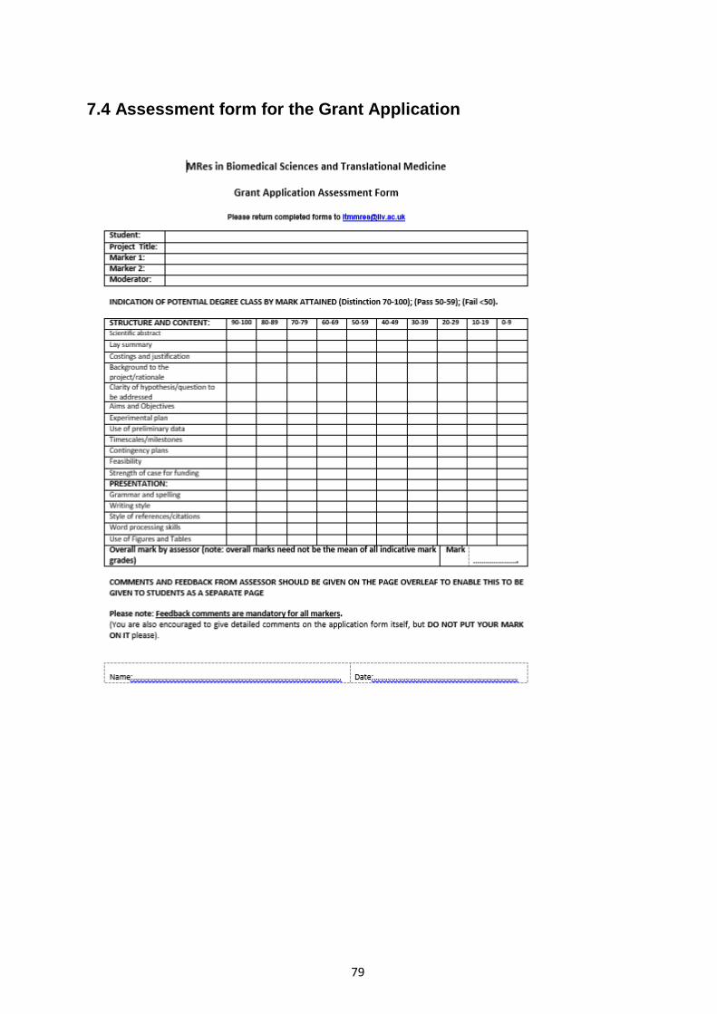
79
7.4 Assessment form for the Grant Application

80

81
7.5 Example Grant Application 1. APPLICANT (Please include full name, title, department and affiliation) Your name and details here 2. TITLE OF RESEARCH PROJECT (no more than 220 characters) Using Caenorhabditis elegans to Fight Human Neurodegenerative Diseases 3. SUMMARY OF RESEARCH
a) For scientifically qualified assessors (no more than 200 words)
In spite of exciting progress, the molecular determinants involved in the aetiology and
susceptibility of various chronic age-related neurodegenerative disorders (NDs) such as
Alzheimer’s disease (AD), Parkinson’s disease (PD), Huntington’s disease (HD), and
amyotrophic lateral sclerosis (ALS), remain mechanistically undefined. Numerous genetic and
pharmacological screens have been conducted in Caenorhabditis elegans disease models to
unravel candidate genes and /or specific cellular pathways pertinent to multiple
neurodegenerative cascades and successfully identified and validated many neuroprotective
factors. The overall aim of this project is to elucidate critical cellular mediators of
neuropathology and neuron vulnerability by exploiting the experimental attributes of C.
elegans and integrating genetic and pharmacological evaluation of a set of experimentally-
delineated genetic modifiers. Multiple transgenic, non-transgenic and toxicant C. elegans ND
models will be generated and optimised by examining their pathological phenotypes. The
bioinformatically prioritised modifiers will then be characterised and validated by phenocopy
using a reverse genetic approach by applying RNA interference (RNAi). In addition, a chemical
reverse genetic screen will be performed to illuminate neuroprotective compounds’ molecular
targets, mechanism of action and affected molecular pathways.
For lay readers (no more than 200 words)
The biological underpinnings of a diverse spectrum of progressive neurological disorders have
remained elusive. Large-scale screens have been performed in model organisms to isolate
genes that either rescue or exacerbate the manifestations of neurodegenerative disorders
(NDs), which in turn yield valuable insights into mediators of disease within cells and reveal
targets for potential therapies.

82
This project will utilise microscopic nematode roundworm, Caenorhabditis elegans to elucidate
the incompletely understood disease mechanisms and genetic susceptibility of multiple NDs.
Worms are used because of their facile genetics and strikingly similar neuronal mechanisms
to the humans. Worms will be manipulated to simulate the cardinal features of several NDs by
inserting human disease proteins, removing or depleting worm version of the disease proteins
and administering toxic compounds, and the animals will be observed for any effects. A
collection of previously identified C. elegans genes already known to influence the severity of
dysfunction will be suppressed in a subsequent screen to discern how they work. In addition,
a set of drugs known to alleviate some disease symptoms will also be applied to reveal their
mechanism of action. Ultimately, the project aims to expand our knowledge of the mechanisms
of NDs.
4. RESEARCH QUESTION
a) What is your research question/hypothesis (no more than 100 words)
This project will address the following fundamental questions: 1. What are the conserved genetic and molecular pathways underlying
neurodegenerative diseases?
2. How do genetic modifiers and pharmacological reagents contribute to
neurodegeneration and neuroprotection?
3. Through what mechanisms do therapeutic candidates mediate their neuroprotective
effects?
4. Do modifier genes have a widespread or disease-specific role in neurodegeneration?
5. Are pathogenic mechanism shared between several or all NDs, irrespective of the
specific disease?
6. Which types of neurons are most vulnerable to alterations of modifier genes?
b) Why is it important? (no more than 250 words)
Neurodegenerative diseases (NDs) pose a tremendous economical and societal burden to our
rapidly ageing populations. Though toxic functions of specific disease proteins have been
ascribed following the identification of many predisposition genes, the lack of mechanistic
insights into why and how these genes contribute or influence the aetiology and progression
of the NDs has severe repercussions for treatment of these crippling diseases since effective
therapeutic agents with disease-modifying or preventive properties remain acutely deficient
and development of new drugs continues to be a challenging job.

83
Definitive insights into the complex mechanistic connections between specific cellular
pathways and specific misfolded protein pathologies are likely to explain the conundrums and
facilitate the development of new disease biomarkers and promising disease-modifying
neuroprotective therapies for timely intervention.
5. DETAILS OF RESEARCH PROJECT: Summarise under the following headings:- a) Aims and objectives of the project b) Work which has led up to the project c) Detailed experimental plan of investigation d) Outcomes e) Contingency plans f) Timetable and milestones NO MORE THAN 2,000 WORDS SHOULD BE USED TO DESCRIBE THE RESEARCH
PROJECT (PLEASE INCLUDE A WORD COUNT) a) Aims and objectives of the project
This project aims to use a combined application of appropriate genetic and toxicant
Caenorhabditis elegans (C. elegans) neurodegenerative diseases (NDs) models to unravel
the incompletely understood key molecular pathways, disease mechanisms and genetic
susceptibility of multiple NDs. The main focus of the investigation is the genetic and
pharmacological characterisation of modifier genes involved in neuropathology and neuron
vulnerability, which contribute toward the identification of putative novel targets and
therapeutic leads.
b) Work which has led up to the project
Essentially, the majority of neurodegenerative disorders (NDs) are characterised by the
mechanistic unifying theme of aggregation, accumulation and deposition of prominent intra-
or extracellular toxic proteinaceous inclusions in a particular population of neurons (1) where
subsequent necrotic or apoptotic neurodegeneration results in irreversible neuronal loss,
synaptic impairments and eventual neuronal death. The use of Caenorhabditis elegans (C.
elegans) as a primary platform to probe the poorly defined disease mechanisms of several
major NDs and a more rapid means towards novel gene and drug discovery has escalated
over the last decade, since worms are amenable to high-throughput genetic, genomic,
proteomic, and drug screening approaches.
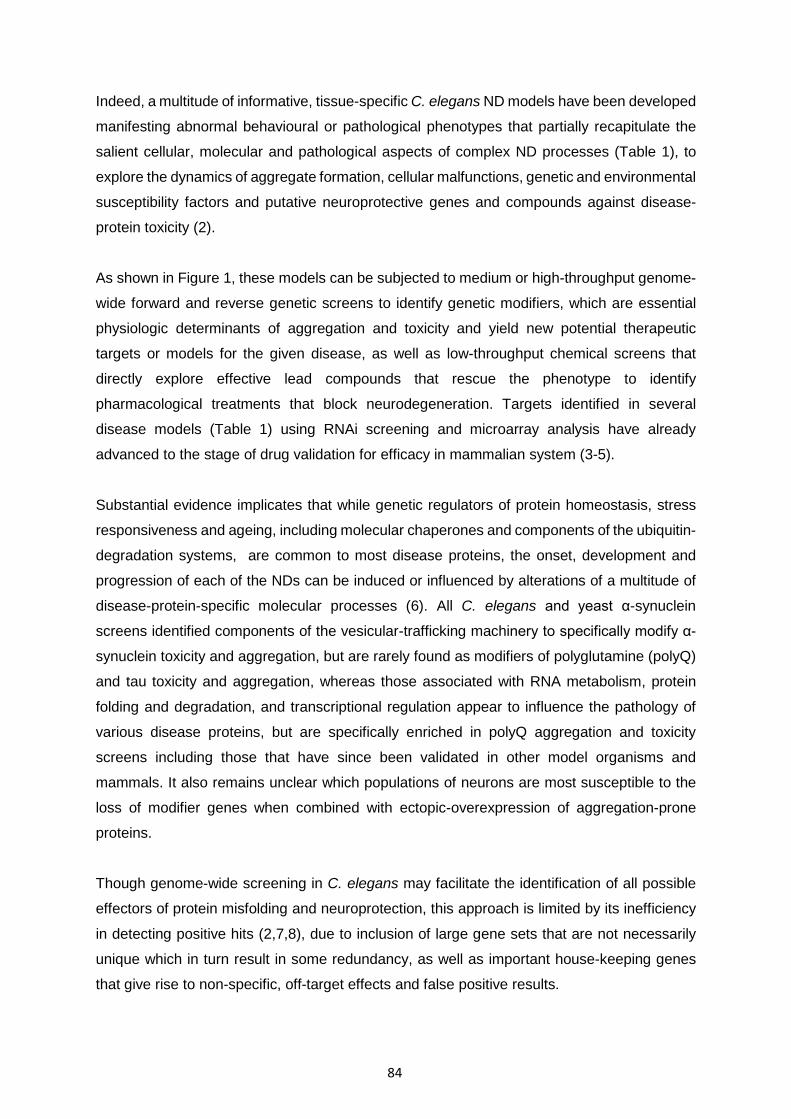
84
Indeed, a multitude of informative, tissue-specific C. elegans ND models have been developed
manifesting abnormal behavioural or pathological phenotypes that partially recapitulate the
salient cellular, molecular and pathological aspects of complex ND processes (Table 1), to
explore the dynamics of aggregate formation, cellular malfunctions, genetic and environmental
susceptibility factors and putative neuroprotective genes and compounds against disease-
protein toxicity (2).
As shown in Figure 1, these models can be subjected to medium or high-throughput genome-
wide forward and reverse genetic screens to identify genetic modifiers, which are essential
physiologic determinants of aggregation and toxicity and yield new potential therapeutic
targets or models for the given disease, as well as low-throughput chemical screens that
directly explore effective lead compounds that rescue the phenotype to identify
pharmacological treatments that block neurodegeneration. Targets identified in several
disease models (Table 1) using RNAi screening and microarray analysis have already
advanced to the stage of drug validation for efficacy in mammalian system (3-5).
Substantial evidence implicates that while genetic regulators of protein homeostasis, stress
responsiveness and ageing, including molecular chaperones and components of the ubiquitin-
degradation systems, are common to most disease proteins, the onset, development and
progression of each of the NDs can be induced or influenced by alterations of a multitude of
disease-protein-specific molecular processes (6). All C. elegans and yeast α-synuclein
screens identified components of the vesicular-trafficking machinery to specifically modify α-
synuclein toxicity and aggregation, but are rarely found as modifiers of polyglutamine (polyQ)
and tau toxicity and aggregation, whereas those associated with RNA metabolism, protein
folding and degradation, and transcriptional regulation appear to influence the pathology of
various disease proteins, but are specifically enriched in polyQ aggregation and toxicity
screens including those that have since been validated in other model organisms and
mammals. It also remains unclear which populations of neurons are most susceptible to the
loss of modifier genes when combined with ectopic-overexpression of aggregation-prone
proteins.
Though genome-wide screening in C. elegans may facilitate the identification of all possible
effectors of protein misfolding and neuroprotection, this approach is limited by its inefficiency
in detecting positive hits (2,7,8), due to inclusion of large gene sets that are not necessarily
unique which in turn result in some redundancy, as well as important house-keeping genes
that give rise to non-specific, off-target effects and false positive results.

85
Few screens have recently used a more focused strategy - a candidate approach/hypothesis-
driven approach to reveal potential modifier genes in a specific pathway based on hypothesis
generated by screens in lower model organisms or existing knowledge of disease mechanisms
and pathways (9,10).
Preliminary work in this laboratory adopted a candidate approach to mine the literature and
compile a collection of experimentally delineated genetic modifiers of protein aggregation,
misfolding and toxicity in C. elegans. Using online databases WormBase
(www.wormbase.org) and GExplore (http://genome.sfu.ca/gexplore/), modifier genes were
further selected for to retrieve only those with known expression in adult neurons that are also
non-RNAi lethal (Table 2) which form the basis for a hypothesis-driven RNAi screen and
subsequent in-depth analyses and mechanistic dissection. Our recent work also used
fluorescence confocal imaging of transgenic C. elegans lines to confirm the promoter
specificity of six neuronal promoters (Fig. 2) which are applicable as means of probing the role
of positive genetic modifiers in the survival, vulnerability and function of specific sets of
neurons and their subsequent validations.
TABLE 1. Summary of all available C. elegans neurodegeneration models. Worms are subjected to molecular, genetic and chemical manipulations to initiate the neurodegenerative cascade. Corresponding pathological phenotypes and identified drug compounds that confer neuroprotection are indicated. Models highlighted in beige are most suitable for this project. Human diseases: AD, Alzheimer’s disease; HD, Huntington’s disease; PD, Parkinson’s disease; ALS, Amyotrophic lateral sclerosis; SMA, Spinal muscular atrophy
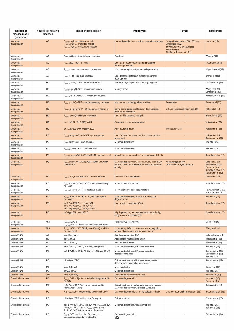
86
Method of disease model
generation
Neurodegenerativediseases
Transgene expression Phenotype Drug References
Molecular manipulation
AD Punc-54::Aβ - constitutive musclemyo-3::Aβ1-42 - inducible muscle
Punc-54::Aβ1-42 - constitutive muscle
Uncoordinated (Unc), paralysis, amyloid formation Ginkgo biloba extract EGb 761 and Ginkgolide A (12)Soya isoflavone glycitein (35)Reserpine (48)Thioflavin T; curcumin (21)
Link et al (15)
Molecular manipulation
AD Psnb-1::Aβ1-42 - inducible pan-neuronal Paralysis Wu et al (12)
Molecular manipulation
AD Paex-3::tau – pan-neuronal Unc, tau phosphorylation and aggregation, neurodegeneration
- Kraemer et al(16)
Molecular manipulation
AD Pmec-7::tau – mechanosensory neurons Mec, tau phosphorylation, neurodegeneration - Miyasaka et al (17)
Molecular manipulation
AD Prgef-1:: PHP tau- pan-neuronal Unc, decreased lifespan, defective neuronal development
- Brandt et al (18)
Molecular manipulation
HD Pmyo-3::polyQ–GFP – inducible muscle Paralysis, age-dependent polyQ aggregation - Caldwell et al (41)
Molecular manipulation
HD Punc-54::polyQ–GFP - constitutive muscle Motility defect - Wang et al (19)Saytal et al (20)
Molecular manipulation
HD Punc-54::DRPLAP::GFP- constitutive muscle - - Yamanaka et al (36)
Molecular manipulation
HD Pmec-3::polyQ–GFP - mechanosensory neurons Mec, axon morphology abnormalities Resveratrol Parker et al (37)
Molecular manipulation
HD Posm-10::polyQ–GFP - chemosensory neurons polyQ aggregation, ASH neuron degeneration, nose-touch defective
Lithium chloride; mithramycin (23) Faber et al (42)
Molecular manipulation
HD Prgef-1::polyQ–GFP - pan-neuronal Unc, motility defects, paralysis - Brignull et al (22)
Molecular manipulation
HD pqe-1(rt13); Htn-Q150(rtIs11) Accelerated neurodegeneration - Voisine et al (23)
Molecular manipulation
HD pha-1(e2123); Htn-Q150(rtIs11) ASH neuronal death Trichostatin (38) Voisine et al (23)
Molecular manipulation
PD Paex-3::α-syn WT and A53T - pan-neuronal Unc, DA dendritic abnormalities, reduced motor movement
- Lakso et al (24)Springer et al (25)
Molecular manipulation
PD Psnb-1::α-syn WT - pan-neuronal Mitochondrial stress - Ved et al (26)
Molecular manipulation
PD Punc-119::α-syn A53T- pan-neuronal Mitochondrial stress - Ved et al (26)
Molecular manipulation
PD Punc-51 ::α-syn WT,A30P and A53T - pan-neuronal Motor/developmental defects, endocytosis defects - Kuwahara et al (27)
Molecular manipulation
PD Pdat-1 ::α-syn WT, A30P, A53T, A56P and A76P –DA neurons
DA neurodegeneration, α-syn accumulation in DA neurons, reduced DA levels, altered DA neuronal activity
Acetaminophen (39)Bromocriptine, Quinpirole (3)
Lakso et al (24)Settivari et al (32)Cao et al (31)Kuwahara et al (27)Karpinar et al (40)
Molecular manipulation
PD Pacr-2::α-syn WT and A53T – motor neurons Reduced motor movement - Lakso et al (24)
Molecular manipulation
PD Pmec-7::α-syn WT and A53T - mechanosensoryneurons
Impaired touch response - Kuwahara et al (27)
Molecular manipulation
PD Punc-54 ::α-syn::GFP - constitutive muscle α-syn misfolding and accumulation - Hamamichi et al (10)Van Ham et al (2)
Molecular manipulation
PD Psnb-1::LRRK2 WT, R1441C, G2019S – pan-neuronal
Mitochondrial stress, reduced DA levels, DA degeneration
- Saha et al (28)
Molecular manipulation
PD eri-1 (mg366);Pdat-1 ::α-syn WT,eri-1 (mg366);Pdat-1 ::α-syn A53Teri-1 (mg366);Pdat-1 ::α-syn A30P
Unc, growth retardation (Gro) - Kuwahara et al (27)
Molecular manipulation
PD pdr-1(lg103); α-syn A53T Highly penetrant, temperature sensitive lethality, early larval arrest phenotype
- Kuwahara et al (27)
Molecular manipulation
ALS Pmyo-3::SOD-1 hsp-16::SOD-1 - body wall muscle or inducible
Paraquat hypersensitivity - Oeda et al (43)
Molecular manipulation
ALS Psnb-1::SOD-1 WT, G85R, H46R/H48Q – YFP –pan-neuronal
Locomotory defects, intra-neuronal aggregation, abnormal processes and synaptic function
- Wang et al (44)
Mutant/RNAi AD sel-12 or hop-1 Egg laying defective (Egl) - Lakowski et al (45)Mutant/RNAi HD pqe-1(rt13) Accelerated neurodegeneration - Voisine et al (23)Mutant/RNAi HD pha-1(e2123) ASH neuronal death - Voisine et al (23)Mutant/RNAi PD lrk-1 (km17), (km41), (tm1898) and (RNAi) Mitochondrial stress, ER stress sensitive - Saha et al (28)
Mutant/RNAi PD pdr-1 (lg103), (XY1046, Parkin KO3) and (RNAi) Mitochondrial stress, ER stress sensitive, decreased life span
- Samann et al (29)Springer et al (25)Ved et al (26)
Mutant/RNAi PD pink-1 (tm1779) Oxidative stress sensitive, neurite outgrowth defects, mitochondrial cristae defects
- Samann et al (29)
Mutant/RNAi PD catp-6 (RNAi) α-syn misfolding - Gitler et al (46)Mutant/RNAi PD djr-1.1 (RNAi) Mitochondrial stress - Ved et al (26)Mutant/RNAi SMA smn-1 (ok355) Neuromuscular function deficits - Briese et al (47)Chemical treatment PD Pdat-1::GFP subjected to 6-hydroxydopamine (6-
OHDA)DA neurodegeneration - Nass et al (30)
Cao et al (31)Chemical treatment PD N2 , Pdat-1::GFP, Pdat-1::α-syn subjected to
Manganese (Mn2+)Oxidative stress, mitochondrial stress, enhanced DA neurodegeneration, reduced DA levels
- Settivari et al (32)
Chemical treatment PD N2, Pcat-2::GFP subjected to MPTP and MPP+ DA neurodegeneration, motility defects, lethality Lisuride, apomorphine, Rottlerin (33) Braungart et al. (33)
Chemical treatment PD pink-1 (tm1779) subjected to Paraquat Oxidative stress - Samann et al (29)
Chemical treatment PD pdr-1 (XY1046), Psnb-1::α-syn WT, Punc-119::α-synA53T, N2, lrk-1 (km17), Psnb-1::LRRK2 WT, R1441C, G2019S subjected to Rotenone
Mitochondrial stress, reduced viability - Ved et al (26)Saha et al (28)
Chemical treatment PD Pdat-1::GFP subjected to Streptomycesvenezuelae secondary metabolite
DA neurodegeneration - Caldwell et al (34)

87
Disease
C. elegans disease model
Functional C. elegans assay
Recapitulation of phenotypes in worms
Tissue-specific expression of mutant disease gene;
mutation/RNAi; compound-induced
WormBase C. elegansgenome
RNAi screen (reverse genetics)
Genetic screen (forward genetics)
Expression profiling Epistatic analysisPharmacological screen
Screening goalsuppressorenhancer
suppressorenhancer
drug sensitivitydrug resistance
EMS
Feeding worms with individual RNAi clones
Mutagenesis
Determine optimal drug concentrations
Phenotype analysis
Confirmation of RNAi clones
Gene mapping Confirmation of drug efficacy
Positive hits identified
Hits prioritisation
Hits validation
Pharmacological intervention in wormsif successful
Mammalian assays and identification of human orthologues
Drug Validation
Secondary analyses in multiple worm models
mechanisms of disease
Pharmacological intervention
gene expression changes
miRNA microarray
Quantitative real-time PCR
Target analysis
Distribution of compounds
Exclude drug effects on growth/development and
aging pathway
genetic complementation and sequencing of
mutant alleles
FIGURE 1. Schematic diagram of the various screening routes for the identification of genetic or pharmacological targets of a C. elegans disease model

88
TABLE 2: Set of 47 modifier genes with known adult neuronal expression and non-RNAi lethality. Assigned functional category, C. elegans gene name, CGC approved name, available description and human orthologue are shown. RNAi feeding library coverage of the modifier genes is also indicated. Four modifier genes are nematode specific and six genes remain uncharacterised. UPS: ubiquitin-proteasome system, ECM: extracellular matrix.
Functional Category Gene name CGC Name Description Human
orthologuesRNAi
library
F08H9.4 - heat shock protein (HSP) of the HSP16 class HSPB6 Vidal
T09B4.10 chn-1 mammalian carboxyl-terminus of Hsc70 interacting protein STUB1 CHIP Ahringer
F55B12.3 sel-10 F-box and WD-repeat-containing protein of CDC4/CUL-1 family of E2-E3 ubiquitin ligases FBXW7 Vidal
Y61A9LA.8 sut-2 Nuclear polyadenylated RNA binding protein ZC3H14 Not available
T05H10.1 - Putative ubiquitin-specific protease USP47 AhringerT26A5.5 - F-box protein JEMMA FBXL19 VidalK09A9.6 - Aspartyl beta-hydroxylase ASPHD2 Ahringer
F47G6.1 dyb-1 ZZ type zinc finger DTNA Vidal
T04D1.3 unc-57 endophilin A, required for synaptic vesicle endocytosis SH3GL2 Vidal
Y22F5A.3 ric-4 Intracellular protein transport, neurotransmitter secretion SNAP-25 Vidal
EEED8.9 pink-1 BRPK/PTEN-induced protein kinase PINK1 Ahringer
T25F10.2 dbl-1 a member of the transforming growth factor beta (TGFbeta) superfamily BMP4 Vidal
M04C9.5 dyf-5 a putative MAP kinase MAK/ICK AhringerK11E8.1 unc-43 type II calcium/calmodulin-dependent protein kinase CaMKII VidalK07A9.2 cmk-1 Ca2+/calmodulin-dependent protein kinase I CaMK1 VidalB0334.8 age-1 functions in an insulin-like signalling (ILS) pathway PI3K Ahringer
W08G11.4 pptr-1 Serine/threonine protein phosphatase 2A PP2A Not available
R11A8.4 sir-2.1 NAD-dependent deacetylase sirtuin-1 SIRT1 AhringerR13H8.1 daf-16 sole C. elegans forkhead box O (FOXO) homologue FOXO4 VidalH21P03.1 mbf-1 Transcription factor MBF1 EDF1 VidalC04G6.3 pld-1 Phospholipase D1 PLD1 VidalF02E9.4 sin-3 SIN3 family of histone deacetylase subunits SIN3 Vidal
F39C12.2 add-1 cytoskeletal protein alpha-adducin ADD1 Vidal
K05B2.3 ifa-4 a nonessential intermediate filament protein IFB-1 Vidal
R05D3.7 unc-116 kinesin-1 heavy chain ortholog KIF5 Ahringer
F47G4.7 smd-1 S-Adenosylmethionine decarboxylase AMD1 VidalC06E7.1 sams-3 S-adenosylmethionine synthetase MAT1A VidalF44E7.2 - p-Nitrophenyl phosphatase PDXP Vidal
Y56A3A.13 nft-1 Carbon-nitrogen hydrolase FHIT Vidal
C30H7.2 - human thioredoxin domain-containing protein 4 precursor ERP44 Vidal
K08F4.7 gst-4 a putative glutathione-requiring prostaglandin D synthase GSTA3 Vidal
F35E8.8 gst-38 unclassified GSTA4 Vidal
C50C3.9 unc-36 alpha2/delta subunit of a voltage-gated calcium channel CACNA2D3 Vidal
R05G6.7 - Porin/voltage-dependent anion-selective channel protein VDAC2 Vidal
Transport ATPase ZK256.1 pmr-1 a Golgi P-type ATPase Ca2+/Mn2+-pump ATP2C1 Not available
Dopamine metabolism T23G5.5 dat-1 a plasma membrane dopamine transporter SLC6A2/A3 VidalLipid metabolism C28C12.7 spp-10 sphingolipid metabolism/lysosomal PSAP VidalRNA metabolism M88.5 - IGF-II mRNA-binding protein IMP IGF2BP2 Vidal
Carbohydrate metabolism K11H3.1 gpdh-2 glycerol 3-phosphate dehydrogenases GPD1L Vidal
T05A12.2 tre-2 putative trehalases of unknown function TREH Vidal
W05E10.4 tre-3 putative trehalases of unknown function TREH VidalC04B4.2 - unclassified None Vidal
R74.3 xbp-1 unclassified None VidalR13A1.8 glb-23 unclassified None VidalF52C9.8 pqe-1 unclassified REXO1 VidalF08C6.3 tag-197 unclassified VPS52 Vidal
Y55F3AM.14 - unclassified None Vidal
Enzymes
Redox/oxidative stress
Voltage-gated ion channels
Trehalose biosynthesis
Unclassified
Chaperone/quality control
Protein turnover/degradation/
UPS
Neurotransmission and signalling
Signal transduction
Transcription cofactor
ECM/Cytoskeletal organisation
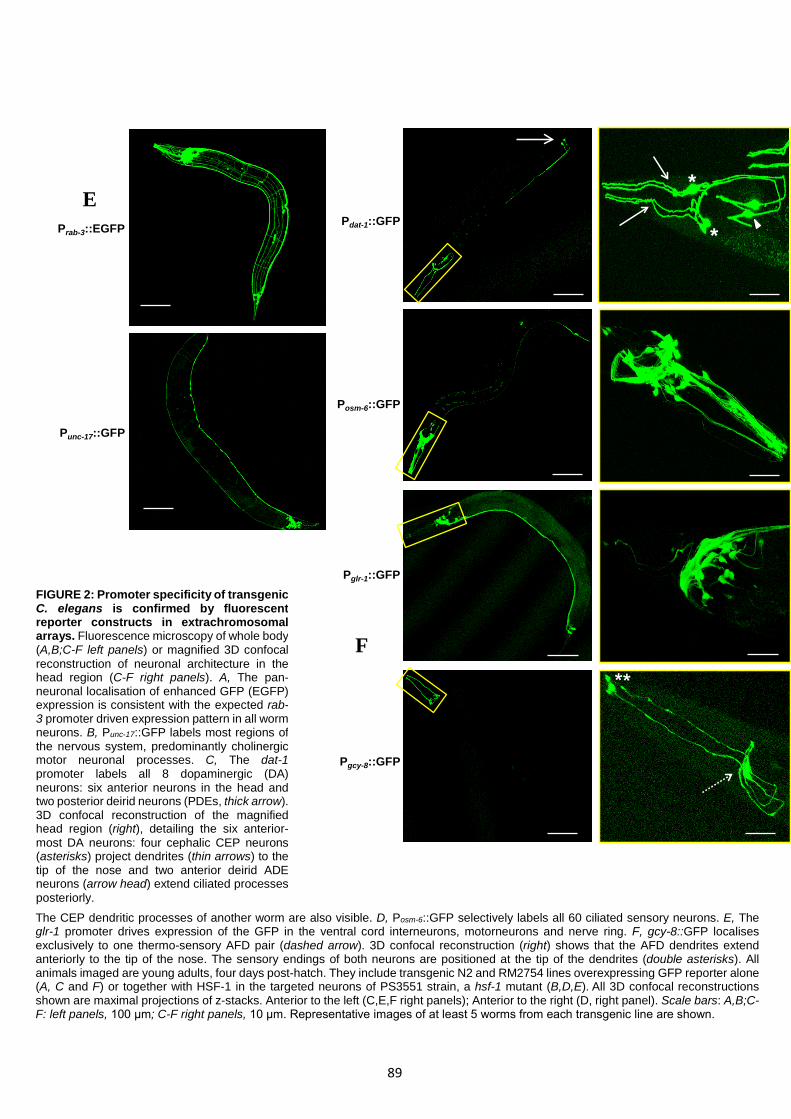
89
Prab-3::EGFP
Punc-17::GFP
Pdat-1::GFP
Posm-6::GFP
Pglr-1::GFP
Pgcy-8::GFP
*
*
FIGURE 2: Promoter specificity of transgenic C. elegans is confirmed by fluorescent reporter constructs in extrachromosomal arrays. Fluorescence microscopy of whole body (A,B;C-F left panels) or magnified 3D confocal reconstruction of neuronal architecture in the head region (C-F right panels). A, The pan-neuronal localisation of enhanced GFP (EGFP) expression is consistent with the expected rab-3 promoter driven expression pattern in all worm neurons. B, Punc-17::GFP labels most regions of the nervous system, predominantly cholinergic motor neuronal processes. C, The dat-1 promoter labels all 8 dopaminergic (DA) neurons: six anterior neurons in the head and two posterior deirid neurons (PDEs, thick arrow). 3D confocal reconstruction of the magnified head region (right), detailing the six anterior-most DA neurons: four cephalic CEP neurons (asterisks) project dendrites (thin arrows) to the tip of the nose and two anterior deirid ADE neurons (arrow head) extend ciliated processes posteriorly. The CEP dendritic processes of another worm are also visible. D, Posm-6::GFP selectively labels all 60 ciliated sensory neurons. E, The glr-1 promoter drives expression of the GFP in the ventral cord interneurons, motorneurons and nerve ring. F, gcy-8::GFP localises exclusively to one thermo-sensory AFD pair (dashed arrow). 3D confocal reconstruction (right) shows that the AFD dendrites extend anteriorly to the tip of the nose. The sensory endings of both neurons are positioned at the tip of the dendrites (double asterisks). All animals imaged are young adults, four days post-hatch. They include transgenic N2 and RM2754 lines overexpressing GFP reporter alone (A, C and F) or together with HSF-1 in the targeted neurons of PS3551 strain, a hsf-1 mutant (B,D,E). All 3D confocal reconstructions shown are maximal projections of z-stacks. Anterior to the left (C,E,F right panels); Anterior to the right (D, right panel). Scale bars: A,B;C-F: left panels, 100 μm; C-F right panels, 10 μm. Representative images of at least 5 worms from each transgenic line are shown.
E
F
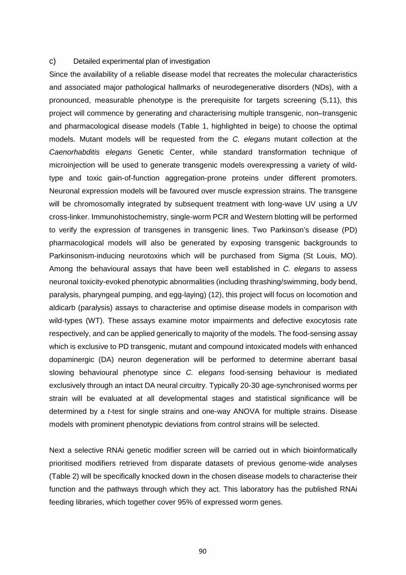
90
c) Detailed experimental plan of investigation
Since the availability of a reliable disease model that recreates the molecular characteristics
and associated major pathological hallmarks of neurodegenerative disorders (NDs), with a
pronounced, measurable phenotype is the prerequisite for targets screening (5,11), this
project will commence by generating and characterising multiple transgenic, non–transgenic
and pharmacological disease models (Table 1, highlighted in beige) to choose the optimal
models. Mutant models will be requested from the C. elegans mutant collection at the
Caenorhabditis elegans Genetic Center, while standard transformation technique of
microinjection will be used to generate transgenic models overexpressing a variety of wild-
type and toxic gain-of-function aggregation-prone proteins under different promoters.
Neuronal expression models will be favoured over muscle expression strains. The transgene
will be chromosomally integrated by subsequent treatment with long-wave UV using a UV
cross-linker. Immunohistochemistry, single-worm PCR and Western blotting will be performed
to verify the expression of transgenes in transgenic lines. Two Parkinson’s disease (PD)
pharmacological models will also be generated by exposing transgenic backgrounds to
Parkinsonism-inducing neurotoxins which will be purchased from Sigma (St Louis, MO).
Among the behavioural assays that have been well established in C. elegans to assess
neuronal toxicity-evoked phenotypic abnormalities (including thrashing/swimming, body bend,
paralysis, pharyngeal pumping, and egg-laying) (12), this project will focus on locomotion and
aldicarb (paralysis) assays to characterise and optimise disease models in comparison with
wild-types (WT). These assays examine motor impairments and defective exocytosis rate
respectively, and can be applied generically to majority of the models. The food-sensing assay
which is exclusive to PD transgenic, mutant and compound intoxicated models with enhanced
dopaminergic (DA) neuron degeneration will be performed to determine aberrant basal
slowing behavioural phenotype since C. elegans food-sensing behaviour is mediated
exclusively through an intact DA neural circuitry. Typically 20-30 age-synchronised worms per
strain will be evaluated at all developmental stages and statistical significance will be
determined by a t-test for single strains and one-way ANOVA for multiple strains. Disease
models with prominent phenotypic deviations from control strains will be selected.
Next a selective RNAi genetic modifier screen will be carried out in which bioinformatically
prioritised modifiers retrieved from disparate datasets of previous genome-wide analyses
(Table 2) will be specifically knocked down in the chosen disease models to characterise their
function and the pathways through which they act. This laboratory has the published RNAi
feeding libraries, which together cover 95% of expressed worm genes.
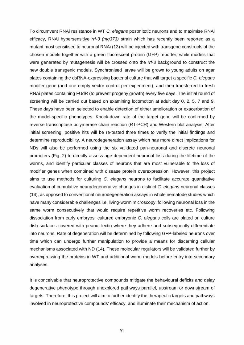
91
To circumvent RNAi resistance in WT C. elegans postmitotic neurons and to maximise RNAi
efficacy, RNAi hypersensitive rrf-3 (mg373) strain which has recently been reported as a
mutant most sensitised to neuronal RNAi (13) will be injected with transgene constructs of the
chosen models together with a green fluorescent protein (GFP) reporter, while models that
were generated by mutagenesis will be crossed onto the rrf-3 background to construct the
new double transgenic models. Synchronised larvae will be grown to young adults on agar
plates containing the dsRNA-expressing bacterial culture that will target a specific C. elegans
modifer gene (and one empty vector control per experiment), and then transferred to fresh
RNAi plates containing FUdR (to prevent progeny growth) every five days. The initial round of
screening will be carried out based on examining locomotion at adult day 0, 2, 5, 7 and 9.
These days have been selected to enable detection of either amelioration or exacerbation of
the model-specific phenotypes. Knock-down rate of the target gene will be confirmed by
reverse transcriptase polymerase chain reaction (RT-PCR) and Western blot analysis. After
initial screening, positive hits will be re-tested three times to verify the initial findings and
determine reproducibility. A neurodegeneration assay which has more direct implications for
NDs will also be performed using the six validated pan-neuronal and discrete neuronal
promoters (Fig. 2) to directly assess age-dependent neuronal loss during the lifetime of the
worms, and identify particular classes of neurons that are most vulnerable to the loss of
modifier genes when combined with disease protein overexpression. However, this project
aims to use methods for culturing C. elegans neurons to facilitate accurate quantitative
evaluation of cumulative neurodegenerative changes in distinct C. elegans neuronal classes
(14), as opposed to conventional neurodegeneration assays in whole nematode studies which
have many considerable challenges i.e. living-worm microscopy, following neuronal loss in the
same worm consecutively that would require repetitive worm recoveries etc. Following
dissociation from early embryos, cultured embryonic C. elegans cells are plated on culture
dish surfaces covered with peanut lectin where they adhere and subsequently differentiate
into neurons. Rate of degeneration will be determined by following GFP-labeled neurons over
time which can undergo further manipulation to provide a means for discerning cellular
mechanisms associated with ND (14). These molecular regulators will be validated further by
overexpressing the proteins in WT and additional worm models before entry into secondary
analyses.
It is conceivable that neuroprotective compounds mitigate the behavioural deficits and delay
degenerative phenotype through unexplored pathways parallel, upstream or downstream of
targets. Therefore, this project will aim to further identify the therapeutic targets and pathways
involved in neuroprotective compounds’ efficacy, and illuminate their mechanism of action.

92
A collection of twelve chemicals (Table1) i.e. antioxidants, kinase inhibitors, dopamine D2
receptor agonists and amyloid-binding compounds (Sigma) that have been previously
identified to confer neuroprotection will be tested in a chemical reverse genetics screen.
Synchronised larvae of disease models expressing GFP in the targeted neurons will be
incubated with and without solubilised drugs for three days in 24-well plates. Each well will
contain a bacteria culture from the RNAi library expressing dsRNA to a C. elegans modifier
gene (12 wells per bacteria), followed by addition of individual or a combination of compounds
per well. The presence or absence of GFP fluorescence will be evaluated in each well to
determine neuronal survival prior to replating on solid media for further behaviour assays for
confirmation. Wells that contain a weak or absent fluorescent signal would indicate that the
modifier is involved in suppression of the compound’s neuroprotective effects. Significance
will be determined using a Fisher’s exact test which is appropriate for small sample sizes.
d) Outcomes
These investigations will help to reveal conserved molecular and therapeutic mechanisms
which contribute not only to the molecular understanding of human NDs but also eventually to
the development of effective therapeutic preventions and interventions for those who are at
genetic risk or are affected clinically.
e) Contingency plans
Project progression may be temporarily slowed by some unexpected events, or potential
difficulties in the operations. In such a case, efforts will be made to tackle the bottleneck as
detailed below:
Difficulties to integrate transgene in the germ-line of the animals.
The difficulty with transgene expression in the C. elegans germ-line resides in uncharacterised
repression mechanisms of this organism and has long been a problem. In the event that no
satisfactory transgene expression can be obtained by conventional microinjection, double
transgenic models will be created by genetic crossing. Alternatively, this problem can also be
circumvented by the new Mos1 techniques developed by Jorgensen Lab.
RNAi knock-down rate cannot be confirmed by PCR
GFP reporter will be used as an alternate to confirm reduced gene expression. Strains with
GFP-labelled neurons will have gradual loss of fluorescence if RNAi have successfully
suppressed gene expressions. Technical challenges of RNAi-based phenotypic analysis arise
from the inherent limitations of RNA (i.e., neuronal intractability, inherent variability in the level
of target mRNA knock-down for different genes, possibility of off-target effects).
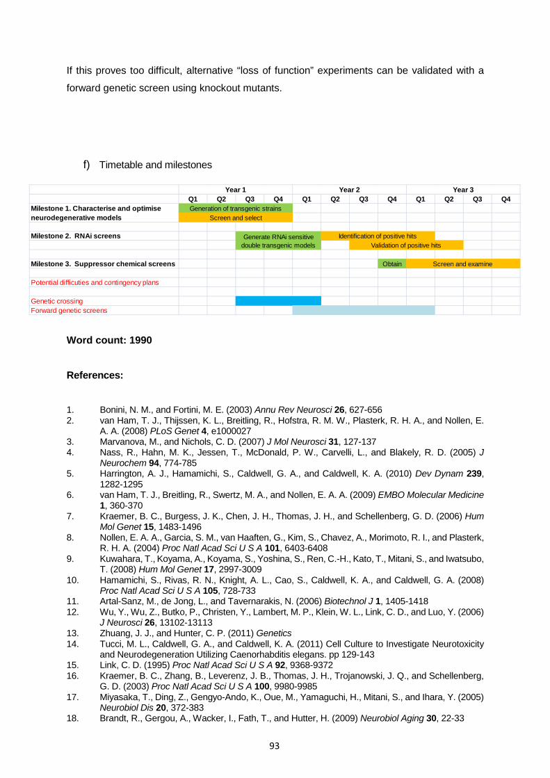
93
If this proves too difficult, alternative “loss of function” experiments can be validated with a
forward genetic screen using knockout mutants.
f) Timetable and milestones
Word count: 1990 References: (12,15-48) 1. Bonini, N. M., and Fortini, M. E. (2003) Annu Rev Neurosci 26, 627-656 2. van Ham, T. J., Thijssen, K. L., Breitling, R., Hofstra, R. M. W., Plasterk, R. H. A., and Nollen, E.
A. A. (2008) PLoS Genet 4, e1000027 3. Marvanova, M., and Nichols, C. D. (2007) J Mol Neurosci 31, 127-137 4. Nass, R., Hahn, M. K., Jessen, T., McDonald, P. W., Carvelli, L., and Blakely, R. D. (2005) J
Neurochem 94, 774-785 5. Harrington, A. J., Hamamichi, S., Caldwell, G. A., and Caldwell, K. A. (2010) Dev Dynam 239,
1282-1295 6. van Ham, T. J., Breitling, R., Swertz, M. A., and Nollen, E. A. A. (2009) EMBO Molecular Medicine
1, 360-370 7. Kraemer, B. C., Burgess, J. K., Chen, J. H., Thomas, J. H., and Schellenberg, G. D. (2006) Hum
Mol Genet 15, 1483-1496 8. Nollen, E. A. A., Garcia, S. M., van Haaften, G., Kim, S., Chavez, A., Morimoto, R. I., and Plasterk,
R. H. A. (2004) Proc Natl Acad Sci U S A 101, 6403-6408 9. Kuwahara, T., Koyama, A., Koyama, S., Yoshina, S., Ren, C.-H., Kato, T., Mitani, S., and Iwatsubo,
T. (2008) Hum Mol Genet 17, 2997-3009 10. Hamamichi, S., Rivas, R. N., Knight, A. L., Cao, S., Caldwell, K. A., and Caldwell, G. A. (2008)
Proc Natl Acad Sci U S A 105, 728-733 11. Artal-Sanz, M., de Jong, L., and Tavernarakis, N. (2006) Biotechnol J 1, 1405-1418 12. Wu, Y., Wu, Z., Butko, P., Christen, Y., Lambert, M. P., Klein, W. L., Link, C. D., and Luo, Y. (2006)
J Neurosci 26, 13102-13113 13. Zhuang, J. J., and Hunter, C. P. (2011) Genetics 14. Tucci, M. L., Caldwell, G. A., and Caldwell, K. A. (2011) Cell Culture to Investigate Neurotoxicity
and Neurodegeneration Utilizing Caenorhabditis elegans. pp 129-143 15. Link, C. D. (1995) Proc Natl Acad Sci U S A 92, 9368-9372 16. Kraemer, B. C., Zhang, B., Leverenz, J. B., Thomas, J. H., Trojanowski, J. Q., and Schellenberg,
G. D. (2003) Proc Natl Acad Sci U S A 100, 9980-9985 17. Miyasaka, T., Ding, Z., Gengyo-Ando, K., Oue, M., Yamaguchi, H., Mitani, S., and Ihara, Y. (2005)
Neurobiol Dis 20, 372-383 18. Brandt, R., Gergou, A., Wacker, I., Fath, T., and Hutter, H. (2009) Neurobiol Aging 30, 22-33
Q1 Q2 Q3 Q4 Q1 Q2 Q3 Q4 Q1 Q2 Q3 Q4
Milestone 2. RNAi screens
Milestone 3. Suppressor chemical screens Obtain
Potential difficuties and contingency plans
Genetic crossingForward genetic screens
Generation of transgenic strains
Year 1 Year 2 Year 3
Milestone 1. Characterise and optimise neurodegenerative models
Screen and examine
Screen and select
Generate RNAi sensitive double transgenic models Validation of positive hits
Identification of positive hits

94
19. Wang, H., Lim, P. J., Yin, C., Rieckher, M., Vogel, B. E., and Monteiro, M. J. (2006) Human Molecular Genetics 15, 1025-1041
20. Satyal, S. H., Schmidt, E., Kitagawa, K., Sondheimer, N., Lindquist, S., Kramer, J. M., and Morimoto, R. I. (2000) Proc Natl Acad Sci U S A 97, 5750-5755
21. Alavez, S., Vantipalli, M. C., Zucker, D. J., Klang, I. M., and Lithgow, G. J. (2011) Nature 472, 226-229
22. Brignull, H. R., Moore, F. E., Tang, S. J., and Morimoto, R. I. (2006) J Neurosci 26, 7597-7606 23. Voisine, C., Varma, H., Walker, N., Bates, E. A., Stockwell, B. R., and Hart, A. C. (2007) PLoS One
2, e504 24. Lakso, M., Vartiainen, S., Moilanen, A. M., Sirvio, J., Thomas, J. H., Nass, R., Blakely, R. D., and
Wong, G. (2003) J Neurochem 86, 165-172 25. Springer, W., Hoppe, T., Schmidt, E., and Baumeister, R. (2005) Hum Mol Genet 14, 3407-3423 26. Ved, R., Saha, S., Westlund, B., Perier, C., Burnam, L., Sluder, A., Hoener, M., Rodrigues, C. M.,
Alfonso, A., Steer, C., Liu, L., Przedborski, S., and Wolozin, B. (2005) J Biol Chem 280, 42655-42668
27. Kuwahara, T., Koyama, A., Gengyo-Ando, K., Masuda, M., Kowa, H., Tsunoda, M., Mitani, S., and Iwatsubo, T. (2006) J Biol Chem 281, 334-340
28. Saha, S., Guillily, M. D., Ferree, A., Lanceta, J., Chan, D., Ghosh, J., Hsu, C. H., Segal, L., Raghavan, K., Matsumoto, K., Hisamoto, N., Kuwahara, T., Iwatsubo, T., Moore, L., Goldstein, L., Cookson, M., and Wolozin, B. (2009) J Neurosci 29, 9210-9218
29. Sämann, J., Hegermann, J., von Gromoff, E., Eimer, S., Baumeister, R., and Schmidt, E. (2009) J Biol Chem 284, 16482-16491
30. Nass, R., Miller, D. M., and Blakely, R. D. (2001) Parkinsonism & Related Disorders 7, 185-191 31. Cao, S., Gelwix, C. C., Caldwell, K. A., and Caldwell, G. A. (2005) J Neurosci 25, 3801-3812 32. Settivari, R., LeVora, J., and Nass, R. (2009) J. Biol. Chem., M109.051409 33. Braungart, E., Gerlach, M., Riederer, P., Baumeister, R., and Hoener, M. C. (2004) Neurodegener
Dis 1, 175-183 34. Caldwell, K. A., Tucci, M. L., Armagost, J., Hodges, T. W., Chen, J., Memon, S. B., Blalock, J. E.,
DeLeon, S. M., Findlay, R. H., Ruan, Q., Webber, P. J., Standaert, D. G., Olson, J. B., and Caldwell, G. A. (2009) PLoS One 4, e7227
35. Gutierrez-Zepeda, A., Santell, R., Wu, Z., Brown, M., Wu, Y., Khan, I., Link, C. D., Zhao, B., and Luo, Y. (2005) BMC Neurosci 6, 54
36. Yamanaka, K., Okubo, Y., Suzaki, T., and Ogura, T. (2004) J Struct Biol 146, 242-250 37. Parker, J. A., Arango, M., Abderrahmane, S., Lambert, E., Tourette, C., Catoire, H., and Neri, C.
(2005) Nat Genet 37, 349-350 38. Bates, E. A., Victor, M., Jones, A. K., Shi, Y., and Hart, A. C. (2006) J Neurosci 26, 2830-2838 39. Locke, C. J., Fox, S. A., Caldwell, G. A., and Caldwell, K. A. (2008) Neurosci Lett 439, 129-133 40. Karpinar, D. P., Balija, M. B., Kugler, S., Opazo, F., Rezaei-Ghaleh, N., Wender, N., Kim, H. Y.,
Taschenberger, G., Falkenburger, B. H., Heise, H., Kumar, A., Riedel, D., Fichtner, L., Voigt, A., Braus, G. H., Giller, K., Becker, S., Herzig, A., Baldus, M., Jackle, H., Eimer, S., Schulz, J. B., Griesinger, C., and Zweckstetter, M. (2009) EMBO J 28, 3256-3268
41. Caldwell, G. A., Cao, S., Sexton, E. G., Gelwix, C. C., Bevel, J. P., and Caldwell, K. A. (2003) Hum Mol Genet 12, 307-319
42. Faber, P. W., Alter, J. R., MacDonald, M. E., and Hart, A. C. (1999) Proc Natl Acad Sci U S A 96, 179-184
43. Oeda, T., Shimohama, S., Kitagawa, N., Kohno, R., Imura, T., Shibasaki, H., and Ishii, N. (2001) Hum Mol Genet 10, 2013-2023
44. Wang, J., Farr, G. W., Hall, D. H., Li, F., Furtak, K., Dreier, L., and Horwich, A. L. (2009) PLoS Genet 5, e1000350
45. Lakowski, B., Eimer, S., Göbel, C., Böttcher, A., Wagler, B., and Baumeister, R. (2003) Development 130, 2117-2128
46. Gitler, A. D., Bevis, B. J., Shorter, J., Strathearn, K. E., Hamamichi, S., Su, L. J., Caldwell, K. A., Caldwell, G. A., Rochet, J. C., McCaffery, J. M., Barlowe, C., and Lindquist, S. (2008) Proc Natl Acad Sci U S A 105, 145-150
47. Briese, M., Esmaeili, B., Fraboulet, S., Burt, E. C., Christodoulou, S., Towers, P. R., Davies, K. E., and Sattelle, D. B. (2009) Hum Mol Genet 18, 97-104
48. Arya, U., Dwivedi, H., and Subramaniam, J. R. (2009) Exp Gerontol 44, 462-466

95
6. SUMMARY OF FINANCIAL SUPPORT REQUESTED Specify the funds required for staff, animals, consumables and travel in the Table below.
YEAR 1 YEAR 2 YEAR 3 STIPEND
£13590 £13590 £13590
ANIMALS
- - -
CONSUMABLES
£10,000 £10,000 £10,000
TRAVEL
£1000 £1000 £1000
TOTAL £24590 £24590 £24590 7. JUSTIFICATION OF FINANCIAL SUPPORT REQUESTED In this section, justify the funds requested for animals, consumables and travel (no more than
200 words).
The applicant will be working full time and participating in the experimental design, application
and analysis. She has gained experience working with C. elegans in our laboratory during
three 10-week Masters-level rotation projects.
The requested funding will cover the purchase for project-specific consumables such as routine
chemicals and labware, the molecular biology techniques outlined above e.g. microinjections,
primary culture of embryonic neurons, as well as obtaining knockout mutants. The collection
of drug compounds required for chemical screening costs around £5000 if to be purchased
from Sigma, therefore contributes to a significant portion of the total costs. In addition, there
are also charges relating to use of communal facilities (i.e. confocal microscope). Finally, funds
requested will cover travel expenses to attend and present research findings at relevant
conferences.

96
7.6 Demonstrator Training Workshop Practical Based Teaching Overview: This session will help postgraduate students to consider in more depth how their role in a practical class complements that of the lecturer and how they can take responsibility for helping the students to get the most from the practical class. Aim: To help participants plan and teach a practical class. Objectives: Following the session the participants will be able to:
• define the purpose of practical-based teaching and discuss their role within it
• discuss the interactions between teaching assistants and other members of staff and students
• prepare themselves prior to a practical class
• identify the skills necessary to operate effectively as a teacher within practical sessions
• understand the range of skills that practical classes can help students to develop
• explore the ways in which language and attitude can affect communication
• critically evaluate a number of incidents occurring in practical classes, and identify appropriate courses of action
• consider how to ensure reliability in marking assessments.

97
Section 8 Attendance Monitoring & Absence Reporting Attendance Monitoring Punctual attendance at all scheduled activities in the MRes course is compulsory and will be monitored carefully. Note that a documented attendance record of at least 70% of all scheduled sessions is required to pass the MRes degree. Experience has shown us that students who are absent from scheduled activities are often having either academic or personal problems. We therefore monitor attendance closely to identify problems as early as possible. The University also has a responsibility to report international students with poor attendance to the UK Border Agency (UKBA) and this could lead to visa problems. Students will be asked to scan their student ID card at each scheduled activity to register their attendance. It is therefore important that students always carry their ID cards with them. If students forget their card, there is a temporary paper register available for signing. Students must scan their card or sign the register BEFORE the lecture commences. Students who arrive after the lecture has begun will not be able to scan/sign in and will be marked as absent. To avoid this, we request that students arrive 10 minutes before the scheduled start time (e.g., at 13:50 for lectures starting at 14:00). Daily laboratory attendance from 9am is required for your research projects. Supervisors are required to monitor this and to report any unauthorised absence to the Programme Administrator. If any scheduled activity or lab work is missed for good reason (e.g. illness, family circumstances, medical appointment) students should complete an absence form no later than 5 days after the last day of absence (see below). Their attendance records will then be amended accordingly. Any student who displays poor attendance will be contacted by their strand convenor and required to explain their absence. If the situation is not resolved, the issue will subsequently be dealt with by the Programme Director, and the Institute Director of Postgraduate Studies. Absence Reporting If for any reason you need to be absent (e.g. other meetings, courses, illness, etc) you should inform your supervisor and strand convenor as soon as possible, at the latest by 09:00 on the day that you will be away from the lab, by telephoning or emailing them. You must provide information on when and why you will be absent, and ask him/her to make arrangements for any ongoing experiments that you cannot complete that day. You should also email the Programme Administrator to formally report your absence. If your absence is for 5 consecutive days or less, then you should use the Self-Certificates as detailed below (see items 1 and 2 below). Absences of longer than 5 consecutive days will require a Medical Certificate authorised by a medical practitioner (see item 3 below). Each of these three forms can be found in the Appendix at the back of this handbook. Note that 20 working days is the maximum that can be lost due to certified illness during the MRes course. If this is exceeded, the affected module(s) may be re-taken the following academic year following a suspension of studies, subject to approval by the Programme Director.

98
1. Self-Certificate of Absence caused by ILLNESS This can be used for a period of 5 consecutive days and does not require the signature of a
medical practitioner. It should be submitted no later than 5 days after the last day of absence. a. Thoroughly complete the Self-Certificate of Absence caused by ILLNESS form. b. Obtain your supervisor’s signature. c. Obtain your strand convenor’s authorisation. d. Submit the form to the Programme Administrator. 2. Self-Certificate of Absence caused by OTHER REASONS (i.e. attending meetings, hospital
appointments, interviews). The form should be submitted no later than 5 days after the last day of absence. a. Thoroughly complete the Self-Certificate of Absence caused by OTHER REASONS
form. b. Obtain your supervisor’s signature. c. Obtain your strand convenor’s authorisation. d. Submit the form to the Programme Administrator. 3. Absence due to Illness – VERIFIED BY A MEDICAL CERTIFICATE
This form and accompanying medical certificate should be submitted no later than 5 days after the last day of absence.
a. Thoroughly complete the Absence due to Illness – VERIFIED BY A MEDICAL
PRACTICTIONER form. b. Obtain your supervisor’s signature. c. Obtain your strand convenor’s authorisation. d. Submit the form and medical certificate to the Programme Administrator. Extenuating Circumstances If you feel that your performance has been adversely affected by circumstances beyond your control, such as illness etc, and wish this to be taken into account, you must fill out the appropriate form in this handbook. This form must be submitted to the Programme Administrator as soon as possible after the event and should be accompanied by all relevant documentation.

99
Section 9 Student Feedback and Representation Feedback on assignments will be given by strand convenors in the first instance. Students will also be able to meet with individual markers for additional feedback on assignments, if desired. We aim to mark all written assignments and provide feedback with 3 weeks of submission. Staff-student consultation will operate in accordance with the University Code of Practice and Guidelines via questionnaires, monthly open meetings with the Programme Director and student representation on the Board of Studies, Curriculum Board and Scrutiny Panel. The Constitution and terms of references for these committees will be found in VITAL under ‘module overview’ in the ‘Programme Management’ folder. Section 10 APPENDIX The following pages contain useful additional forms and information. During Induction you will be asked to sign a declaration of academic integrity form. Cover sheets for all written assignments on the MRes course (Research Project reports, Short Reviews, Referees Report, PhD studentship application) will acknowledge that you have signed this form and understood its content. A copy of the appropriate absence reporting form, signed by you, your supervisor and your strand convenor, should be submitted to the Programme Secretary to formally report any absences. The extenuating circumstances form should be copied, completed and submitted to the Programme Administrator if you feel that your performance has been adversely affected by circumstances beyond your control such as illness etc and wish this to be taken into account. This form must be submitted as soon as possible after the event and should be accompanied by relevant documentation. The campus map shows the location of the various University buildings on campus.

100

101
MRes in Biomedical Sciences and Translational Medicine
Student Self-Certificate of Absence caused by ILLNESS
Name (in full):
Student I.D.:
Strand Name: PLEASE TICK BELOW THE ACTIVITIES YOU HAVE MISSED FROM EACH DAY OF YOUR ABSENCE:
TYPE OF ACTIVITY
Lecture
Strand Specific Activity
Science Skills
Journal Club
Debate
Lab Work
English Class
Other (specify below)
Day 1 of absence
Day 2 of absence
Day 3 of absence
Day 4 of absence
Day 5 of absence
ASSESSMENT(S) AFFECTED (TICK AS APPROPRIATE): my absence prevented my attendance at, or affected my submission of, the following assessed work.
ACTI
VITY
Research Project Report
submission
Research Project Oral
or Poster presentation
Short Review
submission
Referee’s Report
Submission
IP & Comm.
Workshop Business
Plan
PhD Studentship Application submission
Final Portfolio
submission

102
STUDENT: I certify that I was absent from the University through illness during the period:
Date from:
My attendance resumed on:
The reason for illness was:
Student Signature:
Date:
HOW TO SUBMIT THIS FORM: 1. Ask your Supervisor to sign this form (below). 2. Ask your Strand Convenor to sign this form (below). 3. Take the fully completed form to Postgraduate Student Office, Sherrington Building. This must be done
within 5 days of your last day of absence. SUPERVISORS SIGNATURE:
Supervisor’s Name:
Signature:
STRAND CONVENOR’S AUTHORISATION:
Strand Convenor’s Name:
Signature:
FOR OFFICE USE ONLY:
Absence: 1 day [ ] 2 days [ ] 3 days [ ] 4 days [ ] 5 days [ ] more than 5
days [ ]
Number of self-certificates submitted: 1 [ ] 2 [ ] 3 [ ] 4 [ ] more than 4 [ ]

103
MRes in Biomedical Sciences and Translational Medicine
Student Self-Certificate of Absence caused by OTHER REASONS
Name (in full):
Student I.D.:
Strand Name: PLEASE TICK BELOW THE ACTIVITIES YOU HAVE MISSED FROM EACH DAY OF YOUR ABSENCE:
TYPE OF ACTIVITY
Lecture
Strand Specific Activity
Science Skills
Journal Club
Debate
Lab Work
English Class
Other (specify below)
Day 1 of absence
Day 2 of absence
Day 3 of absence
Day 4 of absence
Day 5 of absence
ASSESSMENT(S) AFFECTED (TICK AS APPROPRIATE): my absence prevented my attendance at, or affected my submission of, the following assessed work.
ACTI
VITY
Research Project Report
submission
Research Project Oral
or Poster presentation
Short Review
submission
Referee’s Report
Submission
IP & Comm.
Workshop Business
Plan
PhD Studentship Application submission
Final Portfolio
submission

104
STUDENT: I certify that I was absent from the University during the period:
Date from:
My attendance resumed on:
The reason(s) for my absence were:
ATTACH EVIDENCE CONFIRMING THE REASONS FOR ABSENCE WHERE AVAILABLE
Student Signature:
Date:
HOW TO SUBMIT THIS FORM: 1. Ask your Supervisor to sign this form (below). 2. Ask your Strand Convenor to sign this form (below). 3. Take the fully completed form to Postgraduate Student Office, Sherrington Building. This must be done
within 5 days of your last day of absence. SUPERVISORS SIGNATURE:
Supervisor’s Name:
Signature:
STRAND CONVENOR’S AUTHORISATION:
Strand Convenor’s Name:
Signature:
FOR OFFICE USE ONLY:
Absence: 1 day [ ] 2 days [ ] 3 days [ ] 4 days [ ] 5 days [ ] more than 5
days [ ]
Number of self-certificates submitted: 1 [ ] 2 [ ] 3 [ ] 4 [ ] more than 4 [ ]

105
MRes in Biomedical Sciences and Translational Medicine
Absence due to Illness – VERIFIED BY A MEDICAL CERTIFICATE
Students are to complete this form each time they submit a medical certificate or letter received from a doctor or hospital
Student Name:
Student I.D.:
Strand Name: PLEASE TICK BELOW THE ACTIVITIES YOU HAVE MISSED FROM EACH DAY OF YOUR ABSENCE:
TYPE OF ACTIVITY
Lecture
Strand Specific Activity
Science Skills
Journal Club
Debate
Lab Work
English Class
Other (specify below)
Day 1 of absence
Day 2 of absence
Day 3 of absence
Day 4 of absence
Day 5 of absence
ASSESSMENT(S) AFFECTED (TICK AS APPROPRIATE): my absence prevented my attendance at, or affected my submission of, the following assessed work.
ACTI
VITY
Research Project Report
submission
Research Project Oral
or Poster presentation
Short Review
submission
Referee’s Report
Submission
IP & Comm.
Workshop Business
Plan
PhD Studentship Application submission
Final Portfolio
submission

106
STUDENT: I certify that I was absent from the University through illness during the period:
Date from:
My attendance resumed on:
Student Signature:
Date:
HOW TO SUBMIT THIS FORM: 1. Ask your Supervisor to sign this form (below). 2. Ask your Strand Convenor to sign this form (below). 3. Take the fully completed form AND MEDICAL CERTIFICATE to Postgraduate Student Office, Sherrington
Building. This must be done within 5 days of your last day of absence. SUPERVISORS SIGNATURE:
Supervisor’s Name:
Signature:
STRAND CONVENOR’S AUTHORISATION:
Strand Convenor’s Name:
Signature:
FOR OFFICE USE ONLY:
Absence: 1 day [ ] 2 days [ ] 3 days [ ] 4 days [ ] 5 days [ ] more than 5
days [ ]
Number submitted: 1 [ ] 2 [ ] 3 [ ] 4 [ ] more than 4 [ ]
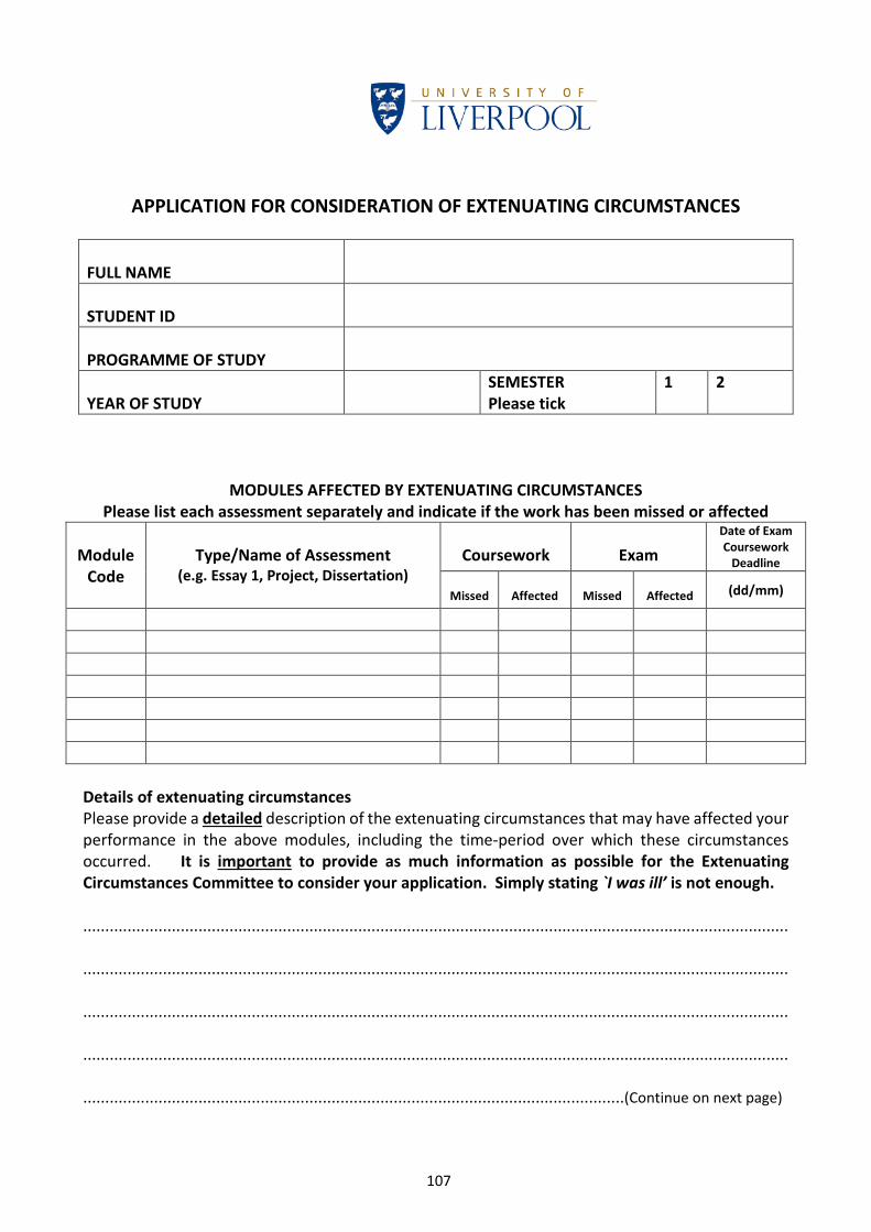
107
APPLICATION FOR CONSIDERATION OF EXTENUATING CIRCUMSTANCES FULL NAME
STUDENT ID
PROGRAMME OF STUDY
YEAR OF STUDY
SEMESTER Please tick
1 2
MODULES AFFECTED BY EXTENUATING CIRCUMSTANCES Please list each assessment separately and indicate if the work has been missed or affected
Module Code
Type/Name of Assessment
(e.g. Essay 1, Project, Dissertation)
Coursework
Exam
Date of Exam Coursework
Deadline
Missed
Affected
Missed
Affected (dd/mm)
Details of extenuating circumstances Please provide a detailed description of the extenuating circumstances that may have affected your performance in the above modules, including the time-period over which these circumstances occurred. It is important to provide as much information as possible for the Extenuating Circumstances Committee to consider your application. Simply stating `I was ill’ is not enough. ............................................................................................................................................................... ............................................................................................................................................................... ............................................................................................................................................................... ............................................................................................................................................................... ..........................................................................................................................(Continue on next page)

108
Details of extenuating circumstances/cont’d ............................................................................................................................................................... ............................................................................................................................................................... ............................................................................................................................................................... ............................................................................................................................................................... ............................................................................................................................................................... Supporting documentation Please list all the supporting documentation of your claim and all documentation should be stapled to this form. Medical claims should be supported by a GP’s medical note or Consultant’s report, other claims should be supported by appropriate documentation (for example, police reports, insurance reports). It is important to be specific with your evidence. For example, a general claim of illness in Semester 1 will not be accepted as evidence for under performance in Semester 2. Examples of the type of evidence that the Committee may expect to see are provided in the CoPA Appendix M Annexe 1: Policy on Extenuating Circumstances: Guidelines for Staff and Students at https://www.liverpool.ac.uk/aqsd/academic-codes-of-practice/code-of-practice-on assessment/ ............................................................................................................................................................... ............................................................................................................................................................... ............................................................................................................................................................... Student declaration I confirm that all the information contained in this statement is accurate and complete to the best of my knowledge. I consent to the information being used by the Extenuating Circumstances Committee, and understand that the information will be treated in the strictest confidence. Signature of student: ……………………................................................... Date: …………………................................ FOR USE BY THE CHAIR OF THE EXTENUATING CIRCUMSTANCES COMMITTEE ONLY I recommend that the following action be taken in respect of this claim: ............................................................................................................................................................. Signature of Chair: .................................................................... Date: …………………………………............

109
Module Titles and Codes
Module Code Module Title BIOM604 Techniques and Frontiers in Biomedical Sciences BIOM603 Transferable Skills
Biology of Cancer BICA601 Biology of Cancer Research Project 1 BICA602 Biology of Cancer Research Project 2 BICA603 Biology of Cancer Research Project 3
Biomedical Imaging and Biosensing BIBS601 Biomedical Imaging and Biosensing Research Project 1 BIBS602 Biomedical Imaging and Biosensing Research Project 2 BIBS603 Biomedical Imaging and Biosensing Research Project 3 Biostatistics (with Health Informatics) BIST601 Biostatistics (with Health Informatics) Research Project 1 BIST602 Biostatistics (with Health Informatics) Research Project 2 BIST603 Biostatistics (with Health Informatics) Research Project 3 Cancer Medicine CAMD601 Cancer Medicine Research Project 1 CAMD602 Cancer Medicine Research Project 2 CAMD603 Cancer Medicine Research Project 3
Cellular and Molecular Physiology CMPH601 Cellular and Molecular Physiology Research Project 1 CMPH602 Cellular and Molecular Physiology Research Project 2 CMPH603 Cellular and Molecular Physiology Research Project 3 Drug Safety DRSA601 Drug Safety Research Project 1 DRSA602 Drug Safety Research Project 2 DRSA603 Drug Safety Research Project 3
Medical Sciences MABI601 Medical Sciences Research Project 1 MABI602 Medical Sciences Research Project 2 MABI603 Medical Sciences Research Project 3
Molecular and Clinical Gastroenterology ANAT701 Gastroenterology Research Project 1 ANAT702 Gastroenterology Research Project 2 ANAT703 Gastroenterology Research Project 3

110
Molecular and Clinical Pharmacology PHAR801 Pharmacology Research Project 1 PHAR802 Pharmacology Research Project 2 PHAR803 Pharmacology Research Project 3 Nanomedicine SYBI601 Nanomedicine and Biostatistics Research Project 1 SYBI602 Nanomedicine and Biostatistics Research Project 2 SYBI603 Nanomedicine and Biostatistics Research Project 3
Neuroscience PHGY601 Neuroscience Research Project 1 PHGY602 Neuroscience Research Project 2 PHGY603 Neuroscience Research Project 3
Stem Cells, Tissues & Disease PHTH601 Stem Cells, Tissues and Disease Research Project 1 PHTH602 Stem Cells, Tissues and Disease Research Project 2 PHTH603 Stem Cells, Tissues and Disease Research Project 3
Women's, Children's & Perinatal Health TMTH601 Women, Children's & Perinatal Health Research Project 1 TMTH602 Women, Children's & Perinatal Health Research Project 2 TMTH603 Women, Children's & Perinatal Health Research Project 3