Embed Size (px)
Citation preview

MR Imaging in Atypical Septic Arthritis of the Knee
and Important Non-Infectious Mimics
Michael Holmes, MD Daria Motamedi, MD
Education/Pictorial Review - Abstract ER_016 [email protected]

Disclosures
• No Disclosures
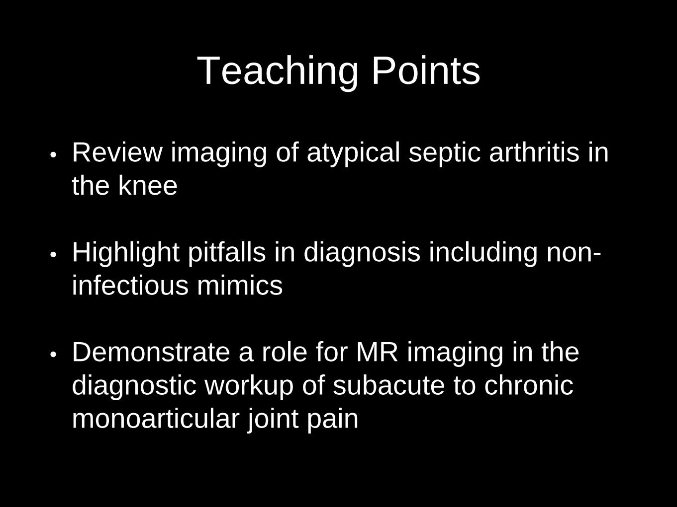
Teaching Points
• Review imaging of atypical septic arthritis in the knee
• Highlight pitfalls in diagnosis including non-
infectious mimics • Demonstrate a role for MR imaging in the
diagnostic workup of subacute to chronic monoarticular joint pain

Typical vs Atypical Septic Arthritis
Infectious process of the joint which commonly involves multiple compartments including the synovium, ligaments, tendons, cartilage, bone and surrounding soft tissues
Pyogenic (Typical) Infection
• Etiology • Bacteria
• “Characteristic Clinical Presentation” • rapid onset • fever • acute pain and swelling • monoarticular (50% of cases involve the knee)
• Diagnosis • Arthrocentesis - first-line step and definitive diagnosis • MRI - not routinely obtained
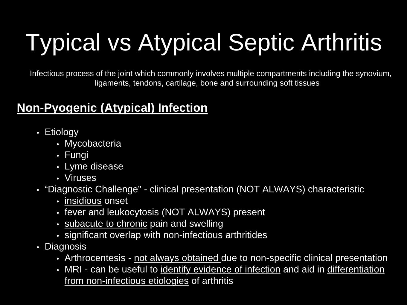
Typical vs Atypical Septic Arthritis
Infectious process of the joint which commonly involves multiple compartments including the synovium, ligaments, tendons, cartilage, bone and surrounding soft tissues
Non-Pyogenic (Atypical) Infection
• Etiology • Mycobacteria • Fungi • Lyme disease • Viruses
• “Diagnostic Challenge” - clinical presentation (NOT ALWAYS) characteristic • insidious onset • fever and leukocytosis (NOT ALWAYS) present • subacute to chronic pain and swelling • significant overlap with non-infectious arthritides
• Diagnosis • Arthrocentesis - not always obtained due to non-specific clinical presentation • MRI - can be useful to identify evidence of infection and aid in differentiation
from non-infectious etiologies of arthritis

MR Imaging - Septic Arthritis
MR imaging plays a greater role in the diagnostic workup of non-pyogenic (atypical) septic arthritis
MRI Findings
• joint effusion (particularly if complex) • intermediate T1/T2 signal
• capsular outpouchings • synovial thickening and intense enhancement • perisynovial edema • marrow edema • +/- osteomyelitis
• confluent low T1 signal, enhancement • joint erosions (+/- enhancement) • cartilage loss • soft tissue edema (+/- enhancement) • abscesses
Septic Arthritis/Osteomyelitis (Staphylococcus aureus)

Imaging/Clinical Mimics - Knee
Several non-infectious etiologies can mimic atypical septic arthritis both clinically and on imaging • Rheumatoid Arthritis • Seronegative Arthropathies
• Psoriatic Arthritis • Reactive Arthritis
• Deposition Disease • Gout • CPPD crystal deposition • Hydroxyapetite deposition
• Mass-Like Arthropathies • Pigmented Villonodular Synovitis • Synovial Osteochondromatosis • Lipoma Aborescens
• Tumors
The following slides will present several cases of non-pyogenic septic arthritis and non-
infectious mimics in the knee to not only demonstrate the similarities and potential pitfalls but also highlight specific clinical and radiologic signs that can aid in diagnosis.
Psoriatic Arthritis
Case 1
• 42 year old female on chronic immunosuppressive therapy presenting with 18 months of worsening pain and swelling of the left knee. Patient recently moved to the U.S. from the Philippines.
Case 1
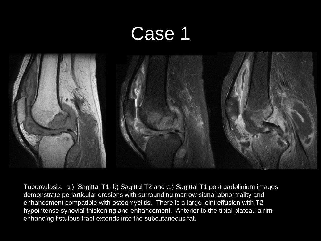
Tuberculosis. a.) Sagittal T1, b) Sagittal T2 and c.) Sagittal T1 post gadolinium images demonstrate periarticular erosions with surrounding marrow signal abnormality and enhancement compatible with osteomyelitis. There is a large joint effusion with T2 hypointense synovial thickening and enhancement. Anterior to the tibial plateau a rim-enhancing fistulous tract extends into the subcutaneous fat.

Tuberculous Arthritis Epidemiology/Risk Factors • close contact to TB • children and elderly • immunocompromised
Clinical Presentation • monoarticular arthritis
• 1-3% of cases of TB • insidious onset • chronic pain and swelling • systemic symptoms (fever, weight loss,
night sweats) • active pulmonary TB (~ 50%)
Imaging • Synovitis
• synovial thickening and enhancement
• low T2 signal (granulomatous inflammation)
• Erosions • peripheral AND central erosions
(different than inflammatory arthritis)
• Cartilage Destruction • late finding (early in pyogenic
infection) • Osteomyelitis
• low T1 signal, enhancement • Abscesses • Sinus Tracts

Case 2
• 42 year old male presenting with 12 month history of gradual worsening pain and asymmetric medial swelling of the right knee. Worked previously cutting roses on a farm.

Case 2
Sporotrichosis. a.) Coronal T2 demonstrates erosive defects in the medial meniscus and posterior medial oblique ligament resulting in a very large parameniscal cyst. b.) Sagittal T2 and c.) sagittal T1 images demonstrates a large popliteal cyst and joint effusion with synovitis. There is patchy reactive marrow edema in the periarticular bone but no confluent T1 hypointensity to suggest osteomyelitis. Extensive subcutaneous edema surrounds the knee.
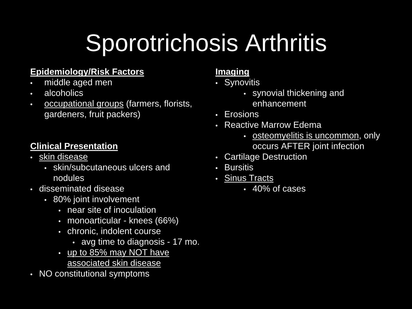
Sporotrichosis Arthritis Epidemiology/Risk Factors • middle aged men • alcoholics • occupational groups (farmers, florists,
gardeners, fruit packers)
Clinical Presentation • skin disease
• skin/subcutaneous ulcers and nodules
• disseminated disease • 80% joint involvement
• near site of inoculation • monoarticular - knees (66%) • chronic, indolent course
• avg time to diagnosis - 17 mo. • up to 85% may NOT have
associated skin disease • NO constitutional symptoms
Imaging • Synovitis
• synovial thickening and enhancement
• Erosions • Reactive Marrow Edema
• osteomyelitis is uncommon, only occurs AFTER joint infection
• Cartilage Destruction • Bursitis • Sinus Tracts
• 40% of cases
Case 3
• 35 year old female from California with history of gradual worsening anterior knee pain, swelling and intermittent fevers following hospitalization for “pneumonia” 4 weeks prior.

Case 3
Coccidiomycosis. a.) Axial CT chest shows a focal area of consolidation within the left upper lobe concerning for infection. b) Sagittal and c.) Axial T2 images demonstrate an infiltrative T2 hyperintense lesion within the inferior patella with extension through the anterior cortex into the prepatellar bursa and Hoffa’s fat pad. There is associated thickening and abnormal signal of the patellar tendon.
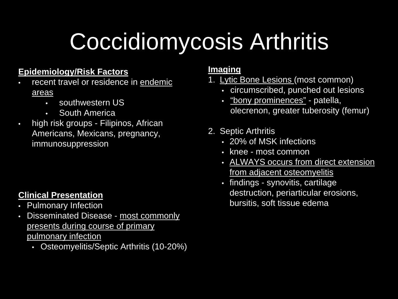
Coccidiomycosis Arthritis Epidemiology/Risk Factors • recent travel or residence in endemic
areas • southwestern US • South America
• high risk groups - Filipinos, African Americans, Mexicans, pregnancy, immunosuppression
Clinical Presentation • Pulmonary Infection • Disseminated Disease - most commonly
presents during course of primary pulmonary infection
• Osteomyelitis/Septic Arthritis (10-20%)
Imaging 1. Lytic Bone Lesions (most common)
• circumscribed, punched out lesions • “bony prominences” - patella,
olecrenon, greater tuberosity (femur) 2. Septic Arthritis
• 20% of MSK infections • knee - most common • ALWAYS occurs from direct extension
from adjacent osteomyelitis • findings - synovitis, cartilage
destruction, periarticular erosions, bursitis, soft tissue edema

Case 4
• 48 year old male with history of HIV (CD4 > 500) presenting with 3 weeks of pain and swelling of the right knee. Synovial fluid analysis was culture negative with elevated WBCs.

Case 4
HIV-Associated Arhritis. a.) Sagittal T1 b) Sagittal T2 and c.) Axial T2 images demonstrate a large joint effusion with associated subcutaneous, intramuscular and deep fascial edema predominantly within the medial soft tissue. No marrow signal abnormality or erosions.
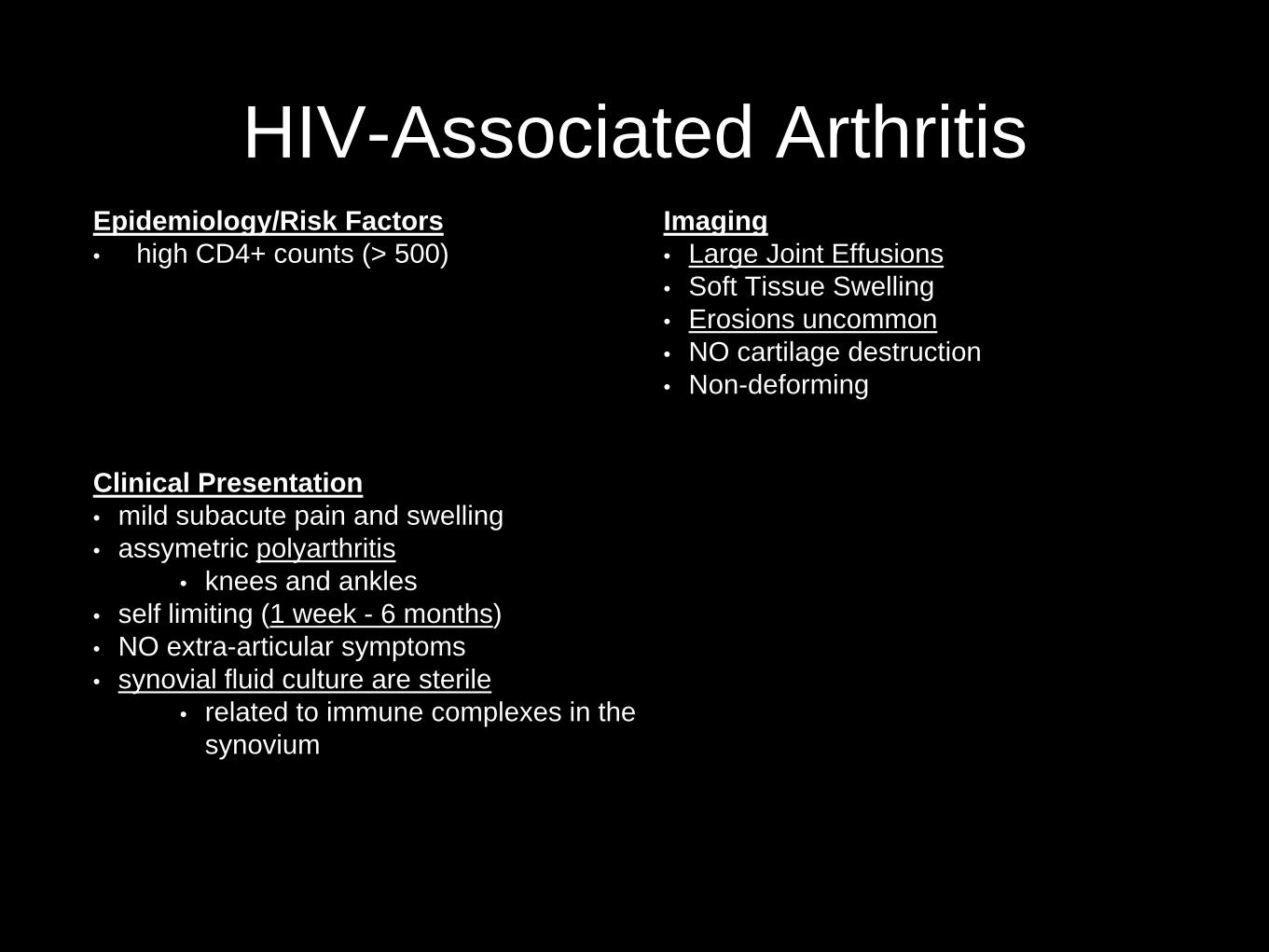
HIV-Associated Arthritis Epidemiology/Risk Factors • high CD4+ counts (> 500) Clinical Presentation • mild subacute pain and swelling • assymetric polyarthritis
• knees and ankles • self limiting (1 week - 6 months) • NO extra-articular symptoms • synovial fluid culture are sterile
• related to immune complexes in the synovium
Imaging • Large Joint Effusions • Soft Tissue Swelling • Erosions uncommon • NO cartilage destruction • Non-deforming

Case 5
• 56 year old female with history of increasing right knee pain with morning stiffness and warmth following a “fall” two year ago. She reports less severe pain in her left knee and bilateral wrists.
Case 5
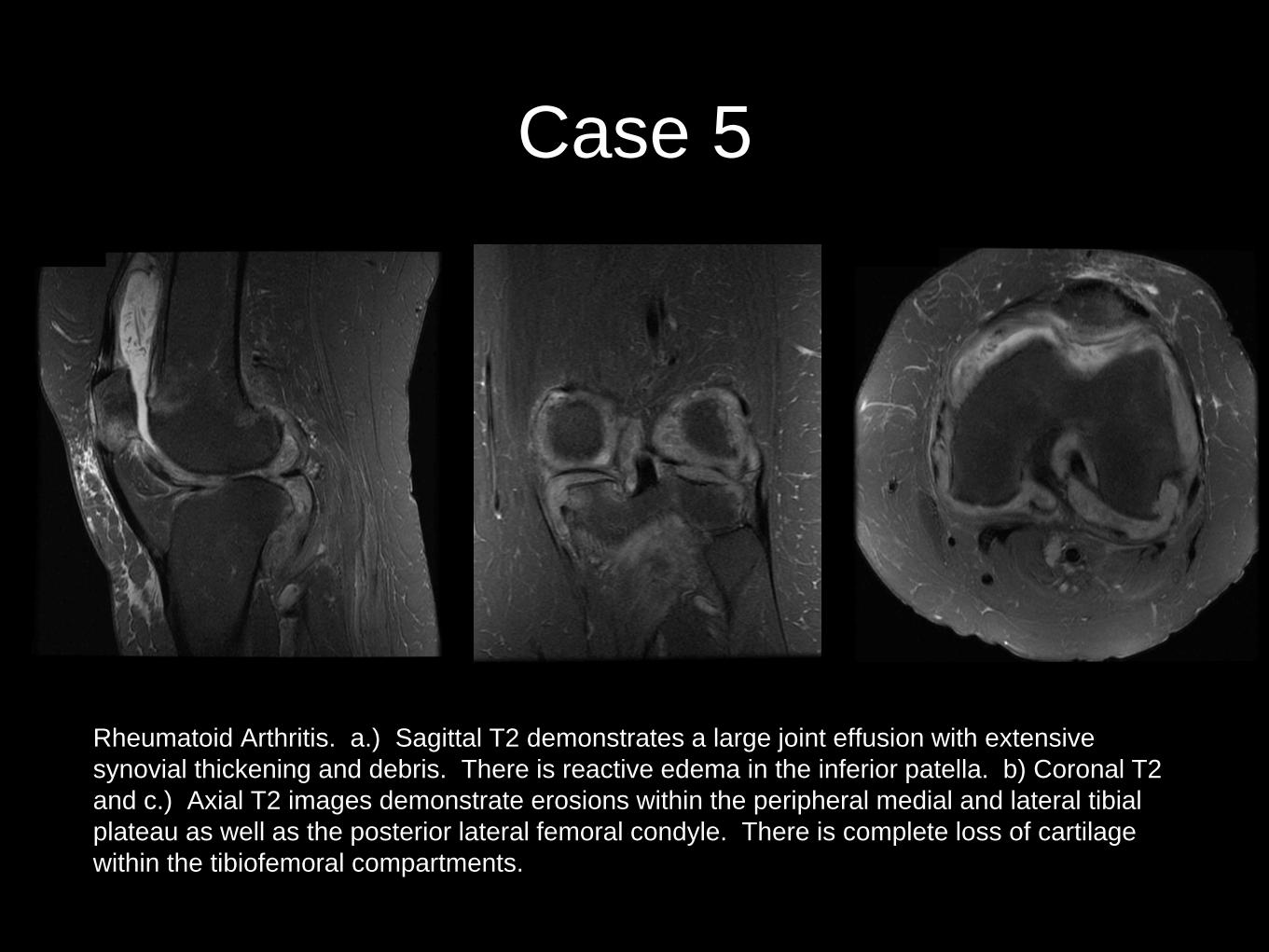
Rheumatoid Arthritis. a.) Sagittal T2 demonstrates a large joint effusion with extensive synovial thickening and debris. There is reactive edema in the inferior patella. b) Coronal T2 and c.) Axial T2 images demonstrate erosions within the peripheral medial and lateral tibial plateau as well as the posterior lateral femoral condyle. There is complete loss of cartilage within the tibiofemoral compartments.

Rheumatoid Arthritis Epidemiology/Risk Factors • prevalence - 1% (5% in Native
American populations) • 3:1 female to male • 4th-5th decade Clinical Presentation • symmetric polyarthritis • usually develops first in proximal hands
and wrists, but can affect feet and large joints
• knees (75% of cases) • insidious onset (weeks to months) • constitutional symptoms are common:
• fatigue • low grade fever
Imaging • large joint effusion
• loose bodies, debris and “rice bodies”
• capsular outpouchings (popliteal cysts) • synovitis
• low T2 signal thickening and enhancement
• peripheral erosions • low T1, high T2 signal with
peripheral rim of enhancement • marrow edema and possible
enhancement
Case 6
• 68 year old female with history of pain and swelling of the left knee with intermittent low grade fever x 3 weeks. Patient reports similar “attacks” in the past. History of back surgery 1 month ago.
Case 6
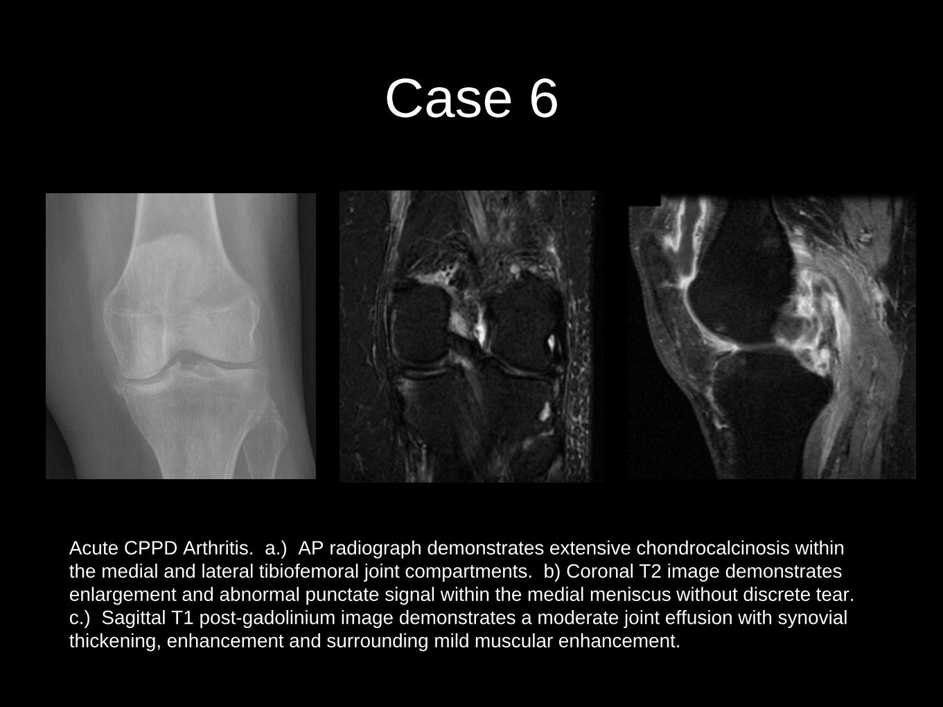
Acute CPPD Arthritis. a.) AP radiograph demonstrates extensive chondrocalcinosis within the medial and lateral tibiofemoral joint compartments. b) Coronal T2 image demonstrates enlargement and abnormal punctate signal within the medial meniscus without discrete tear. c.) Sagittal T1 post-gadolinium image demonstrates a moderate joint effusion with synovial thickening, enhancement and surrounding mild muscular enhancement.

Acute CPPD Arthritis Epidemiology/Risk Factors • prevalence of CPPD - 0.1%
• > 50% in adults > 80 years Clinical Presentation Acute CPPD Arthritis - “Pseudogout”
• episodic acute “attacks” • affects 10-20% of patients with
CPPD • pain, swelling, redness, warmth • fever, leukocytosis • often provoked by trauma, severe
medical illness or surgery (hypocalcemia)
Imaging • Plain Film and CT
• very useful in diagnosis of CPPD • calcification within articular
cartilage, menisci, syovium, tendons and ligaments
• joints not typically involved with OA (patellofemoral, radiocarpal, etc)
• MRI
• Chondrocalcinosis • linear or punctuate areas of
low signal representing susceptibility
• “Pseudogout” • synovitis • capsular outpouchings • perisynovial edema +/-
enhancement • marrow edema
Case 7
• 30 year old female presents with acute onset of pain and swelling of the left knee x4 weeks with multiple episodes of “locking”. She denies history of trauma.
Case 7
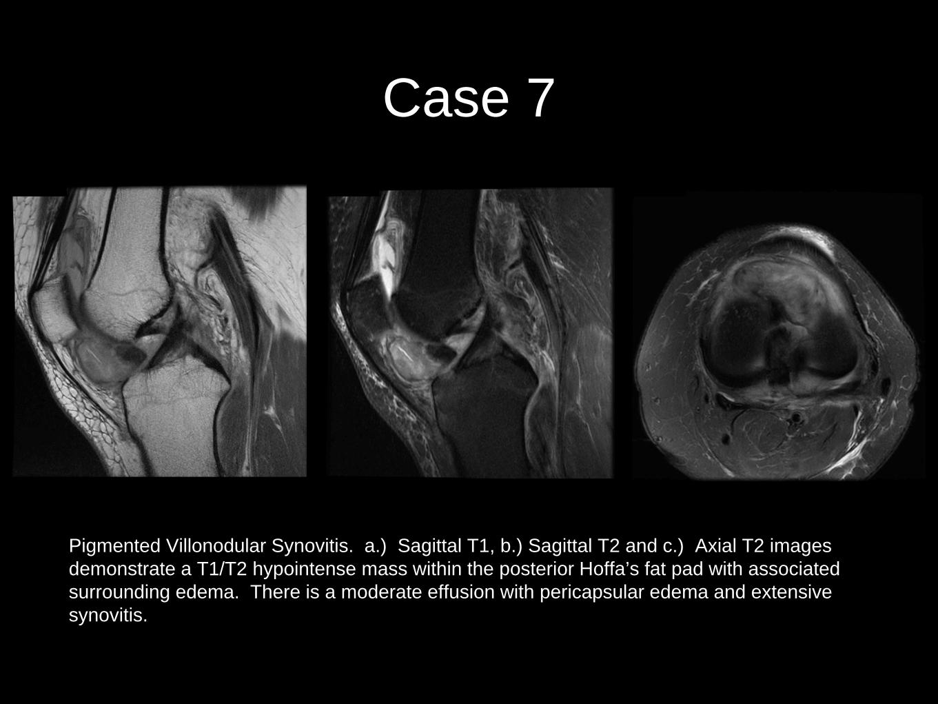
Pigmented Villonodular Synovitis. a.) Sagittal T1, b.) Sagittal T2 and c.) Axial T2 images demonstrate a T1/T2 hypointense mass within the posterior Hoffa’s fat pad with associated surrounding edema. There is a moderate effusion with pericapsular edema and extensive synovitis.

Pigmented Villonodular Synovitis Epidemiology/Risk Factors • prevalence: 11 per 1,000,000 • 3rd and 4th decade Clinical Presentation • monoarticular
• knees (most common) • acute onset • swelling out of proportion to pain • decreased ROM • subjective feeling of joint “locking” or
“catching"
Imaging • synovitis
• irregular, nodular or diffuse thickening of the synovium
• T1 - heterogeneous low signal areas with high signal representing hemorrhage or lipid laden macrophages
• T2* - “blooming” due to hemosiderin
• heterogeneous enhancement • perisynovial edema • joint effusions
• rarely fluid-fluid levels • +/- erosions (50%)
Case 8
• 11 year old male with development of painless swelling of the left knee approximately 3 months following a fall “down 20 stairs”. Diagnostic x-rays at the time of the fall were negative.

Case 8
Lipoma Arborescens. a.) Axial T1 image demonstrates high signal frond-like synovial masses within the suprapatellar joint space associated with a moderate joint effusion. b) Axial T2 and c.) Sagittal T2 fat saturation images demonstrate loss of signal within the frond-like synovial masses compatible with fat.
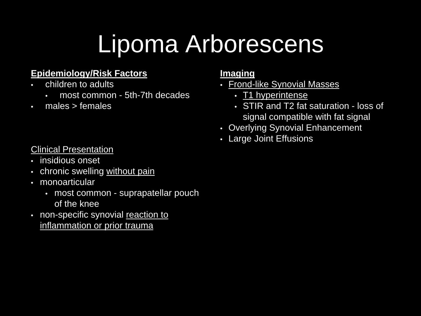
Lipoma Arborescens Epidemiology/Risk Factors • children to adults
• most common - 5th-7th decades • males > females Clinical Presentation • insidious onset • chronic swelling without pain • monoarticular
• most common - suprapatellar pouch of the knee
• non-specific synovial reaction to inflammation or prior trauma
Imaging • Frond-like Synovial Masses
• T1 hyperintense • STIR and T2 fat saturation - loss of
signal compatible with fat signal • Overlying Synovial Enhancement • Large Joint Effusions
Case 9
• 76 year old male with history of metastatic lung cancer, development of left knee pain and swelling x 4 weeks.

Case 9
Osseous Metastases. a.) Coronal T1 image demonstrates a hypointense marrow replacing lesion within the lateral femoral condyle. b) Sagittal T2 image demonstrates T2 fluid signal of the lesion with abnormal surrounding marrow signal. There is a large joint effusion. c.) Axial T1 post-gadolinium image demonstrates peripheral enhancement of the lesion and infiltration of the surrounding marrow.
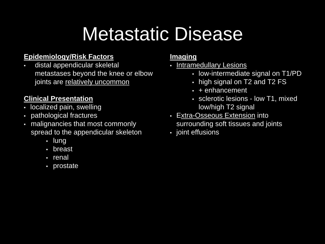
Metastatic Disease Epidemiology/Risk Factors • distal appendicular skeletal
metastases beyond the knee or elbow joints are relatively uncommon
Clinical Presentation • localized pain, swelling • pathological fractures • malignancies that most commonly
spread to the appendicular skeleton • lung • breast • renal • prostate
Imaging • Intramedullary Lesions
• low-intermediate signal on T1/PD • high signal on T2 and T2 FS • + enhancement • sclerotic lesions - low T1, mixed
low/high T2 signal • Extra-Osseous Extension into
surrounding soft tissues and joints • joint effusions

Conclusions 1. MRI is not routinely obtained in the setting of acute pyogenic septic
arthritis but can be very useful in patients presenting with non-characteristic subacute to chronic, monoarticular pain
2. Differential diagnosis for atypical (non-pyogenic) septic arthritis includes several non-infectious arthritides.
3. Although MR imaging of atypical septic arthritis can overlap significantly with non-infectious arthritides there are findings unique to certain etiologies:
• TB - T2 hypointense synovium, soft tissue abscesses, sinus tracts, central AND peripheral erosions
• Sporotrichosis - sinus tracts within the adjacent soft tissue • Coccidiomycosis - osseous lesions • HIV-Associated Arthritis - effusions WITHOUT cartilage
destruction

Conclusions continued… 4. Clinical presentation should help guide image
interpretation. Important clues are often present in the history, physical exam and lab values.
• TB - risk factors, pulmonary disease, constitutional symptoms • Sporotrichosis - occupational exposures, skin disease • Coccidiomycosis - endemic areas, high risk population
groups, pulmonary disease • HIV-Associated Arthritis - HIV with CD4+ > 500
5. Ultimately, tissue sampling (arthrocentesis, synovial biopsy) will
need to be performed to make the definitive diagnosis and guide treatment, but MRI can be useful to suggest the presence of infection (possibly when not expected) and help differentiate from other non-infectious mimics.
References • Graif M, Schweitzer ME, Deely D, Matteucci T. “ The Septic versus Nonseptic Inflamed Joint: MRI characteristics”. Skeletal
Radiology (1999) 28: 616-620
• Resnick D. “Osteomyelitis, Septic Arthritis and Soft Tissue Infection: Organisms”. Diagnosis of Bone and Joint Disorders, 4th Edition. (2002) 2510-2625
• Hollenberg GM, Weinberg EP, Meyers SP. "Lesions within Joints”. Differential Diagnosis in Musculoskeletal MRI (2015) 566-569
• Karchevsky M, Schweitzer ME, Morrison WB, Parellada JA. “MRI Findings of Septic Arthritis and Associated Osteomyelitis in Adults”. AJR (2004) 182: 119-122
• Sanghvi DA, Iyer VR, Deshmukh T, Hoskote S. “MRI Features of Tuberculosis of the Knee.” Skeletal Radiolol (2009) 38: 267-273
• Chowdhary G, Weinstein A, Klein R, Mascarenhas BR. “Sporotrichal Arthritis”. Annals of Rheumatic Diseases (1991) 50:112-114
• Zeppa A, Laorr A, Greenspan A, McGahan JP, Steinbach LS. “ Skeletal coccidiomycosis: Imaging Findings in 19 patients “. Skeletal Radiology (1996) 25: 337-343
• Tehranzadeh, J, Ter-Oganesyan RR, Steinbach LS. “Musculoskeletal Disorders Assocaited with HIV Infection and AIDS. Part II: Non-Infectious Musculoskeletal Conditions”. Skeletal Radiology (2004) 33: 311-320
• Sommer OJ, Klodosek A, Weiler V, Czembirek H, Boeck M, Stiskal M. “Rheumatoid Arthritis: A Practical Guide to State-of-the-Art Imaging, Image Interpretation, and Clinical Implications”. Radiographics (2005) 25: 381-398
• Steinbach LS, Resnick D. “Calcium Pyrophosphate Dihydrate Crystal Deposition Disease”. Current Problems in Diagnostic Radiology (2000) 29(6) 208-227
• Al-Nakshabandi NA, Ryan AG, Choudur H, Torreggiani W, Nicoloau S, Munk PL, Al-Ismail K. “Pigmented Villonodular Synovitis”. Clinical Radiology (2004) 59: 414-420