Embed Size (px)
Citation preview
Prospects & Overviews
Mouse models of colorectal cancer aspreclinical models
Rebecca E. McIntyre1)�, Simon J.A. Buczacki2), Mark J. Arends3) and David J. Adams1)
In this review, we discuss the application of mousemodels
to the identification and pre-clinical validation of novel
therapeutic targets in colorectal cancer, and to the search
for early disease biomarkers. Large-scale genomic, tran-
scriptomic and epigenomic profiling of colorectal carci-
nomas has led to the identification of many candidate
genes whose direct contribution to tumourigenesis is yet
to be defined; we discuss the utility of cross-species
comparative omics-based approaches to this problem.We
highlight recent progress in modelling late-stage disease
using mice, and discuss ways in which mouse models
could better recapitulate the complexity of human cancers
to tackle the problem of therapeutic resistance and
recurrence after surgical resection.
Keywords:.colorectal cancer; disease biomarker; mouse model;
ome and omics; target identification; target validation
Introduction
Colorectal cancer (CRC) accounts for 700,000 deaths and 1.4million newly diagnosed cases globally per annum, making it
the number one cause of non-smoking related cancerdeaths [1]. Most CRCs arise in the epithelium, a processdriven by genetic and/or epigenetic alterations that result inthe formation of premalignant lesions called adenomas (seeTable 1 for list of genes most frequently mutated in CRCs).The transition from normal epithelium to benign adenomais initially driven by alterations that hyperactivate the Wntpathway (>95% CRCs), and this predominantly occursthrough inactivation of the APC gene (>80% CRCs) [2–4].APC is a negative regulator of the Wnt pathway andhyperactivation of the Wnt pathway is critical to both theinitiation and maintenance of the vast majority of CRCs,although pathway activation is finely tuned [2, 5, 6]. Forexample, alteration of many genes that are known toregulate Wnt signalling, such as AXIN2 (Axis inhibitionprotein 2) and AMER1 (APC membrane recruitment 1) arefound in tumours that also harbour APC mutations [2, 7].The Wnt pathway is also critical for the maintenance of theintestinal epithelium, which undergoes complete renewalevery 4–5 days in humans [8]; therefore, it has not beenpossible to target this pathway in cancers without disruptingintestinal regeneration [9].
Activation of the Ras-MAPK (Kirsten rat sarcoma viraloncogene homolog and mitogen activated protein kinase)pathway, usually via point mutations that constitutivelyactivate KRAS or BRAF (v-raf murine sarcoma viral oncogenehomolog B), and inactivation of the TGFb (transforminggrowth factor b) pathway through inactivation of SMAD genefamily members or TGFb receptors, promote the developmentof advanced adenomas or invasive adenocarcinomas [3]. Overtime, a small proportion of advanced adenomas acquirefurther molecular abnormalities that transform them toinvasive and then metastatic carcinomas. Inactivation ofTP53 (Tumour protein p53) or IGF2R (Insulin-like growthfactor 2 receptor) occurs with greater frequency in establishedcarcinomas that invade submucosal layers than in adeno-mas [3, 10]. However the molecular alterations that supportmetastases are poorly understood; at the genomic level atleast, it would appear that there is high concordance betweenprimary CRCs and their matched metastatic lesions, whichsuggests that mechanisms other than gene mutation may beresponsible for progression tometastatic disease—for exampleepigenetic or post-translational modifications [10–12].
DOI 10.1002/bies.201500032
1) Experimental Cancer Genetics, Wellcome Trust Sanger Institute, Hinxton,Cambridge, UK
2) Li Ka Shing Centre, Cancer Research UK Cambridge Institute, Cambridge,UK
3) Edinburgh Cancer Research UK Centre, University of Edinburgh,Edinburgh, UK
*Corresponding author:Rebecca E. McIntyreE-mail: [email protected]
Abbreviations:CRC, colorectal cancer; EGF, epidermal growth factor; GEMM, geneticallyengineered mouse models; HNPCC, hereditary non-polyposis CRC; SNPs,single-nucleotide polymorphisms; Tfh, T follicular helper; TGFb, transforminggrowth factor b.
www.bioessays-journal.com 909Bioessays 37: 909–920,� 2015 The Authors. Bioessays published by WILEY Periodicals, Inc. This is anopen access article under the terms of the Creative Commons Attribution License, which permits use,distribution and reproduction in any medium, provided the original work is properly cited.
Methods,Models
&Techniques
Endoscopic or surgical resection is routinely used for patientswith early premalignant adenomas, as well as for thetreatment of most early stage carcinomas and selectedpatients with late stage or advanced metastatic disease, forwhom chemotherapy (or radiotherapy/chemoradiotherapy forrectal cancers) is also a key treatment modality [13]. Livermetastases of colorectal carcinomas occur in about 50% ofpatients, either at the time of diagnosis or at recurrence, andthis is a major cause of CRC-related deaths [13]. Long-termsurvival of CRC patients is correlated with disease stage atdiagnosis, and the 5-year survival rate for patients withmetastatic CRC is less than 10% [14].
Aside from our lack of understanding of the molecularevents that drive metastases, there are several other plausibleexplanations for our limited success in treating CRC. Firstly,there are currently few biomarkers that are predictive of earlydisease, the likelihood of favourable treatment response,recurrence or the development of metastatic disease [10].Secondly, therapeutic resistance, be it acquired or intrinsic, toseveral licensed colorectal cancer therapies is a majorproblem [15]. For example, overexpression of the epidermalgrowth factor (EGF) receptor is associated with increasedmetastatic potential and poor prognosis in CRC. In thesesubgroups of cancer patients, monoclonal antibodies to the
EGF receptor e.g. Cetuximab and Panitumumab, block EGFbinding and result in tumour regression [16, 17]. Resistance totherapy eventually develops through a variety of mechanismsincluding point mutations in the EGF receptor that inhibitCetuximab binding but not EGF, or activating mutations inKRAS, an intracellular signalling pathway that converges withthe EGF receptor pathway. Finally, the drug discovery pipelineattrition rate for oncology investigational compounds isextremely high. Only 5% of compounds that show promisein pre-clinical studies are eventually approved for clinical useas the majority fail in Phase I/II trials [18]. One of the reasonsfor this may be the extensive use of CRC cell lines andxenograft models derived from them in pre-clinical anti-cancer drug testing (reviewed by [18, 19]). Cell culture does notmodel the interaction of primary tumour cells with surround-ing cells (the tumour microenvironment or stroma), theprocess of organ colonisation by metastatic cancer cells (the‘seed and soil’ hypothesis), and the recruitment and activationof the adaptive and innate immune systems during tumouri-genesis, which are known to exert selective pressures oncancer cells [20–22]. Several cell culture systems have beendeveloped to make allowances for reciprocal cell communi-cation; examples include organoid cultures, the co-culture oftumour epithelial cells and stromal cells or three-dimensional
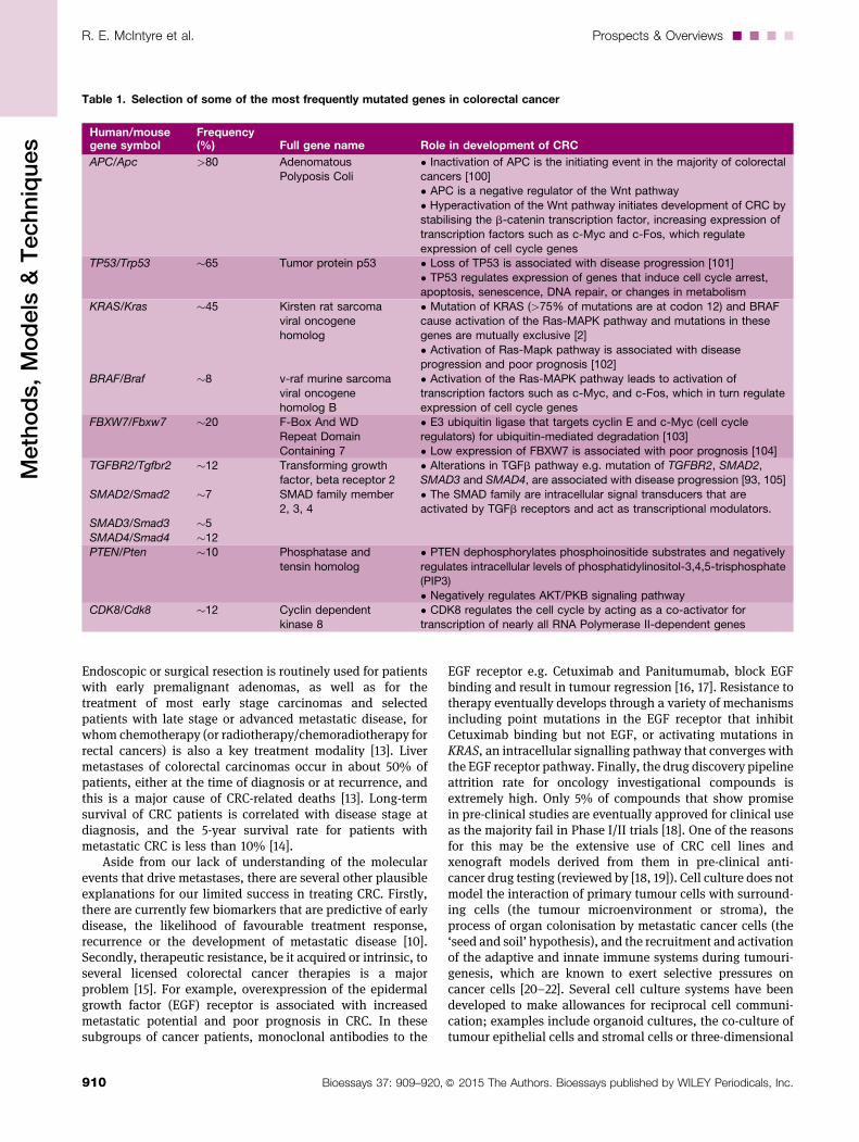
Table 1. Selection of some of the most frequently mutated genes in colorectal cancer
Human/mousegene symbol
Frequency(%) Full gene name Role in development of CRC
APC/Apc >80 AdenomatousPolyposis Coli
• Inactivation of APC is the initiating event in the majority of colorectalcancers [100]• APC is a negative regulator of the Wnt pathway
• Hyperactivation of the Wnt pathway initiates development of CRC bystabilising the b-catenin transcription factor, increasing expression of
transcription factors such as c-Myc and c-Fos, which regulateexpression of cell cycle genes
TP53/Trp53 �65 Tumor protein p53 • Loss of TP53 is associated with disease progression [101]
• TP53 regulates expression of genes that induce cell cycle arrest,apoptosis, senescence, DNA repair, or changes in metabolism
KRAS/Kras
BRAF/Braf
�45
�8
Kirsten rat sarcomaviral oncogenehomolog
v-raf murine sarcomaviral oncogenehomolog B
• Mutation of KRAS (>75% of mutations are at codon 12) and BRAFcause activation of the Ras-MAPK pathway and mutations in thesegenes are mutually exclusive [2]
• Activation of Ras-Mapk pathway is associated with diseaseprogression and poor prognosis [102]
• Activation of the Ras-MAPK pathway leads to activation oftranscription factors such as c-Myc, and c-Fos, which in turn regulateexpression of cell cycle genes
FBXW7/Fbxw7 �20 F-Box And WDRepeat DomainContaining 7
• E3 ubiquitin ligase that targets cyclin E and c-Myc (cell cycleregulators) for ubiquitin-mediated degradation [103]• Low expression of FBXW7 is associated with poor prognosis [104]
TGFBR2/Tgfbr2
SMAD2/Smad2
SMAD3/Smad3
SMAD4/Smad4
�12
�7
�5
�12
Transforming growthfactor, beta receptor 2
SMAD family member2, 3, 4
• Alterations in TGFb pathway e.g. mutation of TGFBR2, SMAD2,SMAD3 and SMAD4, are associated with disease progression [93, 105]
• The SMAD family are intracellular signal transducers that areactivated by TGFb receptors and act as transcriptional modulators.
PTEN/Pten �10 Phosphatase and
tensin homolog
• PTEN dephosphorylates phosphoinositide substrates and negatively
regulates intracellular levels of phosphatidylinositol-3,4,5-trisphosphate(PIP3)• Negatively regulates AKT/PKB signaling pathway
CDK8/Cdk8 �12 Cyclin dependentkinase 8
• CDK8 regulates the cell cycle by acting as a co-activator fortranscription of nearly all RNA Polymerase II-dependent genes
R. E. McIntyre et al. Prospects & Overviews....
910 Bioessays 37: 909–920,� 2015 The Authors. Bioessays published by WILEY Periodicals, Inc.
Methods,Models
&Techniques
cultures [23–25]. While these systems provide importantinsights into disease biology, they are limited in theirability to model the dynamic interplay between tumour andhost. In this review, we summarise the strengths andweaknesses of xenograft models derived from CRC celllines and primary patient derived material, and geneticallymodified mouse models of colorectal cancer. We discussseveral novel applications of these models in cancer genevalidation, biomarker discovery and target validation, anddiscuss their future roles in cross-species comparative ‘omics’approaches to understanding metastasis and therapeuticresistance.
Cell line xenografts for reductionisttarget validation
Xenografting involves the injection of cells or the surgicaltransplantation of primary tissue into immune-deficient micesuch as the nude mouse, which is athymic and does notproduce T-lymphocytes, or the severe combined immune-deficient (SCID) mouse, which has altered adaptive immunitybut normal innate immunity, to prevent rejection. It wasthought that the large number of commercial colorectal celllines available would represent the inter-tumour heteroge-neity of the human disease, but in reality, few cell lines lead toreliable primary tumour growth, and fewer still to naturallymetastatic CRC [26]. Success rates appear to be highest whenusing the colorectal carcinoma cell lines HCT-116 or HT-29, but
it is not yet clear why this is. Xenograft models are usefulfor rapid, reductionist, target validation studies, includingvalidation of the many candidate cancer genes identified byThe Cancer GenomeAtlas (TCGA), however the results must beinterpreted with caution (Table 2, Fig. 1 [27, 28]). For example,the environment of subcutaneous tumours is very differentfrom that of autochthonous colorectal carcinomas (i.e.tumours that originate in the location where they are found)and liver metastases; and the extent to which the speciesmismatch in tumour (human) and stromal (mouse) cellsinfluence tumour growth in vivo is uncertain. CRC stromais composed of macrophages that secrete matrix-degradingfactors such as metalloproteinases, myofibroblasts thatsecrete Wnt ligands, T-cells that secrete proinflammatorycytokines, and endothelial cells and bone-marrow derivedcells, all of which contribute greatly to the growth andprogression of this tumour type [20, 22, 29].
Orthotopic xenografts of CRC cell linesimprove tumour-stromal cell cross-talkand the reproducibility of livermetastases
Microenvironmental differences likely explain why themorphology of subcutaneous CRC cell line xenografts,especially the stromal elements, are often markedly differentfrom the human disease and may explain why they have a low
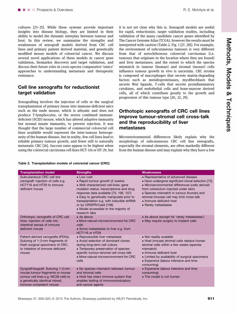
Table 2. Transplantation models of colorectal cancer (CRC)
Transplantation model Strengths Weaknesses
Subcutaneous CRC cell linexenograft: injection of cells e.g.
HCT116 and HT29 to immunedeficient mouse
• Low cost• Rapid tumour growth (2 weeks)
• Well characterized cell-lines: genemutation status, transcriptome and drug
response data available [73, 106, 107]• Easy to genetically manipulate prior totransplantation e.g. with inducible shRNA
or by CRISPR/Cas9 [108]• Model accessible to the majority of
research labs
• Representative of advanced disease• Have undergone significant clonal selection [73]
• Microenvironmental differences (cells derivedfrom colorectum injected under skin)
• Species mismatch in tumour (human) andstromal (mouse) cell may limit cross-talk• Immune deficient host
• Rarely metastasise
Orthotopic xenografts of CRC celllines: injection of cells into
intestinal serosa of immunedeficient mouse
• As above• More natural microenvironment for CRC
cells• Some metastasise to liver e.g. from
HCT116 or HT29
• As above (except for ‘rarely metastasise’)• May require surgery to implant cells
Patient-derived xenografts (PDXs):Suturing of 1–2mm fragments of
fresh surgical specimens of CRC,to intestine of immune deficientmouse
• Reproducible liver metastasis• Avoid selection of dominant clones
during long-term cell culture• Temporary preservation of species-specific tumour-stromal cell cross talk
• More natural microenvironment for CRCcells
• Not readily scalable• Host (mouse) stromal cells replace human
stromal cells within a few weeks (speciesmismatch)• Immune deficient host
• Limited by availability of surgical specimens• Expensive (labour intensive and time
consuming)Syngraft/Isograft: Suturing 1–2mmmouse tumour fragments or mouse
tumour cell lines e.g. MC38 cells toa genetically identical inbred,
immune competent mouse
• No species mismatch between tumourand stromal cells
• Host has intact immune system thatenables testing of immunomodulatory
anti-cancer agents
• Expensive (labour intensive and timeconsuming)
• The model is not human
....Prospects & Overviews R. E. McIntyre et al.
911Bioessays 37: 909–920,� 2015 The Authors. Bioessays published by WILEY Periodicals, Inc.
Methods,Models
&Techniques
metastatic potential [30]. In an attempt to improve the utilityof xenograft models, several investigators have systematicallytested different variables and found that CRC cell line, site ofinjection, mouse age and the genetic background of themouseall markedly affect the frequency of lymph-node or livermetastases [27, 31, 32]. Orthotopic xenografts involve theinjection of differentiated cells to a more natural micro-environment i.e. the serosa of the intestine for CRC cell lines,and appears to result inmore reproducible liver metastasis [31,33]. In addition, orthotopic and subcutaneous xenograftmodels show differing sensitivities to chemotherapeuticagents [30, 31], suggesting that the microenvironment isimportant for both disease progression and therapeutic
response. Recent refinements to the caecalpouch xenograft method include the non-surgical trans-anal injection of cells into thedistal rectum or the microinjection of celllines to the caecum and engineering cells toexpress b-human chorionic gonadotropinto support growth [31, 34].
Patient-derived orthotopicxenografts temporarilypreserve tumour-stromal cellcross-talk and improvesreproducibility of metastasis
Patient derived xenografts (or ‘PDXs’) avoidthe natural selection of dominant clonesand epigenetic and genetic alterations thatoccur during long-term cell culture as wellas temporarily preserving the original,species-specific, tumour-stromal cell inter-actions. The suturing of small fragments of
fresh surgical specimens of CRC liver metastases to thecaecum of immune-deficient mice has been reproducible ingenerating models of metastatic CRC [35, 36]. It was thoughtthat PDXs could be used to test therapies and development ofresistance ahead of patient treatment to help personalizecancer therapy, however the approach requires subculture orserial xenografting which currently takes too long [37]. Anumber of studies have also now reported that hostcells replace the human stroma and vasculature of patientderived xenografts, and in the case of CRCs, this appears tooccur relatively rapidly (3 weeks vs. 9 weeks for mesothe-lioma; [38, 39]).
‘Syngraft’ or ‘Isograft’ models retainspecies-specific cross-talk betweentumour, stroma and immune cells
The two major limitations of all xenograft mouse models ofhuman CRC cell lines are the species mismatch betweentumour and stroma, which likely affects cell communication,and the use of immune-deficient hosts. The role of the immunesystem appears to be particularly important for the develop-ment of CRC; T cell immune infiltrate is an importantpredictive criterion for patient survival [40] and mice withdeletion of Smad4 in T-cells excessively secrete proinflamma-tory cytokines and develop gastrointestinal tumours [41]. Theuse of immune-deficient hosts allows development of tumoursin the absence of an immune infiltrate, which is critical forCRC tumour progression, and also precludes the testing ofimmunomodulatory anti-cancer agents. Grafting tumourfragments or the cell lines derived from them, for exampleMC38 cells (derived from amouse colon adenocarcinoma), to agenetically identical inbred, immune competent mouse(‘syngraft’ or ‘isograft’) is a way of overcoming both of theseproblems [19].
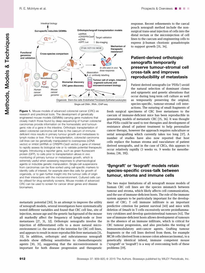
Figure 1. Mouse models of advanced colorectal cancer (CRC) asresearch and preclinical tools. The development of geneticallyengineered mouse models (GEMMs) carrying gene mutations thatclosely match those found by deep-sequencing of human colorectalcarcinomas provide information on the homeostatic and tumouri-genic role of a gene in the intestine. Orthotopic transplantation ofselect colorectal carcinoma cell lines to the caecum of immune-deficient mice results in primary tumour growth and metastases tolymph nodes or liver. Prior to transplantation, colorectal carcinomacell lines can be genetically manipulated to overexpress (cDNAvector) or inhibit (shRNA or CRISPR-Cas9 vector) a gene of interestto rapidly assess its biological role or to validate potential therapeutictargets. Introducing a reporter gene, such as green fluorescentprotein (GFP), to cells prior to transplantation allows in vivomonitoring of primary tumour or metastases growth, which isextremely useful when assessing responses to pharmacologicalagents or inducible genetic manipulation. Single-cell suspensionsfrom carcinomas can be flow-sorted using cell-specific markers toidentify cells of interest, for example stem-like cells for growth oforganoids, or to gain further insight into the tumour cells of originand their interactions with the microenvironment. Cultured cells canbe utilised for drug sensitivity screens. Mouse models of advancedCRC can be used to screen for cancer driver genes and diseasebiomarkers.
R. E. McIntyre et al. Prospects & Overviews....
912 Bioessays 37: 909–920,� 2015 The Authors. Bioessays published by WILEY Periodicals, Inc.
Methods,Models
&Techniques
Genetically modified mouse models ofcolorectal cancer retain species-specificcross-talk between tumour, stroma andimmune cells
Genetically engineered mouse models (GEMM) offer severaladvantages over cancer cell lines as research tools in cancerresearch. Despite their evolutionary distance, the genecontent of the mouse and human genomes has been largelyconserved through evolution [42], and most cancer pathwaysare operative in both species [43]. The genomes of inbredlaboratorymouse strains are well-characterised andmice offera ‘clean’ system in which to test the biological effects ofgenetic alterations or environmental effects; they areessentially homozygous at every locus and are housed in acontrolled environment.
Sequencing of DNA from several tumour types that havedeveloped in mouse or man has recently revealed that humantumours are more heterogeneous than their GEMM-derivedcounterparts [44, 45]. The difference in complexity of mouseand human tumours is likely due to a combination of factors.Humans have a more varied diet and a different microbiomecompared to the laboratorymouse, and so their intestinal cellsare exposed to more exogenous genotoxins [46]. In addition,these gene-environment interactions occur over a longerperiod of time inman; the development of tumours in GEMM islimited by the short lifespan of the mouse or by local researchethics; for example the UK Home Office enforces a tumour sizelimit of around 1 cm2. Furthermore, there is a lack of inter-tumour heterogeneity of GEMM tumours because laboratorymice are inbred and therefore lack the genetic heterogeneitypresent in the outbred human population. Moreover, GEMMtumours are often initiated and maintained by the samegenetic mutation of a strong cancer driver gene, which maylimit the number of pathways to tumourigenesis. While thedifference in complexity of tumours is advantageous whenapplying a cross-species oncogenomics approach to cancergene validation [45], the lack of complexity of mouse tumoursposes a problem when trying to faithfully recapitulate thehuman disease for pre-clinical studies. Some tumour typescan be treated in mouse models of cancer, but it is thoughtthat the heterogeneity of human tumours results in a greaterpotential for resistance to therapy to develop, or for recurrenceafter surgical resection. Nonetheless, GEMMs of CRC thatdevelop autochthonous intestinal tumours can better modelthe dynamic interplay between intestinal tumour cells, stromaand the immune system, as well as the response to therapy,when compared with xenograft models.
Genetically modified mouse models oflate stage CRC—25 years in the making
Table 3 summarises some of the genetically modified mousemodels (GEMM) of CRC that have been developed over the last25 years. The first key mouse model of CRC was the multipleintestinal neoplasia (Min) mouse, which arose from a randomethylnitrosourea (ENU) mutagenesis screen [47, 48]. The
ApcMin/+ mouse was subsequently recognised as a paralog forhuman familial adenomatous polyposis (FAP) syndrome andprovided confirmation of the causal genemutation in sporadicCRCs with 5q21 deletions [49]; later studies showed thatsomatic inactivation of APC is observed in >80% of sporadicCRCs [2].
Hereditary non-polyposis CRC (HNPCC) or Lynch Syn-drome, is the most common inherited, autosomal dominantCRC syndrome and accounts for around 3% of all CRCs.HNPCC is most frequently caused by mutations in MLH1,PMS1, PMS2, MSH2 or MSH6, (MutL Homolog 1, Post-MeioticSegregation Increased 1 and 2 and MutS Homolog 2 and 6,respectively) that encode the enzymatic machinery of DNAmismatch repair. As with sporadic cancers, intestinal cancersfrom HNPCC patients have a high frequency of mutations inAPC, as well as KRAS, TP53 and TGFBR2 [50–52]. Further,approximately 15% of sporadic CRCs display microsatelliteinstability (MSI) secondary to impaired DNA mismatch repair,frequently because of hypermethylation of the MLH1 pro-moter [53]. One of the earliest models that had the potentialto recapitulate HNPCC was a homozygous Msh2�/� deficientmouse [54] (Table 3). Unfortunately, these mice werepredisposed to lymphomas, which limited their use as amodel of HNPCC. One way to restrict genetic modifications totissues of interest is to use a Cre-recombinase driven by atissue-specific promoter. Cre-recombinases catalyse recombi-nation between two loxP sites that are situated in the intronssurrounding a critical exon(s) and result in excision of theDNA between the sites. Using a conditional strategy thatresults in expression of Cre recombinase from the Villinpromoter (intestinal specific gene) a Villin-Cre/Msh2LoxP
mouse was generated that resulted in intestinal adenomasand adenocarcinomas in the absence of lymphoma [55](Table 3).
The short lifespan of theApcMin/+mouse limits the utility ofthe model and it was thought that mice with fewer tumourswould live longer, and that if tumours were restricted to thecolon, they might progress to carcinoma and metastasise [56,57]. A number of different Apc mutant mice with germline orinducible cell-type specific conditional alleles of Apc weredeveloped (seeTable3 forexamplesandforanextensive review,see [58]). Despite evidence of malignant transformation andcolonic invasion, aged Apc mutant mice did not developmetastases, suggesting that to better model the later stages ofdisease in the mouse, additional genetic mutations would berequired (Table 3). Apcmutant mice have been crossed to micewith gene mutations found in later stage disease, includingTP53, KRAS, TGFBR2, FBXW7 and SMAD2-4, in an attempt togenerate more useful pre-clinical models. At best, doublemutants displayedmore adenocarcinomas, but themicedidnotmodel the metastatic disease (see Table 3).
The many different Apc mutant mice that have beencreated have led to greater understanding of the biology ofApc, for example, to ascertain which protein domains andfunctions of Apc are responsible for intestinal tumourinitiation and maintenance; it is widely accepted that Apc’srole in activation of the Wnt pathway is the major contributorto this process [59]. Apc mutant mice have also been used toprobe the effect of environmental factors and drugs onintestinal tumourigenesis and stem cell specific Apc deletion
....Prospects & Overviews R. E. McIntyre et al.
913Bioessays 37: 909–920,� 2015 The Authors. Bioessays published by WILEY Periodicals, Inc.
Methods,Models
&Techniques
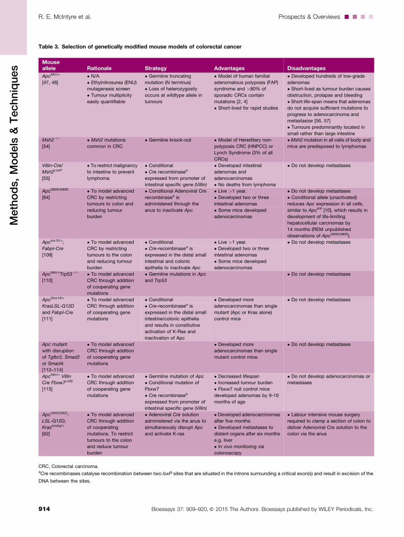
Table 3. Selection of genetically modified mouse models of colorectal cancer
Mouseallele Rationale Strategy Advantages Disadvantages
ApcMin/+
[47, 48]
• N/A
• Ethylnitrosurea (ENU)
mutagenesis screen
• Tumour multiplicity
easily quantifiable
• Germline truncating
mutation (N terminus)
• Loss of heterozygosity
occurs at wildtype allele in
tumours
• Model of human familial
adenomatous polyposis (FAP)
syndrome and >80% of
sporadic CRCs contain
mutations [2, 4]
• Short-lived for rapid studies
• Developed hundreds of low-grade
adenomas
• Short-lived as tumour burden causes
obstruction, prolapse and bleeding
• Short life-span means that adenomas
do not acquire sufficient mutations to
progress to adenocarcinoma and
metastasise [56, 57]
• Tumours predominantly located in
small rather than large intestine
Msh2�/�
[54]
• Msh2 mutations
common in CRC
• Germline knock-out • Model of Hereditary non-
polyposis CRC (HNPCC) or
Lynch Syndrome (3% of all
CRCs)
• Msh2mutation in all cells of body and
mice are predisposed to lymphomas
Villin-Cre/
Msh2LoxP
[55]
• To restrict malignancy
to intestine to prevent
lymphoma
• Conditional
• Cre recombinasea
expressed from promoter of
intestinal specific gene (Villin)
• Developed intestinal
adenomas and
adenocarcinomas
• No deaths from lymphoma
• Do not develop metastases
Apc580S/580S
[64]
• To model advanced
CRC by restricting
tumours to colon and
reducing tumour
burden
• Conditional Adenoviral Cre
recombinasea is
administered through the
anus to inactivate Apc
• Live >1 year. • Do not develop metastases
• Developed two or three
intestinal adenomas
• Conditional allele (unactivated)
reduces Apc expression in all cells,
similar to Apcfl/fl [10], which results in
development of life-limiting
hepatocellular carcinomas by
14 months (REM unpublished
observations of Apc580S/580S)
• Some mice developed
adenocarcinomas
Apclox15/+;
Fabpl-Cre
[109]
• To model advanced
CRC by restricting
tumours to the colon
and reducing tumour
burden
• Conditional • Live >1 year. • Do not develop metastases
• Cre-recombinasea is
expressed in the distal small
intestinal and colonic
epithelia to inactivate Apc
• Developed two or three
intestinal adenomas
• Some mice developed
adenocarcinomas
ApcMin/+Trp53�/�
[110]
• To model advanced
CRC through addition
of cooperating gene
mutations
• Germline mutations in Apc
and Trp53
• Do not develop metastases
Apc2lox14/+
KrasLSL-G12D
and Fabpl-Cre
[111]
• To model advanced
CRC through addition
of cooperating gene
mutations
• Conditional • Developed more
adenocarcinomas than single
mutant (Apc or Kras alone)
control mice
• Do not develop metastases
• Cre-recombinasea is
expressed in the distal small
intestine/colonic epithelia
and results in constitutive
activation of K-Ras and
inactivation of Apc
Apc mutant
with disruption
of Tgfbr2, Smad2
or Smad4.
[112–114]
• To model advanced
CRC through addition
of cooperating gene
mutations
• Developed more
adenocarcinomas than single
mutant control mice.
• Do not develop metastases
ApcMin/+ Villin
Cre Fbxw7(DG)
[115]
• To model advanced
CRC through addition
of cooperating gene
mutations
• Germline mutation of Apc
• Conditional mutation of
Fbxw7
• Cre recombinasea
expressed from promoter of
intestinal specific gene (Villin)
• Decreased lifespan
• Increased tumour burden
• Fbxw7 null control mice
developed adenomas by 9-10
months of age
• Do not develop adenocarcinomas or
metastases
ApcCKO/CKO-
LSL-G12D;
Krastm4tyj/+
[62]
• To model advanced
CRC through addition
of cooperating
mutations. To restrict
tumours to the colon
and reduce tumour
burden
• Adenoviral Cre solution
administered via the anus to
simultaneously disrupt Apc
and activate K-ras
• Developed adenocarcinomas
after five months
• Developed metastases to
distant organs after six months
e.g. liver
• In vivo monitoring via
colonoscopy
• Labour intensive mouse surgery
required to clamp a section of colon to
deliver Adenoviral Cre solution to the
colon via the anus
CRC, Colorectal carcinoma.aCre recombinases catalyse recombination between two loxP sites that are situated in the introns surrounding a critical exon(s) and result in excision of the
DNA between the sites.
R. E. McIntyre et al. Prospects & Overviews....
914 Bioessays 37: 909–920,� 2015 The Authors. Bioessays published by WILEY Periodicals, Inc.
Methods,Models
&Techniques
models lent support to the so-called ‘bottom-up’ model ofadenoma formation [60, 61].
One of the most promising models for both theelucidation of the molecular events that support theformation of distant metastases of colorectal cancer andfor preclinical investigations is a mouse that has simulta-neous, inducible inactivation of Apc and activation of Krasin the adult colon (Table 3). Six months after surgery 20%of ApcCKO/CKO-LSL-Kras mice (G12D; Krastm4tyj/+ allele) micehad developed metastases to distant organs such as theliver [62]. It is important to note that expression of wild-type Apc may be disrupted by introduction of conditionalalleles, which may elevate Wnt signalling levels in tissuesother than the intestine, and which appears to drive theformation of life-limiting hepatocellular tumourigenesisbefore the onset of intestinal tumourigenesis. For exampleApcfl/fl and Apc580S/580S mice show reduced expression ofApc protein throughout their tissues (in the absence of Cre-mediated recombination of alleles), and develop hepatocel-lular carcinomas between 9 and 15months (our unpublishedobservations ofApc580S/580Smice; [63, 64]). Thesefindingsmaypreclude the use of some mouse models in studies that aim tounderstand the events that drive the metastasis of CRC to theliver.
Mouse models of inflammatory boweldisease and colorectal cancer
Inflammatory bowel diseases (IBD) such as ulcerative colitispredispose patients to developing colorectal cancer as a resultof chronic inflammation. It is becoming apparent that thesetumours may develop through alternative TP53 dependentroutes compared with classical APC-driven tumours [65]. Inan attempt to model this disease process mice have beengenerated that are predisposed to intestinal specific inflam-mation, including Il-10�/�, Il-2null and Muc2�/� deficientstrains [66, 67]. Colitis can also be experimentally inducedby the administration of dextran sulfate sodium (DSS) tothe drinking water of mice. In addition to mimicking IBD,mice treated this way also produce intestinal tumours [68].Azoxymethane (AOM) is another commonly used chemicalthat predisposes to cancer experimentally. AOM is thought toenable base mispairings, and when administered alone or incombination with DSS, AOM reliably produces tumouri-genesis in wild type mice [69].
Genetic screens in mice for validation ofcancer ‘driver’ genes
TCGA has identified genes that are frequently mutated inmany different types of malignancy (frequently referred to as‘pan-cancer’ genes) suggesting that they are likely toaccelerate the process [70]. While these genes are obvioustargets for therapy, many are considered intractabledrug targets, including MYC and RAS. Within different cancersub-types there are cancer drivers that could be more
amenable to therapy, however it is difficult to make thedistinction between the ‘driver’ and ‘passenger’ genes(mutations that drive disease progression and mutations thatdo not) in patient specimens because of genetic, epigeneticand environmental heterogeneity. In contrast, mouse models,with their homogeneous genetic background and livingenvironment, can be an efficient tool for validating candidatecancer genes (Figs. 1 and 2). Forward genetic screens in miceusing insertional mutagenesis have aided the identification orvalidation of many drivers of cancer, the relevance of which isassessed by comparing the candidates identified throughscreening with mutations found in the human cancersubtype [45].
One of the first insertional mutagenesis studies to identifynovel genetic drivers of intestinal tumourigenesis involvedrestricting transposition to epithelial cells of the gastro-intestinal tract using a Villin-Cre recombinase system [71].Analysis of the common insertion sites for transposon inneoplasms, adenomas and adenocarcinomas revealed that80% of insertions fell in known CRC genes includingAPC, FBXW7 (F-box and WD repeat domain containing 7),PTEN (phosphatase and tensin homolog), and SMAD4,thus validating the approach. The remainder, includingRSPO2 (R-spondin 2), had not been previously implicated inthe disease. R Spondin is a ligand for LGR4-6 receptors (LGR,leucine-rich repeat containing G protein-coupled receptor),which stimulate the canonical Wnt signalling pathway.We now know that RSPO2 fusion genes occur in a subset ofcolorectal carcinomas and are mutually exclusive with APCmutations [7].
To identify mutations that cooperate with inactivation ofApc and hyperactivation of the Wnt pathway, two independ-ent teams of researchers mobilised the Sleeping Beautytransposon in ApcMin/+ mice or conditional Apcfl/+ mice [6,72]. The most frequently mutated gene (�85%) in both studieswas Apc, consistent with the requirement for loss ofheterozygosity of Apc for intestinal tumour induction.Interestingly, all three mutagenesis screens identified ahigh number of insertions in the WW domain-containingadaptor with coiled-coil gene (Wac), which is also mutated inhuman cancers [6, 71–73]. WAC has recently been shown tobe required for activation of the cell-cycle checkpoint inresponse to DNA damage suggesting that inactivation maycause genomic instability [74]. Both insertional mutagenesisstudies of Apc mutant mice identified common insertions inthe voltage-gated potassium channel subunit Kcnq1 [6, 72].Mutations in voltage-gated ion channel subunits have beenidentified in a variety of human cancers, including colon, andmounting evidence suggests that tumour cells have adepolarised membrane voltage relative to surrounding tissue,which could be exploited for therapy [75]. In support of thisfinding, ApcMin/+Kcnq1�/� double mutant mice developedmore intestinal adenomas than ApcMin/+ control mice, andprogression to aggressive adenocarcinomas was observed.The authors went on to show that reduced KCNQ1 expressioncorrelated with poor survival in patients; however, the exactmechanism of tumour suppression remains unknown [76].These examples prove the utility of transposon-mediatedinsertional mutagenesis for rapid validation of colorectalcancer genes and for colorectal cancer gene discovery.
....Prospects & Overviews R. E. McIntyre et al.
915Bioessays 37: 909–920,� 2015 The Authors. Bioessays published by WILEY Periodicals, Inc.
Methods,Models
&Techniques
ApcMin/+ mice for early diseasebiomarker discovery
Most screening strategies aimed at the early detection ofadenomas and CRC involve endoscopy, and thus have limitedapplicability. Early detection of CRC is critical for diseasemanagement, and so the identification of reliable, earlydisease biomarkers would improve the outcome for manypatients. Mouse models that are predisposed to colorectaltumour formation can be an efficient tool for early diseasebiomarker discovery (Fig. 2). Since early detection of CRC iscritical for disease management, investigators have used micewith germline Apc mutations, which model the initiation ofintestinal tumourigenesis, to screen for early disease bio-markers. Clusterin was a ‘proof-of-principle’ for the cross-species approach to biomarker discovery [77]. Upregulation ofclusterin was reported in adenomas from ApcMin/+ mice andhuman tumours, and was recently shown to be highly-expressed in intestinal stem cells [77, 78]. Secreted clusterin iscytoprotective and its prosurvival function forms the basis ofthe current Phase I/II clinical trials against prostate, lung, andbreast cancers [79].
Cross-species proteomics identifies earlydisease biomarkers
Recent improvements in proteomic profiling methods, basedprimarily on mass spectrometry, have allowed the detectionand identification of peptides in the blood and biologicalfluids of cancer patients (Fig. 2). The plasma of tumour-bearing Apc580S/D mice was screened by mass spectrometry,and cathepsin B and D were found to be elevated comparedwith control mice [80]. Cathepsins are involved in thedegradation of the basement membrane and extracellularmatrix, a key step in tumour invasion and metastasis.Cathepsin B and cathepsin D are thought to associate withtumour grade of CRC and survival suggesting they may beuseful biomarkers of the disease [80, 81]. Similarly, proximalfluid proteome profiling of colon tumours from aged Apc15lox/+
Fabpl-Cre mice resulted in the identification of 192 proteinsthat were more highly excreted by tumours relative to control,including Chitinase 3-like 1 (Chi3l1) [82]. CHI3L1 is thought tobe involved in processes such as inflammation and tissueremodeling, and was shown to be significantly elevated in thesera of patients with adenomas and advanced adenomascompared with control individuals [83].
Cross-species epigenomics identifies epigeneticsignatures of early disease
Epigenomic profiling of adenomas fromApcMin/+mice has alsobeen useful in the identification of patient relevant, earlydisease biomarkers (Fig. 2). Immunoprecipitation of methy-lated DNA followed by sequencing (known as ‘MeDIP-seq’)identified regions of the genome were differentially methy-lated [84]. These regions were mainly enriched for targets ofthe Polycomb Repressive Complex, the subunits of which,together with those for several DNA methyltransferase
complexes, were up-regulated in adenomas. This genome-wide pattern was shown to be partly conserved in humancolon carcinomas, suggesting that an epigenetic signature isestablished early and is retained during progression fromadenoma to carcinoma.
Target identification and validationrequires cancer cell lines, xenograftmodels and GEMM
Not all cancer genes are essential for the sustained growth ofestablished tumours. In addition, tumours may have intrinsictherapeutic resistance, which is believed to arise throughnatural selection of a latent subclone within a tumour, oracquired therapeutic resistance, which arises through acquis-ition of a genetic mutation that allows the cancer cell tosurvive in the presence of the therapeutic agent [15]. It istherefore important to understand both the contribution of a
GENOMICS
EPIGENOMICSPROTEOMICS
TRANSCRIPTOMICS
point mutations
CNVs
small indels
differential expression
alternative splicing
RNA editing
gene fusions
DNA/mRNA methylation
gene silencing histone modifications
TF binding
phosphoproteome
biomarkers
differential expression
ubiquitome
gene activation
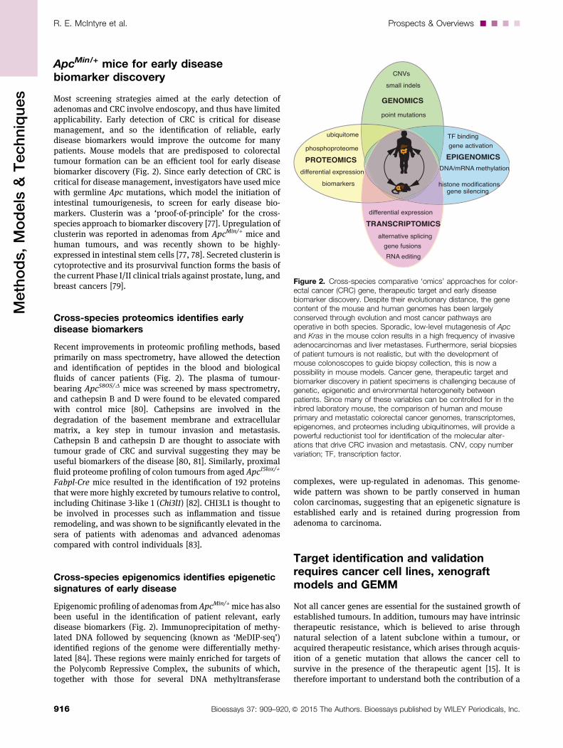
Figure 2. Cross-species comparative ‘omics’ approaches for color-ectal cancer (CRC) gene, therapeutic target and early diseasebiomarker discovery. Despite their evolutionary distance, the genecontent of the mouse and human genomes has been largelyconserved through evolution and most cancer pathways areoperative in both species. Sporadic, low-level mutagenesis of Apcand Kras in the mouse colon results in a high frequency of invasiveadenocarcinomas and liver metastases. Furthermore, serial biopsiesof patient tumours is not realistic, but with the development ofmouse colonoscopes to guide biopsy collection, this is now apossibility in mouse models. Cancer gene, therapeutic target andbiomarker discovery in patient specimens is challenging because ofgenetic, epigenetic and environmental heterogeneity betweenpatients. Since many of these variables can be controlled for in theinbred laboratory mouse, the comparison of human and mouseprimary and metastatic colorectal cancer genomes, transcriptomes,epigenomes, and proteomes including ubiquitinomes, will provide apowerful reductionist tool for identification of the molecular alter-ations that drive CRC invasion and metastasis. CNV, copy numbervariation; TF, transcription factor.
R. E. McIntyre et al. Prospects & Overviews....
916 Bioessays 37: 909–920,� 2015 The Authors. Bioessays published by WILEY Periodicals, Inc.
Methods,Models
&Techniques
disease target to tumourigenesis and the mechanisms bywhich cancers evade targeted therapies. Here, we highlightseveral recent approaches to the identification and validationof novel therapeutic targets using mouse models of CRC.
Although genetic ablation or overexpression of potentialtherapeutic targets in CRC cell lines before xenotransplantationdoes not necessarily mimic pharmacological intervention, nordoes it mimic the biology of autochthonous tumours, it mayprovideashortcut to targetvalidation(Fig. 1). Thisapproachhasrecently been useful for the validation of potential therapeutictargets within the Wnt and TGFb pathways.
Targeting the Wnt pathway—reality or drugdiscovery pipe dream?
Since theWnt–b-catenin signalling pathway is hyperactivatedin 93% of colorectal tumours and high Wnt activity definescolon cancer stem cells, the Wnt pathway is the mostattractive therapeutic target for colorectal cancer [2, 29, 85].Despite this, therapeutic agents that specifically target theWnt pathway have only recently entered clinical trials, andnone has yet been approved. The Wnt inhibitor XAV939inhibits the poly(ADP)-ribosylating (PARP) enzymes tank-yrase 1 and tankyrase 2, which interacts with AXIN (acts withAPC to destruct b-catenin and damper Wnt pathway activity)to promote its ubiquitylation and degradation [86]. Thedevelopment of more selective and potent second-generationtankyrase inhibitors is in progress, however their anti-tumourpotential may not be fully realised owing to their intestinaltoxicity in pre-clinical mouse models [9]. Rosenbluh andcolleagues took a more targeted approach to inhibition of theWnt pathway: they screened 85 cancer cell lines and foundthat b-catenin active cancers are dependent on a signallingpathway involving the transcriptional regulator YAP1 (Yes-associated protein 1) [28]. YAP1 and the transcription factorTBX5 were shown to form a complex with b-catenin, andphosphorylation of YAP1 by the tyrosine kinase YES1 lead tolocalisation of the complex to the promoters of anti-apoptoticgenes such as BIRC5. To determinewhether YAP1was requiredfor HCT116 xenograft growth they transfected cells with adoxycycline-inducible shRNA to YAP1. Three weeks afterinjection, tumour growth was restricted to 80% of controlanimals, suggesting that the YAP1 pathway might be anattractive therapeutic target [28]. Further studies will berequired to confirm whether this effect occurs in vivo, wheremany endogenous ligands that are secreted by stromal cellsare known to modify Wnt pathway activity [22].
Targeting MYC—drugging the undruggable
The transcription factor Myc proto-oncogene protein (Myc) isthought to be a target of the b-catenin–Tcf4 transcriptioncomplex in CRC cells in vitro, in normal crypts in vivo and inintestinal epithelial cells acutely transformed on in vivodeletion of the Apc gene [59, 87, 88]. Array expression analysisrevealed that Myc is required for the majority of b-catenin–Tcf4 transcription complex gene activation following Apc lossin mice. While Myc is required for the formation of intestinal
crypts, it is dispensable for homeostasis of the adult intestinalepithelium [89]. Using a combination of conditional mousemodels, it was shown that simultaneous inactivation of Mycand Apc completely ablated tumour development in micedespite high levels of nuclear b-catenin in the intestinalepithelium [90]. Multiple cancer-associated single-nucleotidepolymorphisms (SNPs) have been mapped to conservedsequences within a 500-kilobase region upstream of theMYC oncogene on human chromosome 8q24, and micelacking a Myc enhancer (Myc-335) that includes human SNPrs6983267 are resistant to intestinal tumours induced by theApcMin mutation [91]. While these studies have establishedMyc as a critical mediator of the early stages of intestinalneoplasia following Apc loss, it has not yet been possible topharmacologically regulate Myc. Instead, a selective small-molecule BET bromodomain inhibitor, JQ1, inhibits proteinsthat are regulatory factors for Myc [92]. Treatment with JQ1resulted in genome-wide downregulation of Myc-dependenttarget genes, reduced proliferation and had anti-cancerproperties in a mouse model of multiple myeloma, a Myc-dependent hematologic malignancy, suggesting that JQ1 mayperform similarly in mouse models of colorectal cancer.
Pharmacological inhibition of TGFBR1 inhibitsmetastasis formation
A large proportion of CRCs display mutational inactivationof the TGFb pathway. However, paradoxically, they arecharacterised by elevated TGFb production. In other cancertypes, such as breast or prostate, which retain a functionalTGFb signalling pathway, TGFb induces a variety ofprometastatic programmes that range from induction ofepithelial-to-mesenchymal-like transition (or ‘EMT’) to expres-sion of genes that allow colonisation of foreign organs [93].During 10 years of follow-up after surgical resection ofcolorectal tumours, only patients with medium or high TGFbexpression in the primary tumour suffered cancer recurrence,whereas all patients bearing TGFb -low stage I, II and IIItumours remained disease-free [94]. When Nude mice wereinoculated with KM12L4a cells that overexpressed TGFb,nearly all developed lung and/or liver metastasis, whereasonly one third of control mice developed metastases. It isthought that the metastatic KM12L4aTGFB1 cells were conferreda survival advantage by suppressing apoptotic stimuliencountered during organ colonization. Moreover, inhibitionof TGFBR1 has been shown to inhibit metastasis formation inmultiple xenograft models [94, 95]. Since the mechanism isthought to involve cross-talk between tumour and stromalcells, it will be important to show that this regulation occurs inautochthonously occurring liver metastases of CRC, such asthose that arise in the conditional ApcCKO/CKO/KrasG12D coloncancer model [62].
Isograft models for validating the role of theimmune system in CRC
In a recent study, Bindea and colleagues examined the spatio-temporal dynamics of 28 different immune cell types (the
....Prospects & Overviews R. E. McIntyre et al.
917Bioessays 37: 909–920,� 2015 The Authors. Bioessays published by WILEY Periodicals, Inc.
Methods,Models
&Techniques
‘immunome’) that were infiltrating colorectal tumours wereanalysed by whole-genome expression, PCR arrays andmulticolored immunohistochemistry [96]. The analysisrevealed that patients with elevations in genes related toMHC-II, B cell co-stimulation, T cells and Tfh cells experienceincreased survival. One chemokine in particular, CXCL13, andone cytokine, IL-21, positively correlated with disease-freesurvival. Bindea and colleagues showed growth accelerationof MC38 isografts in Cxcr5�/� mice (CXCR5 is the receptor forCXCL13) compared to control, and rejection of tumour cellswhen recombinant CXCL13 was injected into the colonicsubmucosa of wild-type mice before transplantation [96].These data confirmed the prognostic value of CXCL13 inassessing CRC tumour burden, and prove the utility of mousemodels in the validation of disease biomarkers.
Conclusions and outlook
In combination with studies of CRC cell lines and organoidmodels, mouse models of late-stage CRC will help us to build acomprehensive picture of the molecular alterations that drivetumour formation and disease progression, through cross-species analysis of the genomes, epigenomes, transcriptomes,proteomes and even immunomes of cancers of mice and men.Mouse models will be critical for reductionist approaches thataim to validate the cancer drivers frommutations identified bythe TCGA.
Human CRC is the result of interactions between genes,including those involved in host immunity, and the environ-ment, including diet and the microbiome [97, 98]. The mouseaffords the ability to investigate the effects of each of thesefactors independently of each other, which is simply notpossible in man. However, recent studies have shown that‘pound for pound’, the average mouse tumour is less complexthan the average human tumour. These sequencing studies,together with the knowledge that lifestyle factors stronglyinfluence the chances of getting cancer, suggest that therelative simplicity of GEMM tumours is due to the lack of gene-environment interactions in the controlled environment of thelaboratory mouse. There is a reasonable body of evidence tosuggest that high red meat intake and alcohol consumptionare risk factors for CRC, but we are probably only justbeginning to understand how our environment affects thecourse of disease [99]. Modelling the full heterogeneity ofhuman tumours will be required to help tackle the problems oftherapeutic resistance and recurrence after surgical resectionin CRC; thismight be better achieved by generating GEMM thatcombine a genetic predisposition to CRC with a diet rich infoods associated with increased risk of CRC.
AcknowledgementREM, SJAB, MJA and DJA were funded by Cancer Research UK.
References
1. Torre LA, Bray F, Siegel RL, Ferlay J, et al. 2015. Global cancerstatistics, 2012. CA Cancer J Clin 65: 87–108.
2. TCGA. 2012. Comprehensive molecular characterization of humancolon and rectal cancer. Nature 487: 330–7.
3. Fearon ER. 2011. Molecular genetics of colorectal cancer. Annu RevPathol 6: 479–507.
4. Sjoblom T, Jones S, Wood LD, Parsons DW, et al. 2006. Theconsensus coding sequences of human breast and colorectal cancers.Science 314: 268–74.
5. Albuquerque C, Breukel C, van der Luijt R, Fidalgo P, et al. 2002. The“just-right” signaling model: APC somatic mutations are selected basedon a specific level of activation of the beta-catenin signaling cascade.Hum Mol Genet 11: 1549–60.
6. March HN, Rust AG, Wright NA, ten Hoeve J, et al. 2011. Insertionalmutagenesis identifies multiple networks of cooperating genes drivingintestinal tumorigenesis. Nat Genet 43: 1202–9.
7. Seshagiri S, Stawiski EW, Durinck S, Modrusan Z, et al. 2012.Recurrent R-spondin fusions in colon cancer. Nature 488: 660–4.
8. Van der Flier LG, Clevers H. 2009. Stem cells, self-renewal,and differentiation in the intestinal epithelium. Annu Rev Physiol 71:241–60.
9. Lau T, Chan E, Callow M, Waaler J, et al. 2013. A novel tankyrasesmall-molecule inhibitor suppresses APC mutation-driven colorectaltumor growth. Cancer Res 73: 3132–44.
10. Walther A, Johnstone E, Swanton C, Midgley R, et al. 2009. Geneticprognostic and predictive markers in colorectal cancer. Nat Rev Cancer9: 489–99.
11. Brannon AR, Vakiani E, Sylvester BE, Scott SN, et al. 2014.Comparative sequencing analysis reveals high genomic concordancebetween matched primary and metastatic colorectal cancer lesions.Genome Biol 15: 454.
12. Jones S, Chen W-D, Parmigiani G, Diehl F, et al. 2008. Comparativelesion sequencing provides insights into tumor evolution.Proc Natl AcadSci USA 105: 4283–8.
13. Van CutsemE, Nordlinger B, Cervantes A. 2010. Advanced colorectalcancer: ESMO clinical practice guidelines for treatment. Ann Oncol Off JEur Soc Med Oncol ESMO 21: v93–v97.
14. Jemal A, Bray F, Center MM, Ferlay J, et al. 2011. Global cancerstatistics. CA Cancer J Clin 61: 69–90.
15. Garraway LA, Janne PA. 2012. Circumventing cancer drug resistancein the era of personalized medicine. Cancer Discov 2: 214–26.
16. Montagut C, Dalmases A, Bellosillo B, Crespo M, et al. 2012.Identification of a mutation in the extracellular domain of the epidermalgrowth factor receptor conferring cetuximab resistance in colorectalcancer. Nat Med 18: 221–3.
17. Valtorta E, Misale S, Sartore-Bianchi A, Nagtegaal ID, et al. 2013.KRAS gene amplification in colorectal cancer and impact on responseto. Int J Cancer J Int Cancer 133: 1259–65.
18. Sharpless NE, Depinho RA. 2006. The mighty mouse: geneticallyengineered mouse models in cancer drug development. Nat Rev DrugDiscov 5: 741–54.
19. Voskoglou-Nomikos T, Pater JL, Seymour L. 2003. Clinical predictivevalue of the in vitro cell line, human xenograft, and mouse allograftpreclinical cancer models. Clin Cancer Res Off J Am Assoc Cancer Res9: 4227–39.
20. Ernst M, Ramsay RG. 2012. Colorectal cancer mouse models:integrating inflammation and the stroma. J Gastroenterol Hepatol 27:39–50.
21. Fidler IJ. 2003. The pathogenesis of cancer metastasis: the “seed andsoil” hypothesis revisited. Nat Rev Cancer 3: 453–8.
22. Medema JP, Vermeulen L. 2011. Microenvironmental regulation ofstem cells in intestinal homeostasis and cancer. Nature 474: 318–26.
23. Huh D, Hamilton GA, Ingber DE. 2011. From 3D cell culture to organs-on-chips. Trends Cell Biol 21: 745–54.
24. Jorgensen C, Sherman A, Chen GI, Pasculescu A, et al. 2009. Cell-specific information processing in segregating populations of Ephreceptor ephrin-expressing cells. Science 326: 1502–9.
25. Sato T, Vries RG, Snippert HJ, van de Wetering M, et al. 2009. SingleLgr5 stem cells build crypt-villus structures in vitro without amesenchymal niche. Nature 459: 262–5.
26. Bedard PL, Hansen AR, Ratain MJ, Siu LL. 2013. Tumourheterogeneity in the clinic. Nature 501: 355–64.
27. Flatmark K, Maelandsmo GM, Martinsen M, Rasmussen H, et al.1990. 2004. Twelve colorectal cancer cell lines exhibit highly variablegrowth and metastatic capacities in an orthotopic model in nude mice.Eur J Cancer Oxf Engl 40: 1593–8.
28. Rosenbluh J, Nijhawan D, Cox AG, Li X, et al. 2012. b-Catenin-drivencancers require a YAP1 transcriptional complex for survival andtumorigenesis. Cell 151: 1457–73.
R. E. McIntyre et al. Prospects & Overviews....
918 Bioessays 37: 909–920,� 2015 The Authors. Bioessays published by WILEY Periodicals, Inc.
Methods,Models
&Techniques
29. Vermeulen L, De Sousa Melo EF, van der Heijden M, Cameron K,et al. 2010. Wnt activity defines colon cancer stem cells and is regulatedby the microenvironment. Nat Cell Biol 12: 468–76.
30. PocardM, Tsukui H, Salmon RJ, Dutrillaux B, et al. 1996. Efficiency oforthotopic xenograft models for human colon cancers. Vivo AthensGreece 10: 463–9.
31. Hackl C, Man S, Francia G, Milsom C, et al. 2013. Metronomic oraltopotecan prolongs survival and reduces liver metastasis in improvedpreclinical orthotopic and adjuvant therapy colon cancer models. Gut62: 259–71.
32. Lavilla-Alonso S, Abo-Ramadan U, Halavaara J, Escutenaire S, et al.2011. Optimized mousemodel for the imaging of tumormetastasis uponexperimental therapy. PLoS ONE 6: e26810.
33. Jin H, Yang Z, Wang J, Zhang S, et al. 2011. A superficial colon tumormodel involving subcutaneous colon translocation and orthotopictransplantation of green fluorescent protein-expressing human colontumor. Tumour Biol J Int Soc Oncodevelopmental Biol Med 32: 391–7.
34. Cespedes MV, Espina C, Garcia-Cabezas MA, Trias M, et al. 2007.Orthotopic microinjection of human colon cancer cells in nude miceinduces tumor foci in all clinically relevant metastatic sites. Am J Pathol170: 1077–85.
35. Fu XY, Besterman JM, Monosov A, Hoffman RM. 1991. Models ofhumanmetastatic colon cancer in nude mice orthotopically constructedby using histologically intact patient specimens. Proc Natl Acad Sci USA88: 9345–9.
36. Rashidi B, Gamagami R, Sasson A, Sun FX, et al. 2000. An orthotopicmouse model of remetastasis of human colon cancer liver metastasis.Clin Cancer Res Off J Am Assoc Cancer Res 6: 2556–61.
37. Garralda E, Paz K, Lopez-Casas PP, Jones S, et al. 2014. Integratednext-generation sequencing and avatar mouse models for personalizedcancer treatment. Clin Cancer Res Off J Am Assoc Cancer Res 20:2476–84.
38. Hylander BL, Punt N, Tang H, Hillman J, et al. 2013. Origin of thevasculature supporting growth of primary patient tumor xenografts.J Transl Med 11: 110.
39. Julien S, Merino-Trigo A, Lacroix L, Pocard M, et al. 2012.Characterization of a large panel of patient-derived tumor xenograftsrepresenting the clinical heterogeneity of human colorectal cancer. ClinCancer Res Off J Am Assoc Cancer Res 18: 5314–28.
40. Galon J, Costes A, Sanchez-Cabo F, Kirilovsky A, et al. 2006. Type,density, and location of immune cells within human colorectal tumorspredict clinical outcome. Science 313: 1960–4.
41. Kim B-G, Li C, Qiao W, Mamura M, et al. 2006. Smad4 signalling in Tcells is required for suppression of gastrointestinal cancer. Nature 441:1015–9.
42. Waterston RH, Lindblad-Toh K, Birney E, Rogers J, et al. 2002. Initialsequencing and comparative analysis of the mouse genome. Nature420: 520–62.
43. Frese KK, Tuveson DA. 2007. Maximizing mouse cancer models. NatRev Cancer 7: 645–58.
44. Francis JC, Melchor L, Campbell J, Kendrick H, et al. 2015. Whole-exome DNA sequence analysis of Brca2- and Trp53-deficient mousemammary gland tumours. J Pathol 236: 186–200.
45. McIntyre RE, van der Weyden L, Adams DJ. 2012. Cancer genediscovery in the mouse. Curr Opin Genet Dev 22: 14–20.
46. Nguyen TLA, Vieira-Silva S, Liston A, Raes J. 2015. How informativeis the mouse for human gut microbiota research? Dis Model Mech 8:1–16.
47. Moser AR, Pitot HC, Dove WF. 1990. A dominant mutation thatpredisposes to multiple intestinal neoplasia in the mouse. Science 247:322–4.
48. Su LK, Kinzler KW, Vogelstein B, Preisinger AC, et al. 1992. Multipleintestinal neoplasia caused by a mutation in the murine homolog of theAPC gene. Science 256: 668–70.
49. Nishisho I, Nakamura Y, Miyoshi Y, Miki Y, et al. 1991. Mutations ofchromosome 5q21 genes in FAP and colorectal cancer patients.Science 253: 665–9.
50. Huang J, Papadopoulos N, McKinley AJ, Farrington SM, et al. 1996.APC mutations in colorectal tumors with mismatch repair deficiency.Proc Natl Acad Sci USA 93: 9049–54.
51. Prolla TA, Baker SM, Harris AC, Tsao JL, et al. 1998. Tumoursusceptibility and spontaneousmutation inmice deficient inMlh1, Pms1and Pms2 DNA mismatch repair. Nat Genet 18: 276–9.
52. Wheeler JM, Bodmer WF, Mortensen NJ. 2000. DNA mismatch repairgenes and colorectal cancer. Gut 47: 148–53.
53. Kane MF, Loda M, Gaida GM, Lipman J, et al. 1997. Methylation ofthe hMLH1 promoter correlates with lack of expression of hMLH1 in
sporadic colon tumors and mismatch repair-defective human tumor celllines. Cancer Res 57: 808–11.
54. Reitmair AH, Schmits R, Ewel A, Bapat B, et al. 1995. MSH2 deficientmice are viable and susceptible to lymphoid tumours. Nat Genet 11:64–70.
55. Kucherlapati MH, Lee K, Nguyen AA, Clark AB, et al. 2010. An Msh2conditional knockout mouse for studying intestinal cancer and testinganticancer agents. Gastroenterology 138: 993–1002.
56. Chen X, Halberg RB, Burch RP, Dove WF. 2008. Intestinal adenoma-genesis involves core molecular signatures of the epithelial-mesen-chymal transition. J Mol Histol 39: 283–94.
57. Halberg RB, Waggoner J, Rasmussen K, White A, et al. 2009. Long-lived Min mice develop advanced intestinal cancers through agenetically conservative pathway. Cancer Res 69: 5768–75.
58. Zeineldin M, Neufeld KL. 2013. More than two decades of Apcmodeling in rodents. Biochim Biophys Acta 1836: 80–89.
59. SansomOJ, Reed KR, Hayes AJ, Ireland H, et al. 2004. Loss of Apc invivo immediately perturbs Wnt signaling, differentiation, and migration.Genes Dev 18: 1385–90.
60. Barker N, Ridgway RA, van Es JH, van de Wetering M, et al. 2009.Crypt stem cells as the cells-of-origin of intestinal cancer. Nature 457:608–11.
61. Roper J, Hung KE. 2012. Priceless GEMMs: genetically engineeredmouse models for colorectal cancer drug development. TrendsPharmacol Sci 33: 449–55.
62. Hung KE, Maricevich MA, Richard LG, Chen WY, et al. 2010.Development of a mouse model for sporadic and metastatic colontumors and its use in assessing drug treatment. Proc Natl Acad Sci USA107: 1565–70.
63. Buchert M, Athineos D, Abud HE, Burke ZD, et al. 2010. Geneticdissection of differential signaling threshold requirements for the Wnt/beta-catenin pathway in vivo. PLoS Genet 6: e1000816.
64. Shibata H, Toyama K, Shioya H, Ito M, et al. 1997. Rapid colorectaladenoma formation initiated by conditional targeting of the Apc gene.Science 278: 120–3.
65. Leedham SJ, Graham TA, Oukrif D, McDonald SAC, et al. 2009.Clonality, founder mutations, and field cancerization in humanulcerative colitis-associated neoplasia. Gastroenterology 132:542–50.
66. Van der Sluis M, De Koning BAE, De Bruijn ACJM, Velcich A, et al.2006. Muc2-deficient mice spontaneously develop colitis, indicatingthat MUC2 is critical for colonic protection. Gastroenterology 131:117–29.
67. Sohn KJ, Shah SA, Reid S, Choi M, et al. 2001. Molecular genetics ofulcerative colitis-associated colon cancer in the interleukin 2- andbeta(2)-microglobulin-deficient mouse. Cancer Res 61: 6912–17.
68. Cooper HS, Murthy S, Kido K, Yoshitake H, et al. 2000. Dysplasia andcancer in the dextran sulfate sodium mouse colitis model. Relevance tocolitis-associated neoplasia in the human: a study of histopathology.Carcinogenesis 21: 757–68.
69. Neufert C, Becker C, Neurath MF. 2007. An inducible mouse model ofcolon carcinogenesis for the analysis of sporadic and inflammation-driven tumor progression. Nat Protoc 2: 1998–2004.
70. Kandoth C, McLellan MD, Vandin F, Ye K, et al. 2013. Mutationallandscape and significance across 12 major cancer types. Nature 502:333–9.
71. Starr TK, Allaei R, Silverstein KAT, Staggs RA, et al. 2009.A transposon-based genetic screen in mice identifies genes alteredin colorectal cancer. Science 323: 1747–50.
72. Starr TK, Scott PM, Marsh BM, Zhao L, et al. 2011. A Sleeping Beautytransposon-mediated screen identifies murine susceptibility genes foradenomatous polyposis coli (Apc)-dependent intestinal tumorigenesis.Proc Natl Acad Sci USA 108: 5765–70.
73. Forbes SA, Bindal N, Bamford S, Cole C, et al. 2011. COSMIC: miningcomplete cancer genomes in the Catalogue of Somatic Mutations inCancer. Nucleic Acids Res 39: D945–50.
74. Zhang F, Yu X. 2011. WAC, a functional partner of RNF20/40, regulateshistone H2B ubiquitination and gene transcription. Mol Cell 41: 384–97.
75. Kale VP, Amin SG, Pandey MK. 2015. Targeting ion channels forcancer therapy by repurposing the approved drugs. Biochim BiophysActa in press. DOI: 10.1016/j.bbamem.2015.03.034
76. Than BLN, Goos JACM, Sarver AL, O’sullivan MG, et al. 2014. Therole of KCNQ1 inmouse and human gastrointestinal cancers.Oncogene33: 3861–68.
77. Chen X, Halberg RB, Ehrhardt WM, Torrealba J, et al. 2003. Clusterinas a biomarker in murine and human intestinal neoplasia. Proc Natl AcadSci USA 100: 9530–5.
....Prospects & Overviews R. E. McIntyre et al.
919Bioessays 37: 909–920,� 2015 The Authors. Bioessays published by WILEY Periodicals, Inc.
Methods,Models
&Techniques
78. Merlos-Suarez A, Barriga FM, Jung P, Iglesias M, et al. 2011. Theintestinal stem cell signature identifies colorectal cancer stem cells andpredicts disease relapse. Cell Stem Cell 8: 511–24.
79. Zoubeidi A, Chi K, Gleave M. 2010. Targeting the cytoprotectivechaperone, clusterin, for treatment of advanced cancer.Clin Cancer ResOff J Am Assoc Cancer Res 16: 1088–93.
80. Hung KE, Faca V, Song K, Sarracino DA, et al. 2009. Comprehensiveproteome analysis of an Apc mouse model uncovers proteinsassociated with intestinal tumorigenesis. Cancer Prev Res Phila Pa 2:224–33.
81. Talieri M, Papadopoulou S, Scorilas A, Xynopoulos D, et al. 2004.Cathepsin B and cathepsin D expression in the progression of colorectaladenoma to carcinoma. Cancer Lett 205: 97–106.
82. Fijneman RJA, de Wit M, Pourghiasian M, Piersma SR, et al. 2012.Proximal fluid proteome profiling of mouse colon tumors revealsbiomarkers for early diagnosis of human colorectal cancer. Clin CancerRes Off J Am Assoc Cancer Res 18: 2613–24.
83. Campo E, Munoz J, Miquel R, Palacin A, et al. 1994. Cathepsin Bexpression in colorectal carcinomas correlates with tumor progressionand shortened patient survival. Am J Pathol 145: 301–9.
84. Grimm C, Chavez L, Vilardell M, Farrall AL, et al. 2013. DNA-methylome analysis of mouse intestinal adenoma identifies a tumour-specific signature that is partly conserved in human colon cancer. PLoSGenet 9: e1003250.
85. De Sousa EMF, Vermeulen L, Richel D, Medema JP. 2011. TargetingWnt signaling in colon cancer stem cells. Clin Cancer Res Off J AmAssoc Cancer Res 17: 647–53.
86. Huang S-MA, Mishina YM, Liu S, Cheung A, et al. 2009. Tankyraseinhibition stabilizes axin and antagonizes Wnt signalling. Nature 461:614–20.
87. He TC, Sparks AB, Rago C, Hermeking H, et al. 1998. Identification ofc-MYC as a target of the APC pathway. Science 281: 1509–12.
88. Van de Wetering M, Sancho E, Verweij C, de Lau W, et al. 2002. Thebeta-catenin/TCF-4 complex imposes a crypt progenitor phenotype oncolorectal cancer cells. Cell 111: 241–50.
89. Bettess MD, Dubois N, Murphy MJ, Dubey C, et al. 2005. C-Myc isrequired for the formation of intestinal crypts but dispensable forhomeostasis of the adult intestinal epithelium.Mol Cell Biol 25: 7868–78.
90. SansomOJ,Meniel VS,MuncanV, PhesseTJ, et al. 2007.Mycdeletionrescues Apc deficiency in the small intestine. Nature 446: 676–9.
91. Sur IK, Hallikas O, Vaharautio A, Yan J, et al. 2012. Mice lacking aMycenhancer that includes human SNP rs6983267 are resistant to intestinaltumors. Science 338: 1360–3.
92. Delmore JE, Issa GC, Lemieux ME, Rahl PB, et al. 2011. BETbromodomain inhibition as a therapeutic strategy to target c-Myc. Cell146: 904–17.
93. Massague J. 2012. TGFbeta signalling in context. Nat Rev Mol Cell Biol13: 616–30.
94. Calon A, Espinet E, Palomo-Ponce S, Tauriello DVF, et al. 2012.Dependency of colorectal cancer on a TGF-beta-driven program instromal cells for metastasis initiation. Cancer Cell 22: 571–84.
95. Zubeldia IG, Bleau A-M, Redrado M, Serrano D, et al. 2013. Epithelialto mesenchymal transition and cancer stem cell phenotypes leading toliver metastasis are abrogated by the novel TGFbeta1-targetingpeptides P17 and P 144. Exp Cell Res 319: 12–22.
96. Bindea G, Mlecnik B, Tosolini M, Kirilovsky A, et al. 2013.Spatiotemporal dynamics of intratumoral immune cells reveal theimmune landscape in human cancer. Immunity 39: 782–95.
97. Santarelli RL, Pierre F, Corpet DE. 2008. Processed meat andcolorectal cancer: a review of epidemiologic and experimentalevidence. Nutr Cancer 60: 131–44.
98. Tjalsma H, Boleij A, Marchesi JR, Dutilh BE. 2012. A bacterial driver-passenger model for colorectal cancer: beyond the usual suspects. NatRev Microbiol 10: 575–82.
99. Crosara Teixeira M, Braghiroli MI, Sabbaga J, Hoff PM. 2014.Primary prevention of colorectal cancer: myth or reality? World JGastroenterol WJG 20: 15060–9.
100. Kinzler KW, Vogelstein B. 1996. Lessons from hereditary colorectalcancer. Cell 87: 159–70.
101. Hamelin R, Laurent-Puig P, Olschwang S, Jego N, et al. 1994.Association of p53 mutations with short survival in colorectal cancer.Gastroenterology 106: 42–48.
102. Bos JL. 1989. Ras oncogenes in human cancer: a review. Cancer Res49: 4682–9.
103. King B, Trimarchi T, Reavie L, Xu L, et al. 2013. The ubiquitin ligaseFBXW7 modulates leukemia-initiating cell activity by regulating MYCstability. Cell 153: 1552–66.
104. Iwatsuki M, Mimori K, Ishii H, Yokobori T, et al. 2010. Loss of FBXW7,a cell cycle regulating gene, in colorectal cancer: clinical significance. IntJ Cancer J Int Cancer 126: 1828–37.
105. Markowitz S, Wang J, Myeroff L, Parsons R, et al. 1995. Inactivationof the type II TGF-beta receptor in colon cancer cells with microsatelliteinstability. Science 268: 1336–8.
106. Garnett MJ, Edelman EJ, Heidorn SJ, Greenman CD, et al. 2012.Systematic identification of genomic markers of drug sensitivity incancer cells. Nature 483: 570–5.
107. Klijn C, Durinck S, Stawiski EW, Haverty PM, et al. 2015.A comprehensive transcriptional portrait of human cancer cell lines.Nat Biotechnol 33: 306–12.
108. Chen S, Sanjana NE, Zheng K, Shalem O, et al. 2015. Genome-wideCRISPR screen in a mouse model of tumor growth and metastasis. Cell160: 1246–60.
109. Robanus-Maandag EC, Koelink PJ, Breukel C, Salvatori DCF, et al.2010. A new conditional Apc-mutant mouse model for colorectalcancer. Carcinogenesis 31: 946–52.
110. Clarke AR, Cummings MC, Harrison DJ. 1995. Interaction betweenmurine germline mutations in p53 and APC predisposes to pancreaticneoplasia but not to increased intestinal malignancy. Oncogene 11:1913–20.
111. Haigis KM, Kendall KR, Wang Y, Cheung A, et al. 2008. Differentialeffects of oncogenic K-Ras and N-Ras on proliferation, differentiationand tumor progression in the colon. Nat Genet 40: 600–8.
112. Hamamoto T, Beppu H, Okada H, Kawabata M, et al. 2002.Compound disruption of smad2 accelerates malignant progression ofintestinal tumors in apc knockout mice. Cancer Res 62: 5955–61.
113. Munoz NM, Upton M, Rojas A, Washington MK, et al. 2006.Transforming growth factor beta receptor type II inactivation induces themalignant transformation of intestinal neoplasms initiated by Apcmutation. Cancer Res 66: 9837–44.
114. Takaku K, Oshima M, Miyoshi H, Matsui M, et al. 1998. Intestinaltumorigenesis in compoundmutant mice of both Dpc4 (Smad4) and Apcgenes. Cell 92: 645–56.
115. Babaei-Jadidi R, Li N, Saadeddin A, Spencer-Dene B, et al. 2011.FBXW7 influences murine intestinal homeostasis and cancer,targeting Notch, Jun, and DEK for degradation. J Exp Med 208:295–312.
R. E. McIntyre et al. Prospects & Overviews....
920 Bioessays 37: 909–920,� 2015 The Authors. Bioessays published by WILEY Periodicals, Inc.
Methods,Models
&Techniques