Embed Size (px)
Citation preview

REVIEW Open Access
Preclinical models of head and necksquamous cell carcinoma for a basicunderstanding of cancer biology and itstranslation into efficient therapiesIngeborg Tinhofer1,2* , Diana Braunholz1,2 and Konrad Klinghammer3
Abstract
Comprehensive molecular characterization of head and neck squamous cell carcinoma (HNSCC) has led to theidentification of distinct molecular subgroups with fundamental differences in biological properties and clinicalbehavior. Despite improvements in tumor classification and increased understanding about the signaling pathwaysinvolved in neoplastic transformation and disease progression, current standard-of-care treatment for HNSCC mostlyremains to be based on a stage-dependent strategy whereby all patients at the same stage receive the sametreatment. Preclinical models that closely resemble molecular HNSCC subgroups that can be exploited for dissectingthe biological function of genetic variants and/or altered gene expression will be highly valuable for translatingmolecular findings into improved clinical care. In the present review, we merge and discuss existing and newinformation on established cell lines, primary two- and three-dimensional ex vivo tumor cultures from HNSCC patients,and animal models. We review their value in elucidating the basic biology of HNSCC, molecular mechanisms oftreatment resistance and their potential for the development of novel molecularly stratified treatment.
Keywords: Preclinical models, Patient-derived xenografts, Patient-derived primary cultures, Organoids,Personalized medicine
BackgroundHead and neck cancer is the seventh most common cancertype by incidence and mortality, with 890,000 new casesand 450,000 deaths worldwide in 2018 [1]. Treatmentremains challenging with current therapies resulting infive-year survival rates below 50% for patients with locallyadvanced disease [2]. Drug resistance and toxicity limit theefficacy of chemotherapeutics such as cis- or carboplatin,5-fluorouracil, and taxanes. The introduction of targetedagents such as cetuximab, nivolumab or pembrolizumab
improved the outcome but did not overcome the problemof primary or acquired treatment resistance in the majorityof patients [3–5]. Only very few biomarkers are currentlyused in clinical practice or have actually proceeded towardsvalidation for routine use [6]. Reliable preclinical modelsare therefore critical to better understand the molecularmechanisms involved in HNSCC treatment resistance andprogression, and to develop more effective therapeuticstrategies.Immortalized cell lines derived from HNSCC tumors
represent a valuable tool for functional analysis of treat-ment resistance. Drug screening in monolayer cell culturesremain the common approach for identifying novel thera-peutic agents. However, three-dimensional (3D) cultureswhich more closely represent tumor tissue architecture andcellular environment [7] might be superior for predicting
© The Author(s). 2020 Open Access This article is licensed under a Creative Commons Attribution 4.0 International License,which permits use, sharing, adaptation, distribution and reproduction in any medium or format, as long as you giveappropriate credit to the original author(s) and the source, provide a link to the Creative Commons licence, and indicate ifchanges were made. The images or other third party material in this article are included in the article's Creative Commonslicence, unless indicated otherwise in a credit line to the material. If material is not included in the article's Creative Commonslicence and your intended use is not permitted by statutory regulation or exceeds the permitted use, you will need to obtainpermission directly from the copyright holder. To view a copy of this licence, visit http://creativecommons.org/licenses/by/4.0/.
* Correspondence: [email protected]é – Universitätsmedizin Berlin, corporate member of Freie UniversitätBerlin, Humboldt-Universität zu Berlin, and Berlin Institute of Health,Department of Radiooncology and Radiotherapy, Berlin, Germany2German Cancer Research Center (DKFZ), Heidelberg and German CancerConsortium (DKTK) Partner Site Berlin, Berlin, GermanyFull list of author information is available at the end of the article
Cancers of theHead & Neck
Tinhofer et al. Cancers of the Head & Neck (2020) 5:9 https://doi.org/10.1186/s41199-020-00056-4

drug efficacy in patients. Indeed, large variations in radi-ation and drug sensitivity have been shown in studies using3D cell cultures, similar to those found with in vivo tumors.Even if 3D cultures are useful to study the interactionsbetween different cell populations, they do not fully repro-duce the complexity of HNSCC. Thus, development ofnovel therapies might ultimately require clinically relevantanimal models of HNSCC that accurately represent thecellular and molecular changes associated with the initi-ation and progression of human cancer. In this respect,carcinogen-induced HNSCC models, transgenic animalsand transplantable xenograft models have entered the fieldof HNSCC research. This review describes the mostly used
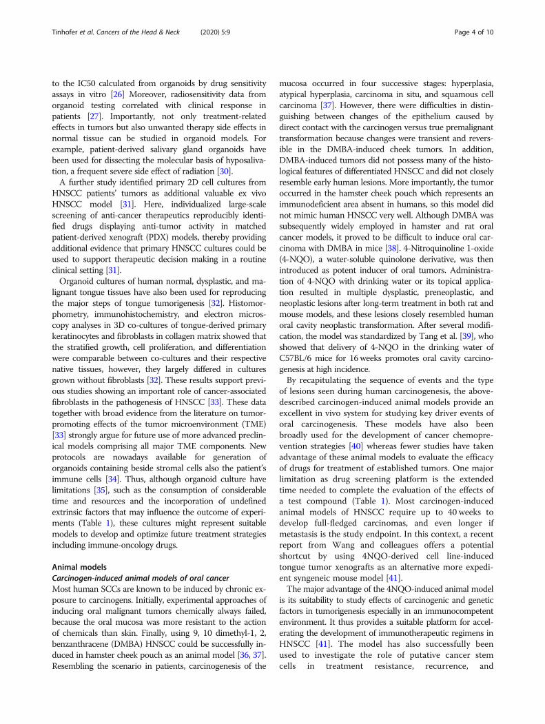
preclinical models of HNSCC (schematically depicted inFig. 1) and gives an overview of their strengths and limita-tions. We also discuss new approaches of personalizedtreatment selection based on these models.
Ex vivo modelsImmortalized HNSCC cell linesFour decades ago, first protocols for ex vivo cultures ofHNSCC cells have been reported [8, 9]. After resolvingprior obstacles such as fibroblast overgrowth and de-pendence on feeder layers with these protocols, HNSCCcell lines were successfully established. Culturetechniques have been further improved since then, and
Fig. 1 Schematic overview of approaches to generate preclinical HNSCC models. a Patient-derived models are mainly generated from surgicaltumor tissue. After mechanical and enzymatic dissociation, tumor cells are grown in vitro as 2D cell monolayers on plastic or 3D spheroidstructures in extracellular matrix (ECM). For generation of patient-derived xenografts (PDX), tumor fragments are transplanted subcutaneously inimmunocompromised mice. Classical patient-derived models are characterized by the absence of human immune and stromal cells. b Geneticallyengineered mouse models of oral squamous cell carcinoma can be generated by selective activation of oncogenes or inactivation of tumorsuppressor genes (TSGs) in epithelial cells. c Delivery of 4-Nitroquinoline 1-oxide in the drinking water of mice over several weeks promotes oralcavity carcinogenesis at high incidence
Tinhofer et al. Cancers of the Head & Neck (2020) 5:9 Page 2 of 10

various HNSCC cell lines stably growing over numerouspassages have been generated. A detailed description ofall available HNSCC cell lines would be beyond thescope of this review. We would thus like to refer thereader to two previous review articles [10, 11]. Since im-mortalized HNSCC cell lines can be easily maintainedand expanded, they have broadly been used to studygenetic alterations and biological responses to chemicaland genetic perturbations, to identify potential moleculartargets, and to develop novel small-molecule and bio-logical therapeutics [12, 13]. More recently, evidence hasbeen provided that these cell lines can also be used forstudying intratumoral heterogeneity and clonal evolutionoccurring under therapy pressure [14]. Data from suchcomprehensive molecular and functional studies in thesemodels have been assembled in libraries like the CancerCell Line Encyclopedia (CCLE), representing a valuablerepository of human cancer diversity [15, 16].Although HNSCC cell lines grown in two-dimensional
(2D) monolayer cultures continue to be importantmodels in the search for new therapeutic approaches forthis disease, they generally suffer from their inability toreflect the histological nature, three-dimensional (3D)architecture and structural and functional differences ofthe tumor in vivo. These limitations significantly influ-ence the informative value of in vitro studies evaluatingthe efficacy of established and novel treatment modal-ities for HNSCC in monolayer cultures. Indeed, notabledifferences in sensitivity of 2D versus 3D cultures fromHNSCC cell lines were reported for radiation [17] anddrug treatment, e.g. with cisplatin [18], cetuximab [18, 19]and the mTOR inhibitor AZD8055 [19]. Comparative mo-lecular analysis of cells growing in 2D versus 3D culturesprovided possible explanations for lower sensitivity of cellsin 3D cultures, like the expression and activation of genesassociated with DNA repair [17], and increased expressionlevels of genes associated with epithelial-mesenchymaltransition and stemness [18] under 3D conditions.Genetic instability and the occurrence of clonal selec-
tion during in vitro culture [20] are further potentiallimitations of cancer cell lines, and can explain why find-ings involving cell lines are often difficult to reproduce.Indeed, comprehensive analysis of strains from thecommonly used MCF7 breast and A549 lung cancer celllines revealed extensive genomic variation across strainswhich was associated with variation in biologicallymeaningful cellular properties. Importantly, when thestrains were tested against 321 anti-cancer compounds,considerably different drug responses were observed,with at least 75% of compounds strongly inhibiting somestrains but being completely inactive in others. Thisstudy clearly underlines the urgent need for improvedex vivo models to support maximally reproduciblecancer research.
Advanced ex vivo models of HNSCCKöpf-Maier and colleagues were the first to establish amethod which allowed human carcinoma cells fromdifferent histological entities including squamous cellcarcinomas (SCCs) of the pharynx to reorganize in vitroto “organoid structures” [7]. They showed that theseorganoid cultures maintained the critical properties ofthe in vivo state, such as the 3D architecture, the growthof heterogeneous cell types from an individual carcinomaand the morphological differentiation under relatively sim-ple experimental conditions [7]. In a subsequent study,the same group demonstrated that these organoid culturescan be used for drug testing, and that the response dataobtained thereof were concordant with patients’ responseto therapy [21]. The authors were the first to proposeorganoid cultures as personalized in vitro drug testingplatform, allowing the prediction of individual chemosen-sitivity of carcinomas within few days [21].Since then, techniques to grow tissues in vitro in 3D
as organotypic structures have been refined. Protocolshave been developed for establishing organoids fromadult and embryonic stem cells which are able to self-organize into 3D structures that reflect the tissue oforigin (for a review see Clevers, 2016 [22]). The firstadult stem cell-derived organoid cultures were estab-lished from mouse intestinal stem cells that were placedin conditions mimicking the intestinal stem cell niche[23]. Conditional reprogramming induced by adding R-spondin-1, epidermal growth factor (EGF) and Nogginto the culture medium, and embedment of the cells inan extracellular matrix-providing basement membranesextract, has been shown to stimulate adult stem cells toself-renew, proliferate and form differentiated offspring,resembling the intestinal epithelium [23–25]. This tech-nique, initially developed to study infected, inflammatoryand neoplastic tissue from the human gastrointestinaltract, has not only been used for the establishment oforganoid cultures from a variety of human normal tissuebut also patient-derived tumor tissue. These studies havesignificantly enlarged and improved the set of availablecancer models.More recently, the early findings by Köpf-Maier and col-
leagues [21] of HNSCC organoid cultures being a suitablein vitro drug testing platform were confirmed by several in-dependent studies. Though considerable differences in thesuccess rates of establishing primary long-term growingorganoid cultures from HNSCC patients were reported(30% [26] versus 65% [27]), all studies so far unanimouslydescribed that organoids retain many properties of theoriginal tumor, including intratumoral heterogeneity [28],mutation profile and protein expression patterns [27, 29].In addition, it was shown that organoids retained theirtumorigenic potential upon xenotransplantation [27]. Re-sponses to drug treatment in vivo were found to be similar
Tinhofer et al. Cancers of the Head & Neck (2020) 5:9 Page 3 of 10
to the IC50 calculated from organoids by drug sensitivityassays in vitro [26] Moreover, radiosensitivity data fromorganoid testing correlated with clinical response inpatients [27]. Importantly, not only treatment-relatedeffects in tumors but also unwanted therapy side effects innormal tissue can be studied in organoid models. Forexample, patient-derived salivary gland organoids havebeen used for dissecting the molecular basis of hyposaliva-tion, a frequent severe side effect of radiation [30].A further study identified primary 2D cell cultures from
HNSCC patients’ tumors as additional valuable ex vivoHNSCC model [31]. Here, individualized large-scalescreening of anti-cancer therapeutics reproducibly identi-fied drugs displaying anti-tumor activity in matchedpatient-derived xenograft (PDX) models, thereby providingadditional evidence that primary HNSCC cultures could beused to support therapeutic decision making in a routineclinical setting [31].Organoid cultures of human normal, dysplastic, and ma-
lignant tongue tissues have also been used for reproducingthe major steps of tongue tumorigenesis [32]. Histomor-phometry, immunohistochemistry, and electron micros-copy analyses in 3D co-cultures of tongue-derived primarykeratinocytes and fibroblasts in collagen matrix showed thatthe stratified growth, cell proliferation, and differentiationwere comparable between co-cultures and their respectivenative tissues, however, they largely differed in culturesgrown without fibroblasts [32]. These results support previ-ous studies showing an important role of cancer-associatedfibroblasts in the pathogenesis of HNSCC [33]. These datatogether with broad evidence from the literature on tumor-promoting effects of the tumor microenvironment (TME)[33] strongly argue for future use of more advanced preclin-ical models comprising all major TME components. Newprotocols are nowadays available for generation oforganoids containing beside stromal cells also the patient’simmune cells [34]. Thus, although organoid culture havelimitations [35], such as the consumption of considerabletime and resources and the incorporation of undefinedextrinsic factors that may influence the outcome of experi-ments (Table 1), these cultures might represent suitablemodels to develop and optimize future treatment strategiesincluding immune-oncology drugs.
Animal modelsCarcinogen-induced animal models of oral cancerMost human SCCs are known to be induced by chronic ex-posure to carcinogens. Initially, experimental approaches ofinducing oral malignant tumors chemically always failed,because the oral mucosa was more resistant to the actionof chemicals than skin. Finally, using 9, 10 dimethyl-1, 2,benzanthracene (DMBA) HNSCC could be successfully in-duced in hamster cheek pouch as an animal model [36, 37].Resembling the scenario in patients, carcinogenesis of the
mucosa occurred in four successive stages: hyperplasia,atypical hyperplasia, carcinoma in situ, and squamous cellcarcinoma [37]. However, there were difficulties in distin-guishing between changes of the epithelium caused bydirect contact with the carcinogen versus true premalignanttransformation because changes were transient and revers-ible in the DMBA-induced cheek tumors. In addition,DMBA-induced tumors did not possess many of the histo-logical features of differentiated HNSCC and did not closelyresemble early human lesions. More importantly, the tumoroccurred in the hamster cheek pouch which represents animmunodeficient area absent in humans, so this model didnot mimic human HNSCC very well. Although DMBA wassubsequently widely employed in hamster and rat oralcancer models, it proved to be difficult to induce oral car-cinoma with DMBA in mice [38]. 4-Nitroquinoline 1-oxide(4-NQO), a water-soluble quinolone derivative, was thenintroduced as potent inducer of oral tumors. Administra-tion of 4-NQO with drinking water or its topical applica-tion resulted in multiple dysplastic, preneoplastic, andneoplastic lesions after long-term treatment in both rat andmouse models, and these lesions closely resembled humanoral cavity neoplastic transformation. After several modifi-cation, the model was standardized by Tang et al. [39], whoshowed that delivery of 4-NQO in the drinking water ofC57BL/6 mice for 16 weeks promotes oral cavity carcino-genesis at high incidence.By recapitulating the sequence of events and the type
of lesions seen during human carcinogenesis, the above-described carcinogen-induced animal models provide anexcellent in vivo system for studying key driver events oforal carcinogenesis. These models have also beenbroadly used for the development of cancer chemopre-vention strategies [40] whereas fewer studies have takenadvantage of these animal models to evaluate the efficacyof drugs for treatment of established tumors. One majorlimitation as drug screening platform is the extendedtime needed to complete the evaluation of the effects ofa test compound (Table 1). Most carcinogen-inducedanimal models of HNSCC require up to 40 weeks todevelop full-fledged carcinomas, and even longer ifmetastasis is the study endpoint. In this context, a recentreport from Wang and colleagues offers a potentialshortcut by using 4NQO-derived cell line-inducedtongue tumor xenografts as an alternative more expedi-ent syngeneic mouse model [41].The major advantage of the 4NQO-induced animal model
is its suitability to study effects of carcinogenic and geneticfactors in tumorigenesis especially in an immunocompetentenvironment. It thus provides a suitable platform for accel-erating the development of immunotherapeutic regimens inHNSCC [41]. The model has also successfully beenused to investigate the role of putative cancer stemcells in treatment resistance, recurrence, and
Tinhofer et al. Cancers of the Head & Neck (2020) 5:9 Page 4 of 10
metastasis. Its potential for developing novel thera-peutic strategies targeting not only the proliferativetumor bulk but also the relatively quiescent subpopu-lation of cancer stem cells has been established [42].
Genetically engineered mouse modelsWhile DNA damage by chemicals occurs randomly,according to the tumor evolution theory random acqui-sition of mutations across the genome is followed byselection of clones harboring genetic changes that facili-tate cell survival and proliferation. Molecular profilingstudies have identified several putative driver genes con-tributing to cancer development in HNSCC. However,these molecular studies did not provide direct evidencefor causality or detailed insight into the biological mech-anisms by which these genes drive tumor development.Although carcinogen-induced animal models can closelyrecapitulate the heterogeneous landscape of genomic al-terations in human primary tumors [43], only a fraction
of these mutations drive tumorigenesis by affecting on-cogenes or tumor suppressor genes, but many mutationsare passengers with no clear contribution to tumordevelopment. These studies also do not reveal whetherdrivers are essential for tumor maintenance and maytherefore be of limited use for designing effective thera-peutic strategies. In contrast, preclinical model systemssuch as genetically engineered mouse models (GEMMs)provide an experimentally tractable approach in whichthe biological effects of specific mutations can be studiedin detail in a controlled genetic background. In the nextchapters, we describe key findings from previous studiesbased on GEMMs in HNSCC.Few GEMMs associated with spontaneous HNSCC
formation in the absence of chronic carcinogen exposurehave been described so far (Table 2). A genetically engi-neered mouse model of oral cancer was first introducedby Schreiber and colleagues [44]. After crossing micetransgenic for the v-Ha-ras gene with transgenic mice
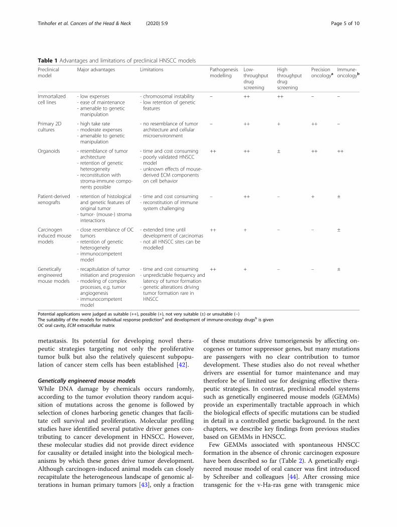
Table 1 Advantages and limitations of preclinical HNSCC models
Preclinicalmodel
Major advantages Limitations Pathogenesismodelling
Low-throughputdrugscreening
Highthroughputdrugscreening
Precisiononcologya
Immune-oncologyb
Immortalizedcell lines
- low expenses- ease of maintenance- amenable to geneticmanipulation
- chromosomal instability- low retention of geneticfeatures
– ++ ++ – –
Primary 2Dcultures
- high take rate- moderate expenses- amenable to geneticmanipulation
- no resemblance of tumorarchitecture and cellularmicroenvironment
– ++ + ++ –
Organoids - resemblance of tumorarchitecture
- retention of geneticheterogeneity
- reconstitution withstroma-immune compo-nents possible
- time and cost consuming- poorly validated HNSCCmodel
- unknown effects of mouse-derived ECM componentson cell behavior
++ ++ ± ++ ++
Patient-derivedxenografts
- retention of histologicaland genetic features oforiginal tumor
- tumor- (mouse-) stromainteractions
- time and cost consuming- reconstitution of immunesystem challenging
– ++ – + ±
Carcinogeninduced mousemodels
- close resemblance of OCtumors
- retention of geneticheterogeneity
- immunocompetentmodel
- extended time untildevelopment of carcinomas
- not all HNSCC sites can bemodelled
++ + – – ±
Geneticallyengineeredmouse models
- recapitulation of tumorinitiation and progression
- modeling of complexprocesses, e.g. tumorangiogenesis
- immunocompetentmodel
- time and cost consuming- unpredictable frequency andlatency of tumor formation
- genetic alterations drivingtumor formation rare inHNSCC
++ + – – ±
Potential applications were judged as suitable (++), possible (+), not very suitable (±) or unsuitable (−)The suitability of the models for individual response predictiona and development of immune-oncology drugsb is givenOC oral cavity, ECM extracellular matrix
Tinhofer et al. Cancers of the Head & Neck (2020) 5:9 Page 5 of 10

that harbored E6/E7 of human papilloma virus (HPV)-16, the development of tumors at the mouth, ear andeye beginning at about 3 months of age was observed[44]. By 6 months, 100% of the bi-transgenic animalshad developed oral tumors while the prevalence in eitherof the two single-transgenic groups was 0% [44]. The pre-requisite of a second genetic hit for tumorigenesis was alsoreported for a transgenic model of K-rasG12D, in which atamoxifen-inducible Cre recombinase under the control ofthe keratin-14 (K14) promoter was used for targeting theendogenous K-ras locus [45]. In the single-transgenemodel, only large papillomas in the oral cavity and hyper-plasias in the tongue were observed after 1 month of tam-oxifen treatment [45]. However, if mice were crossed withfloxed p53 conditional knockout mice, 100% of the com-pound mice developed tongue carcinomas as early as 2weeks after tamoxifen induction [45]. Beside expression ofviral oncogenes E6/E7 and loss of TP53, homozygous dele-tion of the transcription factor krüppel-like-factor 4 (KLF4)[46] and heterozygous deletion of SMAD4 [47] have beenidentified as second genetic hits which in concert with anoncogenic driver mutation promote oral tumor formationat high prevalence (Table 2).Despite recapitulating HNSCC progression, the suit-
ability of the above-described HNSCC models as plat-form for exploring novel molecular targeted treatmentapproaches remains somehow questionable, consideringthat the genetic alterations driving tumorigenesis inthese animals are absent or only rarely found in HNSCCpatients. Overall, mutations in HRAS and KRAS weredetected in only 6 and 0.2% of HNSCC patients, andhomozygous deletion of KLF4 and SMAD4 in 0 and 4%of cases, respectively. Moreover, cases harboring one ofthe compound tumor-prone genotypes of the GEMMsdescribed above have not been identified in The CancerGenome Atlas (TCGA) HNSCC cohort [53]. A GEMMof spontaneous HNSCC more closely resembling the
molecular features of the human disease might be thesingle gene-knockout model of SMAD4 in head andneck epithelia (HN-Smad4del/del) reported by Bornsteinand colleagues [47]. Indeed, although homozygousdeletion is rare, SMAD4 heterozygous loss is detected in30–35% of primary HNSCCs [53, 54] associated withdownregulation of Smad4 expression levels [53]. Morerecently, significant intratumoral heterogeneity of SMAD4loss in primary HNSCC tumors has been reported [54].Interestingly, in ex vivo cultures derived from PDX, thecell subpopulation displaying heterozygous SMAD4 lossby deletion or reduced expression outcompeted cells withwildtype SMAD4 genotype from the parental tumor, sug-gesting a survival advantage of Smad4-deficient cells [54].In further support of the suitability of this single-knockoutGEMM, HNSCC from HN-Smad4del/del mice exhibited in-creased genomic instability [47], which correlated withdownregulated expression and function of genes encodingproteins in the Fanconi anemia/BRCA DNA repair path-way [47], also linked to HNSCC susceptibility in humans[55]. Moreover, both normal head and neck tissue andHNSCC from HN-Smad4del/del mice exhibited severeinflammation which has also been linked to pathogenesisin humans [56] where oral bacteria and inflammatorymediators associated with periodontal disease may be co-factors in the initiation and promotion of oral SCC [57].Since the original report in 2009, the HN-Smad4del/del
model has been used for analyzing in detail the molecu-lar processes involved in HNSCC tumorigenesis. To ourknowledge, it has not yet been exploited for the develop-ment of novel therapeutic strategies. This restraint mightbe explained by a median onset time of 40 weeks fortumor development in this model, a limitation similar tocarcinogen-induced animal models of oral cancer (Table2). The integration of carcinogen treatment to acceleratetumor formation in single-transgene GEMMs might thusrepresent an appropriate way to resolve this limitation,
Table 2 Transgenic models of HNSCC
Transgenic modification Promotor Anatomical site Prevalence Onset (months) Reference
Spontaneous models
E6/E7/mrasR12 K14 OC 100 3–4 [44]
KrasG12/TP53del/del K14 T 100 < 1 [45]
KrasG12/KLF4del/del K14 T 21 < 1 [46]
KrasG12/SMAD4wt/del K14 OC n.a. 3–4 [47]
SMAD4del/del K14 OC 74 10 [47]
4NQO-induced models
GRHL3del/del K14 OC, L, P 40 3–4 [48]
PTENdel/del K14 OC 100 1–2 [49]
miR-211 K14 T n.a. 3 [50, 51]
miR-31 K14 T 80 3 [52]
Abbreviations: K14 keratin 14, OC oral cavity, T tongue, L larynx, P pharynx, n.a. not available
Tinhofer et al. Cancers of the Head & Neck (2020) 5:9 Page 6 of 10
as already successfully demonstrated in studies ofGEMMs harboring a deletion in a tumor suppressorgene (GRHL3 [48], PTEN [49]) or overexpressing onco-genic microRNAs [50–52] (Table 1).
Patient-derived xenograft modelsThe development and improvement of severelyimmune-deficient mouse strains has remarkably in-creased the availability of PDX models for cancerresearch. Successful establishment of HNSCC PDXmodels has been reported by several research groups[58–62]. In our own series, an overall engraftment rateof 48% was observed [60], however, engraftment ratesseemed to largely vary between distinct patient sub-groups [60, 61]. The limiting factors for engraftmenthave not yet been clearly identified. Site of implantationand mouse strains seem to influence the take rate.Moreover, pathological risk factors like tumor histologyand HPV status are important determinants of PDXformation. In general, undifferentiated HPV-negativetumors displaying aggressive growth are more likely toengraft. Accordingly, the rate and kinetics of PDXengraftment have been associated with an unfavorableprognosis of patients [61–63]. Contrary to HPV-negativetumors, HPV-associated HNSCC tumors frequently fail toengraft. Since these tumors are growing at immune-associated sites such as the tonsil or base of tongue, theirtransplantation to immunodeficient mice lacking im-munologic control of virally infected cells bears the risk ofco-transferring Epstein-Barr virus (EBV) positive B-cells.As a result, uncontrolled B-cell proliferation and trans-formation to EBV+ lymphoma frequently occurs [60, 61].Since the proliferation rate of these artificial lymphomas ismuch higher than tumor cell proliferation in transplantedtissue fragments of SCC, the original tumor transplantsare frequently overgrown [60, 61]. Thus, histopathologicvalidation of PDX by a board-certified pathologist isessential to confirm the squamous cell carcinoma hist-ology of the model.The question of how well PDX resemble the primary
patient tumor has been addressed by many groups. Asshown for other tumor entities, established HNSCCmodels in mice display histopathologic features like theoriginal patient tumor [59–62]. Comprehensive geneticanalysis of primary tumors and derived PDX models bynext generation sequencing revealed similar patterns andallelic frequencies of molecular aberrations [60, 62]. Thecorrelation between mutational profiles of original tu-mors and derived models was significantly higher forPDX (R = 0.94) compared to cell lines (R = 0.51) [64].Methylome analysis also showed high concordance be-tween PDX and patient tumors. Indeed, an average ofonly 2.7% of the assayed CpG sites underwent majormethylation changes as a result of transplanting tumors
to mice [65]. Furthermore, gene expression studiesshowed the overall relatedness of parental tumors withtheir PDX, as confirmed by their clustering together inunsupervised hierarchical clustering analysis [59, 60]. Incontrast to increasing evidence for genome and tran-scriptome profiles matching between PDX and primaryHNSCCs, only few data exist for protein expression. Afirst preliminary analysis of PDX tissue using reverse-phase protein array (RPPA) revealed protein profilescomparable to the TCGA HNSCC protein expressiondata [66], suggestive of similarity between original tissueand derived model also at this level.A key feature of PDX is the conservation of a stromal
compartment. Even though human stroma is replacedby mouse stroma within the first passages, an integratedstroma remains which makes the evaluation of com-pounds targeting this compartment or crosstalk betweenstromal compartment and tumor cells possible. Further,tumors grown in mice build up their own tumor vascu-lature which offers the opportunity of evaluating theangiogenic network and interference with compoundstargeting angiogenesis. After model establishment, tu-mors grown in mice can be harvested, vitally frozen andwhenever needed thawed and re-transplanted to mice.Overall, PDX can be considered a suitable method fortumor tissue expansion, and a promising preclinicalmodel system for mechanistic studies and the develop-ment of therapeutic strategies.With the recent advent of immunotherapy in the treat-
ment algorithm of many cancer types including HNSCC,the lack of a functional immune environment in PDX hasbecome a major obstacle to overcome. Different strategieshave been proposed to implement an immune system inimmunodeficient mice. In the landmark study of Mosierand colleagues [67], it was shown that the injection of hu-man peripheral mononuclear cells (PBMCs) resulted in thestable long-term reconstitution of a functional human im-mune system in mice with severe combined immunodefi-ciency (SCID). Thus, immunoproficient PDX models couldbe generated by transfer of patient’s PBMCs into the PDX-bearing mice. However, proper immune cell developmentand T-cell priming are lacking in this approach, resultingin the absence of certain lineages of human immune cellsin mice [67]. More sophisticated immune reconstitutionprotocols subsequently developed are based on the transferof human CD34+ stem cells into NSG mice, as well asimplanting human fetal thymus and liver tissue under thekidney capsule of these mice [68, 69]. This approach re-sulted in the long-term engraftment and systemic reconsti-tution of a complete human immune system includingmultilineage human immune cells consisting of T-, B-,NK-, dendritic cells and macrophages [68, 69]. Unfortu-nately, this method is not feasible for a large number ofPDX due to the complexity of the model. A more
Tinhofer et al. Cancers of the Head & Neck (2020) 5:9 Page 7 of 10
promising procedure has been proposed in melanomawhere tumor-infiltrating T lymphocytes (TILs) isolatedfrom the tumor tissue used for PDX generation were ex-panded in vitro by human interleukin 2 (IL2) before injec-tion in tumor-bearing PDX mice [70].
The potential of PDX models to guide patient treatmentThe value of PDX to guide individual patient treatmentdecision remains to be clarified. In general, patient-to-PDX correlations in different tumor entities comparingtreatment responses between mice and patients havebeen done using retrospective data on clinical outcome.To our knowledge, no such comparisons at sufficientlylarge sample sizes have been performed in HNSCC. Ob-stacles for the worthiness of such approaches comprisedrug dosage in mice which usually reflects the maximumtolerated dose, dose variability within different mousestrains and especially the definition of a clinically mean-ingful endpoint. In the clinical setting, tumor responsesare determined by RECIST. In mice, a very heteroge-neous set of possible endpoints has been used to deter-mine the efficacy of single-drug treatments, includingtumor regression expressed as relative growth inhibition,tumor volume in comparison to a control group, tumorgrowth inhibition and time to endpoint. Further generalmodel limitations are the high cost of PDX establish-ment, varying engraftment rates and times from firsttransplantation to treatment screening results. So far, inour large collection of almost 80 HNSCC PDX modelswe have been unable to establish a predictive value ofdrug-specific tumor responses in the xenograft model.Nevertheless, several companies advertise PDX as a toolto predict treatment response. In 2016, ChampionsOncology launched a feasibility trial (NCT02752932) toexplore the predictive value of PDX. Unfortunately, noresults have been published until now.The main disadvantage of PDX is the prolonged time
needed for model establishment and expansion com-pared to organoids, making their future use as individualdrug screening platform in clinical routine less likely. Inaddition, re-constitution with patient-derived TME com-ponents which are missing in both models generated bycurrent protocols should be achieved much easier inorganoids than in xenograft mouse models. This willallow inclusion of anti-cancer therapies affecting theTME (e.g. everolimus, bevacizumab, anti-PD-1/PD-1 Lantibodies) in future ex vivo screening approaches.
ConclusionsUnderstanding the molecular mechanisms involved inthe treatment resistance and progression of the disease,and elaborating new treatment strategies in preclinicalmodels will significantly contribute to advance clinicalmanagement of HNSCC. While assays using HNSCC
cell lines are essential for understanding HNSCC biologyand drug development, they cannot serve as preclinicalprediction model for the individual cancer patient. Orga-noid cultures resembling more closely the in vivo statemay be more suitable in this respect, despite limitingfactors such as higher costs and more elaborate main-tenance required for these ex vivo cultures. GEMMs areuseful in vivo models to interrogate the role of specificgenes and genetic modifications in the pathogenesis ofHNSCC. However, no single model described so farseems to be perfect for investigation of the pathogenesisand treatment of HNSCC. Though more studies areclearly needed to refine organoids and/or PDX asdiagnostic tools for individual prediction of therapyresponse, it can be envisioned that the combination ofmolecular profiling of tumors together with drug testingin organoid models might significantly advance precisionmedicine in head and neck cancer, and improve thechances for patients to receive a treatment tailored totheir tumor.
AbbreviationsHNSCC: Head and neck squamous cell carcinoma; SCCs: Squamous cellcarcinomas; DMBA: 9, 10 dimethyl-1, 2, benzanthracene; 4-NQO: 4-Nitroquinoline 1-oxide; GEMMs: Genetically engineered mouse models;HPV: Human papilloma virus; TCGA: The Cancer Genome Atlas; PDX: Patient-derived xenograft
AcknowledgementsNot applicable.
Authors’ contributionsAll authors (IT, DB, KK) contributed to literature search, writing of the originaldraft, reviewing and editing of the review. The author(s) read and approvedthe final manuscript.
FundingNot applicable.
Availability of data and materialsNot applicable.
Ethics approval and consent to participateNot applicable.
Consent for publicationThe final version of the article has been approved, and consent forpublication has been given by all authors.
Competing interestsThe authors declare that they have no competing interests.
Author details1Charité – Universitätsmedizin Berlin, corporate member of Freie UniversitätBerlin, Humboldt-Universität zu Berlin, and Berlin Institute of Health,Department of Radiooncology and Radiotherapy, Berlin, Germany. 2GermanCancer Research Center (DKFZ), Heidelberg and German Cancer Consortium(DKTK) Partner Site Berlin, Berlin, Germany. 3Charité – UniversitätsmedizinBerlin, corporate member of Freie Universität Berlin, Humboldt-Universität zuBerlin, and Berlin Institute of Health, Department of Hematology andOncology, Berlin, Germany.
Tinhofer et al. Cancers of the Head & Neck (2020) 5:9 Page 8 of 10
Received: 25 March 2020 Accepted: 15 July 2020
References1. Bray F, Ferlay J, Soerjomataram I, et al. Global cancer statistics 2018:
GLOBOCAN estimates of incidence and mortality worldwide for 36 cancersin 185 countries. CA Cancer J Clin. 2018;68(6):394–424.
2. Siegel RL, Miller KD, Jemal A. Cancer statistics, 2019. CA Cancer J Clin. 2019;69(1):7–34.
3. Ferris RL, Blumenschein G Jr, Fayette J, et al. Nivolumab for recurrentsquamous-cell carcinoma of the head and neck. N Engl J Med. 2016;375(19):1856–67.
4. Seiwert TY, Burtness B, Mehra R, et al. Safety and clinical activity ofpembrolizumab for treatment of recurrent or metastatic squamous cellcarcinoma of the head and neck (KEYNOTE-012): an open-label, multicentre,phase 1b trial. Lancet Oncol. 2016;17(7):956–65.
5. Vermorken JB, Mesia R, Rivera F, et al. Platinum-based chemotherapy pluscetuximab in head and neck cancer. N Engl J Med. 2008;359(11):1116–27.
6. Budach V, Tinhofer I. Novel prognostic clinical factors and biomarkers foroutcome prediction in head and neck cancer: a systematic review. LancetOncol. 2019;20(6):e313–26.
7. Köpf-Maier P, Zimmermann B. Organoid reorganization of human tumorsunder in vitro conditions. Cell Tissue Res. 1991;264(3):563–76.
8. Easty DM, Easty GC, Carter RL, et al. Ten human carcinoma cell lines derivedfrom squamous carcinomas of the head and neck. Br J Cancer. 1981;43(6):772–85.
9. Krause CJ, Carey TE, Ott RW, et al. Human squamous cell carcinoma:establishment and characterization of new permanent cell lines. ArchOtolaryngol. 1981;107(11):703–10.
10. Brenner JC, Graham MP, Kumar B, et al. Genotyping of 73 UM-SCC headand neck squamous cell carcinoma cell lines. Head Neck. 2010;32(4):417–26.
11. Zhao M, Sano D, Pickering CR, et al. Assembly and initial characterization ofa panel of 85 genomically validated cell lines from diverse head and necktumor sites. Clin Cancer Res. 2011;17(23):7248–64.
12. van Harten AM, Poell JB, Buijze M, et al. Characterization of a head and neckcancer-derived cell line panel confirms the distinct TP53-proficient copynumber-silent subclass. Oral Oncol. 2019;98:53–61.
13. Cheng H, Yang X, Si H, et al. Genomic and transcriptomic characterizationlinks cell lines with aggressive head and neck cancers. Cell Rep. 2018;25(5):1332–1345.e5.
14. Niehr F, Eder T, Pilz T, et al. Multilayered omics-based analysis of a head andneck cancer model of cisplatin resistance reveals Intratumoral heterogeneityand treatment-induced clonal selection. Clin Cancer Res. 2018;24(1):158–68.
15. Barretina J, Caponigro G, Stransky N, et al. The cancer cell line encyclopediaenables predictive modelling of anticancer drug sensitivity. Nature. 2012;483(7391):603–7.
16. Ghandi M, Huang FW, Jané-Valbuena J, et al. Next-generationcharacterization of the cancer cell line encyclopedia. Nature. 2019;569(7757):503–8.
17. Storch K, Eke I, Borgmann K, et al. Three-dimensional cell growth confersradioresistance by chromatin density modification. Cancer Res. 2010;70(10):3925–34.
18. Melissaridou S, Wiechec E, Magan M, et al. The effect of 2D and 3D cellcultures on treatment response, EMT profile and stem cell features in headand neck cancer. Cancer Cell Int. 2019;19:16.
19. Ayuso JM, Vitek R, Swick AD, et al. Effects of culture method on response toEGFR therapy in head and neck squamous cell carcinoma cells. Sci Rep.2019;9(1):12480.
20. Ben-David U, Siranosian B, Ha G, et al. Genetic and transcriptional evolutionalters cancer cell line drug response. Nature. 2018;560(7718):325–30.
21. Kopf-Maier P, Kolon B. An organoid culture assay (OCA) for determining thedrug sensitivity of human tumors. Int J Cancer. 1992;51(1):99–107.
22. Clevers H. Modeling development and disease with organoids. Cell. 2016;165(7):1586–97.
23. Sato T, Vries RG, Snippert HJ, et al. Single Lgr5 stem cells build crypt-villusstructures in vitro without a mesenchymal niche. Nature. 2009;459(7244):262–5.
24. Sato T, Stange DE, Ferrante M, et al. Long-term expansion of epithelialorganoids from human colon, adenoma, adenocarcinoma, and Barrett'sepithelium. Gastroenterology. 2011;141(5):1762–72.
25. Liu X, Ory V, Chapman S, et al. ROCK inhibitor and feeder cells inducethe conditional reprogramming of epithelial cells. Am J Pathol. 2012;180(2):599–607.
26. Tanaka N, Osman AA, Takahashi Y, et al. Head and neck cancer organoidsestablished by modification of the CTOS method can be used to predictin vivo drug sensitivity. Oral Oncol. 2018;87:49–57.
27. Driehuis E, Kolders S, Spelier S, et al. Oral mucosal organoids as apotential platform for personalized cancer therapy. Cancer Discov.2019;9(7):852–71.
28. Kijima T, Nakagawa H, Shimonosono M, et al. Three-dimensional organoidsreveal therapy resistance of esophageal and oropharyngeal squamous cellcarcinoma cells. Cell Mol Gastroenterol Hepatol. 2019;7(1):73–91.
29. Driehuis E, Spelier S, Beltrán Hernández I, et al. Patient-Derived Head andNeck Cancer Organoids Recapitulate EGFR Expression Levels of RespectiveTissues and Are Responsive to EGFR-Targeted Photodynamic Therapy. J ClinMed. 2019;8(11):1880.
30. Nagle PW, Hosper NA, Barazzuol L, et al. Lack of DNA damage response atlow radiation doses in adult stem cells contributes to organ dysfunction.Clin Cancer Res. 2018;24(24):6583–93.
31. Chia S, Low JL, Zhang X, et al. Phenotype-driven precision oncology as a guidefor clinical decisions one patient at a time. Nat Commun. 2017;8(1):435.
32. Sawant S, Dongre H, Singh AK, et al. Establishment of 3D co-culture modelsfrom different stages of human tongue tumorigenesis: utility inunderstanding neoplastic progression. PLoS One. 2016;11(8):e0160615.
33. Curry JM, Sprandio J, Cognetti D, et al. Tumor microenvironment in headand neck squamous cell carcinoma. Semin Oncol. 2014;41(2):217–34.
34. Bar-Ephraim YE, Kretzschmar K, Clevers H. Organoids in immunologicalresearch. Nat Rev Immunol. 2020;20(5):279-93.
35. Drost J, Clevers H. Organoids in cancer research. Nat Rev Cancer. 2018;18(7):407–18.
36. Salley JJ. Experimental carcinogenesis in the cheek pouch of the Syrianhamster. J Dent Res. 1954;33(2):253–62.
37. Morris AL. Factors influencing experimental carcinogensis in the hamstercheek pouch. J Dent Res. 1961;40:3–15.
38. Eveson JW. Animal models of intra-oral chemical carcinogenesis: a review. JOral Pathol. 1981;10(3):129–46.
39. Tang XH, Knudsen B, Bemis D, et al. Oral cavity and esophagealcarcinogenesis modeled in carcinogen-treated mice. Clin Cancer Res. 2004;10(1 Pt 1):301–13.
40. Bollag W, Holdener EE. Retinoids in cancer prevention and therapy. AnnOncol. 1992;3(7):513–26.
41. Wang Z, Wu VH, Allevato MM, et al. Syngeneic animal models of tobacco-associated oral cancer reveal the activity of in situ anti-CTLA-4. NatCommun. 2019;10:5546. https://doi.org/10.1038/s41467-019-13471-0.
42. Chen D, Wu M, Li Y, et al. Targeting BMI1(+) cancer stem cells overcomeschemoresistance and inhibits metastases in squamous cell carcinoma. CellStem Cell. 2017;20(5):621–634.e6.
43. Fantini D, Glaser AP, Rimar KJ, et al. A carcinogen-induced mouse modelrecapitulates the molecular alterations of human muscle invasive bladdercancer. Oncogene. 2018;37(14):1911–25.
44. Schreiber K, Cannon RE, Karrison T, et al. Strong synergy between mutantras and HPV16 E6/E7 in the development of primary tumors. Oncogene.2004;23(22):3972–9.
45. Raimondi AR, Molinolo A, Gutkind JS. Rapamycin prevents early onset oftumorigenesis in an oral-specific K-ras and p53 two-hit carcinogenesismodel. Cancer Res. 2009;69(10):4159–66.
46. Abrigo M, Alvarez R, Paparella ML, et al. Impairing squamous differentiationby Klf4 deletion is sufficient to initiate tongue carcinoma developmentupon K-Ras activation in mice. Carcinogenesis. 2014;35(3):662–9.
47. Bornstein S, White R, Malkoski S, et al. Smad4 loss in mice causesspontaneous head and neck cancer with increased genomic instability andinflammation. J Clin Invest. 2009;119(11):3408–19.
48. Georgy SR, Cangkrama M, Srivastava S, et al. Identification of a Novel Proto-oncogenic Network in Head and Neck Squamous Cell Carcinoma. J NatlCancer Inst. 2015;107(9):djv152.
49. Squarize CH, Castilho RM, Abrahao AC, et al. PTEN deficiency contributes tothe development and progression of head and neck cancer. Neoplasia.2013;15(5):461–71.
50. Chen YF, Yang CC, Kao SY, et al. MicroRNA-211 enhances the oncogenicityof carcinogen-induced oral carcinoma by repressing TCF12 and increasingantioxidant activity. Cancer Res. 2016;76(16):4872–86.
Tinhofer et al. Cancers of the Head & Neck (2020) 5:9 Page 9 of 10
51. Chu TH, Yang CC, Liu CJ, et al. miR-211 promotes the progression of head andneck carcinomas by targeting TGFbetaRII. Cancer Lett. 2013;337(1):115–24.
52. Tseng SH, Yang CC, Yu EH, et al. K14-EGFP-miR-31 transgenic mice havehigh susceptibility to chemical-induced squamous cell tumorigenesis that isassociating with Ku80 repression. Int J Cancer. 2015;136(6):1263–75.
53. TCGA Head and Neck Squamous Cell Carcinoma (firehose legacy, n=512).Broad Institute TCGA Genome Data Analysis Center. Firehose Stddata__2016_01_28 run. 2016; Available from: cbioportal.org.
54. Hernandez AL, Wang Y, Somerset HL, et al. Inter- and intra-tumorheterogeneity of SMAD4 loss in head and neck squamous cell carcinomas.Mol Carcinog. 2019;58(5):666–73.
55. Kutler DI, Auerbach AD, Satagopan J, et al. High incidence of head andneck squamous cell carcinoma in patients with Fanconi anemia. ArchOtolaryngol Head Neck Surg. 2003;129(1):106–12.
56. Hunter KD, Parkinson EK, Harrison PR. Profiling early head and neck cancer.Nat Rev Cancer. 2005;5(2):127–35.
57. Feller L, Altini M, Lemmer J. Inflammation in the context of oral cancer. OralOncol. 2013;49(9):887–92.
58. Kimple RJ, Harari PM, Torres AD, et al. Development and characterization ofHPV-positive and HPV-negative head and neck squamous cell carcinomatumorgrafts. Clin Cancer Res. 2013;19(4):855–64.
59. Peng S, Creighton CJ, Zhang Y, et al. Tumor grafts derived from patientswith head and neck squamous carcinoma authentically maintain themolecular and histologic characteristics of human cancers. J Transl Med.2013;11:198.
60. Klinghammer K, Raguse JD, Plath T, et al. A comprehensively characterizedlarge panel of head and neck cancer patient-derived xenografts identifiesthe mTOR inhibitor everolimus as potential new treatment option. Int JCancer. 2015;136(12):2940–8.
61. Facompre ND, Sahu V, Montone KT, et al. Barriers to generating PDXmodels of HPV-related head and neck cancer. Laryngoscope. 2017;127(12):2777–83.
62. Karamboulas C, Bruce JP, Hope AJ, et al. Patient-derived xenografts forprognostication and personalized treatment for Head and neck squamouscell carcinoma. Cell Rep. 2018;25(5):1318–31 e4.
63. Klinghammer K, Otto R, Raguse J-D, et al. Basal subtype is predictive forresponse to cetuximab treatment in patient-derived xenografts ofsquamous cell head and neck cancer. Int J Cancer. 2017;141(6):1215–21.
64. Gao H, Korn JM, Ferretti S, et al. High-throughput screening using patient-derived tumor xenografts to predict clinical trial drug response. Nat Med.2015;21(11):1318–25.
65. Guilhamon P, Butcher LM, Presneau N, et al. Assessment of patient-derivedtumour xenografts (PDXs) as a discovery tool for cancer epigenomics.Genome Med. 2014;6(12):116.
66. Li H, Wheeler S, Park Y, et al. Proteomic characterization of head and neckcancer patient-derived xenografts. Mol Cancer Res. 2016;14(3):278–86.
67. Mosier DE, Gulizia RJ, Baird SM, et al. Transfer of a functional humanimmune system to mice with severe combined immunodeficiency. Nature.1988;335(6187):256–9.
68. Lan P, Tonomura N, Shimizu A, et al. Reconstitution of a functional humanimmune system in immunodeficient mice through combined human fetalthymus/liver and CD34+ cell transplantation. Blood. 2006;108(2):487–92.
69. Melkus MW, Estes JD, Padgett-Thomas A, et al. Humanized mice mountspecific adaptive and innate immune responses to EBV and TSST-1. NatMed. 2006;12(11):1316–22.
70. Jespersen H, Lindberg MF, Donia M, et al. Clinical responses to adoptive T-cell transfer can be modeled in an autologous immune-humanized mousemodel. Nat Commun. 2017;8(1):707.
Publisher’s NoteSpringer Nature remains neutral with regard to jurisdictional claims inpublished maps and institutional affiliations.
Tinhofer et al. Cancers of the Head & Neck (2020) 5:9 Page 10 of 10