Embed Size (px)
Citation preview

IntroductionAlthough the number of reciprocal recombination events atmeiosis is similar for organisms with widely different genomesizes such as the mouse and lily (which have between 20 and50 events), the number of DNA-DNA interactions that arerecognized by RAD51/DMC1p immunocytology at prophaseof meiosis is much higher (250 for the mouse and more than2000 for lily) (Anderson et al., 2001). It follows that most orall of these early interactions do not necessarily function in theformation of reciprocal events. Conceivably, the numbers relateto genome size, 3.3 pg for the mouse and 141 pg for lily.However, the genome sizes differ by a factor of 43, whereasthe number of RAD51/DMC1 foci in the mouse differs onlyby a factor of 10 from the lily. A better correlate appears to bethe length of paired prophase chromosomes, as measured bythe total length per nucleus of the synaptonemal complexes,SCs, which are the axes of the bivalents (about 200 µm for themouse and about 3000 µm for lily). The tentative conclusionis that these foci, which are associated with the chromosomecores and SCs, function in SC formation (Anderson et al.,
2001). Synaptic failure in the absence of RAD51/DMC1 fociin SPO11–/– mice also suggests a role for early nodules insynapsis (Baudat et al., 2000; Romanienko and Camerini-Otero, 2000).
Molecular models for the resolution of DNA-DNAinteractions without reciprocal recombination have beendiscussed by Gilbertson and Stahl (Gilbertson and Stahl, 1996)and involve helicase-topoisomerase activity to resolve jointmolecules. The acquisition of replication protein A, RPA andBloom mutated protein, BLM, at the RAD51/DMC1 sitesmight be the physical manifestation of the models – RPA maystimulate BLM-RecQ helicase activity (Brosh et al., 2000) inconcert with topoisomerase IIIa (Johnson et al., 2000) toresolve the early DNA-DNA interactions at the RAD51/DMC1sites.
The development of reciprocal genetic exchange events atmeiosis in many fungi, plants and animals can be monitored atseveral levels: (1) at the chromosomal level by chiasmata,which are the sites of reciprocal recombination (Jones, 1987);(2) by the recombination nodules, RNs, which correlate with
1611
During mouse meiosis, the early prophase RAD51/DMC1recombination protein sites, which are associated with thechromosome cores and which serve as markers for ongoingDNA-DNA interactions, are in ten-fold excess of theeventual reciprocal recombinant events. Most, if not all, ofthese early interactions are eliminated as prophaseprogresses. The manner in which these sites are eliminatedis the focus of this investigation. We report that these sitesacquire replication protein A, RPA and the Escherichia coliMUTS homologue, MSH4p, and somewhat later the Bloomhelicase, BLM, while simultaneously losing theRAD51/DMC1 component. Eventually the RPA componentis also lost and BLM sites remain. At that time, the MUTLhomologue, MLH1p, which is essential for reciprocalrecombination in the mouse, appears in numbers and
locations that correspond to the distribution of reciprocalrecombination events. However, the MLH1 foci do notappear to coincide with the remaining BLM sites. TheMLH1p is specifically localized to electron-microscope-defined recombination nodules. We consider the possibilitythat the homology-search RAD51/DMC1 complexes areinvolved in homologous chromosome synapsis but thatmost of these early DNA-DNA interactions are laterresolved by the anti-recombination RPA/MSH4/BLM-topoisomerase complex, thereby preventing the formationof superfluous reciprocal recombinant events.
Key words: Meiosis, Recombination proteins, Immunocytology,Synaptonemal complexes, BLM, RPA, MLH1, Mouse,Recombination nodules
Summary
The time course and chromosomal localization ofrecombination-related proteins at meiosis in themouse are compatible with models that can resolvethe early DNA-DNA interactions without reciprocalrecombinationPeter B. Moens 1,*, Nadine K. Kolas 1, Madalena Tarsounas 2, Edyta Marcon 1, Paula E. Cohen 3 andBarbara Spyropoulos 1
1Department of Biology, York University, Toronto, ON, M3J 1P3, Canada2Imperial Cancer Research Fund, South Hall Laboratories, South Mimms, Hertfordshire, England EN6 3LD3Dept of Molecular Genetics, Albert Einstein College of Medicine, New York 10461 USA*Author for correspondence (e-mail: [email protected])
Accepted 11 December 2001Journal of Cell Science 115, 1611-1622 (2002) © The Company of Biologists Ltd
Research Article

1612
genetic and cytological patterns of recombination (Carpenter,1975; Carpenter, 1979; Albini and Jones, 1988); and (3) by theMLH1p sites, which are associated with crossover sites(Anderson et al., 1999)
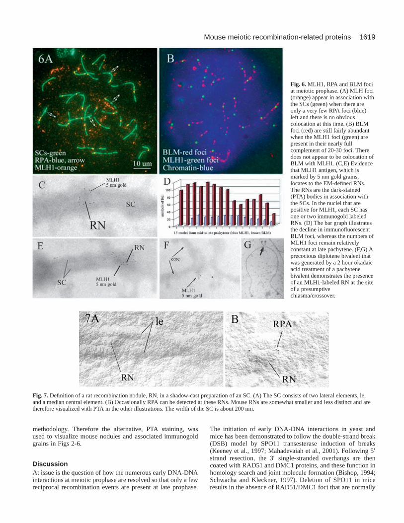
Nodules are SC-associated, electron-microscope-definedstructures that have been reported in the meiocytes of protists,fungi, plants and animals. In early meiotic prophase, ‘nodules’refer to the several hundreds of small dense bodies about 100nm in diameter that are associated with chromosome cores andSCs and contain the RAD51/DMC1 proteins in lily (Andersonet al., 1997), mouse and human (Haaf et al., 1995; Moens etal., 1997; Barlow et al., 1997). Since the correlation of thesestructures with reciprocal recombination is tenuous, they areusually referred to as ‘early nodules’, EN. The late RNs, asoriginally defined by Carpenter (Carpenter, 1975) inDrosophila melanogasteroocyte SCs, correspond in numberand location to reciprocal recombinant events in normal andmutant D. melanogaster. In the rat, ‘late RNs’ are well definedelectron-dense bodies of variable shape and size, 100 to 200nm, located on the mature SC at pachytene stage VII of thespermatogenic pathway but not in earlier pachytene stages I toV (Clermont, 1972; Moens, 1978). Cross-sectioned SCs andwhole-mount, shadow-cast EM preparations show that RNs arelocated on the surface of the SC either along the centralelement or obliquely across the SC (Fig.7). The reportednumber of late RNs (19 to 22 per nucleus) in complete EM-reconstructed rat-pachytene nuclei, their non-randomdistribution and their association with MLH1 (Mut L homolog)protein in rat and mouse (this report), agree with their proposedfunction in reciprocal recombination by Carpenter (Carpenter,1975; Carpenter, 1979). A number of publications haveassigned various proteins to RNs but the assignments have notpreviously been verified by EM demonstration of theseproteins on the RNs.
We report the events in individual mouse and ratspermatocyte nuclei from early to late meiotic prophase interms of chromosome core behavior and associated proteincomplexes. The immunofluorescence observations are refinedand detailed by immunoelectron microscopy of therecombination-related proteins and by EM visualization ofRNs. These observations are interpreted in the context of thechromosome synapsis and reciprocal recombination anddiscussed in relation to reports by others on these events.
Materials and MethodsCell preparationThe nuclear contents of whole-mount spermatocyte were displayed bysurface spreading of a testicular cell suspension on a hypotonic liquidsurface (Counce and Meyer, 1973; Dresser and Moses, 1980). Theintact nuclei became attached to a plastic-coated glass slides and werefixed briefly with a 2% paraformaldehyde with 0.03% SDS topermeabilize the nuclear envelope. After washing and drying in thepresence of the wetting agent, Kodak Photo-Flo 200, the cells wereblocked with goat serum and incubated overnight with primaryantibodies at room temperature (Dobson et al., 1994). For EM, thecells are treated for 10 minutes with 1 µg DNase per ml MEM to makethe SCs and SC-associated proteins more accessible to immunogoldgrains (Moens et al., 1987). After washes, the cells were incubatedfor 1 hour at 37°C with fluorochrome or colloidal gold-conjugatedsecondary antibodies. After washes, the cells were mounted inProLong Antifade (Molecular Probes) for fluorescence microscopy or
the plastic is floated off and transferred to EM grids (Dobson et al.,1994). The EM grids were treated with alcoholic phosphotungstic acid(PTA) or a shadow cast at an angle of 7° with platinum-gold (Moenset al., 1987). For rapid advancement of early prophase cells into thediplotene stage, cells were treated for 2 hours with the phosphataseinhibitor okadaic acid (Tarsounas et al., 1999b).
AntibodiesPolyclonal rabbit antibodies against whole hamster SCs andpolyclonal mouse antibodies against the fusion proteins of the hamster30 kDa chromosome core protein, COR1p, and the 125 kDa synapticprotein SYN1p have been characterized previously (Dobson et al.,1994; Tarsounas et al., 1997; Tarsounas et al., 1999a) and have beenused extensively by others (Plug et al., 1997; Plug et al., 1998; Pittmanet al., 1998). The equivalent rat 30/33 and 125 kDa SC proteins arenamed SCP3p and SCP1p (Heyting et al., 1988). From J. Ingles,University of Toronto, we received the polyclonal rabbit anti-RPAantibody (Henricksen et al., 1994; He et al., 1995), which has alsobeen used for similar experiments on RPA at meiosis (Plug et al.,1997; Plug et al., 1998). From J. Masson (ICRF, UK), we receivedthe recombinant HsRPA with the 14, 32 and 70 kDa subunitsidentified with western blotting. Our mouse, 23LM, produced apolyclonal anti-RPA serum against this recombinant HsRPA withgood fluorescent and EM immunocytology. Centromeres were labeledwith a CREST serum as reported earlier (Dobson at al., 1994). Therabbit anti-BLM antibody was raised against the purified fusionprotein of the last 380 amino acids of BLMp. The antiserumrecognized the appropriate 180 kDa protein in western blots of HeLawhole-cell extracts, and no protein was detected in the BS cell linethat lacked the BLMp C-terminal portion (Moens et al., 2000). Mousefull-length, (His)6-tagged DMC1 and RAD51 proteins wereoverexpressed in Escherichia coli.The Ni-NTA-purified proteins wereinjected into mice and rabbits that had negative pre-immune serum.Both types of sera cross-react with RAD51p and DMC1p, but afterimmune depletion, they were rendered specific for one or the otherantigen. Because it was shown with fluorescence and EM cytology ofthe purified antibodies that the two antigens colocalize on mouse andrat SC-associated early nodules (Tarsounas et al., 1999a), we used,forthis study, the anti-DMC1 serum from our rabbit ‘Patch’ or mouse‘17RB’ to detect the mixed RAD51/DMC1 antigen in core/SC-associated early nodules. These anti-RAD51/DMC1p antibodiesdiffer from those used in a number of earlier reports in that they donot contain anti-SC antibodies, which are common contaminants ofrabbit serum (Ashley et al., 1995; Kovalenko et al., 1996; Moens etal., 1997). The monoclonal anti-MLH1p antibody was obtained fromBD Pharmingen and has been characterized by Edelmann et al.(Edelmann et al., 1996) and Anderson et al. (Anderson et al., 1999).The polyclonal anti-hMSH4 was generated in rabbit and has beencharacterized by Santucci-Darmanin et al. (Santucci-Darmanin et al.,2000). Antibodies against rat testis-specific histone H1t weregenerated in a rabbit with E. coli-expressed H1t protein (Kremer andKistler, 1991; Moens, 1995).
ImagingFluorescence from FITC- or rhodmamine-tagged secondaryantibodies and DAPI-stained chromatin was recorded by CCD cameraor single or multiple exposures of black and white or colour-positivefilm, ASA 400. Slides were scanned at 1,000 dpi and recorded in TIFFformat, and final images were arranged with Adobe PhotoDeluxesoftware. Images were reproduced with an Epson 700 or 870 colourprinter. To determine the colocation of RPA with BLM foci, theimages were recorded on black and white film and one positive andone negative image were superimposed. Electron micrographs ofcores/SCs and immunogold were recorded at various magnifications,1 k to 10 k, and photographically enlarged. The contrast of SCs and
Journal of Cell Science 115 (8)

1613Mouse meiotic recombination-related proteins
nodules in EM preparations was enhanced by staining for 30 minutesin 4% alcoholic PTA followed by a rinse with 95% ethanol. Shadow-casting of SCs was achieved by gold-palladium evaporation at a lowangle of 7° on stationary or rotary moving grids (Moens et al., 1987).
ResultsStaging of meiotic prophase nuclei.For the purpose of this study, it is essential that the successivedevelopmental prophase stages of the mouse male meioticprophase nuclei are accurately defined. The traditional criteriaof SC formation, X-Y pairing and chromosome separation arenot always sufficient by themselves. A greater accuracy can beobtained by a combination of these structural characteristicsand monitoring the presence or absence of proteins thatfunction at different stages of meiotic prophase.
(1) Early prophase: the leptotene stage. With CREST serum,there are 40 immunofluorescent centromeres, which arefrequently arranged in a few groups. The anti-COR1/SCP3antibodies recognize short chromosomal core segments. Thereis no staining of synapsed core segments with anti SYN1/SCP1antibodies. Anti-RAD51 or DMC1 antibodies visualize up to300 core/SC-associated immunofluorescent foci. Otherrecombination-related proteins, BLM, RPA, MSH4 and MLH1are not present or rare. Antibodies against testis-specifichistone 1, H1t, do not cause fluorescence of the chromatin(Moens, 1995).
(2) Early prophase: the synaptic or zygotene stage. Avariable number of centromeres are in pairs. The chromosomecores are single or partially synapsed, and the synapsedsegments are recognized by the anti-SYN1/SCP1 antibodies.The increasing lengths of the synapsed regions are a measureof progression through the synaptic stage. The cores of the Xand Y chromosomes are not distinct from the autosomes. Thenumbers of RAD51/DMC1 are in decline, from about 200 to100 or less. The core and SC-associated RPA and BLM focibecome numerous, up to about 200 per nucleus, and these, too,decline at the late synaptic stage. MSH4 foci appear duringzygotene, but MLH1 foci are not normally present at this time.The partially synapsed bivalents occasionally cause the nucleusto resemble the late prophase diplotene stage. However, atdiplotene the recombination-associated proteins are notnormally present, and the paired centromeres mostly staytogether (unlike the zygotene stage where they often are thelast to pair). Also, the chromatin of diplotene but not zygotenenuclei is H1t positive.
(3) The fully synapsed pachytene stage. Except for anoccasional bivalent that is lagging in synapsis, all theautosomes are fully synapsed so that the immune stainingpatterns of autosomes with anti-SYN1/SCP1 and COR1/SCP3are the same. The X and Y chromosomes are partially paired,but there is not a distinct sex body or sex vesicle associatedwith the sex chromosomes at the early pachytene stage. At thelater pachytene stages, the X and Y are embedded in a sexvesicle. There is a spherical 400 nm double dense body, DDB,associated with the X chromosome, which is visible in EMimages. The number of autosomal RAD51/DMC1 foci declinesto zero, but the foci of the unpaired X chromosome persist intomid or late pachytene. There are 100 or fewer RPA, MSH4 andBLM foci. The MLH1 foci appear for the first time and arepresent in small numbers, up to an average of 26 as reported
by Anderson et al. (Anderson et al., 1999) and in somewhathigher numbers as observed by us. At mid pachytene, thechromatin becomes positive for testis-specific histone 1, H1t.The DDB is no longer associated with the X chromosome.
(4) Late prophase: the repulsion stage. The mainchromosomal characteristic of this stage is the partialseparation of chromosome cores. The remaining pairedsegments have immune staining with an anti-SYN1/SCP1antibody, and there are progressively shorter and fewer of thesesegments. There are no recombination protein foci at the SCsor the cores under normal circumstances. Many of thecentromeres remain in pairs. COR1/SCP1 protein is presentuntil diplotene progresses to metaphase I, at which time thecores become fragmented but the protein remains associatedwith sister centromeres until anaphase II. The chromatin ispositive for antibodies to testis-specific histone H1t. The sexvesicle contains much elongated, sinuous X and Ychromosome cores and is positive for a variety of antibodies(Turner et al., 2000).
Time course of RAD51/DMC1p versus RPA foci onmeiotic chromosome cores/SCsThe RAD51/DMC1 foci of the meiotic prophase chromosomesare the sites of single-strand DNA tails that are active inhomology search and strand exchange (Bishop, 1994;Schwacha and Kleckner, 1997). The presence of RPA proteinin relation to RAD51/DMC1 foci at successive developmentalstages of mouse spermatocyte nuclei at meiotic prophase isillustrated in Fig. 1. The number and intensity ofimmunofluorescent RPA foci increases from the time thechromosome cores first form and synapse at the zygotene stageof meiosis until late in the fully synapsed state at pachytene.The relative proportions of RAD51/DMC1 and RPA foci inindividual nuclei can be demonstrated with simultaneousvisualization of the two types of foci. In Fig. 1A and B, theearly prophase nucleus to the left of the white line is in theleptotene/zygotene stage, judging by the mostly unpairedcentromeres (enhanced red). This nucleus has an abundance ofRAD51/DMC1 foci (about 200) (Fig. 1A), but a maximum ofonly 90 weak RPA foci (Fig. 1B). At a later stage, judging bythe paired centromeres and the aligned foci, there are fewerDMC1 foci (approximately 90) in the nucleus on the right sideof Fig. 1A, while the number of RPA foci has increased to 180in that same nucleus (Fig. 1B).
The comparisons of Fig. 1A with B are reliable since therespective nuclei are in the same preparation and thereforereceived the same treatments. The shift from RAD51/DMC1foci to RPA foci is a general characteristic of developmentalprogression of the prophase nucleus. The replacement is notnecessarily synchronous for individual chromosomes. In Fig.1D, SC#1 has acquired a full complement of RPA foci, and theRAD51/DMC1 component is almost completely eliminatedjudging by the absence of aligned foci in SC#1 of Fig. 1C. Thesame is true for SC#2. On the other hand SC#3 has alignedRAD51/DMC1 foci but few and indistinct RPA foci.
RPA replaces RAD51/DMC1pWith the abundance of the two types of foci along the SCs, it islikely that by chance alone the two types will be close together,

1614
and it is difficult at the level of immunofluorescence resolutionto decide how much actual colocalization exists. At the highresolution of the electron micrographs in Fig. 2, it is possible todistinguish between pure RAD51/DMC1 foci (groups of 10 nmimmunogold grains), pure RPA foci (5 nm gold grains) andmixed foci (colocation of 5 and 10 nm gold grains). Theimmunogold foci of 40 SCs were classified accordingly, and thepercentages of the three types at progressively later prophasestages are shown for nine of the 40 SCs in the bar graph of Fig.1E. It is evident from the brown middle segment of the first barthat most of the RPA protein initially colocalizes with theRAD51/DMC1 foci. The remainder of the bars suggest that theRPA gradually replaces the RAD51/DMC1 component of the
foci to the point where there is no RAD51/DMC1 protein (lastbar in Fig. 1E). While it is possible that the mixed foci arise denovo, the gradient of the percentages of the two types of proteinswould favour a dynamic replacement process. The total numbersof immunofluorescent foci per nucleus for successivedevelopmental stages is demonstrated in Fig. 1F. Many of thesewould be in mixed foci as shown in Fig. 1E, Fig. 2C and Fig. 4.
At high resolution, the abundance of RAD51/DMC1 fociand the paucity of RPA protein at the onset of chromosomesynapsis is illustrated in the electron micrograph of a set ofpartially synapsed cores in Fig. 2A. There are six or sevengroups of 10 nm RAD51/DMC1-associated immunogoldgrains but only one group and a few sporadic 5 nm, RPA-
Journal of Cell Science 115 (8)
Fig. 1. Time course andinteractions of SC-associatedRAD51/DMC1, RPA and BLMprotein in mouse spermatocytes.(A,B) RAD51/DMC1 foci initiateprior to detection of RPA. To theleft of the white line is aspermatocyte early zygotenenucleus with mostly unpairedcentromeres (red), which hasabout 200 RAD51/DMC1 foci(yellow). A nucleus in a later stageof meiotic prophase to the right ofthe line has fewer, about 90, foci.The same two nuclei stained forRPA in Fig. 1B show that the earlyprophase nucleus on the left hasfew and indistinct RPA foci,whereas the pachytene nucleus tothe right of the line has anabundance of RPA foci. (C,D) Theshift from RAD51/DMC1 to RPAfoci is a developmentalprogression for the nucleus as awhole, but details seem to beregulated at the level of theindividual bivalent. SC#1 and 2have acquired a full complementof RPA foci (1D) and have lostmost RAD51/DMC1 foci (1C),whereas SC#3 still hasRAD51/DMC1 foci but few andindistinct RPA foci. (E) Thereplacement of RAD51/DMC1 byRPA is demonstrated from therelative amounts of the twoproteins in individual foci basedon electron microscopy of twotypes of immunogold grains as inFig. 2. The data from nine of 40SCs are presented in a bar graph;the top portion is the percentage offoci with only RPA antigen, themiddle portion represents the fociwith both antigens, and the lower part is the percentage of pure RAD51/DMC1 foci. Each bar represents one SC, and the SCs were sortedaccording to the prophase stage of the nucleus and by the previously documented decline in RAD51/DMC1 foci from zygotene to pachytene(details in Materials and Methods). The composition of the protein complexes change at successively later stages of meiotic prophase. (F) Theline graph shows the number of fluorescent foci at progressively later stages of prophase. RAD51/DMC1 peaks at leptotene. Somewhat later,RPA reaches its maximum and later still, BLM does. These three antigens frequently are present together in individual foci. MLH1 appears atlate prophase and is present in low numbers. Each data point is one nucleus, and the staging of the nuclei is described in the Materials andMethods.

1615Mouse meiotic recombination-related proteins
associated, immunogold grains. The reverse situation exists ata later stage where fully synapsed chromosome cores containfour RPA foci associated with the SC, but there is only oneRAD51/DMC1 focus (Fig. 2B, RAD51/DMC1 10 nmimmunogold grains, RPA 5 nm). At the onset of the pachytenestage, there are occasionally a few centromeric ends ofchromosomes that have not completed synapsis. Such a‘laggard’ is shown in Fig. 2C, which demonstrates thecombination of RAD51/DMC1 and RPA protein in singlenodules, marked as ‘mix’ in the figure. The arrow marks a
small loop of aligned RPA gold grains, which is notuncommon, but the significance is unclear.
RPA at SC-associated transitional or terminal nodulesWhere the chromosome cores are fully synapsed (Fig. 2B), the5 nm gold grains tend to be concentrated on dense nodes of thecentral element of the SC, suggesting that the RPA protein ispresent in some form of nodule (Fig. 2B and insert; Fig. 4D-G). These nodes do not have RAD51/DMC1 protein and
Fig. 2. Immunoelectronmicroscopy of RAD51/DMC1(10 nm gold grains) and RPA(5 nm gold grains) in associationwith chromosome cores/SCs in anearly and later meiotic prophasespermatocyte. (A) At early meioticprophase, there is an abundance ofRAD51/DMC1 foci but relativelyfew RPA foci in association withpartially paired zygotenechromosome cores and SCs.(B; insert) At a later stage,pachytene, there are numerousRPA foci and very fewRAD51/DMC1 foci. The RPA fociare concentrated on centralelement-associated nodules, TN.The width of the SC is about 200nm. (C) At early pachytene, thereare occasionally a few unpairedcentromeric ends (15 nm goldgrains). One of these ‘laggards’ isshown to have RPA associatedwith a single, unpairedchromosome core, indicating thatRPA is not always associated withRAD51/DMC1 protein. The imagefurther demonstrates the presenceof mixed foci containingRAD51/DMC1 and RPA antigen(mix). Incidental is the observationthat RPA gold grains occur as asmall loop (arrow) in this andother electron micrographs. Thewidth of the SC is about 200 nm.

1616
therefore do not conform to the concept of an ‘early nodule’.Since they also do not have the MLH1 protein (see below), theytherefore do not qualify as ‘late nodules’. Few if any of thesenodules will persist and they therefore represent some sort of‘terminal’ or ‘transitional’ nodule, TN.
RPA at the X-Y pseudo autosomal regionThe short Y chromosome becomes fully synapsed with thedistal portion of the long X chromosome during early meioticprophase, but the two are homologous only at their most distalregions, the pseudo-autosomal (PA) region (Fig. 3). In thelaboratory rat and mouse, the PA region has one or morereciprocal recombinant events, which are presumed to regulatethe proper segregation of the X and the Y chromosome at thefirst meiotic division (Soriano et al., 1987). These crossoversare recognized as a chiasma at diplotene and metaphase I. TheRPA antigen, marked by green FITC immunofluorescence inFig. 3B, is clearly present in the terminal segment of the PAregion. The immunofluorescence signal is frequently one of thebrightest FITC foci of the nucleus, and this may be caused bythe presence of two or more proximal foci. Similarly, the PA
region has a pronounced BLM signalas reported previously (Moens et al.,2000).
When the X and the Ychromosomes have a long non-homologous pairing segment, thereis RAD51/DMC1 antigen (10 nmgold grains) along the length of thepaired segment (Fig. 3C) as well asalong the core of the Xchromosomes. However, RPA isconcentrated in the terminal, PA,region (Fig. 3C) and along the Xchromosome core (Fig. 3B). In therat, which has better EM-defined,SC-associated nodules, the RPA can
be detected at the nodule in the PA region (Fig. 3D; Fig. 7B).
RPA and BLMp colocalizeBecause RPA (this report) and BLMp (Walpita et al., 1999;Moens et al., 2000) are frequently present on the smallpseudoautosomal region of the X and Y chromosomes, we testedthe possibility that these two proteins regularly occur together inthe same protein complex. For this purpose, the BLM antigenwas detected by rabbit anti-BLM serum and the secondaryantibody was tagged for fluorescent microscopy or for EMobservation. The RPA antigen was detected by a mouse serumand differentially tagged secondary antibodies. To assess therelative contribution of each antigen to the individual foci and tobe able to quantify the immunogold labeling of foci, we avoidthe use of rabbit antibodies against both BLM and RPA proteinas in Walpita et al. (Walpita et al., 1999). Superposition of thefluorescent images indicates that most RPA foci also have BLMantigen (Fig. 4). Whereas the RPA signals are generally ofsimilar strength (Fig. 4B), the BLM signals vary widely inintensity (Fig. 4A). For this reason, it is difficult to quantify thecontribution of each antigen to the foci by visual evaluation of
Journal of Cell Science 115 (8)
Fig. 3. RPA at the pseudoautosomalregion, PAR, of the X-Y chromosomes.(A) Chromosome cores and centromeresvisualized with fluorescent tags. The Xand Y chromosomes have a short pairedregion, PAR. (B) The same chromosomevisualized with anti-RPA antibody. Theautosomal SCs and the unpaired X-chromosome core have numerous RPAfoci. The PA region frequently has thebrightest RPA focus or foci. Scale: theSCs are about 15 mm in length.(C; insert) At early pachytene there isextensive non-homologous synapsisbetween the distal portions of the X andY chromosomes. RPA is present in themost distal portion, presumably theactual PA region. (D; insert) In the rat,the late RNs are better defined than inthe mouse, and the Figure illustrates agroup of 5 nm RPA gold grains inassociation with a nodule.

1617Mouse meiotic recombination-related proteins
subtle colour differences. Instead, we represent the BLM signalintensity by black dots (negative image) and superimpose thesedots on the white RPA signals (positive image) (Fig. 4C). Thisprocedure clearly demonstrates that at the level of resolution ofimmunofluorescence, most BLM foci colocalize precisely withthe RPA foci, and there is much variation in the intensity of theBLM signals as indicated by the sizes of the BLM signals.
The colocation of RPA and BLM was confirmed inimmunogold-labeled foci, where the two types of gold grains,RPA 5 nm and BLM 10 nm, can be found together in the nodules(Fig. 4E-G). For the antibodies that we use, there are more RPAgold grains per focus than there are BLM gold grains (Fig. 4H).
MSH4 co-locates with RPA in transitional nodulesThe meiosis-specific MSH4p homologue of E. coli MutSp isrequired for synapsis and recombination. In mouse it is
present along the paired chromosome cores at meioticprophase (Fig. 5) (Kneitz et al., 2000) (S. Santucci-Darmanin,P.B.M., F. L. Lespinasse et al., unpublished). MSH4fluorescent foci appear at the time that the early nodules arelosing the RAD51/DMC1 component. Biochemical evidencesuggests that there is an interaction between the two types ofproteins (S. Santucci-Darmanian, P.B.M., F. L. Lespinasse etal., unpublished), but it is relatively rare to find co-locationof the two types of proteins either with fluorescentmicroscopy (Fig. 5C) or with electron microscopy (Fig.5D,E), when the two antigens are differentially labeled.However, it is evident that MSH4 and RPA frequently co-locate to the same foci when observed with fluorescentmicroscopy (Fig. 5A) or electron microscopy (Fig. 5B). It isconcluded that MSH4 is mostly a component of thetransition/terminal nodules. Because the TNs vanish duringmeiotic prophase, it is possible that MSH4, in concert with
Fig. 4. Colocation of RPA andBLM protein in pachytenespermatocytes. (A,B) The samenucleus immunostained for BLM(A) and for RPA (B). (C) The BLMimage of (A) is made into anegative and superimposed on thepositive RPA image of (B). Nearlyall BLM foci colocate with RPAfoci. The relative contributions ofthe two proteins to the individualfoci are semi-quantifiable by thesize and the intensity of theimmunofluorescent signal, and thisis somewhat more efficient thanmultiple colour combinations.(D-G) Electron micrographs of the
mix of RPA 5 nm immunogold grains and 10 nm BLM immunogoldgrains at phosphotungstic acid-stained SCs and nodules. (H) With ourantibodies, there are fewer 10 nm BLM gold grains than there are 5 nmRPA gold grains at the nodules.

1618
RPA, BLM and others, functions in the resolution of earlyDNA-DNA interactions.
Time course of RPA and BLM versus MLH1Immunocytologically detectable MLH1 foci that are inassociation with the SCs appear in the later part of meioticprophase after RAD51/DMC1 foci are no longer present, andtheir numbers, about 23 per nucleus, are small in comparisonwith the numbers of other foci. In general, it appears that RPAfoci are already reduced to small numbers when MLH1 focifirst appear (Fig. 1F). The nucleus in Fig. 6A has four RPAfoci marked by arrows and 22 MLH1 foci, nearly the fullcomplement. There appears to be no positionalcorrespondence between RPA and MLH1 foci. Electronmicroscopy, however, shows the occasional presence of RPAin RNs (Fig. 3D; Fig. 7B). The BLM foci persist longer thanthe RPA foci (Fig. 1F), and they are still present when theMLH1 foci develop (Fig. 6B). From immunofluorescentobservations, there is no clear indication that the two types offoci colocate (Fig. 6B). The BLM foci decline in numbers,whereas the MLH1 foci remain fairly constant (Fig. 6D).Although the MLH1 foci normally remain until the onset of
the diplotene stage in male meiosis, they persist into thediplotene stage of female meiosis.
MLH1 protein is a component of EM-defined laterecombination nodulesEM of late pachytene mouse SCs treated with anti-MLH1monoclonal antibody and secondary goat anti-mouse antibodytagged with 5 nm gold grains show that the MLH1 antigen islocated at the EM-defined late RNs. (Fig. 6C,E-G). Thepresence of the MLH1 antigen is remarkably predictable.Where one SC of a given nucleus is found to have an MLH1-labeled RN, all other SCs of that nucleus predictably have oneor two such RNs. This was confirmed in more than 20 nuclei.The exact localization of an MLH1-labeled RN to the point ofa presumptive chiasma is demonstrated in Fig. 6F and G.Normally RNs are no longer present at the diplotene stage inthe male, but when the diplotene configurations are inducedprecociously by the phosphatase inhibitor okadaic acid inpurified early prophase cells, the MLH1 foci are still presentat diplotene. The platinum/gold shadow casting enhances theappearance of the RNs in the rat (Fig. 7), but the mouse nodulesare somewhat smaller and are less well defined with this
Journal of Cell Science 115 (8)
Fig. 5.The association of MSH4pwith RPA and RAD51/DMC1p.(A) The images of the red MSH4foci and the green RPA foci areslightly offset to betterdemonstrate the colocation of thetwo types of foci. The short whitelines indicate the direction of theoffset and examples of colocatingfoci. The large bright green fociare the centromeres. (B) Thecolocation is also evident withimmune electron microscopy. The10 nm gold particles mark RPAantigen and the 5 nm grains markthe MSH4 antigen. The grains areassociated with a transitionalnodule at the central region of theSC. (C) The association ofRAD51/DMC1 with MSH4 foci isless pronounced, and evidencefrom counts of five completenuclei with some 150 foci eachsuggest that at most 10% of thefoci possibly overlap and that, ingeneral, the MSH4 foci appearafter the Rad51/DMC1 foci havedeclined in numbers. The Xchromosome at the base of (C) has,as usual, prolonged presence ofRAD51/DMC1 foci. (D,E) At theEM level MSH4 foci (5 nm gold)are mostly separate fromRAD51/DMC1 foci (10 nm gold).(E) The MSH4 antigen isassociated with the transitionalnodule, which has no evidence ofRAD51/DMC1 antigen in thisdouble-labeled preparation.
1619Mouse meiotic recombination-related proteins
methodology. Therefore the alternative, PTA staining, wasused to visualize mouse nodules and associated immunogoldgrains in Figs 2-6.
DiscussionAt issue is the question of how the numerous early DNA-DNAinteractions at meiotic prophase are resolved so that only a fewreciprocal recombination events are present at late prophase.
The initiation of early DNA-DNA interactions in yeast andmice has been demonstrated to follow the double-strand break(DSB) model by SPO11 transesterase induction of breaks(Keeney et al., 1997; Mahadevaiah et al., 2001). Following 5′strand resection, the 3′ single-stranded overhangs are thencoated with RAD51 and DMC1 proteins, and these function inhomology search and joint molecule formation (Bishop, 1994;Schwacha and Kleckner, 1997). Deletion of SPO11 in miceresults in the absence of RAD51/DMC1 foci that are normally
Fig. 7. Definition of a rat recombination nodule, RN, in a shadow-cast preparation of an SC. (A) The SC consists of two lateral elements, le,and a median central element. (B) Occasionally RPA can be detected at these RNs. Mouse RNs are somewhat smaller and less distinct and aretherefore visualized with PTA in the other illustrations. The width of the SC is about 200 nm.
Fig. 6.MLH1, RPA and BLM fociat meiotic prophase. (A) MLH foci(orange) appear in association withthe SCs (green) when there areonly a very few RPA foci (blue)left and there is no obviouscolocation at this time. (B) BLMfoci (red) are still fairly abundantwhen the MLH1 foci (green) arepresent in their nearly fullcomplement of 20-30 foci. Theredoes not appear to be colocation ofBLM with MLH1. (C,E) Evidencethat MLH1 antigen, which ismarked by 5 nm gold grains,locates to the EM-defined RNs.The RNs are the dark-stained(PTA) bodies in association withthe SCs. In the nuclei that arepositive for MLH1, each SC hasone or two immunogold labeledRNs. (D) The bar graph illustratesthe decline in immunofluorescentBLM foci, whereas the numbers ofMLH1 foci remain relativelyconstant at late pachytene. (F,G) Aprecocious diplotene bivalent thatwas generated by a 2 hour okadaicacid treatment of a pachytenebivalent demonstrates the presenceof an MLH1-labeled RN at the siteof a presumptivechiasma/crossover.

1620
detected at the cores and SCs of the meiotic prophasechromosomes (Baudat et al., 2000). Because homologouschromosome synapsis is disrupted in these mice, it mightindicate that the RAD51/DMC1 sites function in synapsis(Baudat et al., 2000; Romanienko and Camerini-Otero, 2000).On the basis of the correlation between SC lengths andnumbers of RAD51/DMC1 foci in six plant species withwidely divergent genome sizes, Anderson et al. (Anderson etal., 2001) have also suggested that these early interactionsfunction in chromosome synapsis. Subsequently, most or all ofthe DNA-DNA interactions evidently are resolved withoutreciprocal recombination.
In the double-strand break model, the joint molecules can beresolved in a number of ways by specific enzymatic complexes.Depending on how the two Holliday junctions of a jointmolecule are cleaved, there can be a 1:1 ratio of reciprocal tonon-reciprocal recombinants. Taking into account thepreferential resolution that causes deviations from the 1:1 ratio,there would still be a large excess of reciprocal recombinationsproduced by the 250 RAD51/DMC1 sites in the mouse wherean average of only about 23 reciprocal events are observed. Ithas been argued, however, that the joint molecules can also beresolved without reciprocal recombination along differentpathways (Gilbertson and Stahl, 1996). These pathways requirehelicase and topoisomerase functions, and are therefore ofparticular interest to us because of the observed association ofBloom RecQ helicase with RAD51/DMC1 sites at the cores/SCsof meiotic prophase chromosomes (Moens et al., 2000).
Bloom protein normally functions in chromosome stabilityby the removal of DNA conformations that could lead tochromosome rearrangements or recombination. BLM protein,as a member of the family of RecQ helicases, belongs to a classof anti-recombinases (Constantinou et al., 2000) that couldfunction to resolve the meiotic prophase chromosomeinteraction without reciprocal recombination, analogous to theyeast RecQ helicase SGS1 that can resolve strand invasionevents (Frei and Gasser, 2000). Mutations of the BLM genelead to chromosome rearrangements and characteristically highlevels of sister chromatid exchanges in somatic cells (German,1993; Watt et al., 1996). BLM RecQ helicase andtopoisomerase IIIa interact physically and functionally toenable passage of double-stranded DNA in somatic andmeiotic human cells (Harmon et al., 1999; Johnson et al.,2000). Thus the complex demonstrably suppressesrecombination and is therefore an appropriate candidate for theresolution of early DNA-DNA interactions at meioticprophase.
We observe that RAD51/DMC1 foci become associated withRPA and somewhat later with BLMp. This observation can berelated to the reports that RecQ helicases interact with RPA.Brosh et al. (Brosh et al., 2000) have shown that in vitro thepresence of RPA stimulates the helicase activity of BLMp, andConstantino et al. (Constantino et al., 2000) show that theRecQ helicases WRN and BLM localize to RPA foci, wherethey promote translocation of the Holliday junctions anddissociate recombination intermediates. Similarly, Sakamoto etal. (Sakamoto et al., 2001) report that WRNp colocalizes withRPA foci at sites of induced DNA damage. Taken together,these observations provide support for the hypothesis that earlymeiotic prophase DNA-DNA interactions are resolved, withoutreciprocal recombination, by a protein complex that includes
RPA and BLMp, which probably acts in conjunction withtopoisomerase IIIa.
Although it is well established that MSH4p is a partner ofthe RPA/BLM complex and the TNs (Fig. 5C,D,E), its functionin the complex is uncertain (S. Santucci-Darmanin, P.B.M., F.L. Lespinasse et al., unpublished). At later stages when fewMSH4 foci remain, it has been reported that they colocalizewith MLH1p but here, too, the functions are still speculative(Santucci-Darmanin, 2000).
Origin of recombination nodulesBecause MLH1 protein is associated with recombinant eventsand because the MLH1 foci are correlated in number anddistribution with reciprocal recombinant events, it has beenhypothesized that the MLH1 foci represent the RNs in mice(Anderson et al., 1999). We provide evidence in support of thathypothesis by showing that the MLH1 antigen is present inEM-defined RNs (Fig. 6C,E) and that the RN and the MLH1protein are present at the site of a chiasma (Fig. 6F,G). InMLH1–/– mice, chromosome synapsis is normal. The repulsionof homologues at the onset of meiosis appears normal butsubsequently there is a complete or near complete lack ofchiasmata, and the result is an accumulation of univalents inthe nucleus (Baker, 1996; Edelmann et al., 1996). Apparently,reciprocal recombinant events are mostly absent.
It is evident that most early DNA-DNA interactions that arerecognized as RAD51/DMC1 foci are resolved by midprophase. At a later stage, the foci that correspond to RNs areobserved. The question arises whether (1) a subset of the earlyinteractions persist and become late RNs or (2) whether the lateRNs arise de novo. There is support for both propositions.
(1) The EM data on RAD51/DMC1 and RPA immunogoldgrains in Fig. 1E and Fig. 2 suggest that the earlyRAD51/DMC1 nodules acquire RPA, and eventually noRAD51/DMC1 component is present in the nodules. Thesenodules also acquire BLM and MSH4 protein, and we interpretfrom this that the function of these transitional nodules is theremoval of early DNA-DNA interactions without reciprocalrecombination (see above). There does not seem to be a distinctcolocation of the RPA-BLM foci with the MLH1 foci asdemonstrated in Fig. 6A and B. This would suggest thatinteractions that are being resolved do not become late RNs. Asecond round of SPO11-RAD51/DMC1 function would berequired to initiate new DSBs. There is no clear evidence forthis other than the presence of SPO11 on SCs at mid pachytene(Romanienko and Camerini-Otero, 2000).
(2) Although the RPA and BLM proteins do not show acontinuous presence into the RNs, the MSH4 component hasbeen reported to colocalize with MLH1 (Santucci-Darmanin etal., 2000). This would argue for the progression of a select fewTNs into RNs. We also observe the presence of RPA in someRNs (Fig. 3D; Fig. 7B), but we cannot determine if thisrepresents continuity or newly acquired RPA in the standardcontext of DNA metabolic activity.
These observations on the origins of RNs are clearly relatedto but do not yet resolve the issue of genetic and crossoverinterference. They suggest that in the appropriate organismsor experimental conditions, the immunocytology ofrecombination-related proteins could elucidate the mechanismsof interference that limit and position the crossovers.
Journal of Cell Science 115 (8)

1621Mouse meiotic recombination-related proteins
We gratefully acknowledge the contributions of antibodies for thisstudy: anti-BLM by Raimundo Freire (Facultad de Medicina, Tenerife),rabbit anti-hRPA by Jim Ingles (Banting and Best Institute, Toronto)and rabbit anti-hMNH4 by Veronique Paquis (UMR CNRS, Nice). Theshadow-casting of SCs was performed by York undergraduate student,Mike Sebastian, and York EM technical assistant, Karen Rethoret. Theresearch was funded by NSERC of Canada.
ReferencesAlbini, S. M. and Jones, G. H.(1988). Synaptonemal complex spreading in
Allium cepaand A. fistulosum. II. Pachytene observations: The SC karyotypeand the correspondence of late recombination nodules and chiasma. Genome30, 399-410.
Anderson, L. K., Offenberg, H. H., Verkuijlen, W. M. H. C. and Heyting,C. (1997). RecA-like proteins are components of early meiotic nodules inlily. Proc. Natl. Acad. Sci. USA94, 6868-6873.
Anderson, L. K., Reeves, A., Webb, L. M. and Ashley, T.(1999).Distribution of crossing over on mouse synaptonemal complexes usingimmunofluorescent localization of MLH1 protein. Genetics 151, 1569-1579.
Anderson, L. K., Hooker, K. D. and Stack, S. M.(2001). The distributionof early recombination nodules on zygotene bivalents from plants. Genetics159, 1259-1269.
Ashley, T., Plug, A. W., Xu, J., Solari, A. J., Redy, G., Golub, E. I. andWard, D. C. (1995). Dynamic changes in Rad51 distribution on chromatinduring meiosis in male and female vertebrates. Chromosoma 104, 19-28.
Baker, S. M., Bronner, C. E., Zhang, L., Plug, A. W., Robatzek, M.,Warren, G., Elliott, E. A., Yu, J., Ashley, T., Arnheim, N., Flavell, R. A.and Liskay, R. M. (1995). Male mice defective in the DNA mismatch repairgene PMS2 exhibit abnormal chromosome synapsis in meiosis. Cell 82,309-319.
Barlow, A. L., Benson, F. E., West, S. C. and Hulten, M. A.(1997).Distribution of the Rad51 recombinase in human and mouse spermatocytes.EMBO J. 16, 5207-5215.
Baudat, F., Manova, K., Yuen, J. P., Jasmin, M. and Keeney, S.(2000).Chromosome synapsis defects and sexually dimorphic meiotic progressionin mice lacking spo11. Mol. Cell 6, 989-998.
Bishop, D. K. (1994). RecA homologs Dmc1 and Rad51 interact to formmultiple nuclear complexes prior to meiotic chromosome synapsis. Cell 79,1081-1092.
Brosh, R. M., Jr, Li, J. L., Kenny, M. K., Karow, J. K., Cooper, M. P.,Kureekattil, R. P., Hickson, I. D. and Bohr, V. A. (2000). Replicationprotein A physically interacts with Bloom’s syndrome protein and stimulatesits helicase activity. J. Biol. Chem. 275, 23500-23508.
Carpenter, A. T. C. (1975). Electron microscopy of meiosis in Drosophilamelanogasterfemales II. The recombination nodule – a recombination-associated structure at pachytene? Proc. Natl. Acad. Sci. USA72, 3186-3189.
Carpenter, A. T. C. (1979). Recombination nodules and synaptonemalcomplex in recombination-defective females of Drosophila melanogaster.Chromosoma75, 259-292.
Clermont, Y. (1972). Kinetics of spermatogenesis in mammals: Seminiferousepithelium cycle and spermatogonial renewal. Physiol. Rev. 52, 198-236.
Constantinou, A., Tarsounas, M., Karow, J. K., Brosh, R. M., Bohr, V. A.,Hickson, I. D. and West, S. C.(2000). Werner’s syndrome protein (WRN)migrates Holliday junctions and co-localizes with RPA upon replicationarrest. EMBO reports1, 1-5.
Counce, S. and Meyer, G. F.(1973). Differentiation of the synaptonemalcomplex and the kinetochore in Locustaspermatocytes studied by wholemount electron microscopy. Chromosoma44, 231-253.
Dobson J. M., Pearlman, R. E., Karaiskakis, A., Spyropoulos, B. andMoens, P. B.(1994). Synaptonemal complex proteins: occurrence, epitopemapping and chromosome disjunction. J. Cell Sci. 107, 2749-2760.
Dresser, M. E. and Moses, M. J. (1980). Synaptonemal complex karyotypingin spermatocytes of the Chinese hamster (Cricetulus griseus). IV. Light andelectron microscopy of synapsis and nucleolar development by silverstaining. Chromosoma76, 1-22.
Edelmann, W., Cohen, P. E., Kane, M., Lau, K., Morrow, B., Bennett, S.,Umar, A., Kunkel, T., Cattoretti, G., Changanti, R. et al. (1996). Meioticpachytene arrest in MLH1-deficient mice. Cell 85, 1125-1134.
Frei, C. and Gasser, S. M.(2000). RecQ helicases: the DNA replicationcheckpoint connection. J. Cell Sci. 113, 2641-2646.
German, J. (1993). Bloom syndrome, a Mendelian prototype of somaticmutational disease. Medicine71, 393-406.
Gilbertson, L. A. and Stahl, F. W.(1996). A test of the double-strand breakrepair model for meiotic recombination in Saccharomyces cerevisiae.Genetics144, 27-41.
Haaf, T., Golub, E. I., Reddy, R. G., Radding, G. M. and Ward, D. C.(1995). Nuclear foci of mammalian Rad51 recombination protein in somaticcells after DNA damage and its localization in synaptonemal complexes.Proc. Natl. Acad. Sci. USA92, 2298-2302.
Harmon, F. G., DiGate, R. J. and Kowalczykowsky, S. C.(1999). RecQhelicase and topoisomerase III compromise a novel DNA strand passagefunction: a conserved mechanism for control of DNA recombination. Mol.Cell 3, 611-620.
He, F. G., Henricksen, L. A., Wold, M. S. and Ingles, C. J. (1995). RPAinvolvement in the damage-recognition and incision steps of nucleotideexcision repair. Nature374, 566-569.
Heyting, C., Dietrich, A. J. J., Moens, P. B., Dettmers, R. J., Offenberg,H. H., Redeker, E. J. W. and Vink, A. C. G. (1988). Synaptonemalcomplex proteins. Genome31, 81-87.
Henricksen, L. A., Umbricht, C. B. and Wold, M. S. (1994). Recombinantreplication protein A: expression, complex formation, and functionalcharacterization. J. Biol. Chem. 269, 11121-11132.
Johnson, F. B., Lombard, D. B., Neff, N. F., Mastrangelo, M.-A., Dewolf,W., Ellis, N. A., Marciniak, R. A., Yin, Y., Jaenisch, R. and Guarente,L. (2000). Association of the Bloom Syndrome protein with topoisomeraseIIIa in somatic and meiotic cells. Cancer Res. 60, 1162-1167.
Jones, G. H.(1987). Chiasmata. In Meiosis (ed. P. B. Moens), pp. 213-238.Orlando, USA: Acad. Press. Inc.
Keeney, S., Giroux, C. N. and Kleckner, N. (1997). Meiosis-specific DNAdouble-strand breaks are catalyzed by Spo11, a member of a widelyconserved protein family. Cell 88, 375-384.
Kovalenko, O. V., Plug, A. W., Haaf, T., Gonda, D. K., Ashley, T., Ward, D.C., Radding, C. M. and Golub, E. I. (1996). Mammalian ubiquitin-conjugating enzyme Ubc9 interacts with Rad recombination protein andlocalizes in synaptonemal complexes. Proc. Natl Acad. Sci. USA93, 2958-2963.
Kneitz, B., Cohen, P. E., Avdievich, E., Liyin, Z., Kane, M. F., Hou, H., Jr,Kolodner, R. D., Kucherlapati, R., Pollard, J. W. and Edelmann, W.(2000). MutS homolog 4 localization to meiotic chromosomes is requiredfor chromosome pairing during meiosis in male and female mice. GenesDev. 14, 1085-1097.
Kremer, E. J. and Kistler, W. S. (1991). Localization of mRNA for testisspecific histone H1t by in situ hybridization. Exp. Cell Res. 197, 330-332.
Mahadevaiah, S. K., Turner, J. M., Baudat, F., Rogakou, E. P., de Boer, P.,Blanco-Rodriguez, J., Jasin, M., Keeney, S., Bonner, W. M. andBurgoyne, P. S.(2001). Recombinational DNA double-strand breaks inmice precede synapsis. Nat. Genet. 27, 271-276.
Moens, P. B. (1978). Lateral element cross connections of the synaptonemalcomplex and their relationship to chiasmata in rat spermatocytes. Can. J.Genet. Cytol. 20, 567-579.
Moens, P. B.(1995). Histones H1 and H4 of surface spread chromosomes.Chromosoma104, 169-174.
Moens, P. B., Heyting, C., Dietrich, A. J. J., van Raamsdonk, W. and Chen,Q. (1987). Synaptonemal complex antigen location and conservation. J. CellBiol. 105, 93-103.
Moens, P. B., Chen, D. J., Shen, Z., Kolas, N., Tarsounas, M., Heng, H. H.Q. and Spyropoulos, B.(1997). RAD51 immunocytology in rat and mousespermatocytes and oocytes. Chromosoma106, 207-215.
Moens, P. B., Freire, R., Tarsounas, M., Spyropoulos, B. and Jackson, S.P. (2000). Expression and nuclear localization of BLM, a chromosomestability protein mutated in Bloom’s syndrome, suggest a role inrecombination during meiotic prophase. J. Cell Sci. 113, 663-672.
Pittman, D. L., Cobb, J., Schimenti, K. J., Wilson, L. A., Cooper, D. M.,Brignull, E., Handel, M. A. and Schimenti, J. C. (1998). Meiotic prophasearrest with failure of chromosome synapsis in mice deficient for DMC1, agermline-specific RecA homolog. Mol. Cell 5, 697-705.
Plug, A. W., Peters, A. H. F. M., Yang, X., Keegan, K. S., Hoekstra, M. F.,Baltimore, D., de Boer, P. and Ashley, T.(1997). ATM and RPA in meioticchromosome synapsis and recombination. Nat. Genet. 17, 457-461.
Plug, A. W., Peters, A. H. F. M., Keegan, K. S., Hoekstra, M. F., de Boer,P. and Ashley, T.(1998). Changes in the protein composition of meioticnodules during mammalian meiosis. J. Cell Sci. 111, 413-423.
Romanienko, P. J. and Camerini-Otero, R. D.(2000). The mouse SPO11gene is required for meiotic chromosome synapsis. Mol. Cell 6, 975-987.
Sakamoto, S., Nishikawa, K., Heo, S. J., Goto, M., Furuichi, Y. and

1622
Shimamoto, A. (2001). Werner helicase relocates into nuclear foci inresponse to DNA damaging agents and co-localizes with RPA and RAD51.Genes Cells6, 421-430.
Santucci-Darmanin, S., Walpita, D., Lespinasse, F., Desnuelle, C., Ashley,T. and Paquis-Flucklinger, V. (2000). MSH4 acts in conjunction withMLH1 during mammalian meiosis. FASEB J. 14, 1539-1547.
Soriano, P., Keitges, E. A., Schorderet, D. F., Harbers, K., Gartler, S. M.and Jaenisch, R. (1987). High rate of recombination and double crossoversin the mouse pseudoautosomal region during male meiosis. Proc. Natl.Acad. Sci. USA84, 7218-7220.
Schwacha, A. and Kleckner, N.(1997). Interhomolog bias during meioticrecombination: meiotic functions promote a highly differentiatedinterhomolog-only pathway. Cell 90, 1123-1135.
Tarsounas, M., Pearlman, R. E., Gasser, P. J., Park, M. S. and Moens, P.B. (1997). Protein-protein interactions in the synaptonemal complex. Mol.Biol. Cell 8, 1405-1414.
Tarsounas, M., Morita, T., Pearlman, R. E. and Moens, P. B.(1999a).Rad51 and DMC1 form mixed complexes associated with mouse meiotic
chromosome cores and synaptonemal complexes. J. Cell Biol. 147, 207-219.
Tarsounas, M., Pearlman, R. E. and Moens, P. B.(1999b). Meioticactivation of rat pachytene spermatocytes with okadaic acid: the behaviourof synaptonemal complex components SYN1/SCP1 and COR1/SCP3. J.Cell Sci. 112, 423-434.
Turner, J. M., Mahadevaiah, S. K., Benavente, R., Offenberg, H. H.,Heyting, C. and Burgoyne, P. S. (2000). Analysis of male meiotic ‘sexbody’ proteins during XY female meiosis provide insights into theirfunctions. Chromosoma109, 426-432.
Walpita, D., Plug, A. W., Neff, N. F., German, J. and Ashley, T.(1999).Bloom’s syndrome protein, BLM, colocalizes with replication protein A inmeiotic prophase nuclei of mammalian spermatocytes. Proc. Nat. Acad. Sci.USA96, 5622-5627.
Watt, P. M., Hickson, I. D., Borts, R. H. and Louis, E. J.(1996). SGS1, ahomolog of the Bloom’s and Werner’s Syndrome genes, is required formaintenance of genome stability in Saccharomyces cerevisiae. Genetics144, 935-945.
Journal of Cell Science 115 (8)


![1611[1] PAR245- AC120](https://img.pdfslide.us/doc/110x75/544885ddb1af9f65618b49e0/16111-par245-ac120.jpg)