Embed Size (px)
Citation preview
SHORT REPORT
Meiotic onset is reliant on spatial distribution but independent ofgerm cell number in the mouse ovaryRipla Arora1,*, Emilie Abby2,*, Adam D. J. Ross1, Andrea V. Cantu1, Michael D. Kissner1, Vianca Castro1,Hsin-Yi Henry Ho3, Gabriel Livera2 and Diana J. Laird1,‡
ABSTRACTMouse ovarian germ cells enter meiosis in a wave that propagatesfrom anterior to posterior, but little is known about contribution of germcells to initiation or propagation of meiosis. In a Ror2 mutant withdiminished germ cell number andmigration, we find that overall timingof meiotic initiation is delayed at the population level. We usechemotherapeutic depletion to exclude a profoundly reduced numberof germ cells as a cause for meiotic delay. We rule out sex reversal orfailure to specify somatic support cells as contributors to the meioticphenotype. Instead, we find that anomalies in the distribution of germcells as well as gonad shape in mutants contribute to aberrantinitiation of meiosis. Our analysis supports a model of meioticinitiation via diffusible signal(s), excludes a role for germ cells incommencing the meiotic wave and furnishes the first phenotypicdemonstration of the wave of meiotic entry. Finally, our studiesunderscore the importance of considering germ cell migration defectswhile studying meiosis to discern secondary effects resulting frompositioning versus primary meiotic entry phenotypes.
KEY WORDS: Meiosis, Wave, Gonad, Germ cell, Migration, Ror2
INTRODUCTIONReproduction relies on meiosis, a cell division program for derivinggametes from diploid precursors known as primordial germ cells.Female germ cells enter meiosis during fetal development whereasmales defer until after birth (Spiller and Bowles, 2015). Meiosisinitiates asynchronously in mammalian ovaries, spanning >24 h inmice, and months in humans (Hunt and Hassold, 2008; Spiller andBowles, 2015). In female mice, meiosis initiates at embryonic day(E)13.5 in awave-like pattern, with germ cells located at the anteriorend expressing markers of commitment (Menke et al., 2003; Yaoet al., 2003; Bullejos and Koopman, 2004; Koubova et al., 2006,2014). Upregulation of SYCP3 similarly ensues, marking theinitiation of meiotic prophase I, which comprises leptotene,zygotene, pachytene and diplotene. Expression of pluripotencyfactor OCT4 (also known as POU5F1) is concomitantly quenched
in an anterior to posterior wave (Bullejos and Koopman, 2004). Todate, evidence confirming the wave of meiosis consists of geneexpression changes in an anterior–posterior manner.
Two hypotheses explain the stereotypical wave of meiotic entry;the intrinsic clock model maintains that germ cells undergo adefined number of mitotic divisions, such that the first to proliferateinitiate meiosis first (Ohkubo et al., 1996; McLaren and Southee,1997; McLaren, 2003). Alternatively, in the morphogen model adiffusible meiosis-inducing factor (MIF) must reach the anterior ofthe ovary first to produce a wave. A prime candidate MIF is retinoicacid, which diffuses in from the mesonephros (Bowles et al., 2006),although retinoic-acid-independent mechanisms have also beenproposed (Kumar et al., 2011).
Although it has been insinuated that the meiotic wave is abyproduct of germ cell migration (Menke et al., 2003; Bullejos andKoopman, 2004), there is no evidence that first meiotic entrants atthe anterior ovary migrated farthest or most efficiently (Molyneauxet al., 2001). In apoptosis-resistant Bax mutants, ectopic germ cellslocated furthest anterior (in the adrenals) enter meiosis first, whereasthe posterior ectopic germ cells (in the tail) are least differentiated(Runyan et al., 2008); this suggests that meiotic entry is tied tolocation, resulting from either intrinsic germ cell differentiation orproximity to the source of MIF.
Wnt signaling has been implicated in germ cell development andsex differentiation in mammals. Ovarian somatic cells rely on Wnt4and its effector β-catenin for female sex differentiation and entry ofgerm cells into meiosis (Vainio et al., 1999; Ottolenghi et al., 2007;Liu et al., 2009). In the absence of signaling, gonad somatic cellsadopt a male fate, driving male differentiation in some germ cells,whereas those entering meiosis are delayed (Vainio et al., 1999; Liuet al., 2010; Naillat et al., 2010; Chassot et al., 2011). Signalingmediated by Wnt5a and its receptor Ror2 is key during germ cellmigration and disruption of either diminishes the efficiency withwhich germ cells populate the gonads (Laird et al., 2011;Chawengsaksophak et al., 2012). Ror2 expression in the gonadincreases dramatically at the time of sex differentiation (Arora et al.,2014), whereasWnt5a expression concomitantly becomes restrictedto the testis (Chawengsaksophak et al., 2012).
Here, the study of two Ror2 mutants connects aberrant germ cellmigration to defects in meiosis and supports the diffusion model ofmeiotic entry.
RESULTS AND DISCUSSIONReduced proportion of meiotic germ cells in Ror2 mutantsPrompted by a sharp increase in Ror2 transcript levels coincidentwith sex differentiation and subsequent decline in mouse femalegerm cells (Arora et al., 2014), we examined fetal gonads in a pointmutant (Ror2Y324C, Laird et al., 2011). Ror2Y324C/Y324C ovaries weresmaller and contained significantly fewer germ cells (Fig. 1A–D;Fig. S1A) compared with age-matched controls (WT, includesReceived 31 March 2016; Accepted 13 May 2016
1Department of Ob/Gyn and Reproductive Sciences, Eli and Edythe Broad Centerfor Regeneration Medicine and Stem Cell Research, University of California,San Francisco, 35 Medical Center Way, San Francisco, CA 94143, USA. 2UniversityParis Diderot, Sorbonne Paris Cite, Laboratory of Development of theGonads; CEA,DSV, iRCM, SCSR, LDG; INSERM, Unit of Genetic Stability, Stem cells andRadiation, UMR-967; University Paris-Sud, Fontenay-aux-Roses F-92265, France.3Department of Cell Biology and Human Anatomy, University of California, DavisSchool of Medicine, 4422 Tupper Hall, Davis, CA 95616, USA.*These authors contributed equally to this work
‡Author for correspondence ([email protected])
E.A., 0000-0003-0284-4760; M.D.K., 0000-0001-7603-2402; D.J.L., 0000-0002-4930-0560
2493
© 2016. Published by The Company of Biologists Ltd | Journal of Cell Science (2016) 129, 2493-2499 doi:10.1242/jcs.189910
Journal
ofCe
llScience

phenotypically wild-type and heterozygous animals). Althoughmigration-mediated loss ofRor2Y324C/Y324C germ cells by E11.5 waspreviously established (Laird et al., 2011), persistence until E14.5indicates that proliferation does not compensate for this reduction.Among germ cells that reach Ror2Y324C/Y324C ovaries, the proportionin meiosis at E14.5 was reduced, as assessed by retention of OCT4(Fig. 1E,F) and onset of SYCP3 (Fig. 1G,H). This meiotic delay was
supported by reduced Sycp3, Stra8 and Spo11 transcripts inRor2Y324C/Y324C germ cells at E13.5 (Fig. S1B) and nuclearmorphology at E14.5, which revealed an increased proportion ofgerm cells at preleptotene stage in Ror2Y324C/Y324C and a decreasedproportion at zygotene compared with WT (Fig. 1I,J and Table S1).Delayed initiation did not affect progression of meiosis, ascomparable numbers of germ cells were found across meiotic
Fig. 1. Meiotic entry is delayed inRor2Y324C/Y324Covaries. (A–D) Smaller ovaries and diminished germ cells inRor2Y324C/Y324C; Oct4–GFP at E12.5 (A), E14.5(B), and DAZL at E16.5 (C) and volumemeasurement (D, P=0.01, t-test). (E,F) Higher frequency of VASA+ OCT4+ germ cells in mutant ovaries (P=0.005, t-test).(G,H). Lower frequency of SYCP3+ germ cells in mutant ovaries (P=0.01, t-test). (I) H&E sections of E14.5 ovaries. Arrows indicate stages of meioticprophase. (J,K) Quantification of I. n=6 at E14.5 (J) and n=3 at E18.5 (K). Data represented as mean±s.e.m. G/Gono, gonocytes; PL, pre-leptotene; L, leptotene;Z, zygotene; P, pachytene; lZ/eP, late-zygotene/early-pachytene; MP, mid-pachytene; IP/eD, intermediate-pachytene/early-diplotene; DiploN, naked-diplotene;DiploF, diplotene-forming follicle. Scale bars: 100 µm in A–C; 10 µm in E,G,I.
2494
SHORT REPORT Journal of Cell Science (2016) 129, 2493-2499 doi:10.1242/jcs.189910
Journal
ofCe
llScience

stages at E18.5 (Fig. 1K). Thus, histology and expression studiescorroborate a delay in meiotic initiation at a population level inRor2Y324C/Y324C ovaries. Perinatal lethality of Ror2Y324C/Y324C
embryos (Laird et al., 2011) and inefficient conditional deletionof the Ror2 locus (data not shown) precluded analysis of postnataloocyte and ovary development.
Meiotic initiation is independent of germ cell numberGiven the overall phenotypic similarity between two Ror2 alleles(DeChiara et al., 2000; Takeuchi et al., 2000; Laird et al., 2011), weanalyzed ovaries from knockouts. Ror2−/− ovaries were also smallerthan WT controls and the number of germ cells was decreased(Fig. 2A). Most Ror2−/− ovaries showed a meiotic entry profilesimilar to WT (Fig. 2B), however a reduced frequency of SYCP3+germ cells was observed in one of five knockout ovaries similar toRor2Y324C/Y324C. The same Ror2−/− ovary exhibited a severediminution of germ cells. When all mutants and WT littermateswere considered, a significant correlation was found between germcell number per section and overall frequency of SYCP3 expression(Fig. 2B; r=0.605, P=0.0013) suggesting that either germ cell lossor else severity of the mutant phenotype caused the observed delayin meiotic initiation in mutants.To test if a threshold quantity of germ cells is required for proper
initiation of meiosis, we chemically depleted germ cells in vivousing busulfan. VASA+ germ cells from treated litters were evenlydispersed throughout the ovary (Fig. 2C). At E14.5, althoughbusulfan-mediated reduction in germ cells (across CD1 and FVBgenetic backgrounds) approximated the most severe Ror2 mutants,the overall frequency of meiotic germ cells did not differ fromuntreated controls (Fig. 2D,E). These results rule out thepossibilities that a critical number of germ cells is required torelay MIFs to act permissively for somatic cells to deliver suchsignals.
Meiotic delay in Ror2 mutants is not caused by sex reversalTo assess somatic sex differentiation in Ror2 mutant ovaries, wecompared transcript levels of female markers Wnt4, Foxl2 andRspo1 at E12.5 (Fig. S2A) and granulosa cell number anddistribution by FOXL2 staining at E14.5 (Fig. S2B–D), but nodetectable differences were found. We examined Sox9 at E12.5(Fig. S2A), 3βHSD and AMH at E14.5 (Fig. S2E, data not shown),however, no male markers were detected in Ror2Y324C/Y324C ovaries.Thus, the delay in meiotic entry in Ror2Y324C/Y324C germ cells is notexplained by the loss of female somatic sex differentiation or ectopicmale somatic differentiation.
Aberrant germ cell distribution causes a population-leveldelay in meiotic entry but initiation is normal relative topositionAlthough meiotic delay in most severe Ror2 mutants is notattributable to low germ cell number, a preferential loss ofgerm cells at the anterior extreme in approximately half ofRor2Y324C/Y324C ovaries (Table S2) and one Ror2−/− (Fig. 2B,F)coincided with the meiotic delay (Table S3). Anterior germ celldeficiency was apparent in the testes (data not shown) as well as theovary following colonization of the gonad at E12.5 (Fig. 2G).Ovaries with loss of germ cells in the anterior might simply lack thefirst meiotic entrants (Fig. 2H), which manifests as a delay whenassessed at the population level. Somatic gonad differentiation wasnot disrupted in the anterior region of Ror2Y324C/Y324C ovaries, asrevealed by whole gonad immunostaining for FOXL2 (Fig. S3A)and PECAM (Fig. S3B).
To determine if aberrant positioning of germ cells skews meioticinitiation, we staged prophase I in Ror2Y324C/Y324C mutants inconjunction with the position of each germ cell on the anterior–posterior axis. We partitioned sagittal sections of E14.5 ovaries intoquadrants, with Q1 being the furthest anterior and Q4 being the mostposterior, and scored nuclear morphology of germ cells in eachcompartment (Fig. 3A,B). Whereas germ cells were nearly equallydistributed across quadrants in WT, a relative skewing away fromQ1 and Q2 in Ror2Y324C/Y324C ovaries (Fig. 3C) is consistent withthe anterior depletion observed in Fig. 2. Despite these differences,the frequencies of germ cells in pre-leptotene, leptotene andzygotene stages were similar between mutant and WT in the firsttwo anterior quadrants (Fig. 3D), indicating that the wave of meioticentry is preserved when germ cells are diminished and mislocalized.
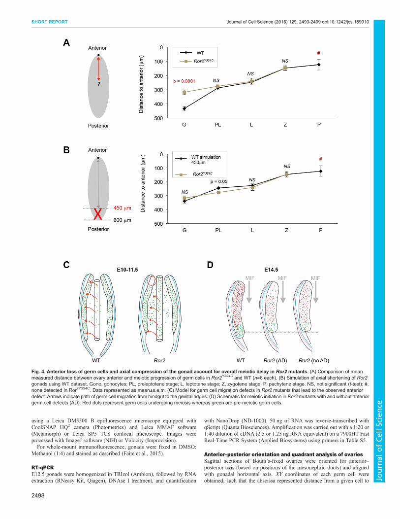
Given the potential discrepancy in the quadrant sizes for mutantswith smaller ovaries, we measured the absolute distance from everygerm cell to the anterior tip of the ovary in each section. In WT, astrong correlation between meiotic stage and distance from theanterior (r=0.869, P<0.0001) reflects the established anterior–posterior wave of meiotic initiation (Fig. 4A). In Ror2Y324C/Y324C
ovaries, distances were identical to WT for preleptotene, leptoteneand zygotene stages; however, a complete loss of pachytene isconcordant with a depletion of germ cells at the anterior tip(Fig. 4A). Most notably, the distance to gonocytes (pre-meioticgerm cells) was significantly shorter in Ror2Y324C/Y324C ovaries(Fig. 4A). This result corroborates proper scheduling of meioticentry according to position in mutants; reduced anterior–posteriordistance of gonocytes reflects a truncated gonad length, as measureddistances in Ror2Y324C/Y324C did not exceed 450 µm, whereasdistances up to 600 µm were found in WT. To model this posteriortruncation, we removed all germ cells in the WT dataset located>450 µm from the most-anterior end of the ovary. The resultingsimulation recapitulated the reduced mean distance to gonocytesobserved in Ror2Y324C/Y324C (Fig. 4B). Together, these anterior–posterior measurements and modeling the absent posterior extremeof the gonad corroborate the hypothesis that aberrant timing ofmeiotic entry in Ror2Y324C/Y324C ovaries results from defects in thedistribution of germ cells as well as gonad morphology.
Ror2 mutant phenotypes link germ cell migration to meioticinitiationAnterior depletion of germ cells in Ror2 ovaries is likely aconsequence of earlier migration defects. Despite absent Wnt5aexpression in the fetal ovary (Chawengsaksophak et al., 2012),similar anterior loss of germ cells in theWnt5a null gonads (data notshown) supports this idea. Strikingly, Sfrp1/2 double knockoutproduces an anterior germ-cell defect in the testis (Warr et al., 2009),highlighting the role of Wnt signaling in germ cell migration aswell as the persistence of early migration defects into sexdifferentiation.
The relationship between migration and positioning of germ cellsin the gonad remains unclear. Germ cell movement along theembryonic anterior–posterior axis is believed to occur passivelywithin the extending hindgut (Buehr et al., 1993; Molyneaux et al.,2001), and subsequent migration occurs in the dorsal and lateraldirections. Upon colonizing the gonad, germ cells proliferate morerapidly and lose their capacity to migrate (Di Carlo and De Felici,2000; Kierszenbaum and Tres, 2001). Hence, the absence of germcells in the gonad anterior could result from an earlier restriction inanterior extension of the hindgut (Fig. 4C,D). Our observations ofRor2 mutants are the first to connect migratory defects tocolonization of a specific region of the gonad.
2495
SHORT REPORT Journal of Cell Science (2016) 129, 2493-2499 doi:10.1242/jcs.189910
Journal
ofCe
llScience

Migration mutants favor gradient hypothesisAccording to the intrinsic clock model, appropriate meiotic entry byanterior–posterior location observed in Ror2 mutants could bemaintained only if the oldest germ cells destined for the anteriorextreme of the gonad entirely failed to reach their target. This
requirement is inconsistent with our observations of Ror2extragonadal germ cells in variable locations along the anterior–posterior axis, not exclusively in the anterior (Fig. S3C). Thediffusible signal model of meiotic initiation more congruouslyexplains the correct entry when indexed by location in Ror2mutants
Fig. 2. Diminished number of germ cells does not delay meiotic initiation. (A) Smaller size and reduced number of germ cells (Oct4–GFP) in whole Ror2−/−
ovaries. (B) Scatter plot shows correlation (r=0.605, P=0.0013) between number of germ cells (VASA+) per section and frequency of SYCP3+ germ cells.(C) Busulfan-treated ovary sections. (D) Frequency of SYCP3+ germ cells at E14.5 did not decrease in busulfan-treated ovaries. (E) Percentage of SYCP3+ germcells was similar between WT, Ror2−/− and busulfan treatment. (F) Data in Fig. 2B re-plotted, segregating ovaries with and without anterior defect (AD) in Rormutants. (G) Anterior depletion of germ cells (VASA, green) inRor2Y324Cmutants. (H) Sagittal sections show germ cells (VASA, green) that have entered meiosis(SYCP3, red) at E14.5. White arrowheads, anterior defects. Images are oriented with anterior at the top. Scale bars: 100 µm in A,G,H; 50 µm in C.
2496
SHORT REPORT Journal of Cell Science (2016) 129, 2493-2499 doi:10.1242/jcs.189910
Journal
ofCe
llScience

with aberrant germ cell distributions (Fig. 4D). Our data support themodel of aMIF that travels from the anterior to the posterior extremeof the ovary. Confirmation of this hypothesis must await moresophisticated lineage labeling and time-lapse imaging of germ cellswith heterogeneous migration trajectories.Our studies suggest that germ cell migration mutants could be
tools to further explore consequences of disruption to the anterior–posterior wave of meiosis. We highlight a need to rule out migrationdefects while studyingmeiosis to discern secondary effects resultingfrom positioning versus primary meiotic entry phenotypes. Finally,we conclude that neither germ cell number, nor localized loss ofgerm cells in the anterior ovary, affects the wave of meiotic entry,supporting a strong role for the niche and somatic gonad in drivingsex differentiation and meiotic initiation in the ovary.
MATERIALS AND METHODSMiceRor2Y324C/Y324C (Laird et al., 2011) and Ror2−/− (Arora et al., 2014) micewere mated with Oct4-ΔPE eGFP (Oct4–GFP) (Boiani et al., 2004) to labelgerm cells. Busulfan (Sigma, B2635) (Douglas et al., 2013) was injectedintraperitoneally (20 mg/ml in 40 μl) in pregnant E11.5 CD1 or FVBfemales crossed with C57/Bl6;Oct4-GFP males. Embryos were dissectedfrom timed matings and tails genotyped by PCR. The dark period was
19.00 h to 05.00 h and day of mating plug was identified as E0.5. All mousework was performed under UCSF Institutional Animal Care and UseCommittee guidelines in an AAALAC approved facility.
Gonad volumes, germ cell counting and meiotic stagingGonadswere fixed inBouin’s or4%paraformaldehyde, dehydrated, embeddedin paraffin, sectioned at 5 µm, and stained with hematoxylin and eosin (CancerDiagnostics). Every fifth or tenth section was analyzed for surface area(Histolab) and volumes were extrapolated based on number and thickness.Meiotic staging was based on nuclear shape and chromatin compaction (Abbyet al., 2016). Total germ cell counts were obtained from every fourth section,and calculated with Abercrombie’s formula correction (Guerquin et al., 2010).
Immunohistochemistry and immunofluorescenceParaformaldehyde-fixed gonads were embedded in paraffin or OCT (Tissue-Tek). Sections were subjected to heat-mediated antigen retrieval in citratebuffer (pH 6). For immunohistochemistry, endogenous peroxidase activitywas inhibited with 3% hydrogen peroxide. For immunohistochemistry andimmunofluorescence, sections were blocked in 2% horse serum, incubatedwith primary antibodies (Table S4) overnight at 4°C, then with peroxidase-conjugated secondary antibody (ImmPRESS™ kit, Vector Laboratories)and DAB (Vector Laboratories) or VIP (Vector Laboratories) or Alexa-Fluor-conjugated secondary antibodies (Invitrogen) and mounted withVectashield DAPI medium (Vector Laboratories). Imaging was performed
Fig. 3. Aberrant distribution of germ cells in the ovary causes meiotic entry defect. (A) Histologic sections with H&E stain at left and heat maps indicatingmeiotic progression on right. (B) Diagram showing division of ovaries into quadrants. (C) Relative frequency of germ cells counted in each quadrant in WTversus Ror2Y324C/Y324C, n=6 embryos. Data represented as mean±s.e.m. (D) Distribution of germ cells scored in each stage of meiotic prophase for eachquadrant, n=6 WT and n=6 Ror2Y324C/Y324C ovaries. Gono, gonocytes; PL, preleptotene stage; L, leptotene stage; Z, zygotene stage; P, pachytene stage.***P<0.001, **P<0.01, *P<0.05 by t-test. Scale bars: 200 µm.
2497
SHORT REPORT Journal of Cell Science (2016) 129, 2493-2499 doi:10.1242/jcs.189910
Journal
ofCe
llScience
using a Leica DM5500 B epifluorescence microscope equipped withCoolSNAP HQ2 camera (Photometrics) and Leica MMAF software(Metamorph) or Leica SP5 TCS confocal microscope. Images wereprocessed with ImageJ software (NIH) or Volocity (Improvision).
For whole-mount immunofluorescence, gonads were fixed in DMSO:Methanol (1:4) and stained as described (Faire et al., 2015).
RT-qPCRE12.5 gonads were homogenized in TRIzol (Ambion), followed by RNAextraction (RNeasy Kit, Qiagen), DNAse I treatment, and quantification
with NanoDrop (ND-1000). 50 ng of RNA was reverse-transcribed withqScript (Quanta Biosciences). Amplification was carried out with a 1:20 or1:40 dilution of cDNA (2.5 or 1.25 ng RNA equivalent) on a 7900HT FastReal-Time PCR System (Applied Biosystems) using primers in Table S5.
Anterior–posterior orientation and quadrant analysis of ovariesSagittal sections of Bouin’s-fixed ovaries were oriented for anterior–posterior axis (based on positions of the mesonephric ducts) and alignedwith gonadal horizontal axis. XY coordinates of each germ cell wereobtained, such that the abscissa represented distance from a given cell to
Fig. 4. Anterior loss of germ cells and axial compression of the gonad account for overall meiotic delay in Ror2 mutants. (A) Comparison of meanmeasured distance between ovary anterior and meiotic progression of germ cells in Ror2Y324C and WT (n=6 each). (B) Simulation of axial shortening of Ror2gonads using WT dataset. Gono, gonocytes; PL, preleptotene stage; L, leptotene stage; Z, zygotene stage; P, pachytene stage. NS, not significant (t-test); #,none detected in Ror2Y324C. Data represented as mean±s.e.m. (C) Model for germ cell migration defects in Ror2 mutants that lead to the observed anteriordefect. Arrows indicate path of germ cell migration from hindgut to the genital ridges. (D) Schematic for meiotic initiation in Ror2mutants with and without anteriorgerm cell defects (AD). Red dots represent germ cells undergoing meiosis whereas green are pre-meiotic germ cells.
2498
SHORT REPORT Journal of Cell Science (2016) 129, 2493-2499 doi:10.1242/jcs.189910
Journal
ofCe
llScience

most anterior point in the gonad section. Anterior–posterior axis length wasdetermined (Histolab) and mean length of Ror2Y324C/Y324C ovary sectionswas used for simulation of shortened axis in WT gonads. For quadrantanalysis, sections were parsed into four equidistant compartments along thehorizontal axis.
StatisticsStatistics were performed in Excel (Microsoft) using the StatPlus plug-in.
AcknowledgementsWe thank Svetlana Altshuler-Keylin and Kevin Ebata for feedback on this work.
Competing interestsThe authors declare no competing or financial interests.
Author contributionsR.A. and D.J.L. conceived the project. R.A., E.A., A.D.J.R., A.V.C., M.D.K. and V.C.did the experiments. H.-Y.H.H. provided reagents and critical feedback. R.A., E.A.,H.-Y.H.H., G.L. and D.J.L. prepared the figures and wrote the manuscript.
FundingCalifornia Institute for Regenerative Medicine [TG TG2-01153] and NationalInstitutes of Health [TG 5T32HD007263-32] to R.A., Irtelis (Commissariat a l’ÉnergieAtomique et aux Énergies Alternatives) and Fondation ARC pour la Recherche sur leCancer to E.A., National Science Foundation fellowship to A.V.C., National Institutesof Health diversity supplement [DP2OD007420 S1] to A.D.J.R., University ofCalifornia, San Francisco Program for Breakthrough Biomedical Research, NationalInstitutes of Health grants [1R21ES023297 and DP2OD007420] and generousphilanthropy to D.J.L. and Institut Universitaire de France support to G.L. Depositedin PMC for release after 12 months.
Supplementary informationSupplementary information available online athttp://jcs.biologists.org/lookup/doi/10.1242/jcs.189910.supplemental
ReferencesAbby, E., Tourpin, S., Ribeiro, J., Daniel, K., Messiaen, S., Moison, D.,Guerquin, J., Gaillard, J-C, Armengaud, J., Langa, F. et al. (2016).Implementation of meiosis prophase I programme requires a conservedretinoid-independent stabilizer of meiotic transcripts. Nat. Commun. 7, 10324.
Arora, R., Altman, E., Tran, N. D. and Laird, D. J. (2014). Novel domains ofexpression for orphan receptor tyrosine kinase Ror2 in the human and mousereproductive system. Dev. Dyn. 243, 1037-1045.
Boiani, M., Kehler, J. and Scholer, H. R. (2004). Activity of the germline-specificOct4-GFP transgene in normal and clone mouse embryos. Methods Mol. Biol.254, 1-34.
Bowles, J., Knight, D., Smith, C., Wilhelm, D., Richman, J., Mamiya, S., Yashiro,K., Chawengsaksophak, K., Wilson, M. J., Rossant, J. et al. (2006). Retinoidsignaling determines germ cell fate in mice. Science 312, 596-600.
Buehr, M., McLaren, A., Bartley, A. and Darling, S. (1993). Proliferation andmigration of primordial germ cells in We/We mouse embryos. Dev. Dyn. 198,182-189.
Bullejos, M. and Koopman, P. (2004). Germ cells enter meiosis in a rostro-caudalwave during development of the mouse ovary. Mol. Reprod. Dev. 68, 422-428.
Chassot, A.-A., Gregoire, E. P., Lavery, R., Taketo, M. M., de Rooij, D. G.,Adams, I. R. and Chaboissier, M. C. (2011). RSPO1/beta-catenin signalingpathway regulates oogonia differentiation and entry into meiosis in themouse fetalovary. PLoS ONE 6, e25641.
Chawengsaksophak, K., Svingen, T., Ng, E. T., Epp, T., Spiller, C. M., Clark, C.,Cooper, H. andKoopman, P. (2012). Loss ofWnt5a disrupts primordial germ cellmigration and male sexual development in mice. Biol. Reprod. 86, 1-12.
DeChiara, T. M., Kimble, R. B., Poueymirou, W. T., Rojas, J., Masiakowski, P.,Valenzuela, D. M. and Yancopoulos, G. D. (2000). Ror2, encoding a receptor-like tyrosine kinase, is required for cartilage and growth plate development. Nat.Genet. 24, 271-274.
Di Carlo, A. and De Felici, M. (2000). A role for E-cadherin in mouse primordialgerm cell development. Dev. Biol. 226, 209-219.
Douglas, N. C., Arora, R., Chen, C. Y., Sauer, M. V. and Papaioannou, V. E.(2013). Investigating the role of tbx4 in the female germline in mice. Biol. Reprod.89, 148.
Faire, M., Skillern, A., Arora, R., Nguyen, D. H., Wang, J., Chamberlain, C.,German, M. S., Fung, J. C. and Laird, D. J. (2015). Follicle dynamics and globalorganization in the intact mouse ovary. Dev. Biol. 403, 69-79.
Guerquin, M.-J., Duquenne, C., Lahaye, J.-B., Tourpin, S., Habert, R. andLivera, G. (2010). New testicular mechanisms involved in the prevention of fetalmeiotic initiation in mice. Dev. Biol. 346, 320-330.
Hunt, P. A. and Hassold, T. J. (2008). Human female meiosis: what makes a goodegg go bad? Trends Genet. 24, 86-93.
Kierszenbaum, A. L. and Tres, L. L. (2001). Primordial germ cell-somatic cellpartnership: a balancing cell signaling act. Mol. Reprod. Dev. 60, 277-280.
Koubova, J., Menke, D. B., Zhou, Q., Capel, B., Griswold, M. D. and Page, D. C.(2006). Retinoic acid regulates sex-specific timing of meiotic initiation in mice.Proc. Natl. Acad. Sci. USA 103, 2474-2479.
Koubova, J., Hu, Y.-C., Bhattacharyya, T., Soh, Y. Q., Gill, M. E., Goodheart,M. L., Hogarth, C. A., Griswold, M. D. and Page, D. C. (2014). Retinoic acidactivates two pathways required for meiosis in mice. PLoS Genet. 10, e1004541.
Kumar, S., Chatzi, C., Brade, T., Cunningham, T. J., Zhao, X. and Duester, G.(2011). Sex-specific timing of meiotic initiation is regulated by Cyp26b1independent of retinoic acid signalling. Nat. Commun. 2, 151.
Laird, D. J., Altshuler-Keylin, S., Kissner, M. D., Zhou, X. and Anderson, K. V.(2011). Ror2 enhances polarity and directional migration of primordial germ cells.PLoS Genet. 7, e1002428.
Liu, C.-F., Bingham, N., Parker, K. and Yao, H. H.-C. (2009). Sex-specific roles ofbeta-catenin in mouse gonadal development. Hum. Mol. Genet. 18, 405-417.
Liu, C.-F., Parker, K. and Yao, H. H.-C. (2010). WNT4/beta-catenin pathwaymaintains female germ cell survival by inhibiting activin betaB in the mouse fetalovary. PLoS ONE 5, e10382.
McLaren, A. (2003). Primordial germ cells in the mouse. Dev. Biol. 262, 1-15.McLaren, A. and Southee, D. (1997). Entry of mouse embryonic germ cells into
meiosis. Dev. Biol. 187, 107-113.Menke, D. B., Koubova, J. and Page, D. C. (2003). Sexual differentiation of germ
cells in XX mouse gonads occurs in an anterior-to-posterior wave. Dev. Biol. 262,303-312.
Molyneaux, K. A., Stallock, J., Schaible, K. and Wylie, C. (2001). Time-lapseanalysis of living mouse germ cell migration. Dev. Biol. 240, 488-498.
Naillat, F., Prunskaite-Hyyrylainen, R., Pietila, I., Sormunen, R., Jokela, T.,Shan, J. and Vainio, S. J. (2010). Wnt4/5a signalling coordinates cell adhesionand entry into meiosis during presumptive ovarian follicle development.Hum.Mol.Genet. 19, 1539-1550.
Ohkubo, Y., Shirayoshi, Y. and Nakatsuji, N. (1996). Autonomous regulation ofproliferation and growth arrest in mouse primordial germ cells studied by mixedand clonal cultures. Exp. Cell Res. 222, 291-297.
Ottolenghi, C., Pelosi, E., Tran, J., Colombino, M., Douglass, E., Nedorezov, T.,Cao, A., Forabosco, A. and Schlessinger, D. (2007). Loss of Wnt4 and Foxl2leads to female-to-male sex reversal extending to germ cells. Hum. Mol. Genet.16, 2795-2804.
Runyan, C., Gu, Y., Shoemaker, A., Looijenga, L. and Wylie, C. (2008). Thedistribution and behavior of extragonadal primordial germ cells in Baxmutant micesuggest a novel origin for sacrococcygeal germ cell tumors. Int. J. Dev. Biol. 52,333-344.
Spiller, C. M. and Bowles, J. (2015). Sex determination in mammalian germ cells.Asian J. Androl. 17, 427-432.
Takeuchi, S., Takeda, K., Oishi, I., Nomi, M., Ikeya,M., Itoh, K., Tamura, S., Ueda,T., Hatta, T., Otani, H. et al. (2000). Mouse Ror2 receptor tyrosine kinase isrequired for the heart development and limb formation. Genes Cells 5, 71-78.
Vainio, S., Heikkila, M., Kispert, A., Chin, N. and McMahon, A. P. (1999). Femaledevelopment in mammals is regulated by Wnt-4 signalling. Nature 397, 405-409.
Warr, N., Siggers, P., Bogani, D., Brixey, R., Pastorelli, L., Yates, L., Dean, C. H.,Wells, S., Satoh, W., Shimono, A. et al. (2009). Sfrp1 and Sfrp2 are required fornormal male sexual development in mice. Dev. Biol. 326, 273-284.
Yao, H. H.-C, DiNapoli, L. and Capel, B. (2003). Meiotic germ cells antagonizemesonephric cell migration and testis cord formation in mouse gonads.Development 130, 5895-5902.
2499
SHORT REPORT Journal of Cell Science (2016) 129, 2493-2499 doi:10.1242/jcs.189910
Journal
ofCe
llScience