Embed Size (px)
Citation preview
Constraint-induced movement therapy (CIMT) is a physi-cal rehabilitation regime that has been previously shownto improve motor function in chronic hemiparetic strokepatients. However, the neural mechanisms supportingrehabilitation-induced motor recovery are poorly under-stood. The goal of this study was to assess motor corticalreorganization after CIMT using functional magneticresonance imaging (fMRI). In a repeated-measuresdesign, 4 incompletely recovered chronic stroke patientstreated with CIMT underwent motor function testing andfMRI. Five age-matched normal subjects were alsoimaged. A laterality index (LI) was determined from thefMRI data, reflecting the distribution of activation inmotor cortices contralateral compared with ipsilateral tothe moving hand. Pre-intervention fMRI showed a lowerLI during affected hand movement of stroke patients (LI =0.23 ± 0.07) compared to controls (LI unaffected patienthand = 0.65 ± 0.10; LI dominant normal hand = 0.65 ±0.11; LI nondominant normal hand = 0.69 ± 0.11; P <0.05) due to trends toward increased ipsilateral motorcortical activation. Motor function testing showed thatpatients made significant gains in functional use of thestroke-affected upper extremity (detected by the MotorActivity Log) and significant reductions in motor impair-ment (detected by the Fugl-Meyer Stroke Scale and theWolf Motor Function Test) immediately after CIMT, andthese effects persisted at 6-month follow-up. The behav-ioral effects of CIMT were associated with a trend towarda reduced LI from pre-intervention to immediately post-intervention (LI = –0.01 ± 0.06; P = 0.077) and 6 monthspost-intervention (LI = –0.03 ± 0.15). Stroke-affectedhand movement was not accompanied by mirror move-
ments during fMRI, and electromyographic measures ofmirror recruitment under simulated fMRI conditionswere not correlated with LI values. These data providepreliminary evidence that gains in motor function pro-duced by CIMT in chronic stroke patients may be associ-ated with a shift in laterality of motor cortical activationtoward the undamaged hemisphere.
Key Words: Stroke—Hemiparesis—Rehabilitation—Magnetic resonance imaging—Motor cortex.
Hemiparesis is the most common acutedeficit of stroke.1 Most hemiparetic patientsexperience some degree of motor recovery
within the first 6 months after stroke.2,3 Post-stroke physical rehabilitation may produce gains inmotor function beyond those occurring sponta-neously.4 However, the neural mechanisms mediat-ing rehabilitation-induced motor recovery are poor-ly understood.
Constraint-induced movement therapy (CIMT) isa physical rehabilitation approach that has beenshown to further motor recovery of the affectedupper extremity in chronic stroke patients withmild to moderate hemiparesis.5–7 CIMT involves theintensive motor training of the stroke-affected limbcoupled with restricted use of the unaffected limb.Determining the neural changes that underliemotor recovery induced by CIMT would help elu-cidate the mechanisms by which physical rehabili-tation can promote post-stroke motor recovery andmay guide the development of new, more effica-cious therapies.
Functional magnetic resonance imaging (fMRI)has proved to be a useful tool for evaluating corti-cal reorganization after stroke. Motor recovery dur-ing the early period after stroke has been shown byfMRI to be associated with shifts in the extent ofactivation in the sensorimotor cortex contralateralrelative to that ipsilateral to affected hand move-ment.8 In patients later after stroke, fMRI has
Motor Recovery and Cortical Reorganizationafter Constraint-Induced Movement Therapy
in Stroke Patients: A Preliminary Study
Judith D. Schaechter, Eduard Kraft, Timothy S. Hilliard, Rick M. Dijkhuizen,Thomas Benner, Seth P. Finklestein, Bruce R. Rosen, and Steven C. Cramer
Copyright © 2002 The American Society of Neurorehabilitation 1
From the Massachusetts General Hospital–NMR Center,Department of Radiology, Charlestown, MA (JDS, RMD, TB,BRR); the University of Ulm Medical School, Ulm, Germany(EK); Northeastern University, Department of Physical Therapy,Boston (TSH); Massachusetts General Hospital, Department ofNeurology, Boston (SPF); and the University of Washington,Department of Neurology, Seattle, WA (SCC).
Address correspondence and reprint requests to Judith D.Schaechter, PhD, PT, Massachusetts General Hospital–NMRCenter, 13th Street, Building 149, Room 2301, Charlestown, MA02129. E-mail: [email protected].
Schaechter JD, Kraft E, Hilliard TS, et al. Motor recovery andcortical reorganization after constraint-induced movement ther-apy in stroke patients: a preliminary study. Neurorehabil NeuralRepair 2002; 16:000–000.
© 2002 American Society of Neurorehabilitation. All rights reserved. Not for commercial use or unauthorized distribution. at SPAULDING REHAB HOSP on June 4, 2008 http://nnr.sagepub.comDownloaded from
revealed that affected hand movement is associat-ed with a posterior shift in the center of activationin contralateral sensorimotor cortex9 and increasedactivation in ipsilateral sensorimotor cortex, ipsilat-eral premotor cortex (PMC), bilateral supplemen-tary motor area (SMA), and the peri-infarct regionin cases of cortical stroke.10,11
A recent pilot study by Levy et al. applied fMRIbefore and after CIMT in 2 cortical stroke patients.12
This study found that gains in motor function of thestroke-affected upper extremity after CIMT wereaccompanied by increased activation in the peri-infarct region in both patients and bilateral sensori-motor cortices in 1 patient. However, neitherpatient could fully perform the motor activationtask at the pre-intervention fMRI session because ofpoor hand motor function. Thus, the observedincreases in cortical activation observed at the post-intervention fMRI session could have been due toimproved performance of the motor activation task.
The goal of the current study was to evaluatecortical reorganization associated with motorrecovery produced by CIMT using fMRI. Weaddressed our concern of a possible contribution ofperformance differences to changes in cortical acti-vation after CIMT by (1) enrolling selected patientswho could perform the fMRI motor activation taskat study entry and (2) controlling task performanceparameters over time. We elected to focus onassessing reorganization in motor cortical areasbecause activation changes in these areas havebeen previously shown to relate to motor recoveryafter stroke.
SUBJECTS AND METHODS
Subjects
Entry criteria for stroke patients were as follows:(1) a single unilateral ischemic stroke > 6 monthsearlier that caused acute hemiplegia or severehemiparesis that included the loss of individuatedfinger movement; (2) the ability to perform the
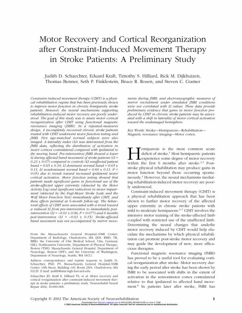
motor task used during fMRI (0.5 Hz, 4-finger flex-ion and extension; see below) with > 50% of fullnormal active range of motion at the metacar-pophalangeal and interphalangeal joints of the 4fingers of the affected hand; (3) affected upperextremity motor status and neuropsychological sta-tus compatible with being able to competently par-ticipate in CIMT and related testing, as describedpreviously;5–7 and (4) premorbid right-hand domi-nance.13 Table 1 gives the demographic and stroke-related data of the 4 stroke patients enrolled in thisstudy (mean age = 57 ± 17 years). Five normal sub-jects (mean age = 57 ± 13 years; range 40 to 71years; 3 female) each with normal neurologicalexamination, no history of stroke, and right-handdominance13 served as controls. Written informedconsent was obtained from all subjects, asapproved by the local institutional review board.
Study Protocol
Consecutive stroke patients participated in 2 weeksof CIMT, preceded and followed by fMRI as well astesting of motor function and surface electromyog-raphy (EMG). There were 2 pre-intervention testingsessions (2 weeks prior and 1 day prior) and 3post-intervention testing sessions (immediatelyafter [within 24 hours], 2 weeks after, and 6 monthsafter), with the exceptions of no fMRI 2 weekspost-intervention and no EMG 6 months post-intervention. Each normal control subject partici-pated in a single fMRI session.
CIMT
CIMT was performed as previously described.5–7
Briefly, the 2-week CIMT period involved intensivetraining of the affected upper extremity andrestricted use of the unaffected upper extremity.There were 10 training sessions on the 5 weekdaysof the 2-week period, each session lasting 4 hours.During these sessions, each patient practiced a set
J. D. Schaechter et al.
2 Neurorehabilitation and Neural Repair 16(4); 2002
Table 1. Demographic and Stroke-Related Data for Stroke Patients
Stroke Patient Age Side of Time Post-Stroke Number (years) Gender Infarct Location Hemiparesis (months)
1 36 Female Left internal capsule Right 112 77 Male Left pons Right 203 55 Male Left frontal cortex Right 124 59 Male Right parietal cortex Left 7
© 2002 American Society of Neurorehabilitation. All rights reserved. Not for commercial use or unauthorized distribution. at SPAULDING REHAB HOSP on June 4, 2008 http://nnr.sagepub.comDownloaded from
of tasks (15 to 25) selected on the basis of individ-ual upper extremity motor abilities and deficits.These tasks included gross and fine functionalmotor skills, such as grasping and using a spoon,and picking up an object with a specified graspand transporting it to a specified target. Each taskwas incrementally modified during the training ses-sions to challenge and improve motor function.Restricted use of the unaffected upper extremityduring the 2-week CIMT period was achieved bywearing a resting hand splint during most wakinghours.
Motor Function Testing
Functional use of the stroke-affected upperextremity was evaluated using the Amount of UseScale of the Motor Activity Log (MAL). This test is astructured interview that evaluates by self-reportthe amount of use of the stroke-affected upperextremity in 30 common activities of daily livingover a specified period of time (scale 0 to 5).5
Motor impairments of the stroke-affected upperextremity were evaluated by the following tests: (1)maximum grip strength (N) of the affected andunaffected hands using a computerized dyna-mometer;14 (2) maximum frequency (Hz) of 4-fin-ger flexion and extension of the affected and unaf-fected hands using the same apparatus and fixedactive range of motion as described below; (3)upper extremity motor section of the Fugl-MeyerStroke Scale (FMSS) (scale 0 to 66);15 and (4) amodified version5 of the timed (s) items of the WolfMotor Function Test (WMFT)16 to evaluate themovement duration of the affected and unaffectedupper extremities.
Each motor function measure of the stroke-affected upper extremity was converted to percent-age of normal function on the basis of either themeasure acquired from the stroke-unaffected upperextremity or the standardized test value of normalfunction.
Electromyographic Testing
To evaluate whether ipsilateral motor corticalactivation might be related to mirror movements,EMG was applied in stroke patients during per-formance of the fMRI motor task (0.5 Hz, 4-fingerflexion and extension; see below) under simulatedscanning conditions. Pre-amplified surface elec-trodes (Biopac Systems, Inc., Santa Barbara, CA)
were positioned bilaterally over the finger flexorand finger extensor muscle groups. EMG data werecollected (1000 samples/s) for the entire durationof the motor paradigm, 1 run during voluntarymovement of the stroke-affected hand, and 1 runduring voluntary movement of the stroke-unaffectedhand.
The EMG data were analyzed for mirror recruit-ment, which we defined as synchronous involun-tary activity of contralateral homologous musclesduring voluntary unilateral muscle recruitment.First, the data were full-wave rectified and band-pass filtered (20 to 250 Hz). Then, the root meansquare (RMS) of contralateral finger flexor andextensor EMG activity coincident with rest and vol-untary movement epochs was calculated. Rest EMGactivity was calculated by taking the average RMSduring all rest epochs. Movement EMG activity wascalculated by taking the average RMS during each2-second flexion and extension phase during allmovement epochs. Phase onset was based on visu-al inspection of the recordings for burst initiation ofvoluntary finger flexor and extensor muscle activi-ty. Finally, a mirror recruitment index for the con-tralateral finger flexors and finger extensors wascalculated by dividing EMG activity of the con-tralateral muscle group during rest epochs by thatduring movement epochs. An index of 1.00 indi-cates no mirror recruitment, an index < 1.00 indi-cates mirror recruitment with increased activity ofthe contralateral muscle group during movementepochs relative to rest epochs, and an index > 1.00indicates increased activity of contralateral musclegroup during rest epochs relative to movementepochs.
Imaging Protocol
Imaging data were acquired at the MassachusettsGeneral Hospital–NMR Center using a 1.5 T MRIscanner (General Electric Signa modified byAdvanced NMR Systems for 2 stroke and 3 controlsubjects; Siemens Sonata for 2 stroke and 2 controlsubjects) and a quadrature head coil. Head motionwas minimized by a bite bar and stabilizing strapsacross the arms and chest. For each subject, 4 setsof blood oxygenation level–dependent (BOLD)functional images were collected using a T2*-weighted gradient-echo, echo planar imaging (EPI)sequence (repetition time [TR] = 2.5 s; echo time[TE] = 50 ms; field of view = 200 × 200 mm2; in-plane resolution = 3.125 mm2; 20 slices; 7-mm slicethickness; 104 images/slice). Two sets of structural
Post-Stroke Therapy and Cortical Reorganization
Neurorehabilitation and Neural Repair 16(4); 2002 3 © 2002 American Society of Neurorehabilitation. All rights reserved. Not for commercial use or unauthorized distribution.
at SPAULDING REHAB HOSP on June 4, 2008 http://nnr.sagepub.comDownloaded from
images were also collected: (1) T1-weighted gradi-ent-echo EPI (TR = 8 s; TE = 39 ms) taken at thesame resolution (3.125 mm2) and in plane with thefunctional images and (2) T1-weighted gradient-echo conventional images (TR = 500 ms; TE = 12ms) taken at high resolution (0.78 mm2) and inplane with the functional images. Slices were ori-ented parallel to the line connecting the anteriorcommissure to the posterior commissure and cov-ered the cerebral hemispheres and the superiorthree quarters of the cerebellum. For each strokepatient, slice positioning applied at the 1st scanningsession was used to fine-tune positioning at all sub-sequent scanning sessions.
During functional imaging, a boxcar function wasused with 7 × 20 second rest epochs and 6 × 20second movement epochs. The motor task wasflexion and extension of the 4 fingers in unison. Tostandardize the kinematics of this motor task, westabilized the shoulders in the neutral position andthe elbows in slight flexion (~15°) with straps.Further, we used a plastic apparatus that stabilizedthe forearms and wrists in the neutral position andthe thumbs in slight flexion and abduction. Thisapparatus also fixed the maximum excursionthrough which the 4 fingers actively flexed andextended. For normal control subjects, maximumexcursion of the 4 fingers was set at full flexion andextension at the metacarpophalangeal and interpha-langeal joints. For patients, maximum excursion ofthe stroke-affected fingers was set on the basis ofthe available active range of motion; this was > 50%of full normal active range of motion in all patients.Maximum excursion of the stroke-unaffected fin-gers was set to match that determined for thestroke-affected fingers. This fixed range of motionwas determined at the 1st scanning session foreach subject and was applied unchanged at all sub-sequent scanning sessions.
During each of 4 functional runs, subjects per-formed the motor task unilaterally, alternatingbetween right (runs 1 and 3) and left (runs 2 and4) hand performance. Auditory cues were deliv-ered pneumatically to a headset worn by each sub-ject. Stimulus presentation software (MacStim ver-sion 2.0) was used to generate a metronome beepat 0.5 Hz throughout each functional run, and “go”and “stop” verbal cues to trigger movement andrest epochs, respectively. The 0.5-Hz beep wasused to pace the motor task during movementepochs. Prior to each functional run, subjects wereinstructed to keep their eyes closed. All subjectswere trained prior to each scanning session toaccurately and consistently perform the hand
motor task. A research investigator standing besidethe scanning bed visually monitored hand motorperformance and possible unintended movements,including mirror movements of the contralateralhand.
Image processing and analysis were performedon Sun SPARC workstations using software devel-oped at the Massachusetts General Hospital–NMRCenter. The raw images were motion corrected,17
BOLD signals were drift corrected and intensitynormalized, and the 2 functional runs collected formovement of the right and left hands were aver-aged. Statistical activation maps were generatedvoxel by voxel using Student’s t test, contrastingimages acquired during rest epochs with thoseacquired during movement epochs. The boxcarfunction was shifted 5 seconds (2 × TR) to accountfor the delay between neuronal activity andincreased cerebral blood flow.18 The statistical acti-vation maps were registered with the correspond-ing structural images. Data sets with excessivemotion artifact, defined as those yielding statisticalactivation maps (P < 0.001) with spuriously activat-ed voxels throughout the brain volume or at leastone quarter of the brain’s circumference, wereeliminated from further analysis.
The quantification of activation in motor corticalareas was conducted by a region-of-interest (ROI)analysis. For each subject, the structural scans wereused to outline 3 bilateral ROIs on the basis ofanatomic landmarks:19,20 primary motor cortex(M1), PMC, and SMA. The M1 encompassed theposterior half of the precentral gyrus and extendedposteriorly midway into the central sulcus. ThePMC was taken as the anterior half of the precen-tral gyrus and extended just rostral of the precen-tral sulcus. The SMA was taken as the medial cor-tex superior to the cingulate gyrus, anterior to themid-precentral gyrus, and extended just rostral ofthe vertical anterior commissure line. Voxels withinthese ROIs that met the following criteria were con-sidered activated: (1) significant difference (P <0.001, Student’s t test, uncorrected) in BOLD signalintensity during rest compared with movementconditions; (2) BOLD signal intensity change < 5%,to exclude voxels with signal coming from drainingveins;21 and (3) clustered with another significantlyactivated voxel, as a means of correcting for multi-ple comparisons.22,23
The extent of activation in each ROI was deter-mined by counting the number of activated voxels.On the basis of these counts, a laterality index (LI)was calculated to provide an estimate of the rela-tive hemispheric activation in motor cortices.10 This
J. D. Schaechter et al.
4 Neurorehabilitation and Neural Repair 16(4); 2002 © 2002 American Society of Neurorehabilitation. All rights reserved. Not for commercial use or unauthorized distribution.
at SPAULDING REHAB HOSP on June 4, 2008 http://nnr.sagepub.comDownloaded from
index was defined as [(cM1 + cPMC + cSMA) – (iM1+ iPMC + iSMA)] / [(cM1 + cPMC + cSMA) + (iM1+ iPMC + iSMA)], where c = contralateral and i =ipsilateral. LI values ranged from +1, indicating thatall motor cortical activation occurred in the hemi-sphere contralateral to the moving hand, to –1,indicating that all motor cortical activation occurredin the hemisphere ipsilateral to the moving hand.
To further examine cortical reorganization afterCIMT, we quantified the magnitude of activation incontralateral M1 of stroke patients. We elected tofocus on this cortical area because previous trans-cranial magnetic stimulation (TMS) studies haveshown that motor gains after CIMT in chronicstroke patients are associated with a change in con-tralateral M1 excitability.24,25 We determined themean percentage BOLD signal intensity change inM1 contralateral to hand movement for all activat-ed voxels and for the most significantly activatedcluster of voxels (defined as the 4 contiguous in-plane voxels with the highest mean significance).
Statistical Analysis
StatView (version 4.5) and SuperANOVA (version1.11) were used for statistical procedures. Paired ttests were used to compare measures acquiredfrom stroke patients at 2 testing sessions. Repeated-measures analysis of variance (ANOVA) tests with 1within-subjects factor (side of hand movement) and1 between-subjects factor (subject group) wereused to examine an interaction effect on brain acti-vation. If a significant interaction was detected,paired and unpaired t tests were used appropriate-ly to test for differences between means. Repeated-measures ANOVA tests with planned comparisonsof means were used to compare motor functiontesting measures in stroke patients over the studyperiod. The Pearson product-moment correlationcoefficient was used to examine correlations. P val-ues of < 0.05 were considered significant. Data arepresented as group means ± standard error of themean.
RESULTS
Motor Function
Figure 1 presents the results of motor functiontesting in stroke patients over the course of thestudy. Motor function test scores were not signifi-cantly different at the 2 pre-intervention sessions
and were therefore averaged. Patient 2 was notavailable for testing at 6 months post-intervention.Pre-intervention, there was markedly reduced useof the affected upper extremity in functional activ-ities (measured by the MAL) and moderate upperextremity motor impairment (measured by gripstrength, frequency of 4-finger flexion and exten-sion, FMSS, and WMFT). MAL scores significantlyincreased from pre-intervention to immediatelypost-intervention (P < 0.01). This behavioral effectwas retained on a group basis at 2 weeks post-intervention (P < 0.01) and at 6 months post-inter-vention (P < 0.01) compared to pre-intervention.However, 1 stroke patient (patient 4) showed littleretention of this behavioral effect at the 6-monthfollow-up testing session compared to earlier post-intervention testing sessions (MAL pre-intervention =28%, immediate post-intervention = 62%, 2-weekpost-intervention = 65%, 6-month post-intervention =39%). Scores on the FMSS significantly increasedfrom pre-intervention to immediately post-interven-tion (P < 0.01); this effect was retained at the 2-week post-intervention (P < 0.01) and 6-monthpost-intervention (P < 0.01) testing sessions.Performance on the WMFT significantly improvedfrom pre-intervention to immediately post-interven-tion (P < 0.01); this effect was slightly reduced butstill significant at the 2-week post-intervention (P <0.05) and 6-month post-intervention (P < 0.05) test-ing sessions. Grip strength was significantlyincreased at 6 months post-intervention comparedto pre-intervention (P < 0.05), but not at earliertesting sessions. There was no significant change inthe frequency of 4-finger flexion and extensionfrom pre-intervention to any post-intervention test-ing session.
EMG testing in stroke patients showed no differ-ence in the mirror recruitment index during affect-ed or unaffected hand movement at the 2 pre-intervention and 2 post-intervention sessions.Therefore, mirror recruitment index values wereaveraged over the 2 respective sessions. Pre-intervention, there was no difference in mirrorrecruitment of contralateral finger flexors or exten-sors during voluntary movement of the affectedhand compared to the unaffected hand (mirrorrecruitment index affected finger flexion = 1.01± 0.01; unaffected finger flexion = 1.00 ± 0.01;affected finger extension = 0.88 ± 0.07; unaffectedfinger extension = 0.94 ± 0.04). Post-intervention,there was also no difference in mirror recruitmentof contralateral finger flexors or extensors duringvoluntary movement of the affected hand com-pared to the unaffected hand (affected finger flex-
Post-Stroke Therapy and Cortical Reorganization
Neurorehabilitation and Neural Repair 16(4); 2002 5 © 2002 American Society of Neurorehabilitation. All rights reserved. Not for commercial use or unauthorized distribution.
at SPAULDING REHAB HOSP on June 4, 2008 http://nnr.sagepub.comDownloaded from
ion = 0.97 ± 0.03; unaffected finger flexion = 1.00± 0.01; affected finger extension = 0.97 ± 0.05;unaffected finger extension = 0.95 ± 0.04).
fMRI
All subjects accurately and consistently per-formed the hand motor task at each scanning ses-sion. No subject exhibited unintended mirrormovements of the contralateral hand during voli-tional unilateral motor task performance at anyscanning session. The fMRI data sets acquired frompatient 3 at the 1st pre-intervention scanning ses-sion and patient 2 during affected hand movementat the immediate post-intervention scanning ses-sion were uninterpretable because of excessivehead motion and therefore not included in subse-quent analysis. Patient 2 was also not available fortesting at the 6-month follow-up.
Activation during the movement of either handof normal control subjects or the unaffected handof stroke patients was predominantly in the con-tralateral hemisphere (i.e., M1, PMC, SMA, andsomatosensory cortex) and ipsilateral cerebellum;more modest activation was variably observed inthese cortical areas of the ipsilateral hemisphere.Before initiating CIMT, activation during stroke-affected hand movement resulted in activation inthese same brain regions, although activation in theipsilateral hemisphere was typically increased.Further, contralateral M1 activation was decreasedin the 2 cortical stroke patients and increased inthe 2 subcortical stroke patients during affectedhand movement compared to unaffected hand
movement. Figure 2 is an example of cortical acti-vation patterns in a normal control subject and astroke patient pre-intervention (cortical stroke,patient 4).
Table 2 presents quantitative results of the extentof activation in motor cortical ROIs, in number ofvoxels, during motor task performance in normalcontrol subjects and stroke patients pre-interven-tion and the resultant LIs. At the 2 pre-interventionfMRI sessions in stroke patients, there was no sig-nificant difference in the extent of activation in anyROI during affected or unaffected hand movement;therefore, average values for each patient weredetermined. There was a significant interactioneffect between subject group and side of handmovement on ipsilateral M1 activation (P < 0.05).Further analysis revealed that there were trendstoward increased ipsilateral M1 activation duringstroke-affected hand movement compared tomovement of the stroke-unaffected hand (P = 0.08)as well as the dominant (P = 0.17) and nondomi-nant (P = 0.10) hands of normal control subjects.There was a nearly significant interaction effectbetween subject group and side of hand movementon ipsilateral SMA activation (P = 0.06). There wasa significant interaction effect between subject groupand side of hand movement on LI (P < 0.05), withfurther analysis revealing a significant decrease inLI during stroke-affected hand movement com-pared to movement of the stroke-unaffected handand either hand of normal control subjects (P <0.05; Fig. 3). There was no correlation between thepre-intervention LI and mirror recruitment indexduring affected or unaffected hand movement instroke patients.
J. D. Schaechter et al.
6 Neurorehabilitation and Neural Repair 16(4); 2002
0
20
40
60
80
100
MAL Grip Freq FMSS WMFT
Pre-InterventionImmediate Post-Intervention2-Week Post-Intervention6-Month Post-Intervention
Per
cent
of N
orm
al F
unct
ion
(%)
Motor Function Test
* *
†
* * * †*†
*
Figure 1. Upper extremity motor func-tion in stroke patients over the studyperiod. The Motor Activity Log evaluatesfunctional use of the stroke-affectedlimb. The other tests evaluate aspects ofstroke-affected limb motor impairment.*P < 0.01. †P < 0.05 compared with preinter-vention.
© 2002 American Society of Neurorehabilitation. All rights reserved. Not for commercial use or unauthorized distribution. at SPAULDING REHAB HOSP on June 4, 2008 http://nnr.sagepub.comDownloaded from
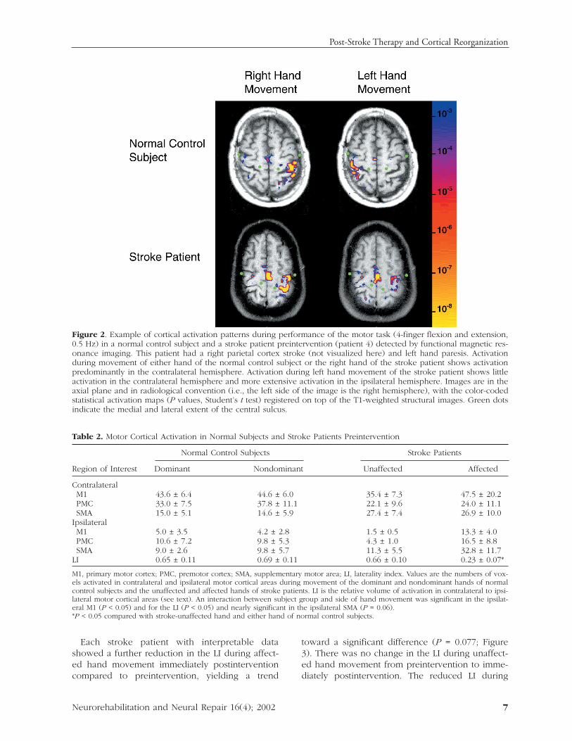
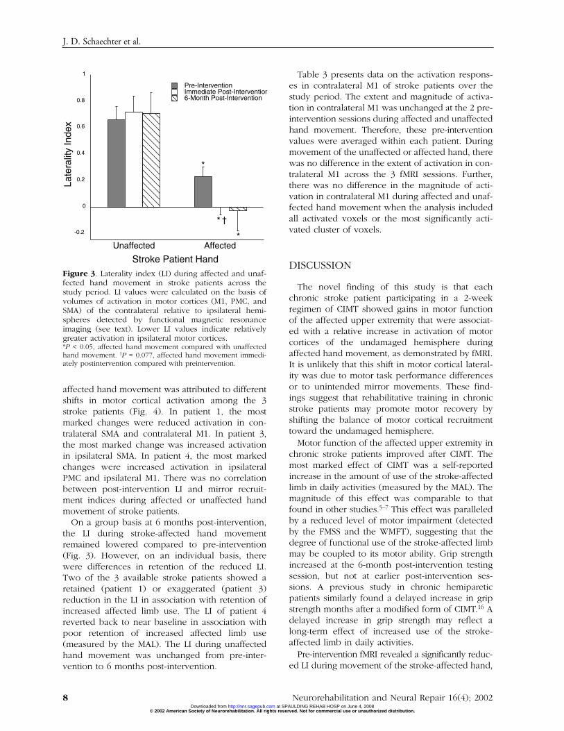
Each stroke patient with interpretable datashowed a further reduction in the LI during affect-ed hand movement immediately postinterventioncompared to preintervention, yielding a trend
toward a significant difference (P = 0.077; Figure3). There was no change in the LI during unaffect-ed hand movement from preintervention to imme-diately postintervention. The reduced LI during
Post-Stroke Therapy and Cortical Reorganization
Neurorehabilitation and Neural Repair 16(4); 2002 7
Figure 2. Example of cortical activation patterns during performance of the motor task (4-finger flexion and extension,0.5 Hz) in a normal control subject and a stroke patient preintervention (patient 4) detected by functional magnetic res-onance imaging. This patient had a right parietal cortex stroke (not visualized here) and left hand paresis. Activationduring movement of either hand of the normal control subject or the right hand of the stroke patient shows activationpredominantly in the contralateral hemisphere. Activation during left hand movement of the stroke patient shows littleactivation in the contralateral hemisphere and more extensive activation in the ipsilateral hemisphere. Images are in theaxial plane and in radiological convention (i.e., the left side of the image is the right hemisphere), with the color-codedstatistical activation maps (P values, Student’s t test) registered on top of the T1-weighted structural images. Green dotsindicate the medial and lateral extent of the central sulcus.
Table 2. Motor Cortical Activation in Normal Subjects and Stroke Patients Preintervention
Normal Control Subjects Stroke Patients
Region of Interest Dominant Nondominant Unaffected Affected
ContralateralM1 43.6 ± 6.4 44.6 ± 6.0 35.4 ± 7.3 47.5 ± 20.2PMC 33.0 ± 7.5 37.8 ± 11.1 22.1 ± 9.6 24.0 ± 11.1SMA 15.0 ± 5.1 14.6 ± 5.9 27.4 ± 7.4 26.9 ± 10.0
IpsilateralM1 5.0 ± 3.5 4.2 ± 2.8 1.5 ± 0.5 13.3 ± 4.0PMC 10.6 ± 7.2 9.8 ± 5.3 4.3 ± 1.0 16.5 ± 8.8SMA 9.0 ± 2.6 9.8 ± 5.7 11.3 ± 5.5 32.8 ± 11.7
LI 0.65 ± 0.11 0.69 ± 0.11 0.66 ± 0.10 0.23 ± 0.07*
M1, primary motor cortex; PMC, premotor cortex; SMA, supplementary motor area; LI, laterality index. Values are the numbers of vox-els activated in contralateral and ipsilateral motor cortical areas during movement of the dominant and nondominant hands of normalcontrol subjects and the unaffected and affected hands of stroke patients. LI is the relative volume of activation in contralateral to ipsi-lateral motor cortical areas (see text). An interaction between subject group and side of hand movement was significant in the ipsilat-eral M1 (P < 0.05) and for the LI (P < 0.05) and nearly significant in the ipsilateral SMA (P = 0.06).*P < 0.05 compared with stroke-unaffected hand and either hand of normal control subjects.
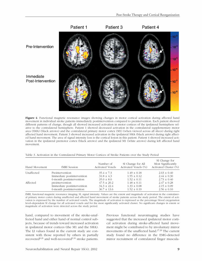
affected hand movement was attributed to differentshifts in motor cortical activation among the 3stroke patients (Fig. 4). In patient 1, the mostmarked changes were reduced activation in con-tralateral SMA and contralateral M1. In patient 3,the most marked change was increased activationin ipsilateral SMA. In patient 4, the most markedchanges were increased activation in ipsilateralPMC and ipsilateral M1. There was no correlationbetween post-intervention LI and mirror recruit-ment indices during affected or unaffected handmovement of stroke patients.
On a group basis at 6 months post-intervention,the LI during stroke-affected hand movementremained lowered compared to pre-intervention(Fig. 3). However, on an individual basis, therewere differences in retention of the reduced LI.Two of the 3 available stroke patients showed aretained (patient 1) or exaggerated (patient 3)reduction in the LI in association with retention ofincreased affected limb use. The LI of patient 4reverted back to near baseline in association withpoor retention of increased affected limb use(measured by the MAL). The LI during unaffectedhand movement was unchanged from pre-inter-vention to 6 months post-intervention.
Table 3 presents data on the activation respons-es in contralateral M1 of stroke patients over thestudy period. The extent and magnitude of activa-tion in contralateral M1 was unchanged at the 2 pre-intervention sessions during affected and unaffectedhand movement. Therefore, these pre-interventionvalues were averaged within each patient. Duringmovement of the unaffected or affected hand, therewas no difference in the extent of activation in con-tralateral M1 across the 3 fMRI sessions. Further,there was no difference in the magnitude of acti-vation in contralateral M1 during affected and unaf-fected hand movement when the analysis includedall activated voxels or the most significantly acti-vated cluster of voxels.
DISCUSSION
The novel finding of this study is that eachchronic stroke patient participating in a 2-weekregimen of CIMT showed gains in motor functionof the affected upper extremity that were associat-ed with a relative increase in activation of motorcortices of the undamaged hemisphere duringaffected hand movement, as demonstrated by fMRI.It is unlikely that this shift in motor cortical lateral-ity was due to motor task performance differencesor to unintended mirror movements. These find-ings suggest that rehabilitative training in chronicstroke patients may promote motor recovery byshifting the balance of motor cortical recruitmenttoward the undamaged hemisphere.
Motor function of the affected upper extremity inchronic stroke patients improved after CIMT. Themost marked effect of CIMT was a self-reportedincrease in the amount of use of the stroke-affectedlimb in daily activities (measured by the MAL). Themagnitude of this effect was comparable to thatfound in other studies.5–7 This effect was paralleledby a reduced level of motor impairment (detectedby the FMSS and the WMFT), suggesting that thedegree of functional use of the stroke-affected limbmay be coupled to its motor ability. Grip strengthincreased at the 6-month post-intervention testingsession, but not at earlier post-intervention ses-sions. A previous study in chronic hemipareticpatients similarly found a delayed increase in gripstrength months after a modified form of CIMT.16 Adelayed increase in grip strength may reflect along-term effect of increased use of the stroke-affected limb in daily activities.
Pre-intervention fMRI revealed a significantly reduc-ed LI during movement of the stroke-affected hand,
J. D. Schaechter et al.
8 Neurorehabilitation and Neural Repair 16(4); 2002
-0.2
0
0.2
0.4
0.6
0.8
1
Unaffected Affected
Pre-InterventionImmediate Post-Intervention6-Month Post-Intervention
Late
ralit
y In
dex
Stroke Patient Hand
*
*
*
†
Figure 3. Laterality index (LI) during affected and unaf-fected hand movement in stroke patients across thestudy period. LI values were calculated on the basis ofvolumes of activation in motor cortices (M1, PMC, andSMA) of the contralateral relative to ipsilateral hemi-spheres detected by functional magnetic resonanceimaging (see text). Lower LI values indicate relativelygreater activation in ipsilateral motor cortices.*P < 0.05, affected hand movement compared with unaffectedhand movement. †P = 0.077, affected hand movement immedi-ately postintervention compared with preintervention.
© 2002 American Society of Neurorehabilitation. All rights reserved. Not for commercial use or unauthorized distribution. at SPAULDING REHAB HOSP on June 4, 2008 http://nnr.sagepub.comDownloaded from
hand, compared to movement of the stroke-unaf-fected hand and either hand of normal control sub-jects, because of trends toward increased activationin ipsilateral motor cortices (the M1 and the SMA).The LI values found in the current study are con-sistent with those reported by others in partiallyrecovered8,26 and well-recovered9,10 stroke patients.
Previous functional neuroimaging studies havesuggested that the increased ipsilateral motor corti-cal activation during stroke-affected hand move-ment might be contributed to by involuntary mirrormovements of the unaffected hand.27,28 The currentstudy found no difference in the EMG-detectedmirror recruitment of contralateral finger muscula-
Post-Stroke Therapy and Cortical Reorganization
Neurorehabilitation and Neural Repair 16(4); 2002 9
Figure 4. Functional magnetic resonance images showing changes in motor cortical activation during affected handmovement in individual stroke patients immediately postintervention compared to preintervention. Each patient showeddifferent patterns of change, though all showed increased activation in motor cortices of the ipsilateral hemisphere rel-ative to the contralateral hemisphere. Patient 1 showed decreased activation in the contralateral supplementary motorarea (SMA) (black arrows) and the contralateral primary motor cortex (M1) (when viewed across all slices) during rightaffected hand movement. Patient 3 showed increased activation in the ipsilateral SMA (black arrows) during right affect-ed hand movement. The area of signal intensity loss is the cortical lesion in this patient. Patient 4 showed increased acti-vation in the ipsilateral premotor cortex (black arrows) and the ipsilateral M1 (white arrows) during left affected handmovement.
Table 3. Activation in the Contralateral Primary Motor Cortices of Stroke Patients over the Study Period
SI Change forNumber of SI Change for All Most Significantly
Hand Movement fMRI Session Activated Voxels Activated Voxels (%) Activated Cluster (%)
Unaffected Preintervention 35.4 ± 7.3 1.49 ± 0.18 2.63 ± 0.40Immediate postintervention 31.8 ± 4.3 1.55 ± 0.12 2.44 ± 0.306-month postintervention 35.0 ± 8.0 1.52 ± 0.11 2.73 ± 0.40
Affected preintervention 47.5 ± 20.2 1.48 ± 0.11 2.47 ± 0.28Immediate postintervention 34.3 ± 22.4 1.33 ± 0.08 2.15 ± 0.096-month postintervention 36.7 ± 13.6 1.52 ± 0.19 2.56 ± 0.10
fMRI, functional magnetic resonance imaging; SI, signal intensity. Values are the extent and magnitude of activation in the contralater-al primary motor cortex during unaffected and affected hand movement of stroke patients across the study period. The extent of acti-vation is expressed by the number of activated voxels. The magnitude of activation is expressed as the percentage blood oxygenationlevel–dependent SI change for all activated voxels and for the most significantly activated cluster. No significant changes in extent ormagnitude of activation were detected across the study period.
recruitment index) during affected compared withunaffected hand movement in stroke patients. Thisfinding suggests that the reduced LI during stroke-affected hand movement was not due to involun-tary mirror movements of the unaffected hand.However, because EMG was conducted offline, wecannot absolutely exclude the possibility that dur-ing fMRI, there was greater involuntary mirrorrecruitment during affected hand movement thanunaffected hand movement, perhaps contributingto the increased activation in ipsilateral motor cor-tical areas during stroke-affected hand movement.This possibility seems unlikely, however, becauseof the observed lack of correlation between the LIand mirror recruitment index during stroke-affect-ed hand movement and because performance ofthe motor activation task online was not observablydifferent from that offline.
Immediately after CIMT, all stroke patients in thissmall sample showed a shift in motor cortical later-ality toward the ipsilateral hemisphere in associa-tion with improved motor function of the affectedlimb. On a group basis, this shift in motor corticallaterality after CIMT reached only the level of anonsignificant trend, likely because of the low sta-tistical power of this pilot study. Further study of alarger cohort of chronic stroke patients is needed todetermine whether the observed laterality shift wefound is representative of brain reorganizationoccurring after CIMT in the population. Among theindividual patients of the current study, the lateral-ity shift was due to different changes in motor cor-tical activation. These differences may reflect therelatively heterogeneous group of stroke patients(e.g., time post-stroke, lesion topography, level ofmotor function) participating in this study. At the 6-month follow-up, the laterality shift was still pres-ent in the same 2 of 3 patients who retainedimproved limb functional use (detected by theMAL) and motor ability (detected by the FMSS andthe WMFT). In contrast, the laterality shift was notretained in the 1 patient who had poor retention ofincreased limb functional use but good retention oflimb motor ability. These directional associationssuggest that motor cortical laterality may be morestrongly linked to stroke-affected limb use than tomotor ability. In contrast to the laterality shift asso-ciated with the stroke-affected hand after CIMT,there was no concurrent laterality shift associatedwith the stroke-unaffected hand. This result sug-gests that repeated testing does not likely accountfor the laterality shift associated with stroke-affect-ed hand movement after CIMT. Further serial fMRIstudies in chronic stroke patients (who do not
receive CIMT) and normal control subjects (whoreceive CIMT) are needed to evaluate the specifici-ty of the relationship between CIMT-induced motorfunction gains and laterality shift associated withthe stroke-affected limb.
Two previous longitudinal functional neuroimag-ing studies conducted early after hemiparetic strokeshowed shifts in relative hemispheric activationduring this phase of motor recovery. Marshall et al.reported a shift in sensorimotor cortical activationtoward the damaged hemisphere in associationwith performance of an activation task thatimproved across scanning sessions.8 In contrast,Calautti et al. reported a hemispheric shift towardthe undamaged hemisphere in association withperformance of an activation task that wasunchanged across scanning sessions.26 Thus, thelaterality shift observed in the current study usingan activation task that was fixed across all scanningsessions suggests that rehabilitation-induced gainsin motor function in chronic stroke patients may bea progression in the cortical processes mediatingmotor recovery early after stroke.
It is unlikely that the relative increase in ipsilat-eral motor cortical activation after CIMT observedin the current study was due to a change in stroke-affected hand task performance. Only patients whocould perform the fMRI motor activation task atstudy entry were enrolled. All subjects performedthe task as trained at all sessions. Previous func-tional neuroimaging studies have shown that themovement parameters of rate,29–31 amplitude,32 andforce33–36 modulate activation in motor cortices.However, we controlled the rate of motor task per-formance by metronome pacing and the amplitudeof finger motion by an apparatus that fixed the endrange of finger flexion and extension. Further,behavioral testing revealed that the maximum fre-quency of 4-finger flexion and extension did notchange over the study period, suggesting that theshift in motor cortical laterality was not likely dueto patients performing the fMRI motor activationtask with more speed after CIMT. Behavioral test-ing also revealed that maximum grip strength wasnot different immediately post-intervention com-pared to pre-intervention, suggesting that increasedforce of task performance does not account for thelaterality shift immediately after CIMT. Behavioraltesting did, however, show improved scores on theFMSS, in part because of reduced synkinesia of thestroke-affected upper extremity. It is possible,therefore, that uncontrolled kinematic details oftask performance may have been different duringpost-intervention fMRI sessions as compared to
J. D. Schaechter et al.
10 Neurorehabilitation and Neural Repair 16(4); 2002 © 2002 American Society of Neurorehabilitation. All rights reserved. Not for commercial use or unauthorized distribution.
at SPAULDING REHAB HOSP on June 4, 2008 http://nnr.sagepub.comDownloaded from
pre-intervention sessions, contributing to the later-ality shift observed after CIMT.
The motor task used in the current fMRI studymay have influenced the results. We previouslyshowed using fMRI that performance of a finemotor task produces a selective activation increasein ipsilateral sensorimotor cortex during movementof a stroke-affected hand compared to a normalcontrol hand yet no activation difference during agross motor task.37 These findings suggest that per-formance of a neurologically challenging motortask during fMRI can differentiate motor corticalstatus on the basis of relative ipsilateral activation.That there were trends toward increased activationin ipsilateral motor cortices during stroke-affectedhand movement before CIMT suggests that thefMRI motor task we used was challenging enoughto probe ipsilateral motor cortices and thus mayhave increased the likelihood of exposing activa-tion changes in motor cortices of the undamagedhemisphere after CIMT.
The current finding of a laterality shift towardipsilateral motor cortices is distinct from the resultsof other studies examining neural changes afterCIMT. Liepert et al. found using TMS that theexcitability of the contralateral motor cortex repre-sentation of the stroke-affected hand increasedimmediately after CIMT.24,25 In contrast, we foundno increase in the extent or magnitude of activationin contralateral M1 after CIMT. The chronicity ofstroke in Liepert et al.’s study25 was greater (mean =~5 years) than in the current study (mean = ~1year), which might have influenced the respon-siveness of motor cortices to increased stroke-affected limb use. Additionally, that no changes inipsilateral motor cortex excitability were detectedin Liepert et al.’s studies24,25 may have beenbecause specific experimental maneuvers that arerequired to elicit ipsilateral motor evoked poten-tials38,39 were not applied. Kopp et al., using elec-troencephalography in stroke patients (chronicityrange 4 to 15 years), found that increased use ofthe affected limb produced by CIMT was associat-ed with a shift in source location associated withaffected hand movement within contralateral motorcortex immediately after therapy and into ipsilater-al motor cortex 3 months later.40 Levy et al., usingfMRI in 2 patients with cortical strokes (4.5 and 9months prior), found that both patients showedincreased perilesional activation, and 1 patientshowed increased bilateral sensorimotor cortexactivation, in association with improved perform-ance of the motor activation task.12 Collectively,these studies raise the possibility that the effect of
CIMT on activity of motor cortices in the damagedand undamaged hemispheres may interact withstroke chronicity, time post-CIMT, and stroketopography. Further, rehabilitation-induced reor-ganization in motor cortices may be differentiallysensitive to detection by various brain-mappingtechnologies and to experimental conditions.
The laterality shift toward ipsilateral motor corticesobserved after CIMT may reflect 1 or more mecha-nisms. One mechanism may be the amplification ofa role that ipsilateral motor cortices normally playin motor control. Studies in normal subjects haveshown that ipsilateral motor cortices participate incontrolling hand motor function, particularly com-plex or precise movements.41–43 Accordingly, theobserved laterality shift may reflect the increasedparticipation of ipsilateral motor cortices in con-trolling stroke-affected limb motor function. Studiesin normal subjects have also shown that motor cor-tices controlling contralateral hand movement con-comitantly inhibit motor cortices of the oppositehemisphere, perhaps to prevent potential interfer-ence by movement of the other hand.44,45 Thus, therelative increase in ipsilateral motor cortical activa-tion during stroke-affected hand movement mayreflect increased inhibition of unaffected handmovement, providing a neural substrate for thebehavioral shift in limb use preference after CIMT.A 2nd mechanism reflected by the laterality shiftafter CIMT may be the unmasking of existing butpreviously less active ipsilateral motor pathways, ashas been suggested to contribute to spontaneousmotor recovery after hemiparetic stroke.10,11,46,47 A3rd possible mechanism stems from animal studiesshowing that experimental stroke induces neuriteoutgrowth and synaptogenesis in the undamagedmotor cortex48,49 and that post-stroke training thatimproves motor skill enhances these structuralchanges.50,51 These animal studies suggest thatCIMT may have enhanced structural reorganizationin ipsilateral motor cortices that occurred sponta-neously after stroke.
In summary, this study provides preliminary evi-dence that in selected chronic stroke patients,improved motor function of the affected upperextremity produced by CIMT is associated withincreased activation in motor cortices of theundamaged hemisphere, relative to that in thedamaged hemisphere. The data suggest that motorcortices of the undamaged hemisphere might bean effective target for new rehabilitative interven-tions directed at improving motor recovery afterstroke. Further studies are required to elucidate therelationship between cortical reorganization and
Post-Stroke Therapy and Cortical Reorganization
Neurorehabilitation and Neural Repair 16(4); 2002 11 © 2002 American Society of Neurorehabilitation. All rights reserved. Not for commercial use or unauthorized distribution.
at SPAULDING REHAB HOSP on June 4, 2008 http://nnr.sagepub.comDownloaded from
post-stroke motor recovery facilitated by physicalrehabilitation.
ACKNOWLEDGMENTS
This study was initiated when Judith D. Schaech-ter was affiliated with Northeastern University andwas supported in part by awards (to Dr.Schaechter) from the Research and DevelopmentFund of Northeastern University, the Marion andJasper Whiting Foundation, and the NeuroRecoveryProgram of Spaulding Rehabilitation Hospital andMassachusetts General Hospital.
REFERENCES
1. Gresham GE, Duncan PW, Stason WB, et al. Post-strokerehabilitation. Clinical practice guideline. Washington,DC: US Department of Health and Human Services, PublicHealth Service, Agency for Health Care Policy andResearch; 1995.
2. Duncan P, Goldstein L, Horner R, Landsman P, Samsa G,Matchar D. Similar motor recovery of upper and lowerextremities after stroke. Stroke 1994;25:1181–8.
3. Nakayama H, Hendrik SJ, Raaschou HO, Olsen TS.Recovery of upper extremity function in stroke patients:the Copenhagen stroke study. Arch Phys Med Rehabil1994;75:394–9.
4. Duncan P. Synthesis of intervention trials to improve motorrecovery following stroke. Top Stroke Rehab 1997;3:1–20.
5. Taub E, Miller NE, Novack TA, et al. Technique to improvechronic motor deficit after stroke. Arch Phys Med Rehabil1993;74:347–54.
6. Kunkel A, Kopp B, Muller G, et al. Constraint-inducedmovement therapy for motor recovery in chronic strokepatients. Arch Phys Med Rehabil 1999;80:624–8.
7. Miltner WHR, Bauder H, Sommer M, Dettmers C, Taub E.Effects of constraint-induced movement therapy onpatients with chronic motor deficits after stroke: a replica-tion. Stroke 1999;30:586–92.
8. Marshall RS, Perera GM, Lazar RM, Krakauer JW,Constantine RC, DeLaPaz RL. Evolution of cortical activa-tion during recovery from corticospinal tract infarction.Stroke 2000;31:656–61.
9. Pineiro R, Pendlebury S, Johansen-Berg H, Matthews PM.Functional MRI detects posterior shifts in primary sensori-motor cortex activation after stroke: evidence of localadaptive reorganization? Stroke 2001;32:1134–9.
10. Cramer SC, Nelles G, Benson RR, et al. A functional MRIstudy of subjects recovered from hemiparetic stroke. Stroke1997;28:2518–27.
11. Cao Y, D’Olhaberriague L, Vikingstad EM, Levine SR,Welch KMA. Pilot study of functional MRI to assess cere-bral activation of motor function after poststroke hemi-paresis. Stroke 1998;29:112–22.
12. Levy CE, Nichols DS, Schmalbrock PM, Keller P, ChakeresDW. Functional MRI evidence of cortical reorganization inupper-limb stroke hemiplegia treated with constraint-induced movement therapy. Am J Phys Med Rehabil2001;80:4–12.
13. Oldfield RC. The assessment and analysis of handedness:the Edinburgh Inventory. Neuropsychologia 1971;9:97–113.
14. Cramer SC, Nelles G, Schaechter JD, Kaplan JD, FinklesteinSP. Computerized measurement of motor performanceafter stroke. Stroke 1997;28:2162–8.
15. Fugl-Meyer AR, Jaasko L, Leyman I, Olsson S, Steglind S.The post-stroke hemiplegic patient: a method for the eval-uation of physical performance. Scand J Rehabil Med1975;7:13–31.
16. Wolf SL, LeCraw DE, Barton LA, Jann BB. Forced use ofhemiplegic upper extremities to reverse the effect oflearned nonuse among chronic stroke and head-injuredpatients. Exp Neurol 1989;104:125–32.
17. Jiang A, Kennedy DN, Baker JR, et al. Motion detection andcorrection in functional MR imaging. Hum Brain Mapp1995;3:224–35.
18. Cohen MS, Bookheimer SY. Localization of brain functionusing magnetic resonance imaging. Trends Neurosci1994;17:268–77.
19. Talairach J, Tournoux P. Co-planar stereotaxic altas of thehuman brain. Stuttgart, Germany: Thieme Medical Pub-lishers; 1988.
20. Duvernoy HM. The human brain: surface, three-dimen-sional sectional anatomy and MRI. New York: Springer-Verlag; 1991.
21. Gati JS, Menon RS, Ugurbil K, Rutt BK. Experimental deter-mination of the BOLD field strength dependence in vesselsand tissue. Magn Reson Med 1997;38:296–302.
22. Forman SD, Cohen JD, Fitzgerald M, Eddy WF, Mintum MA,Noll DC. Improved assessment of significant activation infunctional magnetic resonance imaging (fMRI): use of acluster-size threshold. Magn Reson Med 1995;33:636–47.
23. Xiong J, Gao J-H, Lancaster JL, Fox PT. Clustered pixelsanalysis for functional MRI activation studies of the humanbrain. Hum Brain Mapp 1995;3:287–301.
24. Liepert J, Miltner WH, Bauder H, et al. Motor cortex plas-ticity during constraint-induced movement therapy instroke patients. Neurosci Lett 1998;250:5–8.
25. Liepert J, Bauder H, Miltner WHR, Taub E, Weiller C.Treatment-induced cortical reorganization after stroke inhumans. Stroke 2000;31:1210–6.
26. Calautti C, Leroy F, Guincestre JY, Marie RM, Baron JC.Sequential activation brain mapping after subcorticalstroke: changes in hemispheric balance and recovery.Neuroreport 2001;12:3883–6.
27. Weiller C, Ramsay SC, Wise RJS, Friston KJ, FrackowiakRSJ. Individual patterns of functional reorganization in thehuman cerebral cortex after capsular infarction. AnnNeurol 1993;33:181–9.
28. Wittenberg GF, Bastian AJ, Dromerick AW, Thach WT,Powers WJ. Mirror movements complicate interpretation ofcerebral activation changes during recovery from subcorti-cal infarction. Neurorehabil Neural Repair 2000;14:213–21.
29. VanMeter JW, Maisog JM, Zeffiro TA, Hallet M, HerscovitchP, Rapoport SI. Parametric analysis of functional neuroim-ages: application to a variable-rate motor task. Neuroimage1995;2:273–83.
30. Rao SM, Bandettini PA, Binder JR, et al. Relationshipbetween finger movement rate and functional magneticresonance signal change in human primary motor cortex. JCereb Blood Flow Metab 1996;16:1250–4.
31. Sadato N, Ibanez V, Deiber MP, Campbell G, Leonardo M,Hallet M. Frequency-dependent changes of regional cere-bral blood flow during finger movements. J Cereb BloodFlow Metab 1996;16:23–33.
32. Waldvogel D, van Gelderen P, Kenji I, Hallett M. The effectof movement amplitude on activation in functional mag-netic resonance imaging studies. J Cereb Blood Flow Metab1999;19:1209–12.
33. Dettmers C, Fink GR, Lemon RN, et al. Relation betweencerebral activity and force in the motor areas of the humanbrain. J Neurophysiol 1995;74:802–15.
J. D. Schaechter et al.
12 Neurorehabilitation and Neural Repair 16(4); 2002 © 2002 American Society of Neurorehabilitation. All rights reserved. Not for commercial use or unauthorized distribution.
at SPAULDING REHAB HOSP on June 4, 2008 http://nnr.sagepub.comDownloaded from
34. Wexler BE, Fulbright RK, Lacadie CM, et al. An fMRI studyof the human cortical motor system response in increasingfunctional demands. Magn Reson Imaging 1997;15:385–96.
35. Thickbroom GW, Phillips BA, Morris I, Byrnes ML,Mastaglia FL. Isometric force-related activity in sensorimo-tor cortex measured with functional MRI. Exp Brain Res1998;121:59–64.
36. Cramer SC, Weisskoff R, Schaechter JD, et al. Motor cortexactivation is related to force of squeezing. Hum BrainMapp 2002;16:197–205.
37. Cramer SC, Nelles G, Schaechter JD, Kaplan JD, FinklesteinSP, Rosen BR. A functional MRI study of three motor tasksin the evaluation of stroke recovery. Neurorehabil NeuralRepair 2001;15:1–8.
38. Wasserman EM, Pascual-Leone A, Hallet M. Cortical motorrepresentation of the ipsilateral hand and arm. Exp BrainRes 1994;100:121–32.
39. Ziemann U, Borgheresi A, Yaseen Z, et al. Dissociation ofthe pathways mediating ipsilateral and contralateral motor-evoked potentials in human hand and arm muscles. JPhysiol 1999;518:895–906.
40. Kopp B, Kunkel A, Muhlnickel W, Villringer K, Taub E, FlorH. Plasticity in the motor system related to therapy-inducedimprovement of movement after stroke. Neuroreport1999;10:807–10.
41. Chen R, Gerloff G, Hallett M, Cohen LG. Involvement ofthe ipsilateral motor cortex in finger movements of differ-ent complexities. Ann Neurol 1997;41:247–54.
42. Schluter ND, Rushworth MFS, Passingham RE, Mills KR.Temporary interference in human premotor cortex sug-gests dominance for the selection of movements: a studyusing transcranial magnetic stimulation. Brain 1998;121:785–99.
43. Ehrsson HH, Fagergren A, Jonsson T, Westling G,Johansson RS, Forssberg H. Cortical activity in precision-versus power-grip tasks: an fMRI study. J Neurophysiol2000;83:528–36.
44. Ferbert A, Priori A, Rothwell JC, Day BL, Colebatch JG,Marsden CD. Interhemispheric inhibition of the humanmotor cortex. J Physiol 1992;453:525–46.
45. Allison JD, Meador KJ, Loring DW, Figueroa RE, Wright JC.Functional MRI cerebral activation and deactivatioin duringfinger movement. Neurology 2000;54:135–42.
46. Weiller C, Chollet F, Friston KJ, Wise RJS, Frackowiak RSJ.Functional reorganization of the brain in recovery fromstriatocapsular infarction in man. Ann Neurol 1992;31:463–72.
47. Caramia MD, Palmieri MG, Giacomimi P, Iani C, Dally L,Silvestrini M. Ipsilateral activation of the unaffected motorcortex in patients with hemiparetic stroke. Clin Neurophysiol2000;111:1990–6.
48. Jones TA, Schallert T. Use-dependent growth of pyramidalneurons after neocortical damage. J Neurosci 1994;14:2140–52.
49. Stroemer RP, Kent TA, Hulsebosch CE. Neocortical neuralsprouting, synaptogenesis, and behavioral recovery afterneocortical infarction in rats. Stroke 1995;26:2135–44.
50. Jones TA, Chu CJ, Grande LA, Gregory AD. Motor skillstraining enhances lesion-induced structural plasticity in themotor cortex of adult rats. J Neurosci 1999;19:10153–63.
51. Biernaskie J, Corbett D. Enriched rehabilitative trainingpromotes improved forelimb motor function and enhanceddendritic growth after focal ischemic injury. J Neurosci2001;21:5272–80.
Post-Stroke Therapy and Cortical Reorganization
Neurorehabilitation and Neural Repair 16(4); 2002 13 © 2002 American Society of Neurorehabilitation. All rights reserved. Not for commercial use or unauthorized distribution.
at SPAULDING REHAB HOSP on June 4, 2008 http://nnr.sagepub.comDownloaded from


![Upper Limb Treatment Principles in Intensive Functional ...currentneurobiology.org/neurobiology/upper-limb-treatment-principles-in-intensive...CIMT [H-CIMT], bimanual intensive movement](https://img.pdfslide.us/doc/110x75/5ea2417b6d256b24c6549424/upper-limb-treatment-principles-in-intensive-functional-cimt-h-cimt-bimanual.jpg)