Embed Size (px)
Citation preview
1454 VOLUME 15 | NUMBER 10 | OCTOBER 2012 nature neurOSCIenCe
a r t I C l e S
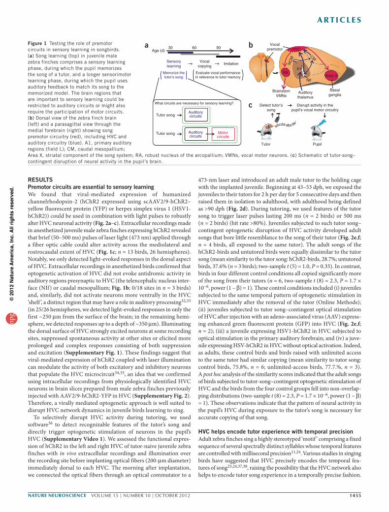
The cultural transmission of behavior involves observation of a behav-ioral model followed by imitation of the observed behavior. How the brain encodes the formative sensory experience provided by the behavioral model is not well understood. Although sensory structures are undoubtedly activated during observation of the model, premo-tor structures that have a role in generating imitative behaviors can also be activated during observation of another animal’s behavior1–5. This has led to speculation that premotor circuits may help encode sensory information about the model that is important to subsequent behavioral imitation6–9. Birdsong is a culturally transmitted vocal behavior with strong parallels to human speech learning, including the obligatory auditory experience of a vocal model during a juvenile sensitive period followed by a phase of vocal copying10–13. Juvenile male zebra finches first listen to and memorize the song of an adult male tutor during a sensory learning phase (Fig. 1a; ~30–60 days post-hatching (dph)) and then engage in vocal practice to emulate this memorized song model during a partially overlapping and more prolonged phase of sensorimotor learning (~45–90 dph)11. In addi-tion, the brain of the male zebra finch contains well-described audi-tory and song motor pathways that are thought to be critical to these two phases of learning (Fig. 1b)13,14. Nonetheless, how the experience of the tutor song is initially encoded in the brain of the juvenile and how this information interacts with song motor circuits to guide song development is unclear.
One possibility is that the auditory memory of the tutor song is encoded in forebrain structures that are analogous to the second-ary and tertiary auditory cortices of mammals (Fig. 1b). In sup-port of this idea, vocal imitation is impaired after pharmacological manipulation of these secondary auditory regions of juvenile zebra finches during tutoring15, and mapping studies of activity-dependent gene expression and electrophysiological responses in adult zebra finches have suggested that neurons in these regions could encode a
long-lasting representation of the tutor song16–19. However, these findings do not address whether encoding the tutor song also requires activity in downstream structures, including motor structures that directly control singing (Fig. 1b). Indeed, secondary auditory regions provide direct and indirect input to the telencephalic nucleus HVC20,21, a premotor structure that is essential for song generation22 and that contains neurons that encode precise timing information for song patterning and respond to the auditory presentation of a tutor song23–25. Moreover, exposing a juvenile zebra finch to a tutor song can trigger rapid structural and functional changes to synapses in its HVC that correlate with the quality of its subsequent song imita-tion26. The finding that exposure to a tutor song can rapidly alter the HVC network suggests a possible role for HVC in encoding the tutor song experience.
One challenge to testing this idea is that juvenile zebra finches often interleave periods of singing and other forms of vocal activity with periods of listening to a tutor song. Consequently, although pharmacological manipulations either upstream or downstream of HVC can affect the quality of song copying15,27, it is unclear whether these effects are the result of interference with vocal premotor activ-ity, auditory activity evoked by the tutor song or auditory feedback activity evoked by the pupil’s own singing. To examine whether HVC has a critical role in encoding the experience of the tutor song, we sought a method that would allow us to disrupt HVC activity only when the pupil listened to his tutor’s song but not at other times, including during periods of vocal rehearsal (Fig. 1c). The transgenic expression of light-activated cation channels (channelrhodopsins) provides a means for the precise spatiotemporal control of neural activity without the potential confound of activating fibers of passage that can accompany electrical stimulation or the typically prolonged (minutes to hours) modulation of neural activity accompanying pharmacological manipulations28–31.
1Department of Neurobiology, Duke University Medical Center, Durham, North Carolina, USA. 2Department of Organismic and Evolutionary Biology, Harvard University, Boston, Massachusetts, USA. 3Neuroscience Program, Wellesley College, Wellesley, Massachusetts, USA. Correspondence should be addressed to R.M. ([email protected]).
Received 3 April; accepted 6 August; published online 16 September 2012; doi:10.1038/nn.3206
Motor circuits are required to encode a sensory model for imitative learningTodd F Roberts1, Sharon M H Gobes2,3, Malavika Murugan1, Bence P Ölveczky2 & Richard Mooney1
Premotor circuits help generate imitative behaviors and can be activated during observation of another animal′s behavior, leading to speculation that these circuits participate in sensory learning that is important to imitation. Here we tested this idea by focally manipulating the brain activity of juvenile zebra finches, which learn to sing by memorizing and vocally copying the song of an adult tutor. Tutor song–contingent optogenetic or electrical disruption of neural activity in the pupil′s song premotor nucleus HVC prevented song copying, indicating that a premotor structure important to the temporal control of birdsong also helps encode the tutor song. In vivo multiphoton imaging and neural manipulations delineated a pathway and a candidate synaptic mechanism through which tutor song information is encoded by premotor circuits. These findings provide evidence that premotor circuits help encode sensory information about the behavioral model before shaping and executing imitative behaviors.
npg
© 2
012
Nat
ure
Am
eric
a, In
c. A
ll rig
hts
rese
rved
.
nature neurOSCIenCe VOLUME 15 | NUMBER 10 | OCTOBER 2012 1455
a r t I C l e S
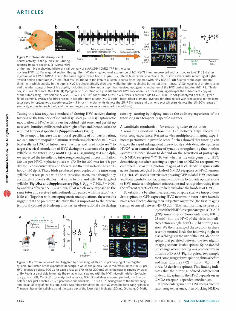
RESULTSPremotor circuits are essential to sensory learningWe found that viral-mediated expression of humanized channelrhodopsin-2 (hChR2 expressed using scAAV2/9-hChR2–yellow fluorescent protein (YFP) or herpes simplex virus 1 (HSV1-hChR2)) could be used in combination with light pulses to robustly alter HVC neuronal activity (Fig. 2a–c). Extracellular recordings made in anesthetized juvenile male zebra finches expressing hChR2 revealed that brief (50–500 ms) pulses of laser light (473 nm) applied through a fiber optic cable could alter activity across the mediolateral and rostrocaudal extent of HVC (Fig. 1c; n = 13 birds, 26 hemispheres). Notably, we only detected light-evoked responses in the dorsal aspect of HVC. Extracellular recordings in anesthetized birds confirmed that optogenetic activation of HVC did not evoke antidromic activity in auditory regions presynaptic to HVC (the telencephalic nucleus inter-face (NIf) or caudal mesopallium; Fig. 1b; 0/18 sites in n = 3 birds) and, similarly, did not activate neurons more ventrally in the HVC ‘shelf ’, a distinct region that may have a role in auditory processing32,33 (in 25/26 hemispheres, we detected light-evoked responses in only the first ~250 µm from the surface of the brain; in the remaining hemi-sphere, we detected responses up to a depth of ~350 µm). Illuminating the dorsal surface of HVC strongly excited neurons at some recording sites, suppressed spontaneous activity at other sites or elicited more prolonged and complex responses consisting of both suppression and excitation (Supplementary Fig. 1). These findings suggest that viral-mediated expression of hChR2 coupled with laser illumination can modulate the activity of both excitatory and inhibitory neurons that populate the HVC microcircuit34,35, an idea that we confirmed using intracellular recordings from physiologically identified HVC neurons in brain slices prepared from male zebra finches previously injected with AAV2/9-hChR2-YFP in HVC (Supplementary Fig. 2). Therefore, a virally mediated optogenetic approach is well suited to disrupt HVC network dynamics in juvenile birds learning to sing.
To selectively disrupt HVC activity during tutoring, we used software36 to detect recognizable features of the tutor’s song and directly trigger optogenetic stimulation of neurons in the pupil’s HVC (Supplementary Video 1). We assessed the functional expres-sion of hChR2 in the left and right HVC of tutor-naive juvenile zebra finches with in vivo extracellular recordings and illumination over the recording site before implanting optical fibers (200-µm diameter) immediately dorsal to each HVC. The morning after implantation, we connected the optical fibers through an optical commutator to a
473-nm laser and introduced an adult male tutor to the holding cage with the implanted juvenile. Beginning at 43–53 dph, we exposed the juveniles to their tutors for 2 h per day for 5 consecutive days and then raised them in isolation to adulthood, with adulthood being defined as >90 dph (Fig. 2d). During tutoring, we used features of the tutor song to trigger laser pulses lasting 200 ms (n = 2 birds) or 500 ms (n = 2 birds) (hit rate >80%). Juveniles subjected to such tutor song–contingent optogenetic disruption of HVC activity developed adult songs that bore little resemblance to the song of their tutor (Fig. 2e,f; n = 4 birds, all exposed to the same tutor). The adult songs of the hChR2-birds and untutored birds were equally dissimilar to the tutor song (mean similarity to the tutor song: hChR2-birds, 28.7%; untutored birds, 37.6% (n = 3 birds); two-sample t (5) = 1.0, P = 0.35). In contrast, birds in four different control conditions all copied significantly more of the song from their tutors (n = 6, two-sample t (8) = 2.3, P = 1.7 × 10⁻⁶, power (1 – β) = 1). These control conditions included (i) juveniles subjected to the same temporal pattern of optogenetic stimulation in HVC immediately after the removal of the tutor (Online Methods); (ii) juveniles subjected to tutor song–contingent optical stimulation of HVC after injection with an adeno-associated virus (AAV) express-ing enhanced green fluorescent protein (GFP) into HVC (Fig. 2e,f; n = 2); (iii) a juvenile expressing HSV1-hChR2 in HVC subjected to optical stimulation in the primary auditory forebrain; and (iv) a juve-nile expressing HSV-hChR2 in HVC without optical activation. Indeed, as adults, these control birds and birds raised with unlimited access to the same tutor had similar copying (mean similarity to tutor song: control birds, 75.8%, n = 6; unlimited-access birds, 77.7.%, n = 3). A post hoc analysis of the similarity scores indicated that the adult songs of birds subjected to tutor-song–contingent optogenetic stimulation of HVC and the birds from the four control groups fell into non-overlap-ping distributions (two-sample t (8) = 2.3, P = 1.7 × 10⁻⁶, power (1 – β) = 1). These observations indicate that the pattern of neural activity in the pupil’s HVC during exposure to the tutor’s song is necessary for accurate copying of that song.
HVC helps encode tutor experience with temporal precisionAdult zebra finches sing a highly stereotyped ‘motif ’ comprising a fixed sequence of several spectrally distinct syllables whose temporal features are controlled with millisecond precision11,24. Various studies in singing birds have suggested that HVC precisely encodes the temporal fea-tures of song23,24,37,38, raising the possibility that the HVC network also helps to encode tutor song experience in a temporally precise fashion.
Age (d)a b
c
30
Sensorylearning
Auditorycircuits
Auditorycircuits
Motorcircuits
Memorize thetutor’s song
Evaluate vocal performancein reference to tutor memory
What circuits are necessary for sensory learning?
60
Vocalcopying
Vocalpremotor HVC
RAA1
NlfArea X
NC
M
CM
BrainstemVMNs
Detect tutor’ssong
′a abcde′bcde
Tutor Pupil
Disrupt activity in thepupil’s vocal motor circuitry
Auditorythalamus
Basalganglia
Tutor song
Tutor song
Imitation
90Figure 1 Testing the role of premotor circuits in sensory learning in songbirds. (a) Song learning (top) in juvenile male zebra finches comprises a sensory learning phase, during which the pupil memorizes the song of a tutor, and a longer sensorimotor learning phase, during which the pupil uses auditory feedback to match its song to the memorized model. The brain regions that are important to sensory learning could be restricted to auditory circuits or might also require the participation of motor circuits. (b) Dorsal view of the zebra finch brain (left) and a parasagittal view through the medial forebrain (right) showing song premotor circuitry (red), including HVC and auditory circuitry (blue). A1, primary auditory regions (field L); CM, caudal mesopallium; Area X, striatal component of the song system; RA, robust nucleus of the arcopallium; VMNs, vocal motor neurons. (c) Schematic of tutor-song–contingent disruption of neural activity in the pupil’s brain.
npg
© 2
012
Nat
ure
Am
eric
a, In
c. A
ll rig
hts
rese
rved
.
1456 VOLUME 15 | NUMBER 10 | OCTOBER 2012 nature neurOSCIenCe
a r t I C l e S
Testing this idea requires a method of altering HVC activity during tutoring on the time scale of individual syllables (~100 ms). Optogenetic modulation of HVC activity can lag behind light onset and persist up to several hundred milliseconds after light offset and, hence, lacks the required temporal specificity (Supplementary Fig. 1).
To attempt to increase the temporal specificity of our perturbation, we implanted monopolar platinum stimulating electrodes (0.1 MΩ) bilaterally in HVC of tutor-naive juveniles and used software36 to target electrical stimulation of HVC during the utterance of a specific syllable in the tutor’s song motif (Fig. 3a). Beginning at 43–53 dph, we subjected the juveniles to tutor song–contingent microstimulation (20 µA per HVC, biphasic pulses at 170 Hz for 200 ms) for 4 h per day for 5 consecutive days and then raised them in isolation to adult-hood (>90 dph). These birds produced poor copies of the tutor song syllable that was paired with the microstimulation, even though they accurately copied syllables that came before and after the targeted syllable (Fig. 3b,c and Supplementary Fig. 3; F4,14 = 7.508, P = 0.001 by analysis of variance, n = 4 birds, all of which were exposed to the same tutor and received microstimulation paired with the tutor’s syl-lable ‘c’). Together with our optogenetic manipulations, these results suggest that the premotor structure that is important to the precise temporal control of birdsong also has an observational role during
sensory learning by helping encode the auditory experience of the tutor song in a temporally specific manner.
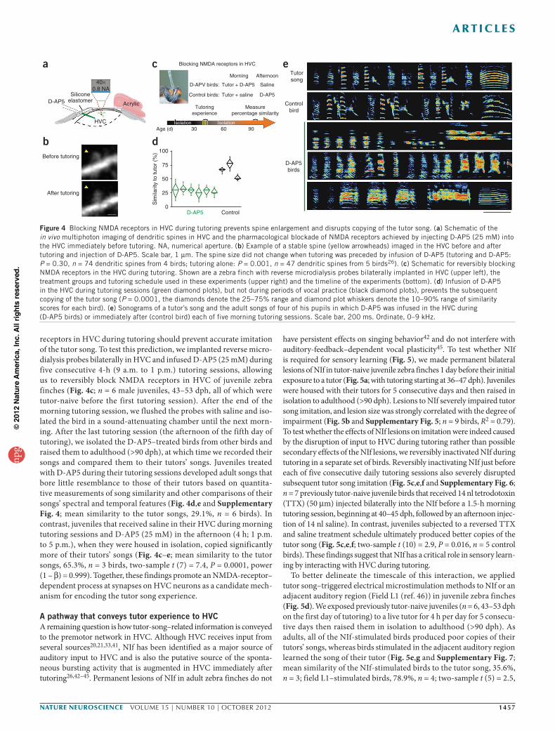
A candidate mechanism for encoding tutor experienceA remaining question is how the HVC network helps encode the tutor song experience. Recent in vivo multiphoton imaging experi-ments performed in juvenile zebra finches showed that tutoring can trigger the rapid enlargement of previously stable dendritic spines in HVC26, a structural correlate of synaptic strengthening that in other systems has been shown to depend on the activation of postsynap-tic NMDA receptors39,40. To test whether the enlargement of HVC dendritic spines after tutoring is dependent on NMDA receptors, we combined in vivo multiphoton imaging of HVC dendritic spines with acute pharmacological blockade of NMDA receptors on HVC neurons (Fig. 4a). We used a lentivirus expressing GFP to label HVC neurons and their dendritic spines, cranial windowing to provide optical access to HVC under a multiphoton microscope and retrograde tracing from the efferent targets of HVC to help visualize the borders of HVC.
To establish a baseline measurement of spine size, we imaged den-dritic spines on GFP-expressing HVC neurons in tutor-naive juvenile male zebra finches during their subjective nighttime (the first imaging session occurred between 43–53 dph). The next morning, we pressure
injected the NMDA receptor antagonist D-AP5 ((2R)-amino-5-phosphonopentanoate; 100 nl, 25 mM) into the HVC of the birds immedi-ately before a single, brief (~1.5 h) tutoring ses-sion. We then reimaged the neurons in these recently tutored birds the following night to assess changes in the size of the HVC dendritic spines that persisted between the two nightly imaging sessions (stable spines). Spine size did not change when tutoring was preceded by an infusion of D-AP5 (Fig. 4b; paired, two-sample t test comparing relative spine brightness before and after tutoring t (72) = 1.0, P = 0.3, n = 4 birds, 73 dendritic spines). This finding indi-cates that the tutoring-induced enlargement of dendritic spines in the HVC depends on an NMDA receptor–dependent mechanism.
If spine enlargement in HVC helps encode tutor song experience, then blocking NMDA
hChR2 inHVC
a
DorsalAnterior
LTV
HVC
b c
Tutor song–contingentoptogenetic disruption of HVC
Age (d) 30 60 90Isolation Isolation
Measurepercentage similarityd
Tutorsong
Pupilsong
(control)
Pupilsong
(hChR2)
e100
75
50
Sim
ilarit
y to
tuto
r (%
)
25
Controlbirds
hChR2birds
0
f
Figure 2 Optogenetic disruption of neural activity in the pupil’s HVC during tutoring impairs copying. (a) Dorsal view of the finch brain showing bilateral viral delivery of scAAV2/9-hChR2-YFP to the song nucleus HVC. (b) Parasagittal section through the HVC showing neuronal expression of hChR2-YFP immunoreacted with antibodies to GFP 11 d after injection of scAAV-hChR2-YFP into the same region. Scale bar, 100 µm. LTV, lateral telencephalic ventricle. (c) In vivo extracellular recording of light-evoked action potentials (473 nm, 500 ms, 10 trials) in the HVC of a juvenile zebra finch injected with HSV-hChR2. (d) Sketch of the experimental timeline in which activity in the pupil’s HVC is optogenetically disrupted while the tutor is singing but not at other times. (e) Sonograms of a tutor’s song and the adult songs of two of his pupils, including a control and a pupil that received optogenetic activation of the HVC during tutoring (hChR2). Scale bar, 200 ms. Ordinate, 0–9 kHz. (f) Optogenetic disruption of a juvenile finch’s HVC only when its tutor is singing disrupts the subsequent copying of the tutor’s song ((two-sample t8 = 2.3, P = 1.7 × 10⁻⁶ for hChR2-birds (n = 4) versus control birds (n = 6) (20–25 songs analyzed per bird); green filled diamond, average for birds raised in isolation from a tutor (n = 3 birds); black filled diamond, average for birds raised with free access to the same tutor used for optogenetic experiments (n = 3 birds); the diamonds denote the 25–75% range and diamond plot whiskers denote the 10–90% range of similarity scores for each bird, and the learning outcomes were measured in adulthood).
c
Tutorsong
a b c d e
Pupilsong
aSyllabledetection
abc* *de abcde
Tutor Pupil
Stimulator
b
Syllables
100
75
50
25
0
Acc
urat
e im
itatio
n (%
)
a b c d e
Figure 3 Microstimulation of HVC triggered by tutor song syllable disrupts copying of the targeted syllable. (a) Sketch of the experimental design in which the pupil’s HVC is microstimulated (20 µA per HVC, biphasic pulses, 300 µs for each phase at 170 Hz for 200 ms) while the tutor is singing syllable c. (b) Pupils are not able to imitate the syllable that is paired with the HVC microstimulation (syllable c; F4,14 = 7.508, P = 0.001 by analysis of variance, 90–100 syllables analyzed per bird, n = 4 birds; notched box plot denotes 25–75 percentile and whiskers, 1.5 s.d.). (c) Sonograms of the tutor’s song and the adult song of one his pupils that was microstimulated in the HVC when the tutor sang syllable c. The green bar under syllable c and the scale bar at the lower right indicate 130 ms. Ordinate, 0–9 kHz.
npg
© 2
012
Nat
ure
Am
eric
a, In
c. A
ll rig
hts
rese
rved
.
nature neurOSCIenCe VOLUME 15 | NUMBER 10 | OCTOBER 2012 1457
a r t I C l e S
receptors in HVC during tutoring should prevent accurate imitation of the tutor song. To test this prediction, we implanted reverse micro-dialysis probes bilaterally in HVC and infused D-AP5 (25 mM) during five consecutive 4-h (9 a.m. to 1 p.m.) tutoring sessions, allowing us to reversibly block NMDA receptors in HVC of juvenile zebra finches (Fig. 4c; n = 6 male juveniles, 43–53 dph, all of which were tutor-naive before the first tutoring session). After the end of the morning tutoring session, we flushed the probes with saline and iso-lated the bird in a sound-attenuating chamber until the next morn-ing. After the last tutoring session (the afternoon of the fifth day of tutoring), we isolated the D-AP5–treated birds from other birds and raised them to adulthood (>90 dph), at which time we recorded their songs and compared them to their tutors’ songs. Juveniles treated with D-AP5 during their tutoring sessions developed adult songs that bore little resemblance to those of their tutors based on quantita-tive measurements of song similarity and other comparisons of their songs’ spectral and temporal features (Fig. 4d,e and Supplementary Fig. 4; mean similarity to the tutor songs, 29.1%, n = 6 birds). In contrast, juveniles that received saline in their HVC during morning tutoring sessions and D-AP5 (25 mM) in the afternoon (4 h; 1 p.m. to 5 p.m.), when they were housed in isolation, copied significantly more of their tutors’ songs (Fig. 4c–e; mean similarity to the tutor songs, 65.3%, n = 3 birds, two-sample t (7) = 7.4, P = 0.0001, power (1 – β) = 0.999). Together, these findings promote an NMDA-receptor– dependent process at synapses on HVC neurons as a candidate mech-anism for encoding the tutor song experience.
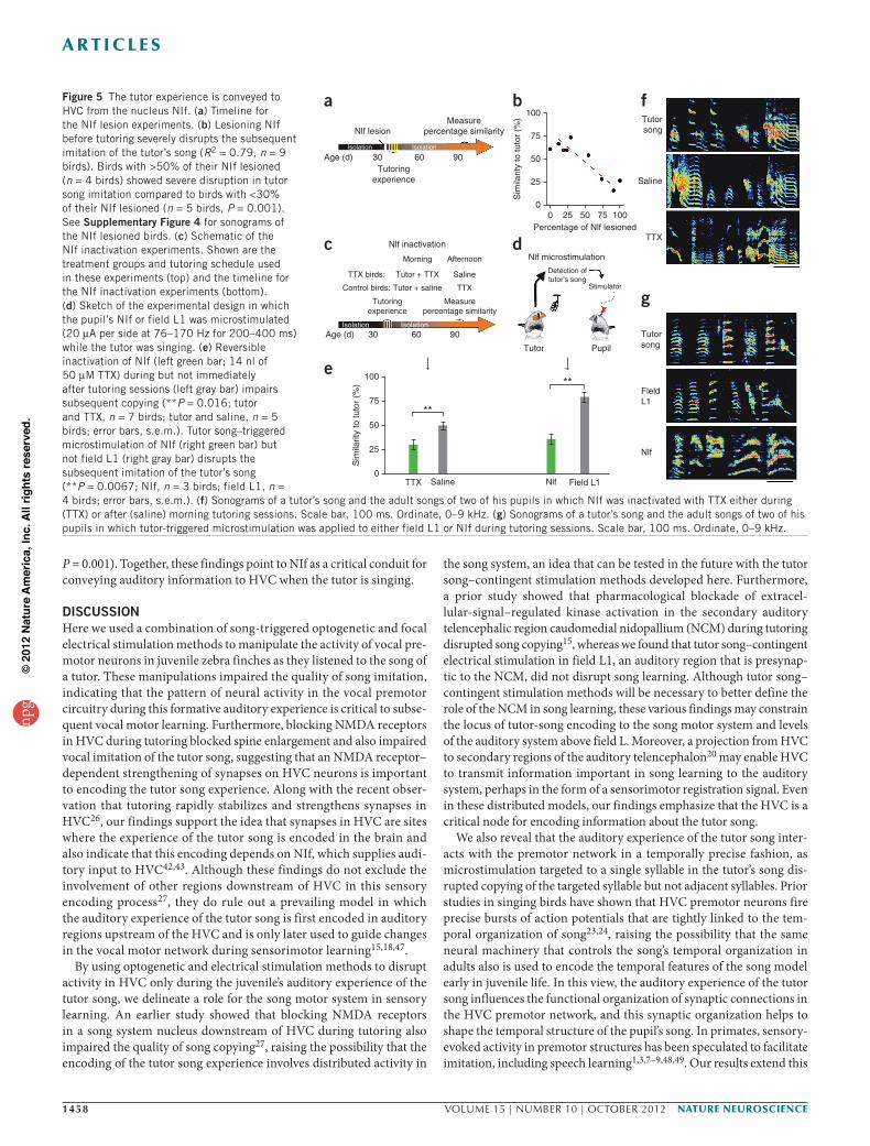
A pathway that conveys tutor experience to HVCA remaining question is how tutor-song–related information is conveyed to the premotor network in HVC. Although HVC receives input from several sources20,21,33,41, NIf has been identified as a major source of auditory input to HVC and is also the putative source of the sponta-neous bursting activity that is augmented in HVC immediately after tutoring26,42–45. Permanent lesions of NIf in adult zebra finches do not
have persistent effects on singing behavior42 and do not interfere with auditory-feedback–dependent vocal plasticity45. To test whether NIf is required for sensory learning (Fig. 5), we made permanent bilateral lesions of NIf in tutor-naive juvenile zebra finches 1 day before their initial exposure to a tutor (Fig. 5a; with tutoring starting at 36–47 dph). Juveniles were housed with their tutors for 5 consecutive days and then raised in isolation to adulthood (>90 dph). Lesions to NIf severely impaired tutor song imitation, and lesion size was strongly correlated with the degree of impairment (Fig. 5b and Supplementary Fig. 5; n = 9 birds, R2 = 0.79). To test whether the effects of NIf lesions on imitation were indeed caused by the disruption of input to HVC during tutoring rather than possible secondary effects of the NIf lesions, we reversibly inactivated NIf during tutoring in a separate set of birds. Reversibly inactivating NIf just before each of five consecutive daily tutoring sessions also severely disrupted subsequent tutor song imitation (Fig. 5c,e,f and Supplementary Fig. 6; n = 7 previously tutor-naive juvenile birds that received 14 nl tetrodotoxin (TTX) (50 µm) injected bilaterally into the NIf before a 1.5-h morning tutoring session, beginning at 40–45 dph, followed by an afternoon injec-tion of 14 nl saline). In contrast, juveniles subjected to a reversed TTX and saline treatment schedule ultimately produced better copies of the tutor song (Fig. 5c,e,f; two-sample t (10) = 2.9, P = 0.016, n = 5 control birds). These findings suggest that NIf has a critical role in sensory learn-ing by interacting with HVC during tutoring.
To better delineate the timescale of this interaction, we applied tutor song–triggered electrical microstimulation methods to NIf or an adjacent auditory region (Field L1 (ref. 46)) in juvenile zebra finches (Fig. 5d). We exposed previously tutor-naive juveniles (n = 6, 43–53 dph on the first day of tutoring) to a live tutor for 4 h per day for 5 consecu-tive days then raised them in isolation to adulthood (>90 dph). As adults, all of the NIf-stimulated birds produced poor copies of their tutors’ songs, whereas birds stimulated in the adjacent auditory region learned the song of their tutor (Fig. 5e,g and Supplementary Fig. 7; mean similarity of the NIf-stimulated birds to the tutor song, 35.6%, n = 3; field L1–stimulated birds, 78.9%, n = 4; two-sample t (5) = 2.5,
bBefore tutoring
After tutoring
ec Blocking NMDA receptors in HVC
D-APV birds:
Control birds:
Age (d) 30 60 90
Morning Afternoon
Saline
D-AP5
Tutor + D-AP5
Tutoringexperience
Isolation Isolation
Measurepercentage similarity
Tutor + saline
a
HVC
D-AP5Silicone
elastomer Acrylic
40×0.8 NA
d100
75
50
25
ControlD-AP50
Sim
ilarit
y to
tuto
r (%
)
Tutorsong
Controlbird
D-AP5birds
Figure 4 Blocking NMDA receptors in HVC during tutoring prevents spine enlargement and disrupts copying of the tutor song. (a) Schematic of the in vivo multiphoton imaging of dendritic spines in HVC and the pharmacological blockade of NMDA receptors achieved by injecting D-AP5 (25 mM) into the HVC immediately before tutoring. NA, numerical aperture. (b) Example of a stable spine (yellow arrowheads) imaged in the HVC before and after tutoring and injection of D-AP5. Scale bar, 1 µm. The spine size did not change when tutoring was preceded by infusion of D-AP5 (tutoring and D-AP5: P = 0.30, n = 74 dendritic spines from 4 birds; tutoring alone: P = 0.001, n = 47 dendritic spines from 5 birds26). (c) Schematic for reversibly blocking NMDA receptors in the HVC during tutoring. Shown are a zebra finch with reverse microdialysis probes bilaterally implanted in HVC (upper left), the treatment groups and tutoring schedule used in these experiments (upper right) and the timeline of the experiments (bottom). (d) Infusion of D-AP5 in the HVC during tutoring sessions (green diamond plots), but not during periods of vocal practice (black diamond plots), prevents the subsequent copying of the tutor song (P = 0.0001, the diamonds denote the 25–75% range and diamond plot whiskers denote the 10–90% range of similarity scores for each bird). (e) Sonograms of a tutor’s song and the adult songs of four of his pupils in which D-AP5 was infused in the HVC during (D-AP5 birds) or immediately after (control bird) each of five morning tutoring sessions. Scale bar, 200 ms. Ordinate, 0–9 kHz.
npg
© 2
012
Nat
ure
Am
eric
a, In
c. A
ll rig
hts
rese
rved
.
1458 VOLUME 15 | NUMBER 10 | OCTOBER 2012 nature neurOSCIenCe
a r t I C l e S
P = 0.001). Together, these findings point to NIf as a critical conduit for conveying auditory information to HVC when the tutor is singing.
DISCUSSIONHere we used a combination of song-triggered optogenetic and focal electrical stimulation methods to manipulate the activity of vocal pre-motor neurons in juvenile zebra finches as they listened to the song of a tutor. These manipulations impaired the quality of song imitation, indicating that the pattern of neural activity in the vocal premotor circuitry during this formative auditory experience is critical to subse-quent vocal motor learning. Furthermore, blocking NMDA receptors in HVC during tutoring blocked spine enlargement and also impaired vocal imitation of the tutor song, suggesting that an NMDA receptor– dependent strengthening of synapses on HVC neurons is important to encoding the tutor song experience. Along with the recent obser-vation that tutoring rapidly stabilizes and strengthens synapses in HVC26, our findings support the idea that synapses in HVC are sites where the experience of the tutor song is encoded in the brain and also indicate that this encoding depends on NIf, which supplies audi-tory input to HVC42,43. Although these findings do not exclude the involvement of other regions downstream of HVC in this sensory encoding process27, they do rule out a prevailing model in which the auditory experience of the tutor song is first encoded in auditory regions upstream of the HVC and is only later used to guide changes in the vocal motor network during sensorimotor learning15,18,47.
By using optogenetic and electrical stimulation methods to disrupt activity in HVC only during the juvenile’s auditory experience of the tutor song, we delineate a role for the song motor system in sensory learning. An earlier study showed that blocking NMDA receptors in a song system nucleus downstream of HVC during tutoring also impaired the quality of song copying27, raising the possibility that the encoding of the tutor song experience involves distributed activity in
the song system, an idea that can be tested in the future with the tutor song–contingent stimulation methods developed here. Furthermore, a prior study showed that pharmacological blockade of extracel-lular-signal–regulated kinase activation in the secondary auditory telencephalic region caudomedial nidopallium (NCM) during tutoring disrupted song copying15, whereas we found that tutor song–contingent electrical stimulation in field L1, an auditory region that is presynap-tic to the NCM, did not disrupt song learning. Although tutor song– contingent stimulation methods will be necessary to better define the role of the NCM in song learning, these various findings may constrain the locus of tutor-song encoding to the song motor system and levels of the auditory system above field L. Moreover, a projection from HVC to secondary regions of the auditory telencephalon20 may enable HVC to transmit information important in song learning to the auditory system, perhaps in the form of a sensorimotor registration signal. Even in these distributed models, our findings emphasize that the HVC is a critical node for encoding information about the tutor song.
We also reveal that the auditory experience of the tutor song inter-acts with the premotor network in a temporally precise fashion, as microstimulation targeted to a single syllable in the tutor’s song dis-rupted copying of the targeted syllable but not adjacent syllables. Prior studies in singing birds have shown that HVC premotor neurons fire precise bursts of action potentials that are tightly linked to the tem-poral organization of song23,24, raising the possibility that the same neural machinery that controls the song’s temporal organization in adults also is used to encode the temporal features of the song model early in juvenile life. In this view, the auditory experience of the tutor song influences the functional organization of synaptic connections in the HVC premotor network, and this synaptic organization helps to shape the temporal structure of the pupil’s song. In primates, sensory-evoked activity in premotor structures has been speculated to facilitate imitation, including speech learning1,3,7–9,48,49. Our results extend this
Measurepercentage similarity
Tutoringexperience
Nlf lesion
Isolation Isolation
906030Age (d)
a100
75
50
25
Sim
ilarit
y to
tuto
r (%
)
00 25 50 75 100
Percentage of Nlf lesioned
b
Nlf microstimulation
Detection oftutor’s song
Stimulator
PupilTutor
d
Measurepercentage similarity
Tutoringexperience
Nlf inactivation
Morning
TTX birds:
Control birds:
Afternoon
SalineTutor + TTX
TTXTutor + saline
Isolation Isolation906030Age (d)
c
100
75
50
25
0TTX Nlf Field L1Saline
Sim
ilarit
y to
tuto
r (%
) **
**
e
Tutorsong
FieldL1
Nlf
g
Tutorsong
Saline
TTX
fFigure 5 The tutor experience is conveyed to HVC from the nucleus NIf. (a) Timeline for the NIf lesion experiments. (b) Lesioning NIf before tutoring severely disrupts the subsequent imitation of the tutor’s song (R2 = 0.79, n = 9 birds). Birds with >50% of their NIf lesioned (n = 4 birds) showed severe disruption in tutor song imitation compared to birds with <30% of their NIf lesioned (n = 5 birds, P = 0.001). See Supplementary Figure 4 for sonograms of the NIf lesioned birds. (c) Schematic of the NIf inactivation experiments. Shown are the treatment groups and tutoring schedule used in these experiments (top) and the timeline for the NIf inactivation experiments (bottom). (d) Sketch of the experimental design in which the pupil’s NIf or field L1 was microstimulated (20 µA per side at 76–170 Hz for 200–400 ms) while the tutor was singing. (e) Reversible inactivation of NIf (left green bar; 14 nl of 50 µM TTX) during but not immediately after tutoring sessions (left gray bar) impairs subsequent copying (**P = 0.016; tutor and TTX, n = 7 birds; tutor and saline, n = 5 birds; error bars, s.e.m.). Tutor song–triggered microstimulation of NIf (right green bar) but not field L1 (right gray bar) disrupts the subsequent imitation of the tutor’s song (**P = 0.0067; NIf, n = 3 birds; field L1, n = 4 birds; error bars, s.e.m.). (f) Sonograms of a tutor’s song and the adult songs of two of his pupils in which NIf was inactivated with TTX either during (TTX) or after (saline) morning tutoring sessions. Scale bar, 100 ms. Ordinate, 0–9 kHz. (g) Sonograms of a tutor’s song and the adult songs of two of his pupils in which tutor-triggered microstimulation was applied to either field L1 or NIf during tutoring sessions. Scale bar, 100 ms. Ordinate, 0–9 kHz.
npg
© 2
012
Nat
ure
Am
eric
a, In
c. A
ll rig
hts
rese
rved
.

nature neurOSCIenCe VOLUME 15 | NUMBER 10 | OCTOBER 2012 1459
a r t I C l e S
view, providing evidence that premotor circuits initially function in an observational mode to help store information about the behavio-ral model. Later in development, this information could help instruct these same circuits when they operate to shape and execute the motor programs underlying behavioral imitation.
METHODSMethods and any associated references are available in the online version of the paper.
Note: Supplementary information is available in the online version of the paper.
AcknowledgmentSThe authors thank H. Greenside, S. Lisberger, D. Purves, K. Tschida and F. Wang for reading and commenting on the manuscript, K. Hamaguchi (Duke University Medical Center) for data analysis software support and design of microdialysis probes, T. Otchy, K. Bohon, A. Bae and S. Lotfi for technical assistance and assistance with data processing and J. Baltzegar for animal husbandry and laboratory support. This research was supported by grants from the National Science Foundation (R.M.), the US National Institutes of Health (R.M. (R01 DC02524) and B.P.Ö. (R01 NS066408)), a Rubicon fellowship from the Netherlands Organization for Scientific Research (NWO) to S.M.H.G. and grants from the Klingenstein, Sloan and McKnight Foundations (B.P.Ö.).
AUtHoR contRIBUtIonSThis manuscript combines the synergistic results of two independently conceived and executed research studies arriving at similar conclusions. T.F.R. and R.M. designed all experiments involving tutor song-contingent optogenetic or electrical manipulations of HVC activity, in vivo multiphoton imaging of HVC dendritic spines, pharmacological manipulations of NMDA receptors in HVC during tutoring and tutor song-contingent microstimulation of NIf and Field L. T.F.R. conducted all of the aforementioned experiments and analyzed resultant data with input from R.M., S.M.H.G. and B.P.Ö. designed, executed and analyzed the results of experiments involving permanent or reversible NIf lesions. M.M. adapted optogenetic methods for use in the zebra finch and characterized the effects of hChR2 activation on HVC neurons in brain slices. T.F.R. and R.M. wrote the manuscript with extensive input from B.P.Ö. and S.M.H.G. All authors read and commented on the manuscript.
comPetIng FInAncIAl InteReStSThe authors declare no competing financial interests.
Published online at http://www.nature.com/doifinder/10.1038/nn.3206. Reprints and permissions information is available online at http://www.nature.com/reprints/index.html.
1. Dehaene-Lambertz, G. et al. Functional organization of perisylvian activation during presentation of sentences in preverbal infants. Proc. Natl. Acad. Sci. USA 103, 14240–14245 (2006).
2. Ferrari, P.F., Gallese, V., Rizzolatti, G. & Fogassi, L. Mirror neurons responding to the observation of ingestive and communicative mouth actions in the monkey ventral premotor cortex. Eur. J. Neurosci. 17, 1703–1714 (2003).
3. Gallese, V., Fadiga, L., Fogassi, L. & Rizzolatti, G. Action recognition in the premotor cortex. Brain 119, 593–609 (1996).
4. Prather, J.F., Peters, S., Nowicki, S. & Mooney, R. Precise auditory-vocal mirroring in neurons for learned vocal communication. Nature 451, 305–310 (2008).
5. Prather, J.F., Peters, S., Nowicki, S. & Mooney, R. Persistent representation of juvenile experience in the adult songbird brain. J. Neurosci. 30, 10586–10598 (2010).
6. Arbib, M.A., Liebal, K. & Pika, S. Primate vocalization, gesture, and the evolution of human language. Curr. Anthropol. 49, 1053–1063 discussion 1063–1076 (2008).
7. Iacoboni, M. et al. Cortical mechanisms of human imitation. Science 286, 2526–2528 (1999).
8. Rizzolatti, G. & Arbib, M.A. Language within our grasp. Trends Neurosci. 21, 188–194 (1998).
9. Rizzolatti, G., Fogassi, L. & Gallese, V. Neurophysiological mechanisms underlying the understanding and imitation of action. Nat. Rev. Neurosci. 2, 661–670 (2001).
10. Doupe, A.J. & Kuhl, P.K. Birdsong and human speech: common themes and mechanisms. Annu. Rev. Neurosci. 22, 567–631 (1999).
11. Immelman, K. Song development in the zebra finch and other estrildid finches. in Bird Vocalizations (ed. Hinde, R.A.), 61–74 (Cambridge University Press, 1969).
12. Marler, P. & Tamura, M. Culturally transmitted patterns of vocal behavior in sparrows. Science 146, 1483–1486 (1964).
13. Mooney, R. Neural mechanisms for learned birdsong. Learn. Mem. 16, 655–669 (2009).
14. Hahnloser, R.H. & Kotowicz, A. Auditory representations and memory in birdsong learning. Curr. Opin. Neurobiol. 20, 332–339 (2010).
15. London, S.E. & Clayton, D.F. Functional identification of sensory mechanisms required for developmental song learning. Nat. Neurosci. 11, 579–586 (2008).
16. Bolhuis, J.J., Hetebrij, E., Den Boer-Visser, A.M., De Groot, J.H. & Zijlstra, G.G. Localized immediate early gene expression related to the strength of song learning in socially reared zebra finches. Eur. J. Neurosci. 13, 2165–2170 (2001).
17. Gobes, S.M. & Bolhuis, J.J. Birdsong memory: a neural dissociation between song recognition and production. Curr. Biol. 17, 789–793 (2007).
18. Phan, M.L., Pytte, C.L. & Vicario, D.S. Early auditory experience generates long-lasting memories that may subserve vocal learning in songbirds. Proc. Natl. Acad. Sci. USA 103, 1088–1093 (2006).
19. Terpstra, N.J. & Bolhuis, J.J. & den Boer-Visser, A.M. An analysis of the neural representation of birdsong memory. J. Neurosci. 24, 4971–4977 (2004).
20. Akutagawa, E. & Konishi, M. New brain pathways found in the vocal control system of a songbird. J. Comp. Neurol. 518, 3086–3100 (2010).
21. Bauer, E.E. et al. A synaptic basis for auditory-vocal integration in the songbird. J. Neurosci. 28, 1509–1522 (2008).
22. Nottebohm, F., Stokes, T.M. & Leonard, C.M. Central control of song in the canary, Serinus canarius. J. Comp. Neurol. 165, 457–486 (1976).
23. Hahnloser, R.H., Kozhevnikov, A.A. & Fee, M.S. An ultra-sparse code underlies the generation of neural sequences in a songbird. Nature 419, 65–70 (2002).
24. Long, M.A. & Fee, M.S. Using temperature to analyse temporal dynamics in the songbird motor pathway. Nature 456, 189–194 (2008).
25. Nick, T.A. & Konishi, M. Neural song preference during vocal learning in the zebra finch depends on age and state. J. Neurobiol. 62, 231–242 (2005).
26. Roberts, T.F., Tschida, K.A., Klein, M.E. & Mooney, R. Rapid spine stabilization and synaptic enhancement at the onset of behavioural learning. Nature 463, 948–952 (2010).
27. Basham, M.E., Nordeen, E.J. & Nordeen, K.W. Blockade of NMDA receptors in the anterior forebrain impairs sensory acquisition in the zebra finch (Poephila guttata). Neurobiol. Learn. Mem. 66, 295–304 (1996).
28. Boyden, E.S., Zhang, F., Bamberg, E., Nagel, G. & Deisseroth, K. Millisecond-timescale, genetically targeted optical control of neural activity. Nat. Neurosci. 8, 1263–1268 (2005).
29. Nagel, G. et al. Light activation of channelrhodopsin-2 in excitable cells of Caenorhabditis elegans triggers rapid behavioral responses. Curr. Biol. 15, 2279–2284 (2005).
30. Yizhar, O., Fenno, L.E., Davidson, T.J., Mogri, M. & Deisseroth, K. Optogenetics in neural systems. Neuron 71, 9–34 (2011).
31. Zhang, F. et al. Multimodal fast optical interrogation of neural circuitry. Nature 446, 633–639 (2007).
32. Kelley, D.B. & Nottebohm, F. Projections of a telencephalic auditory nucleus-field L-in the canary. J. Comp. Neurol. 183, 455–469 (1979).
33. Vates, G.E., Broome, B.M., Mello, C.V. & Nottebohm, F. Auditory pathways of caudal telencephalon and their relation to the song system of adult male zebra finches. J. Comp. Neurol. 366, 613–642 (1996).
34. Wild, J.M., Williams, M.N., Howie, G.J. & Mooney, R. Calcium-binding proteins define interneurons in HVC of the zebra finch (Taeniopygia guttata). J. Comp. Neurol. 483, 76–90 (2005).
35. Mooney, R. & Prather, J.F. The HVC microcircuit: the synaptic basis for interactions between song motor and vocal plasticity pathways. J. Neurosci. 25, 1952–1964 (2005).
36. Tumer, E.C. & Brainard, M.S. Performance variability enables adaptive plasticity of ‘crystallized’ adult birdsong. Nature 450, 1240–1244 (2007).
37. Vu, E.T., Mazurek, M.E. & Kuo, Y.C. Identification of a forebrain motor programming network for the learned song of zebra finches. J. Neurosci. 14, 6924–6934 (1994).
38. Yu, A.C. & Margoliash, D. Temporal hierarchical control of singing in birds. Science 273, 1871–1875 (1996).
39. Engert, F. & Bonhoeffer, T. Dendritic spine changes associated with hippocampal long-term synaptic plasticity. Nature 399, 66–70 (1999).
40. Matsuzaki, M., Honkura, N., Ellis-Davies, G.C. & Kasai, H. Structural basis of long-term potentiation in single dendritic spines. Nature 429, 761–766 (2004).
41. Fortune, E.S. & Margoliash, D. Parallel pathways and convergence onto HVc and adjacent neostriatum of adult zebra finches (Taeniopygia guttata). J. Comp. Neurol. 360, 413–441 (1995).
42. Cardin, J.A., Raksin, J.N. & Schmidt, M.F. Sensorimotor nucleus NIf is necessary for auditory processing but not vocal motor output in the avian song system. J. Neurophysiol. 93, 2157–2166 (2005).
43. Coleman, M.J. & Mooney, R. Synaptic transformations underlying highly selective auditory representations of learned birdsong. J. Neurosci. 24, 7251–7265 (2004).
44. Hahnloser, R.H. & Fee, M.S. Sleep-related spike bursts in HVC are driven by the nucleus interface of the nidopallium. J. Neurophysiol. 97, 423–435 (2007).
45. Roy, A. & Mooney, R. Song decrystallization in adult zebra finches does not require the song nucleus NIf. J. Neurophysiol. 102, 979–991 (2009).
46. Fortune, E.S. & Margoliash, D. Cytoarchitectonic organization and morphology of cells of the field L complex in male zebra finches (Taenopygia guttata). J. Comp. Neurol. 325, 388–404 (1992).
47. Bolhuis, J.J. & Gahr, M. Neural mechanisms of birdsong memory. Nat. Rev. Neurosci. 7, 347–357 (2006).
48. Arbib, M.A. From grasp to language: embodied concepts and the challenge of abstraction. J. Physiol. Paris 102, 4–20 (2008).
49. Fabbri-Destro, M. & Rizzolatti, G. Mirror neurons and mirror systems in monkeys and humans. Physiology (Bethesda) 23, 171–179 (2008).
npg
© 2
012
Nat
ure
Am
eric
a, In
c. A
ll rig
hts
rese
rved
.
nature neurOSCIenCe doi:10.1038/nn.3206
ONLINE METHODStutoring and comparison of adult pupil songs to tutor songs. Juvenile male zebra finches, obtained from the Duke University or Harvard University breeding facility, were isolated from adult male song tutors at 7–12 dph and then exposed to a song tutor for 5 consecutive days starting at 40–53 dph. Before tutoring, juvenile male zebra finches were housed in nesting groups in sound-attenuation chambers and cared for by one to three adult female zebra finches. Juvenile males were removed from the nesting groups and separately housed in sound-attenuation chambers starting at 34–40 dph. After 5 days of tutoring, the juvenile zebra finches were raised to adulthood (>90 dph) in visual and acoustic isola-tion from other birds. Songs were recorded with microphones (Shure SM 93) preamplified and saved to a computer using Sound Analysis Pro (http://ofer.sci.ccny.cuny.edu/sound_analysis_pro) or with custom-written software (Labview, National Instruments). The adult song of each bird was then compared to the song of its tutor to measure song imitation. We quantified the amount of song that juvenile birds copied from their tutor using the percentage similarity score for whole-motif comparisons or the percentage local similarity score (percent-age accuracy imitation) for syllable-level comparisons using Sound Analysis Pro (α = 0.05). Standard parametric and nonparametric statistical methods were used to calculate significant differences (α = 0.01), and a retrospective power analysis was used to determine the inferential power of our analyses (1 – β > 0.95; see Supplementary table 1 for a list of experimental manipulations and outcomes). Experimental procedures were conducted in accordance with the US National Institutes of Health guidelines and were reviewed by the Duke University Medical Center Animal Care and Use Committee (IACUC) or the Harvard University IACUC.
Viral, tracer and ibotenic acid injections. Male zebra finches were anesthe-tized using isoflurane inhalation (2%) and placed in a stereotaxic apparatus. Target sites in the brain were located using stereotaxic coordinates and mul-tiunit neural recordings. A glass pipette attached to a pressure injection unit (Drummond Nanoject II, Drummond Scientific, Broomall, Pennsylvania, United States) was used to deliver the virus or a neural tracer to target brain regions. For behavioral optogenetic experiments in HVC, we used a self- complementary AAV expressing hChR2 under the control of cytomegalovirus (CMV) promoter (scAAV2/9-hChR2-YFP, UNC Vector Core, custom prepared) or HSV1 expressing hChR2 (HSV1-hChR2 BioVex). scAAV2/9-hChR2-YFP (600–700 nl) and HSV1-hChR2 (400 nl) injections were made into the HVC 5–6 d before in vivo electrophysiological recordings and fiber optic cable implantation. For in vitro optogenetic experiments in HVC, we used AAV2/9-hChR2-Venus or -mCherry (Penn Vector Core) injected 40–60 d before cutting brain slices to drive expression of hChR2. For in vivo multiphoton imaging of dendritic spines in HVC, we used a lentivirus expressing eGFP under the control of the Rous sar-coma virus long terminal repeat50. Lentiviral injections (1 µl) were made 15–20 d before imaging, and retrograde tracer injections were made into the two targets of the HVC 5–7 d before imaging (Fast Blue (Polysciences Inc.) to area X and Alexa Fluor 594–conjugated dextran amines (Invitrogen) to the robust nucleus of the arcopallium). For neurotoxic lesions of NIf, the location of NIf was veri-fied electrophysiologically by recording antidromic responses to stimulation in the nucleus HVC (bipolar stimulation electrodes, 200-µs pulses of ~500 µA at 1 Hz). Twenty-three nanoliters of 1% ibotenic acid (Asc-041, Ascent Scientific, Princeton, New Jersey, United States) dissolved in 0.1 M NaOH were then injected bilaterally into the NIf using a Nanoject II injector.
tutor song–triggered optogenetics. One to two days before their tutoring expe-rience, juvenile male zebra finches (41–51 dph) previously injected in HVC with a virus expressing hChR2 were anesthetized with isoflurane (2%), and multi-unit neural recordings were used to assess light-evoked optogenetic responses (473-nm light; Ikecool, IKE-473-200-OP) in HVC. In a subset of these birds, multiunit recordings from HVC auditory afferents, NIf and the caudal meso-pallium were also combined with optical stimulation of HVC to examine whether optogenetic stimulation of HVC was capable of antidromically exciting NIf and caudal mesopallium. Only birds with light-evoked responses in at least three different recordings sites in each HVC were implanted with fiber optic cables (200-µm diameter core, 0.37 NA; Thor Labs (BFL37)) and used as pupils in subsequent tutor song–triggered behavioral experiments. Fiber optic cable guide cannulae (PlasticsOne, C315GS-4-SP guide 26GA cut 2 mm below the
pedestal) were implanted immediately dorsal to HVC. Fiber optic cables were connected to a diode-pumped solid-state 473-nm laser (Ikecool, IKE-473-200-OP) through a 1 × 2 fiber optic commutator (Doric Lenses, FRJ_1x2i_FC-2FC). Custom software36 was used to detect components of the tutor’s song and trigger optical stimulation (200–500 ms, 5–8 mW/mm2 per hemisphere) of HVC. Pupils were tutored for 2 h per day for 5 consecutive days and then raised in isolation to adulthood.
A separate group of birds was used as optogenetic controls and subjected to one of the four following conditions: (i) juvenile birds were tutored 2 h per day for 5 consecutive days. Immediately after each tutoring session and out of earshot of the juvenile, an audio recording of the tutor’s singing behavior from that day’s tutor-ing session was played back to voice recognition software to trigger optogenetic stimulation of the pupil’s HVC. This approach ensured that the juveniles received a pattern and amount of optogenetic stimulation in HVC that was highly similar to the tutor song–contingent stimulation group except that the stimulation was not coincident with the tutor song experience. (ii) Juveniles were subjected to tutor song–contingent optical stimulation of HVC after injection into the HVC with AAV virus expressing eGFP. (iii) A juvenile was subjected to tutor-song–con-tingent optical stimulation in the primary auditory forebrain after injection in HVC with HSV expressing hChR2. (iv) A juvenile was tutored after injection in HVC with HSV expressing hChR2 without optical activation.
In a blind post hoc analysis of learning outcomes, data from all experimental and control birds were pooled and found to be bimodally distributed, constituting two nonoverlapping groups. One group (n = 4) showed very low similarity to the tutor, and the other group (n = 6) showed very high similarity to the tutor. All of the birds that received tutor song–contingent optogenetic stimulation of HVC fell into the population with very low similarity to the tutor song, whereas all of the birds that received any of our four control manipulations fell into the second population with very high similarity to the tutor song. A two-sample t test was used to examine statistical differences between these two groups.
tutor song–triggered microstimulation. One to two days before tutoring, iso-late juvenile male zebra finches (41–51 dph) were anesthetized with isoflurane (2%) and placed in a stereotaxic holder, and multiunit neural recordings were used to identify target structures in the brain (HVC, NIf or field L1; HVC was identified by its characteristic bursting activity, NIf was identified by antidromic stimulation from the HVC, and field L1 implants were placed anterior to the NIf). Platinum monopolar electrodes (0.1 MΩ; MPI) were bilaterally implanted in HVC, NIf or field L1 and secured in place with dental acrylic. A small ground-ing screw was then implanted over the cerebellum. Stimulating electrodes and the grounding screw were then wired to a custom-built adaptor and secured with additional dental acrylic. Custom software36 was used to detect specific acoustic features associated with a given syllable in the tutor’s song and trigger electrical bilateral stimulation of the HVC, NIf or field L1 (200 400 ms, 20 µA per hemisphere, 73–170 Hz biphasic pulses; A-M Systems isolated pulse stimulator model 2100). Pupils were tutored for 4 h per day for 5 consecutive days starting at 43–53 dph and then raised in isolation to adulthood.
In vivo multiphoton imaging. Measurement of changes to dendritic spines on HVC neurons using in vivo multiphoton imaging was conducted as previously described26 with additional modifications described below. Cranial windows were bilaterally implanted over the HVC of isolate juvenile male zebra finches (43–53 dph) that were previously injected with a lentivirus in the HVC express-ing GFP and retrograde tracers in the targets of the HVC, robust nucleus of the arcopallium and area X. A 200-µm gap between the custom cut glass coverslip covering the HVC and the skull at the caudal border of the HVC was covered with Kwik-Sil (MPI) to allow targeted infusion of D-AP5 under the coverslip with a glass pipette. The juvenile zebra finches used for these experiments were maintained in a reversed day-night cycle, and images of the HVC neurons and their dendritic spines were obtained during the bird’s subjective night-time with a multiphoton microscope (Zeiss LSM 510). Immediately before the beginning of the bird’s subjective daytime, the bird was briefly anesthetized with isoflurane (2%), and a glass pipette filled with D-AP5 (25 mM) attached to a pressure injection unit (Drummond, Nanoject II) was advanced at 45° to the pial surface to the center of the HVC. One-hundred nanoliters of D-AP5 was injected in the center of the HVC bilaterally. Immediately after recovery from anesthesia (~5 min) a tutor was placed with the isolate bird for 1.5 h.
npg
© 2
012
Nat
ure
Am
eric
a, In
c. A
ll rig
hts
rese
rved
.
nature neurOSCIenCedoi:10.1038/nn.3206
The following evening, the same neurons, stretches of dendrite and dendritic spines were reimaged under the multiphoton microscope.
Reverse microdialysis. Custom designed reverse microdialysis probes (K. Hamaguchi, Duke University Medical Center) with a 200-µm diameter dialysis membrane of which 200 µm was exposed (Spectra/Por; 13 kD molecu-lar weight cutoff) were bilaterally implanted into HVC and secured to the skull with dental acrylic 1–2 days before tutoring. Pupils received morning tutor-ing sessions 4 h per day for 5 consecutive days starting at 43–53 dph. D-AP5 (25 mM) was dialyzed into HVC during morning tutoring sessions, and saline was dialyzed into HVC in the afternoon when the birds were not with their tutor. Control birds received the opposite treatment: saline was dialyzed into the HVC during morning tutoring sessions, and D-AP5 was dialyzed into the HVC in the afternoon when the birds were not with their tutor. After 5 days of tutor exposure, birds were raised to adulthood in isolation from other finches in sound-attenuating chambers. Post mortem histological analyses were used to confirm placement of the probe.
transient inactivation. Three to four days before the tutoring experiments (age range 38–42 dph), birds were anesthetized, and small holes were made in the skull above the NIf bilaterally. A head holder was implanted on the anterior part of the skull as described previously51. The craniotomies were covered with Kwik-Kast (World Precision Instruments, Sarasota, Florida, United States).
In the morning and afternoon of the experimental days, birds were placed in a foam restraint, and the head holder was attached to the stereotaxic apparatus for approximately 10 min. Kwik-Kast was removed from the craniotomies, and TTX (14 nl, 50 µM; T5651, Sigma, St. Louis, Missouri, United States) or PBS was injected bilaterally into the NIf using a Nanoject II. Based on previous studies51, we estimated the inactivation radius resulting from the TTX injections to be <200 µm. Visual inspection of the fluid level in the injection pipette confirmed successful drug injection. Dye-conjugated dextrans (D-22912 or D-22910, Molecular Probes, Eugene, Oregon, United States) were co-injected with TTX for post hoc verification of the injection site.
In vitro intracellular recordings from HVc neurons. Forty to sixty days after injection of AAV expressing hChR2, birds were anesthetized with isoflurane (5%) and decapitated. The brain was quickly removed and moved into a solution of ice cold artificial cerebrospinal fluid. 400-µm sagittal brain slices including the HVC were cut using a vibratome (Leica, VT 1000s). Borosilicate glass electrodes (80–200 MΩ) filled with 2 M potassium acetate and 5% Neurobiotin were used to obtain sharp intracellular recordings. Membrane potential recordings were ampli-fied with an Axoclamp 2B amplifier (Axon Instruments) in bridge mode, low-pass filtered at 1–3 kHz and digitized at 10 kHz. Data were collected using a data acquisition board (National Instruments) controlled by custom Labview software. The different HVC cell types (Area X projecting neurons (HVCX), neurons
projecting to the robust nucleus of the arcopallium (HVCRA) and interneurons (HVCINT)) were identified by their response to families of current pulses52 (−600 to +1,000 pA, 500 ms duration). Short collimated light pulses (3–100 ms duration) at 473 nm (3–5 mW/mm2) were delivered to the HVC by a 200-µm diameter fiber optic cable coupled to a diode-pumped solid-state laser (model BL473T3-150, Shanghai Laser and Optics). Electrophysiological data were analyzed offline using custom-written MATLAB software (K. Hamaguchi and M.M.).
Histology. Birds were anesthetized with 0.08 ml natriumpentobarbital (Nembutal, intramuscular injection) and subsequently perfused with PBS, followed by fixa-tion with 4% paraformaldehyde in PBS (PFA). Brains were dissected out and post-fixed in 4% paraformaldehyde at 4 °C overnight. Parasagittal sections (50 µm) were cut on a Vibratome (Leica). Tissue sections were mounted and stained with cresyl violet to reconstruct the location of implanted dialysis probes, stimulating electrodes or fiber optic cables. Injection sites for the TTX inactiva-tion experiments were verified in alternate brain slices by fluorescence micro-scopy (Supplementary Fig. 5). The remaining slices were stained with cresyl violet, and the location of the NIf was confirmed based on nucleus shape and size and its orientation between the anatomical landmarks lamina mesopallialis and lamina pallio-subpallialis53 (Supplementary Fig. 5a). Photomicrographs of fluorescent injection sites were superimposed on their alternate cresyl vio-let sections using Adobe Photoshop to determine the location of the injection (Supplementary Fig. 5b–e). We measured the distance between the center of the injection and the center of the NIf with ImageJ (NIH) software (left hemisphere: 194 ± 38 µm (s.e.m.); right hemisphere: 189 ± 36 µm; Supplementary Fig. 5e). Outlier analyses using z-scores confirmed that the centers of the injections rela-tive to the center of the NIf were not significantly different from the group mean in any of the birds for both hemispheres, and, thus, all birds were included for further statistical analyses. For lesions, location and size were determined by out-lining the area of visually damaged tissue (based on loss of neurons and gliosis53) on photomicrographs of cresyl violet–stained sections with Spot Basic image capture software. Lesion size was expressed as percentage of intact NIf size53. All analyses were performed blind to experimental treatment.
50. Roberts, T.F., Klein, M.E., Kubke, M.F., Wild, J.M. & Mooney, R. Telencephalic neurons monosynaptically link brainstem and forebrain premotor networks necessary for song. J. Neurosci. 28, 3479–3489 (2008).
51. Ölveczky, B.P., Andalman, A.S. & Fee, M.S. Vocal experimentation in the juvenile songbird requires a basal ganglia circuit. PLoS Biol. 3, e153 (2005).
52. Dutar, P., Vu, H.M. & Perkel, D.J. Multiple cell types distinguished by physiological, pharmacological and anatomic properties in nucleus HVC of the adult zebra finch. J. Neurophysiol. 80, 1828–1838 (1998).
53. Cardin, J.A., Raksin, J.N. & Schmidt, M.F. Sensorimotor nucleus NIf is necessary for auditory processing but not vocal motor output in the avian song system. J. Neurophysiol. 93, 2157–2166 (2005).
npg
© 2
012
Nat
ure
Am
eric
a, In
c. A
ll rig
hts
rese
rved
.