Embed Size (px)
Citation preview
Thin Solid Films 284-285 (1996) 678-682
Morphology and binding of spontaneously assembled multilayers of calix-4-resorcinarenes
Frank Davis, Michael Gerber, Neil Cowlam, Charles J.M. Stirling Centre for Molecular Materials, University of Shefield, Shefield S3 7HF, UK
Abstract
Calix-4-resorcinarenes form multilayers (7-40 layers) by self-assembly from hydrophobic solvents such as hexane on various substrates, Hydrophilic solvents such as ethanol reverse this process. Infrared and X-ray photoelectron spectroscopy, together with X-ray and neutron reflectometry measurements are described. Comparison is made with similar measurements on Langmuir-Blodgett films of these materials. The aim of this work was to study the structure and morphology of the multilayers in the context of a model which has been proposed for their bilayer structure and which is based on the X-ray crystal structure of calix-4-resorcinarene 1. These multilayers have been found to bind glutaric acid selectively.
Keywork Self-assembly; Multilayer; Calixarenes; X-ray and neutron reflectometry
1. Introduction
Calix-4-resorcinarenes such as 1 (Scheme 1) are macro-
cyclic tetramers formed by the acid-catalysed condensation
of resorcinol and aldehydes [ I]. These interesting com-
pounds display three main features: eight hydrophilic phe-
nolic groups arranged into a rigid bowl conformation [ 21; a
cavity which is capable of the binding many species [ 31 and four long hydrophobic sidechains. Previous work on these
species has included the binding of sugars to these com-
pounds in bulk [ 31, and their examination as monolayers on
water [4] and as multilayers deposited by the Langmuir-
Blodgett (LB) technique [5]. We have constructed self- assembled monolayers of calix-4-resorcinarene thiol 3 on
gold [ 2 ] and demonstrated their ability to bind polyhydroxy
compounds such as Vitamin C. Gold-sulphide monolayers of
substituted calix-4-resorcinarenes have also been studied as
sensors for chlorinated solvents [ 61. Previous work from these laboratories has shown that sim-
ple calix-4-resorcinarenes such as 1 deposit spontaneously from hydrophobic solvents such as hexane, in which thecalix-
4-resorcinarene is known to be aggregated [ 33, to form films which have shown to be approximately 40 monolayers thick by FAIR measurements [ 71. These films are stable for several days in non-hydrogen bonding solvents and in water, but are rapidly removed by solvents such as acetone, ethanol and DMSO. The precise structure of these multilayers has not yet
0040-6090/96/$15.00 8 1996 Elsevier Science S.A. All rights reserved
SSD10040-6090(95)08419-3
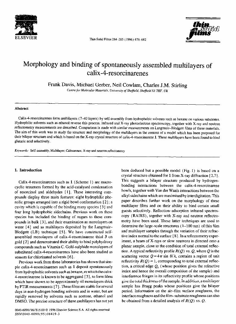
been deduced but a possible model (Fig. 1) is based on a
crystal structure obtained for 1 from X-ray diffraction [ 2,7].
This suggests a bilayer structure produced by hydrogen-
bonding interactions between the calix-4-resorcinarene
bowls, together with Van der Waals interactions between the
alkyl sidechains which are maximised by interdigitation. This
paper describes further work on the morphology of these
multilayer films and on their ability to bind certain small
guests selectively. Reflection-adsorption infrared spectros-
copy (RAIRS), together with X-ray and neutron reflectro-
metry have been used. These latter techniques are used to
determine the large-scale structures ( l-100 nm) of thin film
and multilayer samples through the variation of their refrac-
tive index normal to the surface [ 81. In a reflectometry exper-
iment, a beam of X-rays or slow neutrons is directed onto a
planar sample, close to the condition of total external reflec-
tion. A typical reflectivity profile R( Q) vs. Q, where Q is the
scattering vector Q = 4rr sin 6/h, contains a region of unit reflectivity R(Q) = 1, corresponding to total external reflec-
tion, a critical edge Q, (whose position gives the refractive index and hence the overall composition of the sample) and
interference fringes in its reflectivity profile whose positions
give the total thickness of the sample. In addition, a multilayer
sample has Bragg peaks whose positions give the bilayer period. Information on the air-film surface roughness, the interface roughness and the film-substrate roughness can also
be obtained from a detailed analysis of R( Q) vs. Q.
F. Davis et al. /Thin Solid Films 284-28s (19%) 678-682 619
l.R=-c,,H,,x=H. 2.R=-C,,H,,X=OH. 3.R= dCH,),,,.SHX=H.
HO OH
H°CHzYH OH 4
HOOP--COOH 6
HO-CH
OH 5
HOO/&OOH
7
HOOCVCY HOOC+O;
COP,"
HOOC
COOH
11
'COOH
HOOCmc:
Scheme 1.
2. Experimental
<
8
The self-assembled multilayers were obtained by a tech- nique described previously [ 71. A clean glass microscope slide was coated with a thermally evaporated 100 nm layer of aluminium or gold and was placed in a 10 mm01 dm- 3 solution of 1 or 2 in n-hexane for 16 h. The sample was then removed repeatedly, soaked in clean solvent for at least 1 h and finally dried. Longer exposures or cleaning times did not appear to have significant effects on the film thickness. The films were stable to soaking in hexane, even with sonication. FI’IR measurements were made using a Perkin-Elmer 1725 X spectrometer, fitted with an MCT detector and an IT80 reflection accessory for RAIRS. Contact angles were meas- ured by placing at least five 1 pl drops of water on the film. X-ray photoelectron spectra (XPS) were measured on a VG Clam 2 spectrometer. X-ray reflectometry experiments were made using a conventional Siemens 8:20 diffractometer with Cu Ka radiation. Neutron reflectometry experiments were made on the CRISP instrument at the ISIS neutron source, Rutherford Appleton Laboratory, using a time-of-flight method and a “white” incident beam at a fixed grazing angle of incidence. Bulk solutions of 1 with guests were made by stirring 10 mm01 dmd3 solutions in hexane with excess of solid guest and subsequent filtration [ 91. They were shown
I Al I Fig. 1. Proposed structure of the self-assembled calix-kesorcinarene
multilayer.
by PTIR and ‘H NMR to contain the guest, complexed in a (0.9-l. 1) : 1 ratio.
3. Results and discussion
3.1. Structure
A multilayer of 1 on aluminium/glass was assembled as described. XPS [ 101 showed the multilayer to have the cal- culated C:O and C-H:C-O ratios, indicating that no solvent was incorporated. The contact angle of the layer was 90”, lower than that for an octadecane thiol monolayer ( 105”) [ 111 but much higher than that of a t-calix-4-resorcinarene thiol monolayer (30”) [ 21. This indicates that the hydrocar- bon sidechains are at the surface, but not as well packed together as those found in a simple thiol monolayer [ 111, probably because of the large macrocyclic headgroup. RAIRS measurements and comparison with self-assembled
680 F. Davis et al. /Thin Solid Films 284-285 (1996) 678682
00 1000 2000 600
Wnvenumber (rme’)
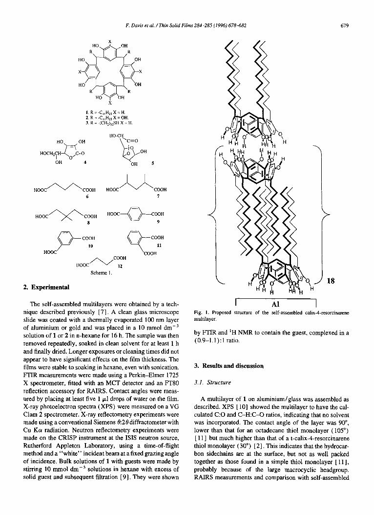
Fig. 2. RAIRS spectra of (a) 1.5 layers of 1 deposited on Al by the LB
technique; (b) a self-assembled multilayer of 1 on Al.
monolayers of 3 and LB films of 1 showed the multilayer to
be composed of about 38 monolayers (Fig. 2). A typical X-
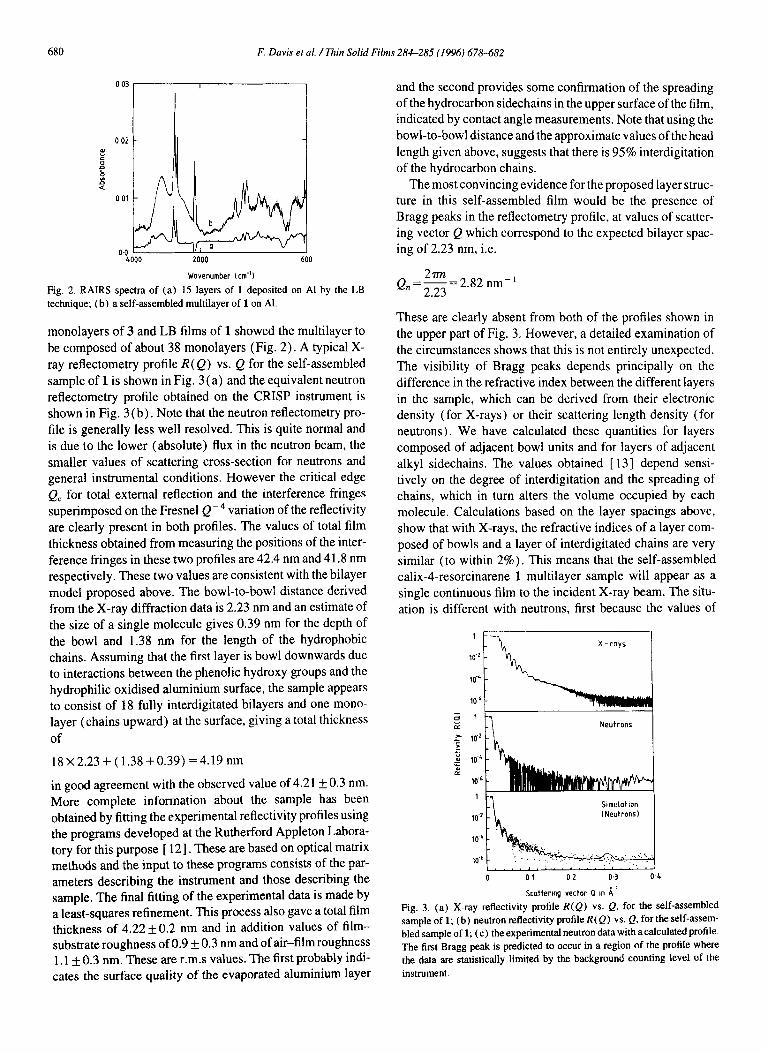
ray reflectometry profile R(Q) vs. Q for the self-assembled
sample of 1 is shown in Fig. 3 (a) and the equivalent neutron reflectometry profile obtained on the CRISP instrument is
shown in Fig. 3 (b) . Note that the neutron reflectometry pro-
file is generally less well resolved. This is quite normal and
is due to the lower (absolute) flux in the neutron beam, the
smaller values of scattering cross-section for neutrons and general instrumental conditions. However the critical edge
Q, for total external reflection and the interference fringes superimposed on the Fresnel Q -4 variation of the reflectivity
are clearly present in both profiles. The values of total film
thickness obtained from measuring the positions of the inter-
ference fringes in these two profiles are 42.4 nm and 41.8 nm respectively. These two values are consistent with the bilayer
model proposed above. The bowl-to-bowl distance derived from the X-ray diffraction data is 2.23 nm and an estimate of
the size of a single molecule gives 0.39 nm for the depth of
the bowl and 1.38 nm for the length of the hydrophobic chains. Assuming that the first layer is bowl downwards due
to interactions between the phenolic hydroxy groups and the hydrophilic oxidised aluminium surface, the sample appears to consist of 18 fully interdigitated bilayers and one mono-
layer (chains upward) at the surface, giving a total thickness
of
18 X 2.23 + ( 1.38 + 0.39) = 4.19 nm
in good agreement with the observed value of 4.21 f 0.3 nm.
More complete information about the sample has been obtained by fitting the experimental reflectivity profiles using
the programs developed at the Rutherford Appleton Labora- tory for this purpose [ 121. These are based on optical matrix methods and the input to these programs consists of the par-
ameters describing the instrument and those describing the sample. The final fitting of the experimental data is made by a least-squares refinement. This process also gave a total film thickness of 4.22 f 0.2 nm and in addition values of film- substrate roughness of 0.9 f 0.3 nm and of air-film roughness 1.1 f 0.3 nm. These are r.m.s values. The first probably indi- cates the surface quality of the evaporated aluminium layer
and the second provides some confirmation of the spreading of the hydrocarbon sidechains in the upper surface of the film, indicated by contact angle measurements. Note that using the
bowl-to-bowl distance and the approximate values of the head
length given above, suggests that there is 95% interdigitation
of the hydrocarbon chains.
The most convincing evidence for the proposed layer struc- ture in this self-assembled film would be the presence of Bragg peaks in the reflectometry profile, at values of scatter-
ing vector Q which correspond to the expected bilayer spac-
ing of 2.23 nm, i.e.
27m Q,=E=2.82 nm-’
These are clearly absent from both of the profiles shown in
the upper part of Fig. 3. However, a detailed examination of
the circumstances shows that this is not entirely unexpected. The visibility of Bragg peaks depends principally on the
difference in the refractive index between the different layers
in the sample, which can be derived from their electronic density (for X-rays) or their scattering length density (for
neutrons). We have calculated these quantities for layers composed of adjacent bowl units and for layers of adjacent
alkyl sidechains. The values obtained [ 131 depend sensi- tively on the degree of interdigitation and the spreading of chains, which in turn alters the volume occupied by each
molecule. Calculations based on the layer spacings above, show that with X-rays, the refractive indices of a layer com-
posed of bowls and a layer of interdigitated chains are very
similar (to within 2%). This means that the self-assembled calix-4-resorcinarene 1 multilayer sample will appear as a
single continuous film to the incident X-ray beam. The situ- ation is different with neutrons, first because the values of
10-L -
106 -
;i l- z -7, Neutrons
I\ Simulotlon 1 INeutrons
srotter1ng vector (1 I” A ’ Fig. 3. (a) X-ray reflectivity profile R(Q) vs. Q. for the self-assembled
sample of 1; (b) neutron reflectivity profile R(Q) vs. Q, for the self-assem- bled sample of 1; (c) the experimental neutron data with a calculated profile.
The first Bragg peak is predicted to occur in a region of the profile where the data are statistically limited by the background counting level of the
instrument.
F. Davis et al. /Thin Solid Films 284-285 (1996) 678682 681
scattering length density are not related to atomic number
and second because the value for hydrogen has a different
sign from those of carbon and oxygen. The difference
between the refractive indices of the two layers is therefore
slightly greater for a neutron beam than for X-rays. Unfor- tunately, the poorer statistical quality of the neutron beam
data means that the Bragg peaks cannot be positively identi- fied from the currently avai!able data in Fig. 3. This is shown
in Fig. 3(c) in which the neutron reflectivity profile calcu- lated for the proposed multilayer structure is superimposed
as a continuous line on the actual data points from the neutron experiment. It is clear there is good agreement between the
model and the data down to a reflectivity value
R(Q) = 1 X 10e4, but beyond this, the poor statistical quality
of the neutron data points do not allow any further detail to be seen in the profile. In addition, the intrinsic background
counting level of the instrument, which corresponds to avalue R(Q) = 1 X 10⁻⁶,hasbeenreachedforvaluesofQr2nm-’.
In this region it is not possible for the first Bragg peak from
the multilayer at Q 2 2.82 nm- ’ to be detected.
Thus to summarise, the X-ray and neutron reflectometry experiments on the calix-4-resorcinarene sample have both
provided data on its structure which are consistent with the
results from the spectroscopic experiments. They are also
completely consistent with the model that has been proposed
for the structure of this self-assembled multilayer film. How-
ever, conclusive evidence for the bilayer structure in the form
of Bragg peaks in the reflectivity profiles has not yet emerged. In X-ray reflectometry this is because of an inherent lack of
contrast within the constituent layers of the sample, while for
neutrons the limitation is an instrumental one. Ways of cir-
cumventing these problems, in the future, will be discussed in the conclusions.
A pyrogallol/dodecanal tetramer 2 was also assembled into a multilayer under similar conditions. Comparison of C-
H stretching intensities from RAIRS data indicated that either
the film was much thinner (about 5 monolayers) or that its alkyl sidechains were at a much steeper angle to the substrate.
The contact angle was lower (78”) for this system, indicating
a more disordered surface [ 111.
3.2. Binding
Attempts were made to incorporate guests into these calix- 4-resorcinarene multilayers. Previous results [ 71 have shown that the multilayers, when placed in aqueous solutions of Vitamin C 4 and glucuronolactone 5 (Scheme 1)) did not
incorporate the guests, unlike the gold-thiol monolayers [ 21.
This is thought to be due to difficulty of penetration of the
matrix by the guests. Smaller guests were therefore used, which are derivatives of glutaric acid. These were known to
form a 1: 1 adduct with 1 in bulk solution [ 91. Solutions ( 10 mm01 dmp3) of 1: 1 adducts of 1 with acids 6,7 or 8, were made using previously described methods [9], and self- assembled multilayers were made on aluminium-coated microscope slides. RAIRS measurements showed that only
O-110 -
00 4000 2000 600
Wovenumber (cm-'1
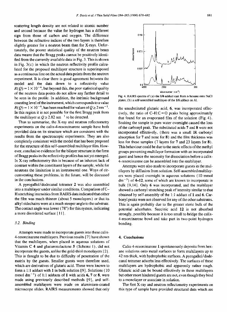
Fig. 4. RAIRS spectra of (a) the l/6 adduct cast from n-hexane onto NaCl
plates; (b) a self-assembled multilayer of the l/6 adduct on Al.
the unsubstituted glutaric acid, 6, was incorporated effec-
tively, the ratio of C-H:C = 0 peaks being approximately
that found for an evaporated film of the solution (Fig. 4).
Soaking the sample in pure water overnight caused the loss of the carbonyl peak. The substituted acids 7 and 8 were not
incorporated effectively, (there was a small IR carbonyl absorption for 7 and none for 8) and the film thickness was
less for these samples (7 layers for 7 and 23 layers for 8).
This behaviour could be due to the steric effects of the methyl
groups preventing multilayer formation with an incorporated
guest and hence the necessity for dissociation before a calix- 4-resorcinarene can be assembled into the multilayer.
Attempts were also made to incorporate guests in the mul- tilayers by diffusion from solution. Self-assembled multilay-
ers were placed overnight in aqueous solutions (10 mmol
dme3) of 4-12, some of which are known to incorporate in bulk [9,14]. Only 6 was incorporated, and the multilayer showed a carbonyl stretching peak of intensity similar to that
obtained by self-assembly of the 1: 1 adduct of 1 and 6. Car-
bony1 peaks were not observed for any of the other substrates. This is again probably due to the greater steric bulk of the
potential adsorbates. Succinic acid 12 is not absorbed
strongly, possibly because it is too small to bridge the calix- 4-resorcinarene bowl and take part in two-point hydrogen
bonding.
4. Conclusions
Calix-4-resorcinarene 1 spontaneously deposits from hex-
ane solutions onto metal surfaces to form multilayers up to
42 nm thick, with hydrophobic surfaces. A pyrogallol/dode-
canal tetramer adsorbs less effectively. The surfaces of these multilayers are hydrophobic and apparently rather rough.
Glutaric acid can be bound effectively in these multilayers but other more hindered guests are not, even though they bind to a monolayer or associate in solution,
The first X-ray and neutron reflectometry experiments on this type of sample have provided structural data which are
682 F. Davis et al. /Thin Solid Films 284-285 (1996) 678-682
consistent with both spectroscopic results and with the model
proposed for the structure of the multilayer. It has not yet proved possible to obtain completely conclusive evidence for
the repeating bilayer structure in the sample by observation
of Bragg peaks in the reflectivity profiles. This is due to an
inherent lack of contrast between the constituent layer of the
sample with X-rays and instrumental limitations with neu-
trons. These initial reflectometry experiments are sufficiently
encouraging to suggest ways of circumventing these prob-
lems. Contrast between layers can be enhanced for X-rays by incorporating heavy atoms into the molecule. This will be
done by making the resorcinol/lO-undecenal tetramer and
subsequent addition of bromine to the alkenyl group. For neutrons the problems are less tractable. An experiment 100
times longer than that in Fig. 3(b) would be needed to pro- duce a lo-fold increase in the statistics of the data points. In
various parts of these molecules, protons will be replaced by
deuterons to increase contrast.
Acknowledgements
We thank Dr T. Richardson and V.C. Smith for supplying
LB films, Dr. L. O’Toole for XPS measurements and the
University of Sheffield for support of this work. The neutron reflectometry experiments were performed under the aegis of
the E.P.S.R.C’s neutron beam program and the authors thank Dr. D. Bucknall for his assistance.
References
[ I] CD. Gutsche. Calirarenes, Royal Society of Chemistry, Monographs
in Supramolecular Chemistry, 1989.
[2] H. Adams, F. Davis and C.J.M. Stirling, J. Chem. Sot., Chem. Commun., ( 1994) 2521.
[3] Y. Aoyama, Y. Tanake and T. Kunitake, J. Am. Chem. Sot., III ( 1989) 5397.
[4] K. Ohto, Y. Tanake, Y. Aoyama and T. Kunitake, Thin Solid Films, I79 (1989) 21.
[S] K. Kurihara, K. Ohto, Y. Tanake, Y. Aoyama and T. Kunitake, J. Am.
Chem. Sot., 113 (1991) 444.
[6] E.U.T. VanVelzen, J.F.J. Engbersenand D.N. Reinhoudt,J. Am. Chem. sot., 116 (1994) 3597.
[7] F. Davis and C.J.M. Stirling, J. Am. Chem. Sot.. 117 (1995) 10385.
[S] J. Penfold and R.K. Thomas, J. Phys: Condens. Matt., 2 ( 1990) 1369.
[ 91 Y. Tanake, Y. Kato and Y. Aoyama, J. Am. Chem. Sot., 112 ( 1990)
2807.
[lo] P. Laibinis. M. Fox, J. Folkers and G.M. Whitesides, Langmuir, 7
(1991) 3167.
[ 1 I] A. Ulman, An Introduction to Ultrathin Organic Films, Academic
Press, New York, 1991.
[ 121 1. Penfold, Rutherford-Appleton Report, RAL-88-088, 1988.
[ 131 M. Gerber, M. Phil. Thesis, University of Sheffield (1996), in
preparation.
[ 141 D.A. Leigh, P. Linnane, R.G. Pritchard and G. Jackson, J. Chem. Sot., Chem. Commun., ( 1994) 389.


![Development of Calix[4]arene-Functionalized](https://img.pdfslide.us/doc/110x75/61ab1bfbbc68120d180622ab/development-of-calix4arene-functionalized-.jpg)