Embed Size (px)
Citation preview
Morphological characterization of Zn-Based Nanostructured Thin Films
A. Gomes1 ,T. Frade1 and Isabel D. Nogueira2
1Center of Materials and Molecular Sciences, Department of Chemistry and Biochemistry of Faculty of Science of the University of Lisbon, Campo Grande Ed. C8, 1749-016 Lisbon, Portugal
2ICEMS/Instituto Superior Tecnico-Universidade Tecnica de Lisboa, Av. Rovisco Pais, 1049-001 Lisbon, Portugal
Zn-based nanostructures research has been greatly increased in the last years because of their huge technological applications, namely in wastewater treatments and photovoltaic cells.
Electrodeposition is one of the most versatile and low cost technique used to obtain nanostrutured materials. The nanomaterial characteristics, such as morphology, could be tailored and improved by modification of deposition parameters used, like current profile, bath composition, temperature, and substrate.
The Field Emission-Scanning Electron Microscopy (FEG-SEM) is a powerful microscopic technique that helps us to understand modifications in morphological characteristics of the electrodeposits and correlated them to its electric, catalytic and electrochemical properties.
Keywords Zn-based nanostructures; Electrodeposition; Field emission-scanning electron microscopy
1. Introduction
Electrodeposition of zinc based films in constant and pulsed regimes of electrolysis was performed and the obtained electrodeposits were examined by field emission-scanning electron microscopy. The electrodeposition is one of the most effective methods for the synthesis of nanostructures of metals and semiconductors with desired morphology and characteristics for many technological applications [1]. There are several variables that may be tailored specifically the electrolysis regime, the bath composition, pH, solution stirring and temperature [2]. Most of the depositions that will be discussed here were performed at acidic or near neutral solutions. Electrodeposition by pulse current, is a very versatile method, since more experimental conditions can be changed such as current density, duty cycle and pulse frequency with huge effect on the deposits characteristics: composition, structure, morphology and porosity [3]. The electrodeposits of zinc on different metals, namely steel, have special relevance due to their anticorrosive properties by the formation of passive layers in contact with air, and in aqueous solution. The corrosive resistance of the films is strongly connected to the morphological characteristics of metallic films that could be tailored by using additives in the bath, such as surfactants or oxide nanoparticles. The adsorption of surfactants aggregates or adsorption of nanoparticles onto electrodes can have large effects on the kinetics of the electron transfer and consequently on the electrodeposition process. The effect on the electron transfer rates includes blocking of the active sites by the surfactants, and electrostatic interactions between electroactive species and adsorbed surfactants [4]. Due to those effects it is possible to modify the growth mode of the crystals and tailor the morphology and structure of the electrodeposits. In a previous work cationic, anionic and non-ionic surfactants have been used for studying the effect of the charge of headgroups on the electrodeposition process. It was conclude that the use of the surfactant leads to a decrease in the grain size and changes in the film’s morphology and the zinc texture [5]. Due to the co-deposition process of oxide nanoparticles and metallic film, metal matrix nanocomposites can be successfully prepared [6]. It is expected that by electrolysis of plating solutions in which micron or sub-micron size particles are suspended, it is possible to obtain solid materials with improved and/or combined properties, which make them interesting for applications such as environmental remediation. Composites containing occluded TiO2 particles are suitable materials, due to the semiconducting properties of TiO2, with applications as photocatalysts, particularly in the treatment of polluted water. In recent years research efforts are underway to develop more powerful methods than those currently applied in wastewater treatment. Thus, the search for new efficient methods for the degradation of pollutants is a priority. Electrochemical and photoelectrochemical degradation can be suitable and low-cost alternatives to those used presently [7]. The implementation of these methods is closely linked with the development of stable, non-pollutant, cheap and electrocatalytic/ photocatalytic electrode materials. Composite electrodes of Ti/Zn-TiO2 have been successfully prepared by us in recent years [8]. In addition, we have tried with little success the implementation of these electrodes in the pharmaceutical degradation by photoassisted electrochemical processes. To improve the photo ability of matrix we have transformed the Zn matrix into wurtzite-structured ZnO. With a wide band gap (3.35 eV) [9], ZnO has been used as an environmental photocatalyst for water purification with the aid of artificial light source. Among the techniques used for the oxidation of Zn films, heat treatment in air seems to be the simplest due to relative low melting point of Zn. The conversion process depends on the crystallinity and orientation of as-deposited Zn. We have already achieved the formation of nanocrystalline ZnO films with higher surface area [10] and, successfully used this strategy to prepare Ti/ZnO-TiO2 photoelectrodes for the degradation of the AO7 dye and ibuprofen present in
Current Microscopy Contributions to Advances in Science and Technology (A. Méndez-Vilas, Ed.)
© 2012 FORMATEX 1146
simulated wastewater [10,11]. It should be highlighted that during the annealing process the intermetallic phases formation also occurs. To overcome this undesirable formation, we decide to prepare ZnO by cathodic electrodeposition from zinc nitrate solutions onto conductive glass substrates. In literature, a variety of ZnO nanostructures have been reported by electrodeposition process. Among them the 1D ZnO arrays attracting intensive research because they provide high charge transport making them suitable for many practical applications [12]. Previously to the growth of vertically aligned 1D ZnO nanostructures, the preliminary stage of electrodeposition is crucial to overall process. In this work, ZnO nanostructured electrodes were prepared using the two-step electrodeposition method constituted by the formation of a seed or buffer layer on first stage followed by a second stage related to the ZnO growth at 70 ºC. The deposition of a seed layer has shown to promote the vertical alignment of the ZnO nanostructures [13]. The aim of the present work is to show that using Field emission-scanning electron microscopy (FEG-SEM) it is possible to correlate the morphology of the electrodeposits with the deposition conditions used. This analytical technique allows direct visualization of metallic and semiconducting surface structures with a three-dimensional appearance and high resolution, without artifacts. This technique of analysis is widely used due to its simplicity, and versatility [1].
2. Experimental
Zn deposits that have been prepared by galvanostatic pulsed electrolysis, from acidic zinc sulphate solutions, on a stainless steel substrate. The electrodeposition was carried out in a two-compartment glass cell using a stainless steel disc as substrate, a platinum spiral as the counter electrode and a commercial saturated calomel electrode as the reference electrode. Before the experiments, the stainless steel discs were mechanically polished to a mirror finish. The electrochemical cell was connected to a EG & G Princeton Applied Research potentiostat/galvanostat model 263. The electrolyte was prepared dissolving ZnSO4.7H2O in Millipore Milli-Q ultra pure water. Separately, cetyl trimethyl ammonium bromide (CTAB) was added to this base solution. The experiments were carried out at room temperature and the deposition was performed under magnetic stirring. N2 was bubbled through the solution for 15 min before each experiment and during the deposition, a weak flux of N2 was maintained. For the nanocomposite film preparation, the electroplating bath was made up by ZnSO4·7H2O, MgSO4, H3BO3 and TiO2 (AEROXIDE® TiO2 P25, particle size approx. 25 nm, with 80% anatase and 20% rutile). The bath pH was adjusted to 4 by adding a H2SO4 diluted solution. A glass cell with two compartments was used, with a Zn plate as sacrificial counter electrode and a commercial Ag/AgCl as reference. The working electrodes were Ti discs. The deposition was performed by galvanostatic pulsed electrolysis under magnetic stirring (150 rpm) at room temperature for 100 min in order to obtain an average charge value of 30 C. When finished, the electrode was removed from the cell, rinsed with Millipore Milli-Q ultra pure water and dried under nitrogen atmosphere for 5–10 min at room temperature. The as-deposited Zn–TiO2 films were annealed in air at 450 °C, using a heating rate of 5 °C min−1, held for 3 h, and then cooled to room temperature. Figure 1 shows a representative pulse-current waveform where the pulse plating parameters are the pulse length ton, the time between two pulses toff, the pulse height ip and the average current density ia, defined as in Eq. (1).
ia=(ip·ton)/(ton+toff) (1)
Pulse height imposed provokes the depletion of ions near the cathode. During toff, ions migrate to the interfacial region and when ton occurs the cycle repeats. The duty cycle is the ratio of pulse duration (on-time) and sum of on- and off-time and frequency is the inverse of the sum of ton and toff. The ZnO nanostructured films were prepared potentiostatically on fluorine doped tin oxide (FTO) by application of −1.0 V vs. Ag/AgCl for 60 min from an electrolytic bath constituted by 0.01 M Zn(NO3)2 + 0.005 M KCl, at 70 °C. Preceding this, a seed layer was prepared by application of −1.3 V vs. Ag/AgCl for 30 s, at 70 ºC or a buffer layer by application of 67 μA for 30 min, at room temperature. The electrochemical depositions were carried out using a Voltalab PGZ 100 potentiostat/ galvanostat of Radiometer, with automatic data acquisition. The buffer layer formed by Zn(OH)2 was annealed in air at 450 °C for1 h, and then cooled to room temperature. The films morphology and elemental composition were investigated by field-emission scanning electron microscopy (FEG-SEM, JEOL 7001 F). X-ray diffraction (XRD) analysis of the electrodeposits was carried out using a Philips X-ray diffractometer (model PW 1710) operating with a monochromatic Cu Kα radiation in Bragg–Brentano geometry. The diffractograms were obtained in the 2θ range from 20 to 80°, using a 0.02° step and an acquisition time of 2 s/step.
Current Microscopy Contributions to Advances in Science and Technology (A. Méndez-Vilas, Ed.)
© 2012 FORMATEX 1147
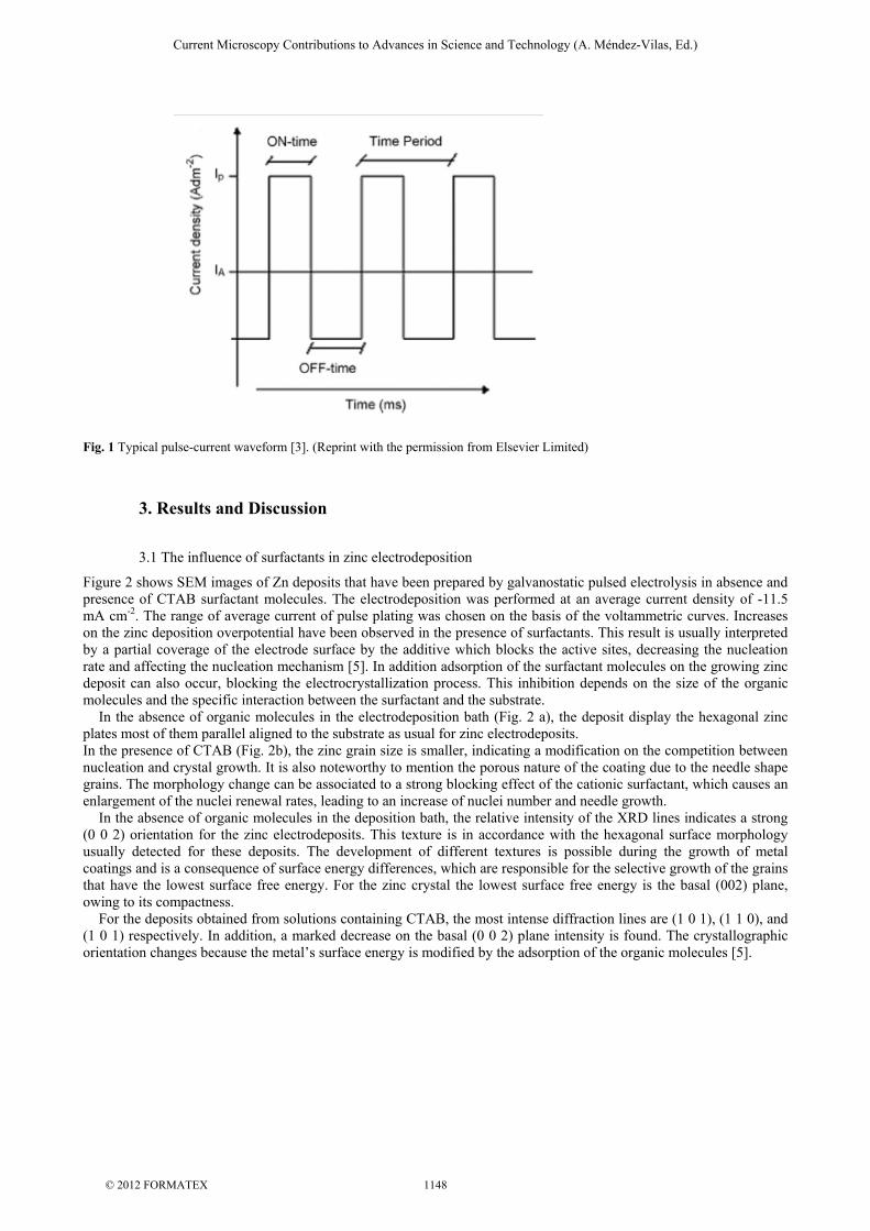
Fig. 1 Typical pulse-current waveform [3]. (Reprint with the permission from Elsevier Limited)
3. Results and Discussion
3.1 The influence of surfactants in zinc electrodeposition
Figure 2 shows SEM images of Zn deposits that have been prepared by galvanostatic pulsed electrolysis in absence and presence of CTAB surfactant molecules. The electrodeposition was performed at an average current density of -11.5 mA cm-2. The range of average current of pulse plating was chosen on the basis of the voltammetric curves. Increases on the zinc deposition overpotential have been observed in the presence of surfactants. This result is usually interpreted by a partial coverage of the electrode surface by the additive which blocks the active sites, decreasing the nucleation rate and affecting the nucleation mechanism [5]. In addition adsorption of the surfactant molecules on the growing zinc deposit can also occur, blocking the electrocrystallization process. This inhibition depends on the size of the organic molecules and the specific interaction between the surfactant and the substrate. In the absence of organic molecules in the electrodeposition bath (Fig. 2 a), the deposit display the hexagonal zinc plates most of them parallel aligned to the substrate as usual for zinc electrodeposits. In the presence of CTAB (Fig. 2b), the zinc grain size is smaller, indicating a modification on the competition between nucleation and crystal growth. It is also noteworthy to mention the porous nature of the coating due to the needle shape grains. The morphology change can be associated to a strong blocking effect of the cationic surfactant, which causes an enlargement of the nuclei renewal rates, leading to an increase of nuclei number and needle growth. In the absence of organic molecules in the deposition bath, the relative intensity of the XRD lines indicates a strong (0 0 2) orientation for the zinc electrodeposits. This texture is in accordance with the hexagonal surface morphology usually detected for these deposits. The development of different textures is possible during the growth of metal coatings and is a consequence of surface energy differences, which are responsible for the selective growth of the grains that have the lowest surface free energy. For the zinc crystal the lowest surface free energy is the basal (002) plane, owing to its compactness. For the deposits obtained from solutions containing CTAB, the most intense diffraction lines are (1 0 1), (1 1 0), and (1 0 1) respectively. In addition, a marked decrease on the basal (0 0 2) plane intensity is found. The crystallographic orientation changes because the metal’s surface energy is modified by the adsorption of the organic molecules [5].
Current Microscopy Contributions to Advances in Science and Technology (A. Méndez-Vilas, Ed.)
© 2012 FORMATEX 1148
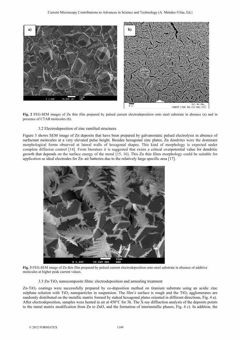
Fig. 2 FEG-SEM images of Zn thin film prepared by pulsed current electrodeposition onto steel substrate in absence (a) and in presence of CTAB molecules (b).
3.2 Electrodeposition of zinc ramified structures
Figure 3 shows SEM image of Zn deposits that have been prepared by galvanostatic pulsed electrolysis in absence of surfactant molecules at a very elevated pulse height. Besides hexagonal zinc plates, Zn dendrites were the dominant morphological forms observed at lateral walls of hexagonal shapes. This kind of morphology is expected under complete diffusion control [14]. From literature it is suggested that exists a critical overpotential value for dendritic growth that depends on the surface energy of the metal [15, 16]. This Zn thin films morphology could be suitable for application as ideal electrodes for Zn–air batteries due to the relatively large specific area [17]. Fig. 3 FEG-SEM image of Zn thin film prepared by pulsed current electrodeposition onto steel substrate in absence of additive molecules at higher peak current values.
3.3 Zn-TiO2 nanocomposite films: electrodeposition and annealing treatment
Zn-TiO2 coatings were successfully prepared by co-deposition method on titanium substrate using an acidic zinc sulphate solution with TiO2 nanoparticles in suspension. The film’s surface is rough and the TiO2 agglomerates are randomly distributed on the metallic matrix formed by staked hexagonal plates oriented in different directions, Fig. 4 a). After electrodeposition, samples were heated in air at 450°C for 3h. The X-ray diffraction analysis of the deposits points to the metal matrix modification from Zn to ZnO, and the formation of intermetallic phases, Fig. 4 c). In addition, the
a) b)
Current Microscopy Contributions to Advances in Science and Technology (A. Méndez-Vilas, Ed.)
© 2012 FORMATEX 1149
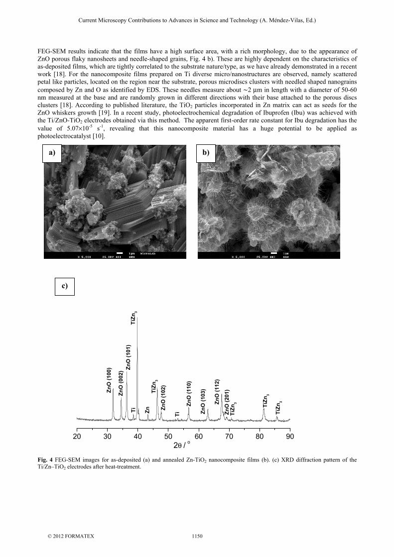
FEG-SEM results indicate that the films have a high surface area, with a rich morphology, due to the appearance of ZnO porous flaky nanosheets and needle-shaped grains, Fig. 4 b). These are highly dependent on the characteristics of as-deposited films, which are tightly correlated to the substrate nature/type, as we have already demonstrated in a recent work [18]. For the nanocomposite films prepared on Ti diverse micro/nanostructures are observed, namely scattered petal like particles, located on the region near the substrate, porous microdiscs clusters with needled shaped nanograins composed by Zn and O as identified by EDS. These needles measure about ∼2 μm in length with a diameter of 50-60 nm measured at the base and are randomly grown in different directions with their base attached to the porous discs clusters [18]. According to published literature, the TiO2 particles incorporated in Zn matrix can act as seeds for the ZnO whiskers growth [19]. In a recent study, photoelectrochemical degradation of Ibuprofen (Ibu) was achieved with the Ti/ZnO-TiO2 electrodes obtained via this method. The apparent first-order rate constant for Ibu degradation has the value of 5.07×10⁻⁵ s-1, revealing that this nanocomposite material has a huge potential to be applied as photoelectrocatalyst [10].
Fig. 4 FEG-SEM images for as-deposited (a) and annealed Zn-TiO2 nanocomposite films (b). (c) XRD diffraction pattern of the Ti/Zn–TiO2 electrodes after heat-treatment.
c)
20 30 40 50 60 70 80 90
TiZn
2θ / o
ZnO
(100
)
ZnO
(002
) ZnO
(101
)Ti
TiZn
3
TiZn
3
ZnO
(102
)
ZnO
(110
)
ZnO
(103
)
ZnO
(112
)
ZnO
(201
)Ti
Zn3 TiZn
3
TiZn
3
b) a)
Current Microscopy Contributions to Advances in Science and Technology (A. Méndez-Vilas, Ed.)
© 2012 FORMATEX 1150
3.4 1D ZnO nanostructures: Two-step electrodeposition
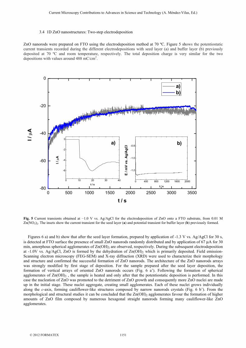
ZnO nanorods were prepared on FTO using the electrodeposition method at 70 ºC. Figure 5 shows the potentiostatic current transients recorded during the different electrodepositions with seed layer (a) and buffer layer (b) previously deposited at 70 ºC and room temperature, respectively. The total deposition charge is very similar for the two depositions with values around 488 mC/cm2.
Fig. 5 Current transients obtained at −1.0 V vs. Ag/AgCl for the electrodeposition of ZnO onto a FTO substrate, from 0.01 M Zn(NO3)2. The insets show the current transient for the seed layer (a) and potential transient for buffer layer (b) previously formed. Figures 6 a) and b) show that after the seed layer formation, prepared by application of -1.3 V vs. Ag/AgCl for 30 s, is detected at FTO surface the presence of small ZnO nanorods randomly distributed and by application of 67 μA for 30 min, amorphous spherical agglomerates of Zn(OH)2 are observed, respectively. During the subsequent electrodeposition at -1.0V vs. Ag/AgCl, ZnO is formed by the dehydration of Zn(OH)2 which is primarily deposited. Field emission-Scanning electron microscopy (FEG-SEM) and X-ray diffraction (XRD) were used to characterize their morphology and structure and confirmed the successful formation of ZnO nanorods. The architecture of the ZnO nanorods arrays was strongly modified by first stage of deposition. For the sample prepared after the seed layer deposition, the formation of vertical arrays of oriented ZnO nanorods occurs (Fig. 6 a’). Following the formation of spherical agglomerates of Zn(OH)2 , the sample is heated and only after that the potentiostatic deposition is performed. In this case the nucleation of ZnO was promoted to the detriment of ZnO growth and consequently more ZnO nuclei are made up in the initial stage. These nuclei aggregate, creating small agglomerates. Each of these nuclei grows individually along the c-axis, forming cauliflower-like structures composed by narrow nanorods crystals (Fig. 6 b’). From the morphological and structural studies it can be concluded that the Zn(OH)2 agglomerates favour the formation of higher amounts of ZnO film composed by numerous hexagonal straight nanorods forming many cauliflower-like ZnO agglomerates.
0 500 1000 1500 2000 2500 3000 3500-80
-60
-40
-20
0
I / μ
A
t / s
a) b)
0 10 20 30 40
-6
-4
-2
0
I / μ
A
t / s
a)
0 400 800 1200 1600 2000
-1.2
-1.0
-0.8b)
E / m
V vs
. Ag/
AgC
l
t / s
Current Microscopy Contributions to Advances in Science and Technology (A. Méndez-Vilas, Ed.)
© 2012 FORMATEX 1151
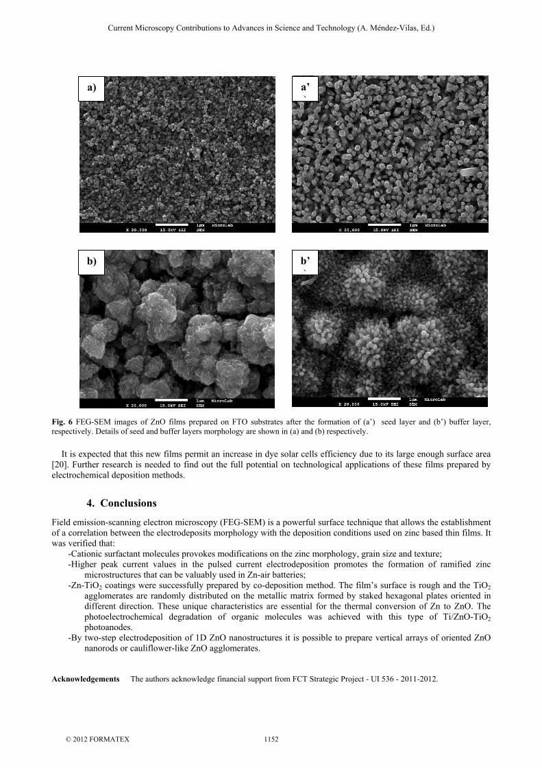
Fig. 6 FEG-SEM images of ZnO films prepared on FTO substrates after the formation of (a’) seed layer and (b’) buffer layer, respectively. Details of seed and buffer layers morphology are shown in (a) and (b) respectively.
It is expected that this new films permit an increase in dye solar cells efficiency due to its large enough surface area [20]. Further research is needed to find out the full potential on technological applications of these films prepared by electrochemical deposition methods.
4. Conclusions
Field emission-scanning electron microscopy (FEG-SEM) is a powerful surface technique that allows the establishment of a correlation between the electrodeposits morphology with the deposition conditions used on zinc based thin films. It was verified that:
- Cationic surfactant molecules provokes modifications on the zinc morphology, grain size and texture; - Higher peak current values in the pulsed current electrodeposition promotes the formation of ramified zinc
microstructures that can be valuably used in Zn-air batteries; - Zn-TiO2 coatings were successfully prepared by co-deposition method. The film’s surface is rough and the TiO2
agglomerates are randomly distributed on the metallic matrix formed by staked hexagonal plates oriented in different direction. These unique characteristics are essential for the thermal conversion of Zn to ZnO. The photoelectrochemical degradation of organic molecules was achieved with this type of Ti/ZnO-TiO2 photoanodes.
- By two-step electrodeposition of 1D ZnO nanostructures it is possible to prepare vertical arrays of oriented ZnO nanorods or cauliflower-like ZnO agglomerates.
Acknowledgements The authors acknowledge financial support from FCT Strategic Project - UI 536 - 2011-2012.
a) a’)
b’)
b)
Current Microscopy Contributions to Advances in Science and Technology (A. Méndez-Vilas, Ed.)
© 2012 FORMATEX 1152
References
[1] Bicelli LP, Bozzini B, Mele C, D'Urzo L. A Review of Nanostructural Aspects of Metal Electrodeposition. Int. J. Electrochem. Sci. 2008;3:356 – 408.
[2] Frade T, Bouzon V, Gomes A, da Silva Pereira MI. Pulsed-reverse current electrodeposition of Zn and Zn-TiO2 nanocomposite films. Surf. Coat. Tech. 2010;204 :3592-3598.
[3] Chandrasekar MS, Pushpavanam M. Pulse and pulse reverse plating- Conceptual, advantages and applications. Electrochim. Acta 2008;53:3313–3322.
[4] J.F. Rusling, Molecular aspects of electron transfer at electrodes in micellar solutions Coll. Surf. A 1997;123/124:81-88. [5] Gomes A, da Silva Pereira MI. Pulsed electrodeposition of Zn in the presence of surfactants. Electrochim. Acta 2006;51:1342–
1350. [6] Hovestad R, Janssen LJJ. Electrochemical codeposition of inert particles in a metallic matrix. J. Appl. Electrochem. 1995;25:519-
527. [7] Skoumal M, Cabot PL, Centellas F, Arias C, Rodriguez RM, Garrido JA, Brillas E. Mineralization of paracetamol by ozonation
catalyzed with Fe2+, Cu2+ and UVA light. Appl. Cat. B:Environ. 2006;66:228–240. [8] Gomes A, da Silva Pereira MI, Mendonça MH, Costa FM. Zn–TiO2 composite films prepared by pulsed electrodeposition. J.
Solid State Electrochem. 2005;9: 190–196. [9] Bard AJ, Stratmann, Licht S (eds) Encyclopedia of Electrochemistry, Vol. 6, Semiconductor Electrodes and
Photoelectrochemistry, Wiley-VCH, Weinheim; 2002. [10] Gomes A, Frade T, Lobato K, Melo Jorge ME, da Silva Pereira MI, Círiaco L, Lopes A. Annealed Ti/Zn-TiO2 nanocomposites
tested as photoanodes for the degradation of Ibuprofen. J. Solid State Electrochem. 2012;16:2061-2069. [11] Frade T, Gomes A, da Silva Pereira M I, Lopes A, Ciríaco L. Photoelectrodegradation of AO7 dye by ZnO-TiO2 nanocomposite
films. Quim. Nova 2012; 35:30-34. [12] Yang M, Yin G, Huang Z, Kang Y, Liao X, Wang H. Preparation and Optical Properties of Biomimic Hierarchical ZnO
Column Arrays. Cryst. Growth Des. 2009;9:707–714. [13] Xu S, Wang ZL. One-Dimensional ZnO Nanostructures: Solution Growth and Functional Properties. Nano. Res. 2011;4:1013–
1098. [14] Paunovic M, Schlesinger M. Fundamentals of Electrochemical Deposition. Electrochemical Society Series, John Wiley & Sons,
1998. [15] Diggle JW, Despić AR, Bockris JO’M. The mechanism of the dendritic electrocrystallization of zinc. J. Electrochem. Soc.
1969;116:1503–1514 [16] Nikolić ND, Branković G, Lačnjevac UČ. Formation of two-dimensional (2D) lead dendrites by application of different
regimes of electrolysis. J. Solid State Electrochem. 2012;16:2121–2126. [17] Hsu P-C, Seol S-K, Lo T-N, Liu C-J, Wang C-L, Lin C-S, Hwu Y, Chen CH, Chang L-W, Je JH, Margaritondo G.. Hydrogen
Bubbles and the Growth Morphology of Ramified Zinc by Electrodeposition. J. Electrochem. Soc, 2008;155:D400-D407. [18] Frade T, Melo Jorge ME, Gomes A. Annealed Zn–TiO2 nanocomposites electrodeposited: Effect of the substrate. Surf.
Coat.Tech. 2012;206:3459–3466. [19] Deguchi T, Imai K, Iwasaki M, Tada H, Ito S. Photocatalytically Highly Active Nanocomposite Films Consisting of TiO2
Particles and ZnO Whiskers Formed on Steel Plates. J. Electrochem. Soc. 2000; 147:2263-2267. [20] Baxter JB. Commercialization of dye sensitized solar cells: Present status and future research needs to improve efficiency,
stability, and manufacturing. J. Vac. Sci. Technol. A 2012; 30: 020801-1-020801-19.
Current Microscopy Contributions to Advances in Science and Technology (A. Méndez-Vilas, Ed.)
© 2012 FORMATEX 1153