Embed Size (px)
Citation preview
Morphologic and Morphometric Analyses ofAcetic Acid-induced Colitis in Rats afterTreatment with Enemas from MyracrodruonUrundeuva Fr. All. (Aroeira do Sertao)
Lusmar Veras Rodrigues,1 Francisco Valdeci Ferreira,2 F. Sergio P. Regadas,1 Delcio Matos4
and Glauce Socorro de Barros Viana3*1Department of Surgery, Federal University of Ceara, Rua Cel. Nunes de Melo, 1127 60.431–930 Fortaleza, Brazil2Department of Pathology, Federal University of Ceara, Rua Cel. Nunes de Melo, 1127 60.431–930 Fortaleza, Brazil3Department of Pharmacology, Federal University of Ceara, Rua Cel. Nunes de Melo, 1127 60.431–930 Fortaleza, Brazil4Department of Surgery, Federal University of Sao Paulo (EPM), Sao Paulo, Brazil.
The present work showed the effects of Myracrodruon urundeuva Fr. All., popularly known as ‘aroeira’(AE), in the form of enemas prepared from the stem bark, on several morphologic and morphometricparameters after acetic acid-induced colitis in rats. Enemas from 5-ASA were used as standard while thevehicle, carboxymethylcellulose, was used as a control. The results of the morphological evaluationshowed that on day 1 acetic acid produced significantly more necrosis in the groups treated with AE(10% and 20%) or 5-ASA than the controls. However, on day 60, there were more caliciform andabsorptive cells in the treated groups compared with the controls. A significantly higher number of eosi-nophil and mononuclear cells and also collagen deposition in the controls compared with the treatedgroups were observed on day 60. However, a higher number of polymorphonuclear cells was detected onday 60 only in the AE treated group but not in the 5-ASA group. These data indicate that animals trea-ted with AE or 5-ASA showed complete epithelial tissue regeneration, while in the controls chronicinflammatory exudate persisted and tissue regeneration occurred through fibrosis. Copyright � 2002John Wiley & Sons, Ltd.
Keywords: acetic acid-induced colitis; Myracrodruon urundeuva; crude extract..
INTRODUCTION
Several experimental models are used for studyingnonspecific ulcerative colitis whose aetiology andaetiopathogeny are still unknown. One of these models,diffuse colitis induced by acetic acid (MacPherson andPfeifer, 1978) shows good reproducibility, showinglesions similar to those of human ulcerative colitis andis useful for screening substances for the treatment ofnonspecific ulcerative colitis.
Intrarectal administration of diluted acetic acidproduces diffuse and intense inflammation of the colonbesides neutrophil infiltration, haemorrhage, necrosis anddenuding of the epithelium (Yamada et al., 1991a; andLowe and Noronha-Blob, 1993). Both experimentalcolitis and human inflammatory bowel disease arecharacterized by an increased colonic blood flow.Neutrophils are thought to play a role in the pathogenesisof inflammatory bowel diseases such as ulcerative colitis,since prominent neutrophil infiltration has been observedin the inflamed colonic mucosa of patients with this
pathology (Higa et al., 1997). Other data (Sekizuka et al.,1988) suggest a temporal relationship between colonicblood flow and the extent of neutrophil infiltration.
In Northeast Brazil, several medicinal plants are usedas antiinflammatory agents for the treatment of gastro-intestinal diseases. This is the case of Myracrodruonurundeuva, a plant belonging to the Anacardiaceaefamily and largely used in gynaecology and also as ageneral antiinflammatory. Preliminary studies (Rao et al.,1986, 1987; Menezes and Rao, 1988) showed potentanalgesic and antiinflammatory activities in the hydro-alcoholic and aqueous extracts from the plant stem bark,and also antiulcer activity (Viana et al., 1997).
The objective of the present paper was to study, from amacroscopic and morphometric point of view, the effectof M. urundeuva on the acetic acid-induced diffuse colitisin rats.
MATERIALS AND METHODS
Plant material and enema preparation. The plant wascollected at Iguatu, Brazil in June 1995. The excicateae(no. 14.999) is deposited at the Prisco Bezerra Herbariumat the Federal University of Ceara. Enemas were preparedfrom 10% and 20% ‘aroeira’ stem bark aqueous extracts(AE) as follows: 100 or 200 g of ground stem bark
PHYTOTHERAPY RESEARCHPhytother. Res. 16, 267–272 (2002)Published online in Wiley InterScience (www.interscience.wiley.com). DOI: 10.1002/ptr.841
Copyright � 2002 John Wiley & Sons, Ltd.
* Correspondence to: Dr G. S. B. Viana, Faculty of Medicine, FederalUniversity of Ceara, Rua Barbosa de Freitas, 130/1100, Fortaleza, Brazil.Email: [email protected]/grant sponsor: Brazilian National Research Council (CNPq).Contract/grant sponsor: Research Support Foundation (FUNCAP) Ceara,Brazil.
Received 8 May 2000Accepted 3 November 2000
respectively were suspended in 500 mL of distilled waterand boiled for 10 min. After cooling, the material wasfiltered through filter paper. Then, 5 or 10g ofcarboxymethylcellulose were added to the 10% and20% extracts, respectively. Residues left from this firstextraction were submitted to a second extraction with200 mL of water, and the filtrates added to the firstextraction plus glycerol (12.5 or 25 mL). Finally, to eachaqueous extract concentration an alcoholic solution ofnipagin (0.25 or 0.5g) was added, and the volumescompleted to 500 mL. 5-ASA was prepared at 12% anddiluted in vehicle similar to that used for ‘aroeira’enemas. For colitis induction, a 10% acetic acid solution,pH 2.3, was prepared.
Colitis induction. All animals were submitted to colitisinduction through the rectal instillation of 0.5mL of a10% acetic acid solution, after ethyl ether anaesthesia,according to the method previously described (MacPher-son and Pfeifer, 1978). Before induction, animal colonswere cleaned with 20 mL of saline enema and rectal anddescendent colon endoscopic examinations were per-formed.
Experimental procedure. Male Wistar rats were main-tained under a 12 h light/dark cycle at 25°C with freeaccess to water and food. All experiments wereperformed according to the Guide of Care and Use ofLaboratory Animals from the U.S. Department of Healthand Human Services (1985). Animals were randomlydistributed into four groups of 70 animals each, and 24 hafter the induction of colitis were treated daily with 1 mLenemas of 10% and 20% AE, 5-amino-2-hydroxybenzoicacid (5-ASA), as standard, and carboxymethylcellulose(vehicle, as control) until the day of killing. Subgroups of10 rats, from each of the four groups above, were killedon days 1, 3, 5, 7, 14, 21 and 60 after colitis induction.Animals (5 per cage) were weighed daily, and thepresence of diarrhoea or blood in the faeces wasregistered. On the day of killing, the animals wereanaesthetized with ethyl ether, and submitted to lapar-atomy with the withdrawal of the descendent colon,rectum and anus. Every tissue was photographed andfixed in formalin solution and processed for histologicalstudies (H-E and Masson tricromic staining procedures).The presence of oedema, haemorrhage, superficial lesion,exudate, crypt abscess, crypt disappearance, ulcer, focaland segmental necroses, pseudomembranes, glandular
microcysts, focal and segmental epithelial regeneration,glandular regeneration and collagen was registered.
Morphometric evaluation. Morphometric evaluationwas performed with an occular containing a 25-pointmesh geometrically distributed. The occular was coupledto a light microscope with 40� amplification and a finalamplification of 400 diameters (Weibel et al., 1966;Underwood, 1968). Before histological analyses, thecolon wall thickness and the ulcer depth were measured.Then, the number of caliciforme and absorptive cells,hemaces, cellular debris, necrosis, polymorphonuclearcells, eosinophil, lymphocytes, mononuclear cells, plas-mocytes and collagen was registered. In this case, thesection was examined longitudinally at the site of anulcer or at a site of an intense inflammatory process andparameters were registered on five fields followed by fiveother fields situated distally and proximally, respectively,from the area.
Statistical analysis. Statistical analysis of the data on thepercentage of ulcer cases on each day of experiment wascarried out by a nonparametric test such as the chi-squarefor contingency tables. In all other cases, one-wayANOVA followed by the Scheffe test as the post hoctest were used for comparing the four groups for eachparameter studied on each day of the experiment. Thelevel of significance was set at 0.05.
RESULTS
No helminths or protozoans were observed in the animalsduring the experiments. The bacteria present were takenfrom the gram-negative (Escherichia coli, Enterobacterspp, Citrobacter spp, Klebsiella spp, Serratia sp andPseudomonas spp) and gram-positive types (Enterococ-cus spp, Bacillus spp, Staphylococcus spp and Strepto-coccus sp). Endoscopic findings, 24h after colitisinduction, included oedema and hyperaemia (14.30%),shallow ulcers with or without pseudomembranes (25%),and extended and deep ulcers with or without necrosis(60.7%). Animals showed diarrhoea (AE 10%, 91.43%;AE 20%, 82.86%; 5-ASA: 85,71%; and vehicle, 82.86%)and blood in the faeces (AE 10%, 37.16%; AE 20%,34.29%; 5-ASA, 62.86%; and vehicle: 52.86%). Animalmortality was also registered (AE 10%, 11.43%; AE
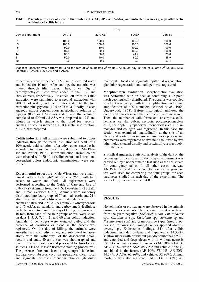
Table 1. Percentage of cases of ulcer in the treated (10% AE, 20% AE, 5-ASA) and untreated (vehicle) groups after aceticacid-induced colitis in rats
��� �� �������
����
��� �� ��� �� ����� ������
� ����� ����� ����� ������ ����� ����� ����� ������ ���� ��� ����� �����! !�� "��� ����� ������# ��! ��� ##�# !����� ���� #��� "��� ��"� ��� ��� ��� �!��
� � $ ��� �����$$ %�$ ������& �$�' �� �$ �� (� )����� �& (� *���� + !� �,� -� &�� "�. �� ������� �& (� *���� + �����)��� ��� ������ ����� ��& �����,�
268 L. V. RODRIGUES ET AL.
Copyright � 2002 John Wiley & Sons, Ltd. Phytother. Res. 16, 267–272 (2002)
20%, 2.86%; 5-ASA: 2.86%; vehicle:10%) (data notshown).
With the exception of crypt abcesses and glandularregeneration, the morphological aspects showed signifi-cant differences in the study period. However, thepresence of oedema, haemorrhage, exudate, ulcers,pseudomembranes, focal epithelial regeneration andcollagen were very important parameters for groupcomparison. The percentage of cases of ulcer at severaltimes in the treated animals and controls are shown in
Table 1. Significant differences were observed on day 60in the 10% and 20% AE and 5-ASA treated groupscompared with the controls.
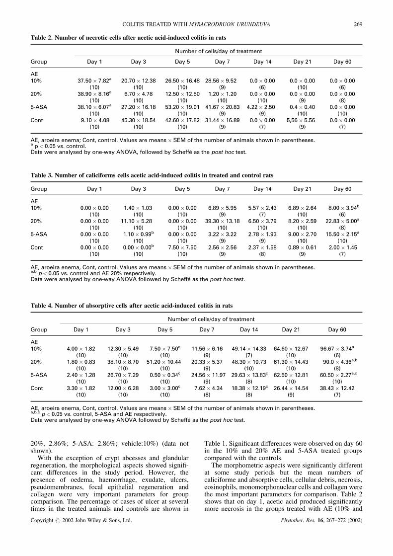
The morphometric aspects were significantly differentat some study periods but the mean numbers ofcaliciforme and absorptive cells, cellular debris, necrosis,eosinophils, monomorphonuclear cells and collagen werethe most important parameters for comparison. Table 2shows that on day 1, acetic acid produced significantlymore necrosis in the groups treated with AE (10% and
Table 2. Number of necrotic cells after acetic acid-induced colitis in rats
/��0� �� ����$1&�� �� �� ���
���� ��� � ��� � ��� � ��� ! ��� �# ��� �� ��� "�
����� �!���� !� �� ���!�� ���� �"���� �"�# � ��"� ���� ���� ���� ���� ���� ���� ����
)��, )��, )��, )�, )", )��, )",��� � ���� ��"� "�!�� #�! ������ ����� ����� ���� ���� ���� ���� ���� ���� ����
)��, )��, )��, )��, )��, )�, ) ,����� � ���� "��!� �!���� �"�� ������ ����� #��"!� ��� � #���� ���� ��#� ��#� ���� ����
)��, )��, )��, )�, )�, )��, )��,2�� ����� #�� #����� � ��# #��"�� �!� � ���##� �"� � ���� ���� �.�"� ���" ���� ����
)��, )��, )��, )�, )!, )�, )!,
��. ���� �����3 2�� . ��� ��� �����$ �� ����$� ��4 �� �� ���0� �� �����$ $��%� � ���� ��$�$�� �� ���� *$� ��� ����� � %�� �����$�& 0� ����%�� �/-��. �����%�& 0� �������5 �$ �� ���� ��� �$ �
Table 3. Number of caliciforms cells acetic acid-induced colitis in treated and control rats
���� ��� � ��� � ��� � ��� ! ��� �# ��� �� ��� "�
����� ����� ���� ��#�� ���� ����� ���� "� �� ���� ���!� ��#� "� �� ��"# ���� ���#0
)��, )��, )��, )�, )!, )��, )",��� ����� ���� ������ ��� ����� ���� ������ ���� "���� ��!� ���� ���� ��� �� �����
)��, )��, )��, )��, )��, )��, ) ,����� ����� ���� ����� ����0 ����� ���� ����� ���� ��! � ���� ����� ��!� ������ �����
)��, )��, )��, )�, )�, )��, )��,2�� ����� ���� ����� ����0 !���� !��� ���"� ���" ���!� ��� �� �� ��"� ����� ��#�
)��, )��, )��, )�, ) , )�, )!,
��. ���� �����. 2�� . ��� ��� �����$ �� ����$� ��4 �� �� ���0� �� �����$ $��%� � ���� ��$�$��.0 �� ���� *$� ��� �� ��& �� ��� �$��� *������ � %�� �����$�& 0� ����%�� �/-�� �����%�& 0� �������5 �$ �� ���� ��� �$ �
Table 4. Number of absorptive cells after acetic acid-induced colitis in rats
/��0� �� ����$1&�� �� �� ���
���� ��� � ��� � ��� � ��� ! ��� �# ��� �� ��� "�
����� #���� �� � ������ ��#� !���� !���� ����"� "��" #���#� �#��� "#�"�� ���"! �"�"!� ��!#�
)��, )��, )��, )�, )!, )��, )",��� �� �� �� � � ���� �!� ������ ���## ������ ���! # ���� ���!� "����� �#�#� ����� #��"�.0
)��, )��, )��, )�, )��, )��, ) ,����� ��#�� ��� �"�!�� !��� ����� ���#� �#��"� ����! ���"�� ��� �� "����� ��� � "����� ���!�.�
)��, )��, )��, )�, ) , )��, )��,2�� ����� �� � ������ "�� ����� ����� !�"�� #��# � �� � ������ �"�##� �#��# � �#�� ���#�
)��, )��, )��, ) , ) , )�, )!,
��. ���� �����. 2�� . ��� ��� �����$ �� ����$� ��4 �� �� ���0� �� �����$ $��%� � ���� ��$�$��.0.� �� ���� *$� ��� ��. ����� ��& �� �$��� *������ � %�� �����$�& 0� ����%�� �/-�� �����%�& 0� �������5 �$ �� ���� ��� �$ �
COLITIS TREATED WITH MYRACRODRUON URUNDEUVA 269
Copyright � 2002 John Wiley & Sons, Ltd. Phytother. Res. 16, 267–272 (2002)
20%) and 5-ASA than in the controls. However, on day60 there were more caliciforme and absorptive cells inthe treated groups compared with the controls. In the caseof the 20% AE treated group this fact was alreadymanifest on days 5 and 7 for absorptive and caliciformecells, respectively. Although not significant, all groupsshowed higher number of both types of cells, on day 21,compared with the controls (Tables 3 and 4).
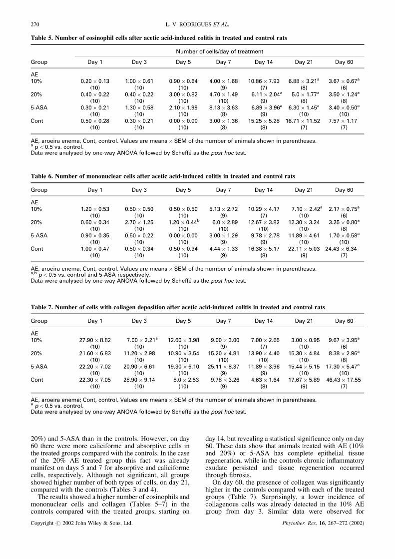
The results showed a higher number of eosinophils andmononuclear cells and collagen (Tables 5–7) in thecontrols compared with the treated groups, starting on
day 14, but revealing a statistical significance only on day60. These data show that animals treated with AE (10%and 20%) or 5-ASA has complete epithelial tissueregeneration, while in the controls chronic inflammatoryexudate persisted and tissue regeneration occurredthrough fibrosis.
On day 60, the presence of collagen was significantlyhigher in the controls compared with each of the treatedgroups (Table 7). Surprisingly, a lower incidence ofcollagenous cells was already detected in the 10% AEgroup from day 3. Similar data were observed for
Table 5. Number of eosinophil cells after acetic acid-induced colitis in treated and control rats
/��0� �� ����$1&�� �� �� ���
���� ��� � ��� � ��� � ��� ! ��� �# ��� �� ��� "�
����� ����� ���� ����� ��"� ����� ��"# #���� ��" ��� "� !��� "� � ����� ��"!� ��"!�
)��, )��, )��, )�, )!, ) , )",��� ��#�� ���� ��#�� ���� ����� �� � #�!�� ��#� "���� ���#� ���� ��!!� ����� ���#�
)��, )��, )��, )��, )�, ) , ) ,����� ����� ���� ����� ��� ����� ���� ���� ��"� "� �� ���"� "���� ��#�� ��#�� �����
)��, )��, )��, ) , )�, )��, )��,2�� ����� ��� ����� ���� ����� ���� ����� ���" ������ ��� �"�!�� ����� !��!� ���!
)��, )��, )��, ) , ) , )!, )!,
��. ���� �����. 2�� . ��� ��� �����$ �� ����$� ��4 �� �� ���0� �� �����$ $��%� � ���� ��$�$�� �� ��� *$� ��� ����� � %�� �����$�& 0� ����%�� �/-�� �����%�& 0� �������5 �$ �� ���� ��� �$ �
Table 6. Number of mononuclear cells after acetic acid-induced colitis in treated and control rats
���� ��� � ��� � ��� � ��� ! ��� �# ��� �� ��� "�
����� ����� ���� ����� ���� ����� ���� ����� ��!� ������ #��! !���� ��#�� ���!� ��!��
)��, )��, )��, )�, )!, )��, )",��� ��"�� ���# ��!�� ���� ����� ��##0 "��� �� � ���"!� �� � ������ ���# ����� �� ��
)��, )��, )��, )��, )��, )��, ) ,����� ����� ���� ����� ���� ����� ���� ����� ���� ��! � ��! ��� �� #�"� ��!�� ��� �
)��, )��, )��, )�, )�, )��, )��,2�� ����� ��#! ����� ���# ����� ���# #�##� ���� �"�� � ���! ������ ���� �#�#�� "��#
)��, )��, )��, )�, ) , )�, )!,
��. ���� �����. 2�� . ��� ��� �����$ �� ����$� ��4 �� �� ���0� �� �����$ $��%� � ���� ��$�$��.0 �� ��� *$� ��� �� ��& ����� �$��� *������ � %�� �����$�& 0� ����%�� �/-�� �����%�& 0� �������5 �$ �� ���� ��� �$ �
Table 7. Number of cells with collagen deposition after acetic acid-induced colitis in treated and control rats
���� ��� � ��� � ��� � ��� ! ��� �# ��� �� ��� "�
����� �!���� � � !���� ����� ���"�� ��� ����� ���� !���� ��"� ����� ���� ��"!� �����
)��, )��, )��, )�, )!, )��, )",��� ���"�� "� � ������ ��� ������ ���# ������ #� � ������ #�#� ������ #� # �� � ���"�
)��, )��, )��, )��, )��, )��, ) ,����� ������ !��� ������ "�"� ������ "��� ������ ��! ��� �� ���" ���##� ���� �!���� ��#!�
)��, )��, )��, )�, )�, )��, )��,2�� ������ !��� � ���� ���# ��� ���� ��! � ���" #�"�� ��"# �!�"!� �� � #"�#�� �!���
)��, )��, )��, )�, ) , )�, )!,
��. ���� �����3 2�� . ��� ��� �����$ �� ����$� ��4 �� �� ���0� �� �����$ $��%� � ���� ��$�$�� �� ��� *$� ��� ����� � %�� �����$�& 0� ����%�� �/-�� �����%�& 0� �������5 �$ �� ���� ��� �$ �
270 L. V. RODRIGUES ET AL.
Copyright � 2002 John Wiley & Sons, Ltd. Phytother. Res. 16, 267–272 (2002)
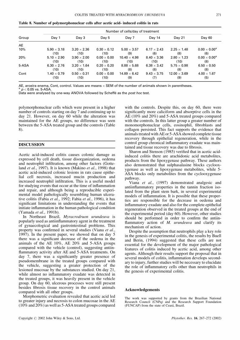
polymorphonuclear cells which were present in a highernumber of controls starting on day 7 and continuing up today 21. However, on day 60 while the alteration wasmaintained for the AE groups, no difference was seenbetween the 5-ASA treated group and the controls (Table8).
DISCUSSION
Acetic acid-induced colitis causes colonic damage asexpressed by cell death, tissue disorganization, oedemaand neutrophil infiltration, among other factors (Gotte-land et al., 1997). It is known (Makino et al., 1998) thatacetic acid-induced colonic lesions in rats cause epithe-lial cell necrosis, increased mucin production andincreased neutrophil infiltration. This is a useful modelfor studying events that occur at the time of inflammationand repair, and although being a reproducible experi-mental model pathologically resembling human ulcera-tive colitis (Fabia et al., 1992; Fabia et al., 1996), it hassignificant limitations in understanding the events thatinitiate inflammation in the human pathological condition(Yamada et al., 1991b).
In Northeast Brazil, Myracrodruon urundeuva ispopularly used as antiinflammatory agent in the treatmentof gynaecological and gastrointestinal problems. Thisproperty was confirmed in several studies (Viana et al.,1997). In the present paper, we showed that on day 5there was a significant decrease of the oedema in theanimals of the AE 10%, AE 20% and 5-ASA groupscompared with the vehicle (control), suggesting antiin-flammatory activity after AE and 5-ASA treatments. Onday 7, there was a significantly greater presence ofpseudomembrane in the treated groups compared withthe vehicle, suggesting a greater protection of thelesioned mucosae by the substances studied. On day 21,while almost no inflammatory exudate was detected inthe treated groups, it was heavily present in the vehiclegroup. On day 60, ulcerous processes were still presentbesides fibrosis tissue recovery in the control animalscompared with all other groups.
Morphometric evaluation revealed that acetic acid ledto greater injury and necrosis to colon mucosae in the AE(10% and 20%) as well as 5-ASA treated groups compared
with the controls. Despite this, on day 60, there weresignificantly more caliciform and absorptive cells in theAE (10% and 20%) and 5-ASA treated groups comparedwith the controls. In this latter group a greater number ofmonomorphonuclear cells, eosinophil, fibroblasts andcollagen persisted. This fact supports the evidence thatanimals treated with AE or 5-ASA showed complete tissuerecovery through epithelial regeneration, while in thecontrol group chronical inflammatory exudate was main-tained and tissue recovery was due to fibrosis.
Sharon and Stenson (1985) verified that in acetic acid-induced colitis there are arachidonic acid metabolites,products from the lipoxygenase pathway. These authorsalso demonstrated that sulphasalasine blocks cycloox-ygenase as well as lipoxygenase metabolites, while 5-ASA blocks only metabolites from the cyclooxygenasepathway.
Viana et al., (1997) demonstrated analgesic andantiinflammatory properties in the tannin fraction iso-lated from the plant stem bark, in several experimentalmodels of inflammation. It is possible that these proper-ties are responsible for the decrease in oedema andinflammatory exudate and also for the complete epithelialregeneration observed in the treated groups at the end ofthe experimental period (day 60). However, other studiesshould be performed in order to confirm the antiin-flammatory action of M. urundeuva and clarify itsmechanism of action.
Despite the assumption that neutrophils play a key rolein the genesis of experimental colitis, the results by Buelland Berin, (1994) suggested that these cells are notessential for the development of the major pathologicalfeatures of colitis induced by acetic acid, among otheragents. Although their results support the proposal that inseveral models of colitis, inflammation develops second-ary to injury, further studies will be necessary to elucidatethe role of inflammatory cells other than neutrophils inthe genesis of experimental colitis.
Acknowledgements
The work was supported by grants from the Brazilian NationalResearch Council (CNPq) and the Research Support Foundation(FUNCAP) from the state of Ceara, Brazil.
Table 8. Number of polymorphonuclear cells after acetic acid- induced colitis in rats
/��0� �� ����$1&�� �� �� ���
���� ��� � ��� � ��� � ��� ! ��� �# ��� �� ��� "�
����� ����� ��� ����� ���" ����� ���� ����� ���! "��!� ��#� ����� ��# ����� �����
)��, )��, )��, )�, )", ) , )",��� ����� ���� ����� ���� ����� ���� ���#�� #�� �#�� ���" �� �� ���� ����� �����
)��, )��, )��, )��, )��, )��, ) ,����� "���� ��!" ����� ��"# ����� ���� � � ��" "�� � ��#� ��!�� ��� "���� ����
)��, )��, )��, ) , ) , ) , ) ,2�� ��#�� ��!� ����� ���� ����� ���� �#� �� "�#� ��#�� ��!� ������ ��"� #���� ��"!
)��, )��, )��, )�, )!, )�, )�,
��. ���� �����. 2�� . ��� ��� �����$ �� ����$� ��4 �� �� ���0� �� �����$ $��%� � ���� ��$�$�� �� ���� *$� �������� � %�� �����$�& 0� ����%�� �/-�� �����%�& 0� �������5 �$ �� ���� ��� �$ �
COLITIS TREATED WITH MYRACRODRUON URUNDEUVA 271
Copyright � 2002 John Wiley & Sons, Ltd. Phytother. Res. 16, 267–272 (2002)
REFERENCES
6���� 4�. 6�� 42� ���#� /�� ������&����&���� �� ��� � �� �� ������ �7��� 2����$�� �� �$�� $ ��� ��� ��&��$ �� ������� �� ��� $ � �� � � �� ��� ����8 ��!�9�� �
:�0� ;. �<;�7�0 �. =���5 � ;. 4�>���& �. ��&�$$�� ;����"� ?�� ��� �� ��$�� ����$�� $����� � ��� ���&��&���& ��� $ � �� � � � � ��� ��8 #�"9#���
:�0� ;. =���� ;. �<;�7�0 �. ��&�$�� ;. ���� 6.6��'��> �� ����� ��� � ��&��&���& ��� $ � �� � 8� ���&��0�� ������� �� ��&�� �� ��� � ����� *���� $� �� � ��� ��8 ���9����
�� ����& 4. @����A B. 6��$� -. �� ��� ���!� C� �� *������ �� 0��&�� � ������� �� ��� $� ������ ������ ��8���9����
D'� �. � � ?. /�%� E� ���!� �*���� �� �� �� ��� ����� ����$ � �� �� ��'���$$ �� ��� � ��&��&���&��� $ � ���� ���� � ���� ����� �� ��8 �"#9�" �
F�%� �2. /������6��0 F� ����� ?�� ���>��� � ��� ��� ��0 �. /C2 ��""�. ������� �$ �����' � ��� � ��&��&���& ��� $� ����� ������� ��8 �9 ��
4��C��$�� 6;. C���� 2@� ��! � ������� �� ��&�� �� ��&���$� ��� $ � � $� �������� ��8 ���9����
4�>�� 4. B���� � E. B � G. ?�0� � G� ��� � ����� �������� �� �� � �0��A��� $������ ��& ��& ��� � ��&�&���& ������ ��$��$ � � $������� ���� ���� ���������8 �!9��"�
4���A�$ �4�. ;�� ��� �� � ����� �� ��� ����� � �������)����, �� '�$ �� �$ ��� ��$ � ���� �! � ��� ��� ��� ��8 ���9����
;�� ��/. 4���A�$ �4�. ��$�������$ :�. ����&� :2;.:�� ���$ 42� �� "� ����� �� ��� ����� � ������� ��'��
)����, � ������� �� ��� $� �! � ��� ��� ��� ��8�" �
;�� ��. ���� ��6. 4���A�$ �4�. ��&���� 4�?� �� !�� �&�$ �� �� �� �����'��� �� * � �� ��� ������ ������� ��'�� BB� �H����$ �� �� � �! � ��� ������ ��8 ��9 ���
��>A�>� �. �$��� 46. F 4. �� �� ��. ���'� �/� �� �B�I���� ����&���& � �$ ��� ������� � �� � 8��� �� ��� ����$� ���� ����� ���" �8 ��� 9���#�
����� C. � ��$�� =:� �� �� 4� �0��$� �� ����&��� ��&� ��� � ��& ��� $ � � $� ���� ����� ���" 8 ��9"��
J�&�%��& ��� ��" � � �����'�. � �� H��� � *� �*����� �� �� ���$ �� ��$� � ��� ��� �8 �"�9����
J � ���� ��� �� D��� � ��& D���� ��*��$� B�$ � � ��F�0�� �� ����� ;�$����$� �� �� �������� ����� ��#������ ����� $� ��� #� � ��� %�� �$ &�'� ��� "������� J� ���� ��� �� D��� � ��& D���� ��*��$8=�$��' ��. �2�
���� ��6. 6��&�� 4�4. 4��� F2. ���A� :��� 4�C.4� �$ :@�. ;0�� ;�� ���!� ����'�$� ��& �� �I����� �� ����� $ �� �� ���� ��� �� ��� �" �� �� ���� ������� :� ���� ��"����� ��� ��8 �� 9����
=�0�� �;. G$ �� ��. ������ =:� ��""� C�� ��� $ ������'��� �� ��&$ �� ������� � �� ���'�� � #��� �����8 ��9� �
E���&� ?. ������ ;�. ���'� �/. ��'����� ?. ;$���46� ������ 4$���$ �� � ���� �$ ��� � ��&��&���&����$�$ �����$�� �����0� � ��& �I���� ��8 ����� 0���& I�%� �� � ��"���� ���8 ���9����
E���&� ?. K������ 6@. ������ ;�. �$���46� ����0�;��� �� ��� ����$ � ��� � ��&��&���& ��� $ � � $�(�)�������� �8 ���9#���
272 L. V. RODRIGUES ET AL.
Copyright � 2002 John Wiley & Sons, Ltd. Phytother. Res. 16, 267–272 (2002)