Embed Size (px)
Citation preview
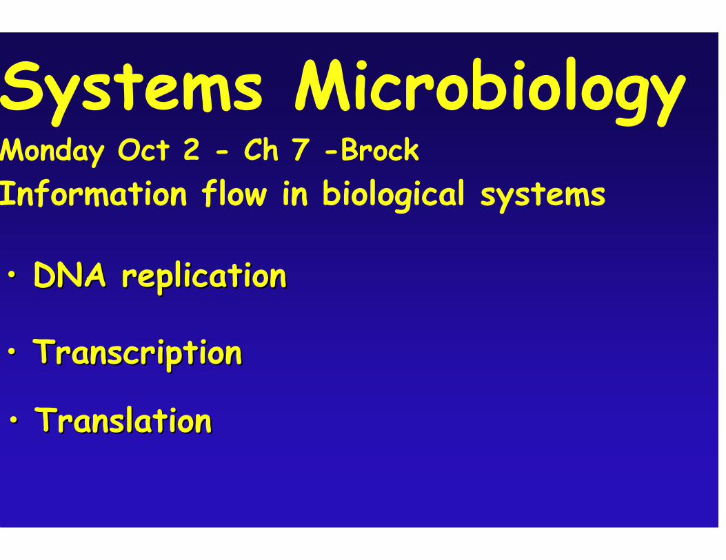
•• TranscriptionTranscription
•• TranslationTranslation
Systems Microbiology Monday Oct 2 - Ch 7 -Brock Information flow in biological systems
•• DNA replicationDNA replication
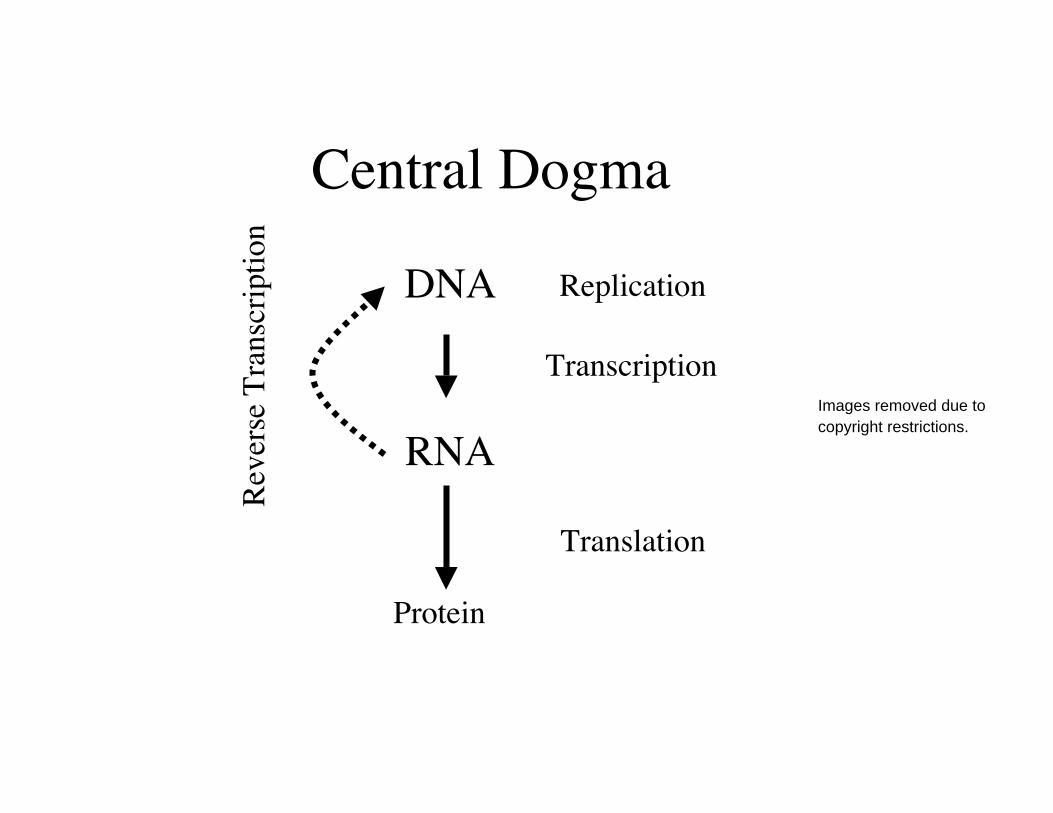
Central Dogma
DNA
RNA
Protein
Replication
Transcription
Translation
Reve
rse
Tran
scrip
tion
Images removed due tocopyright restrictions.
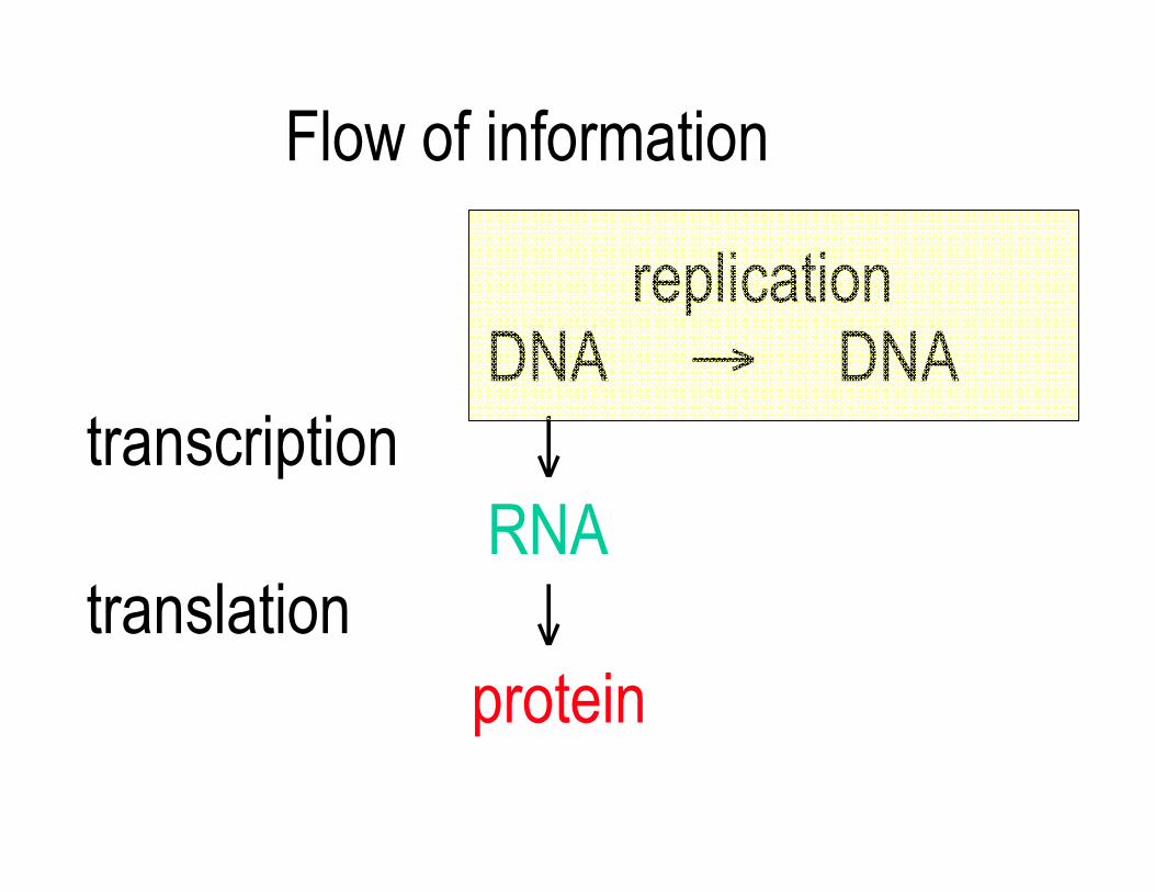
Flow of information
replication DNA → DNAtranscription ↓ RNAtranslation ↓ protein
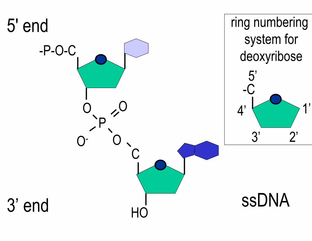
5' end ring numbering system for
-P-O-C deoxyribose 5’
-C 1’O O 4’
P O- O 3’ 2’
C
3’ end HO ssDNA
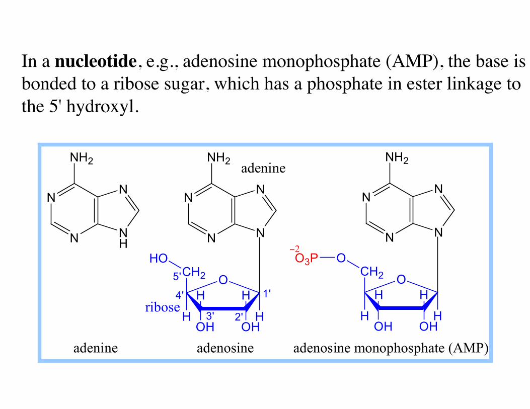
In a nucleotide, e.g., adenosine monophosphate (AMP), the base isbonded to a ribose sugar, which has a phosphate in ester linkage tothe 5' hydroxyl.
N
N N
N
NH2
adenine adenosine adenosine monophosphate (AMP)
O
OH OH
HH
H
CH2
H
HO
N
N N H
N
NH2
N
N N
N
NH2
O
OH OH
HH
H
CH2
H
OO3P −2
ribose
5'
adenine
4'
3' 2'
1'
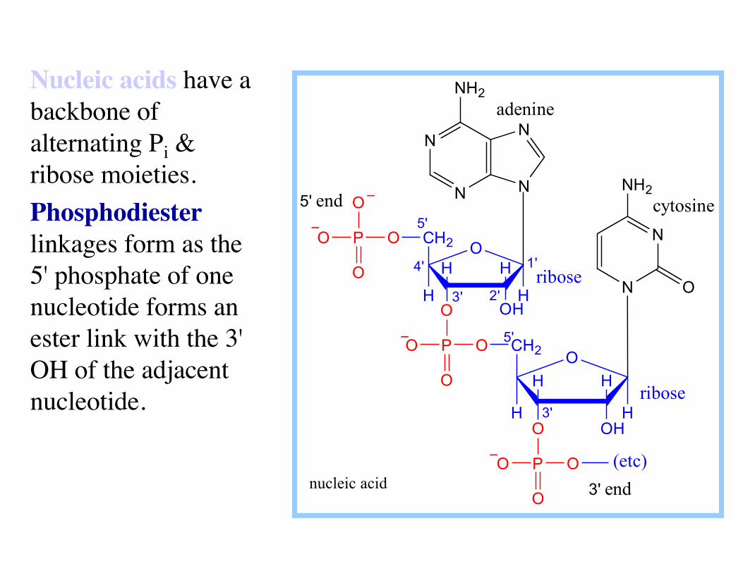
Nucleic acids have a backbone of alternating Pi & ribose moieties. Phosphodiesterlinkages form as the5' phosphate of onenucleotide forms an ester link with the 3' OH of the adjacentnucleotide.
N
N N
N
NH2
O
OH O
HH
H
CH2
H ribose
adenine
P
O
O O O
OH O
HH
H
CH2
H
N
N
NH2
O
P
O
O O
OP
O
O
O
−
−
−
cytosine 5'
4'
3' 2'
1'
ribose 3'
5'
3' end
5' end −
(etc) nucleic acid
N H
H
H
H
O H
H
N
NHCH3
H
O
N NN
NN
NNN
NN
Backbone
Backbone
Backbone
Backbone
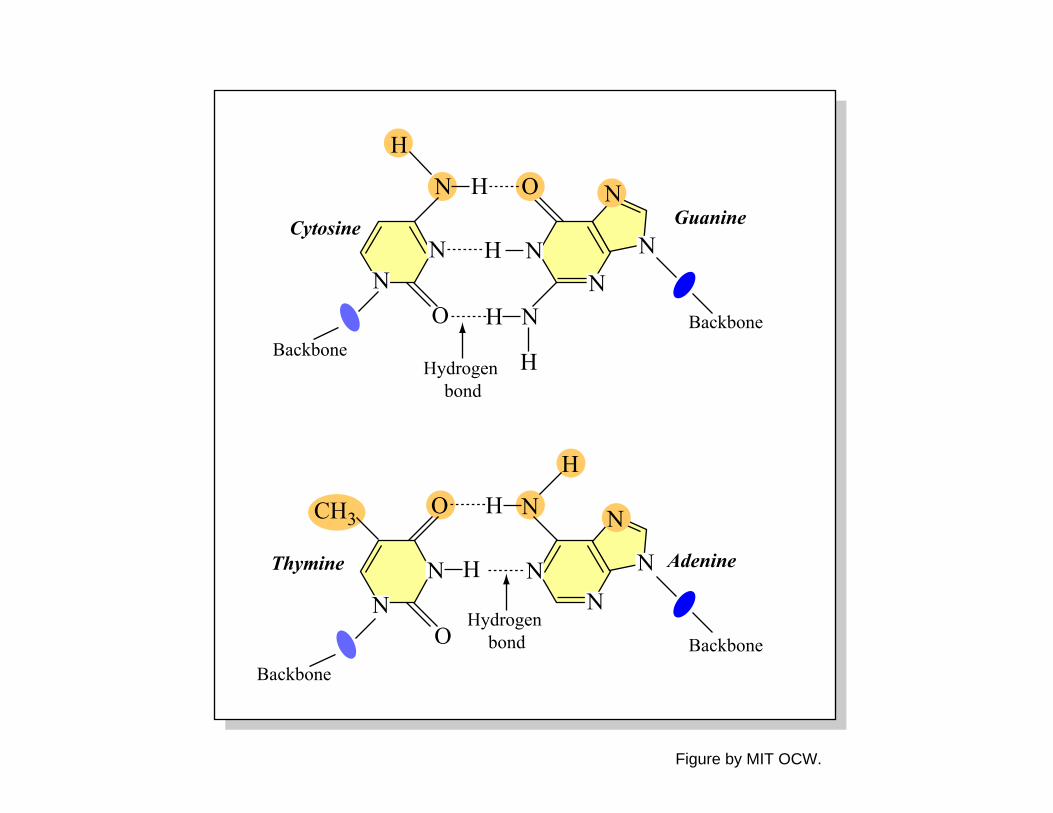
Cytosine Guanine
Hydrogen bond
Hydrogen bond
Thymine Adenine
O N
NO
Figure by MIT OCW.

Diagram of genetic structure removed due to copyright restrictions. See Figure 7-4 in Madigan, Michael, and John Martinko. Brock Biology of Microorganisms. 11th ed. Upper Saddle River, NJ: Pearson Prentice Hall, 2006. ISBN: 0131443291.
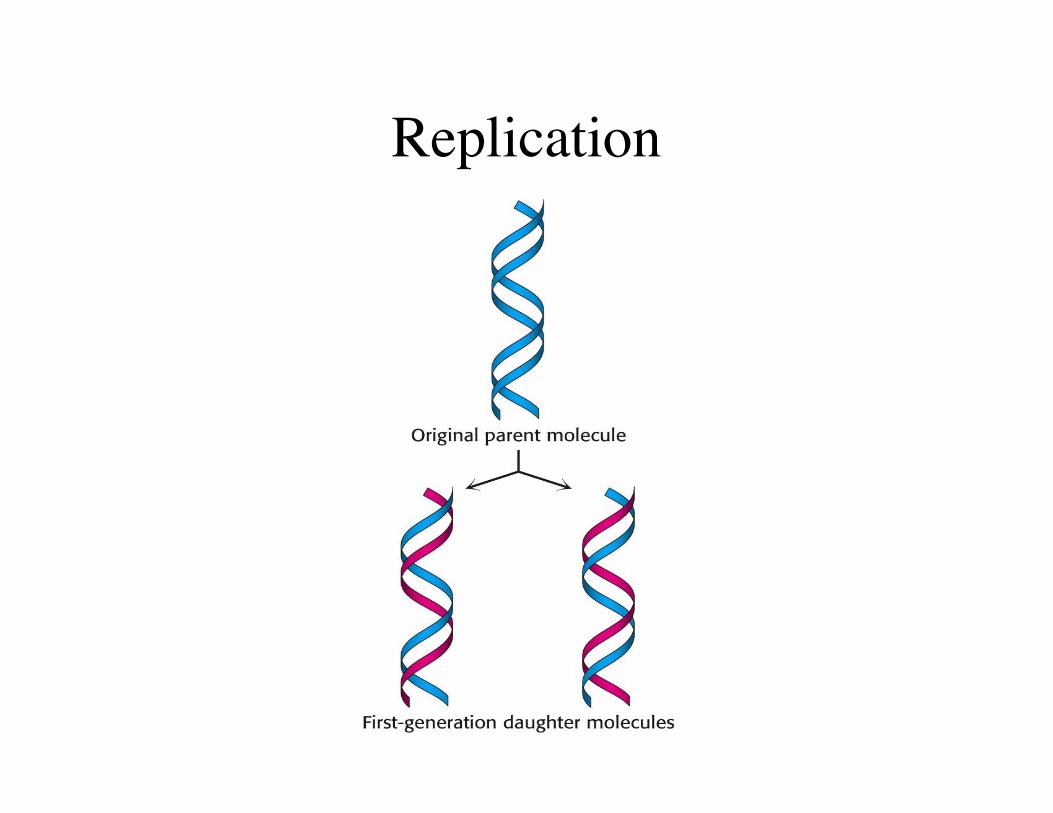
Replication
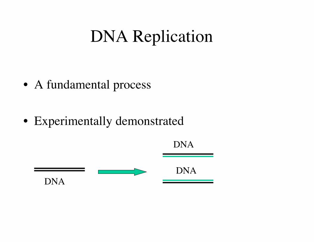
DNA Replication
• A fundamental process
• Experimentally demonstrated
DNA
DNA
DNA
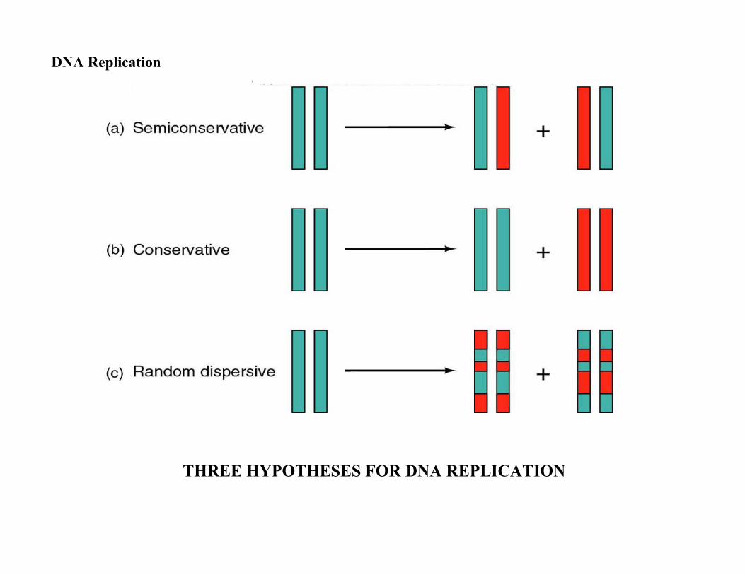
DNA Replication
THREE HYPOTHESES FOR DNA REPLICATION
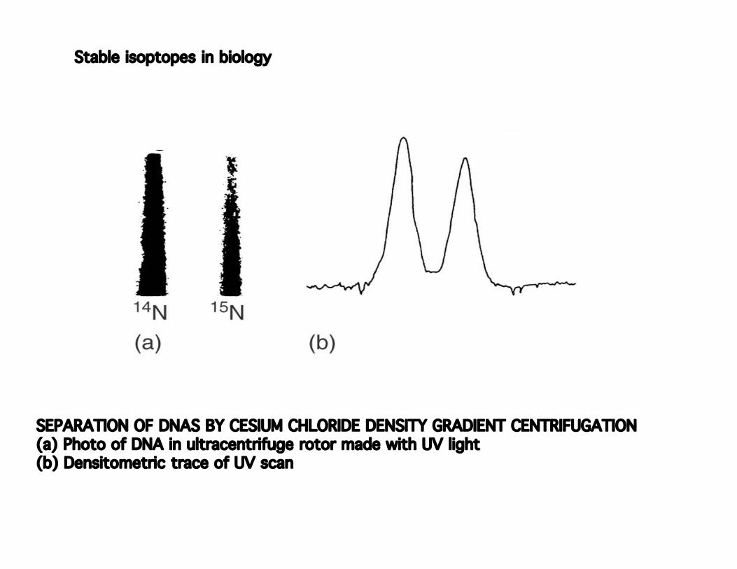
Stable isoptopes in biology
SEPARATION OF DNAS BY CESIUM CHLORIDE DENSITY GRADIENT CENTRIFUGATION (a) Photo of DNA in ultracentrifuge rotor made with UV light (b) Densitometric trace of UV scan

15 15D= N labeled 1. Grow E coli so DNA uniformly N labeled
14 14L= N labeled N labeled to growth media and observe result
PREDICTED DENSITIES OFNEWLY REPLICATED DNAMOLECULES ACCORDING
TO THE THREE HYPOTHESESABOUT DNA REPLICATION
2. Add over several generations of growth
Image of experimental results removed due to copyright restrictions. See Meselson, and Stahl. "The Replication of DNA in Escherichia coli." PNAS 44 (1958): 674, f. 4.
RESULTS OF CsCl GRADIENTULTRACENTRIFUGATIONEXPERIMENT SHOWINGDISTRIBUTION OF DNA
DENSITY IN E. coli CELLSAFTER 0 TO 4.1
GENERATIONS OF GROWTH
THIS EXPERIMENT ESTABLISHEDTHAT DNA REPLICATION IS
SEMICONSERVATIVE
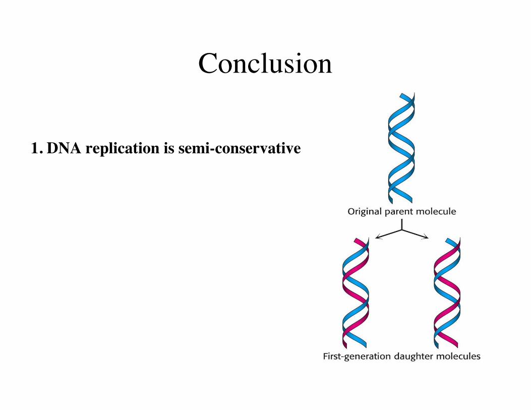
Conclusion
1. DNA replication is semi-conservative
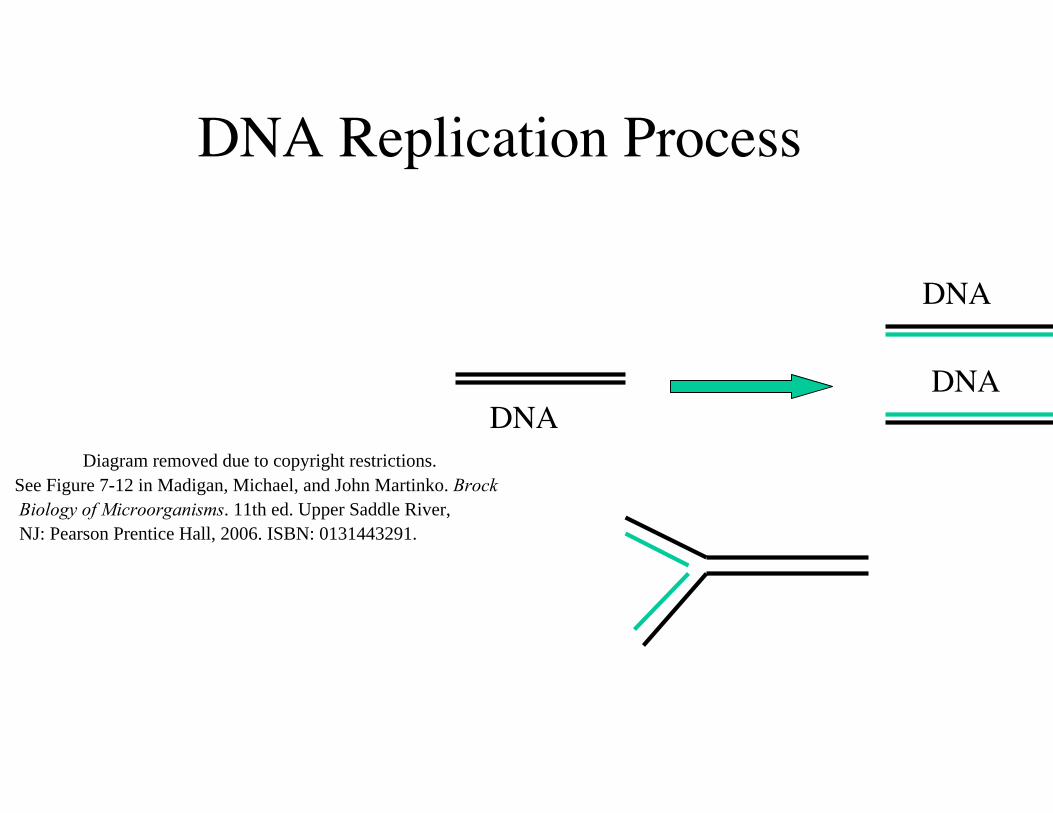
DNA Replication Process
DNA
DNADNA
Diagram removed due to copyright restrictions. See Figure 7-12 in Madigan, Michael, and John Martinko. Brock Biology of Microorganisms. 11th ed. Upper Saddle River, NJ: Pearson Prentice Hall, 2006. ISBN: 0131443291.
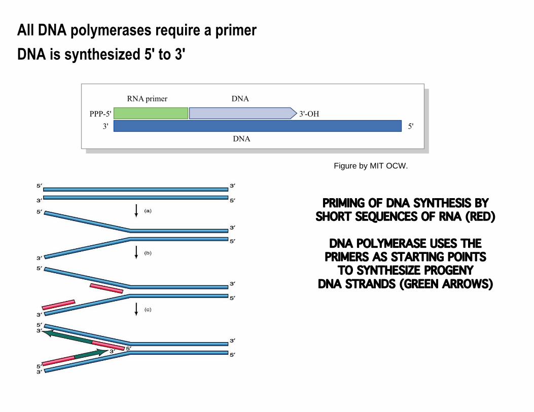
All DNA polymerases require a primer
DNA is synthesized 5' to 3'
PRIMING OF DNA SYNTHESIS BYSHORT SEQUENCES OF RNA (RED)
DNA POLYMERASE USES THEPRIMERS AS STARTING POINTS
TO SYNTHESIZE PROGENYDNA STRANDS (GREEN ARROWS)
RNA primer
PPP-5' 3'-OH3'
DNA
DNA5'
Figure by MIT OCW.

Table of the major enzymes involved in DNA replication in bacteria removed due to copyright restrictions. See Table 7-3 in Madigan, Michael, and John Martinko. Brock Biology of Microorganisms. 11th ed. Upper Saddle River, NJ: Pearson Prentice Hall, 2006. ISBN: 0131443291.

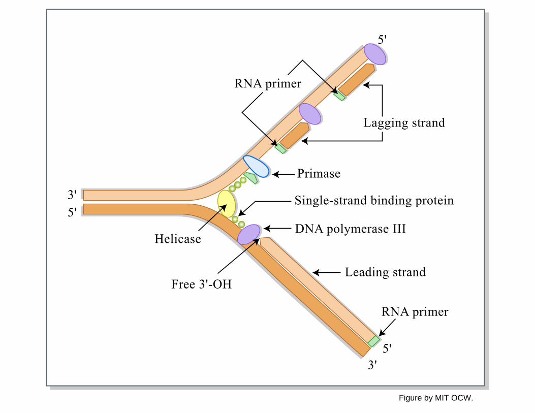
Helicase & topoisomerase
Unwind & remove supercoils in duplex DNA
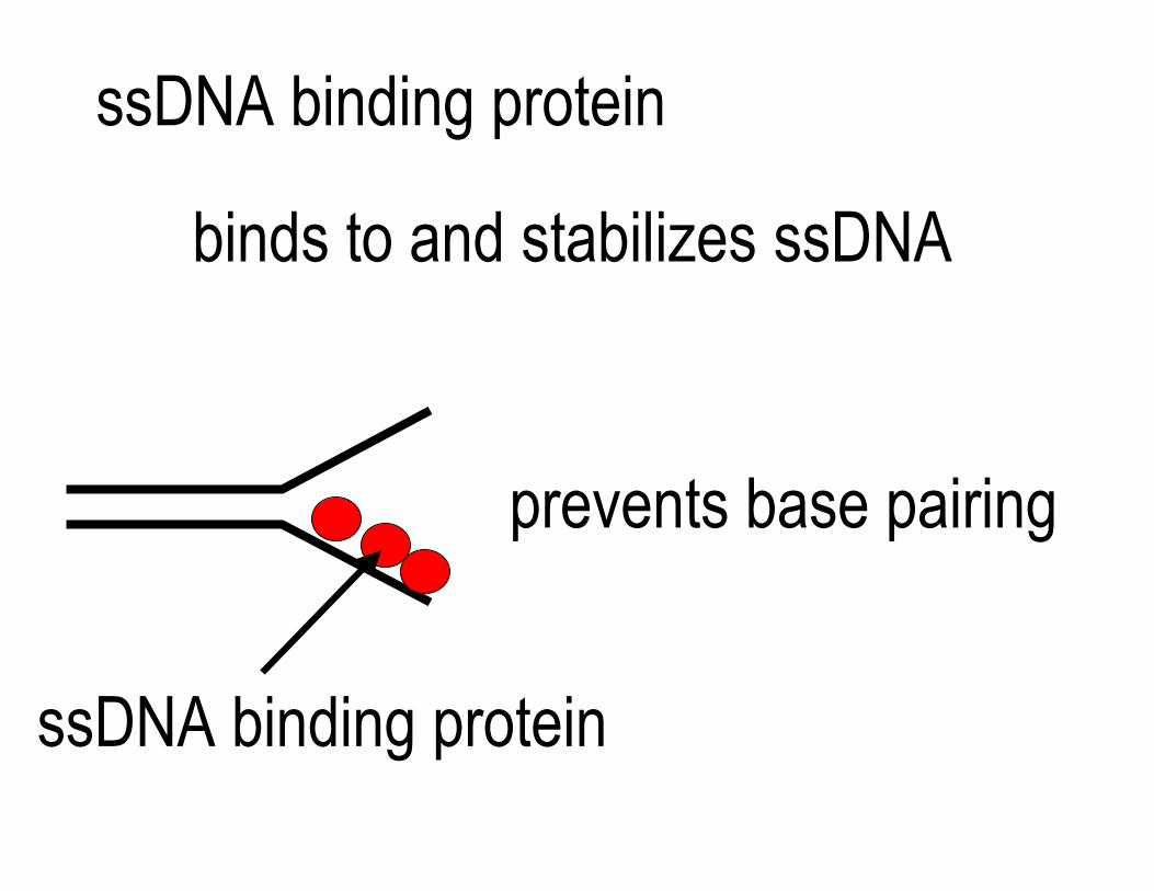
ssDNA binding protein
binds to and stabilizes ssDNA
prevents base pairing
ssDNA binding protein
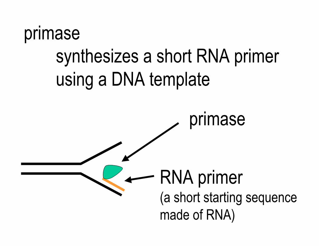
primasesynthesizes a short RNA primer using a DNA template
primase
RNA primer (a short starting sequence made of RNA)
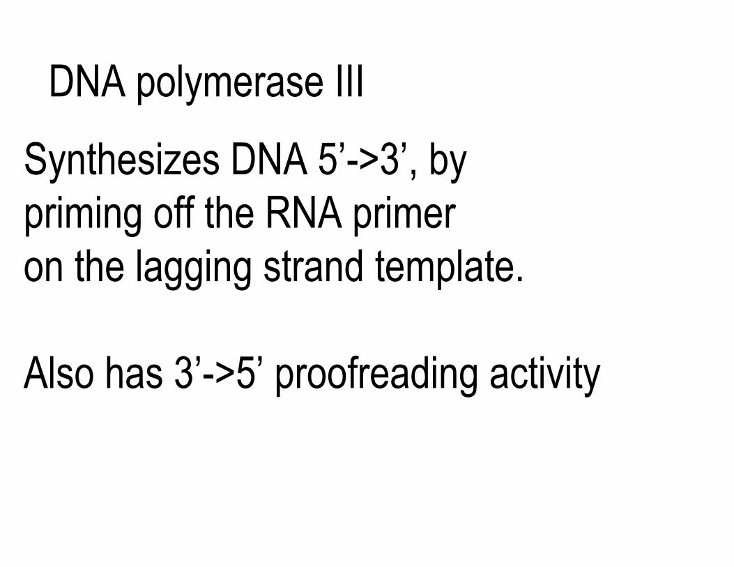
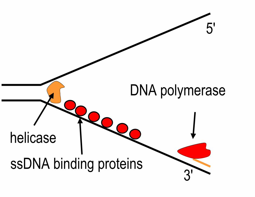
DNA polymerase III
Synthesizes DNA 5’->3’, by priming off the RNA primer on the lagging strand template.
Also has 3’->5’ proofreading activity
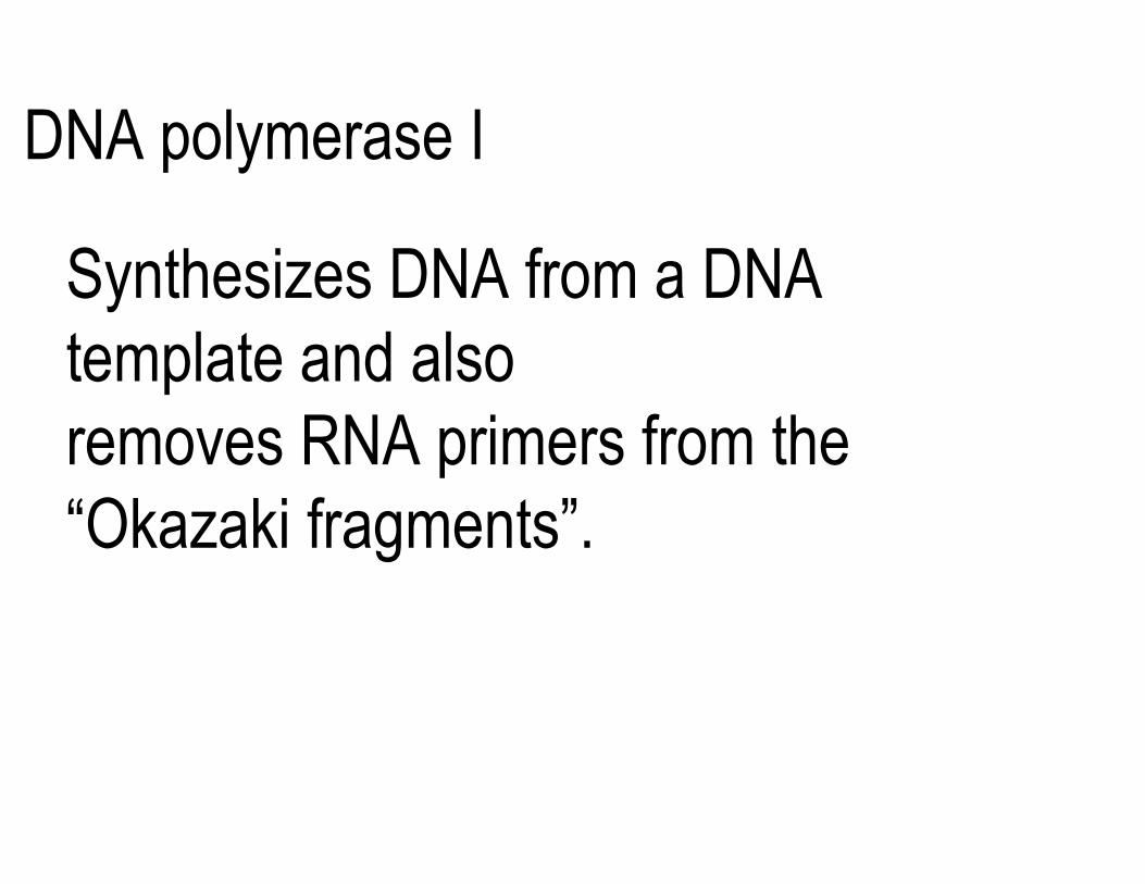
DNA polymerase I
Synthesizes DNA from a DNA template and also removes RNA primers from the “Okazaki fragments”.
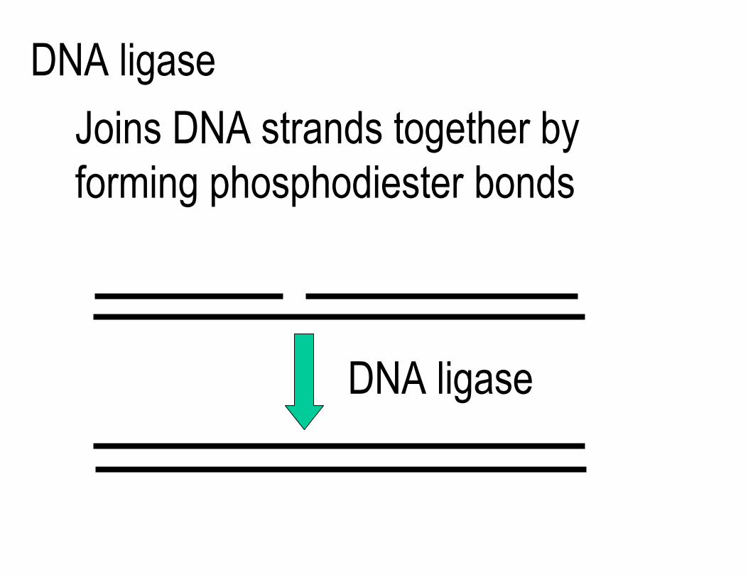
DNA ligase
Joins DNA strands together by forming phosphodiester bonds
DNA ligase
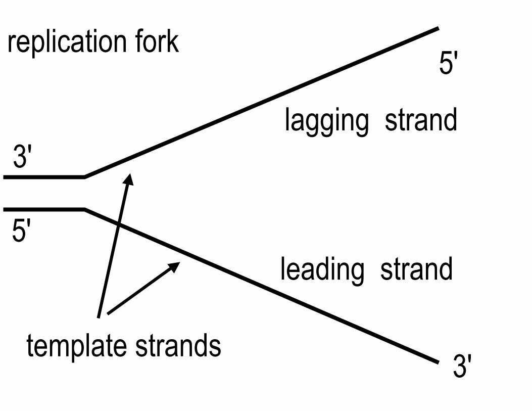
replication fork 5'
lagging strand 3'
5'leading strand
template strands 3'
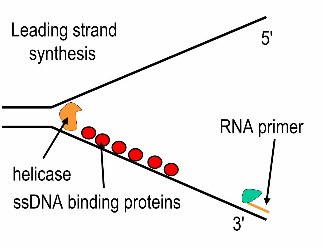
Leading strand 5'synthesis
RNA primer
helicase
ssDNA binding proteins3'
helicase
ssDNA binding proteins
DNA polymerase
3'
5'
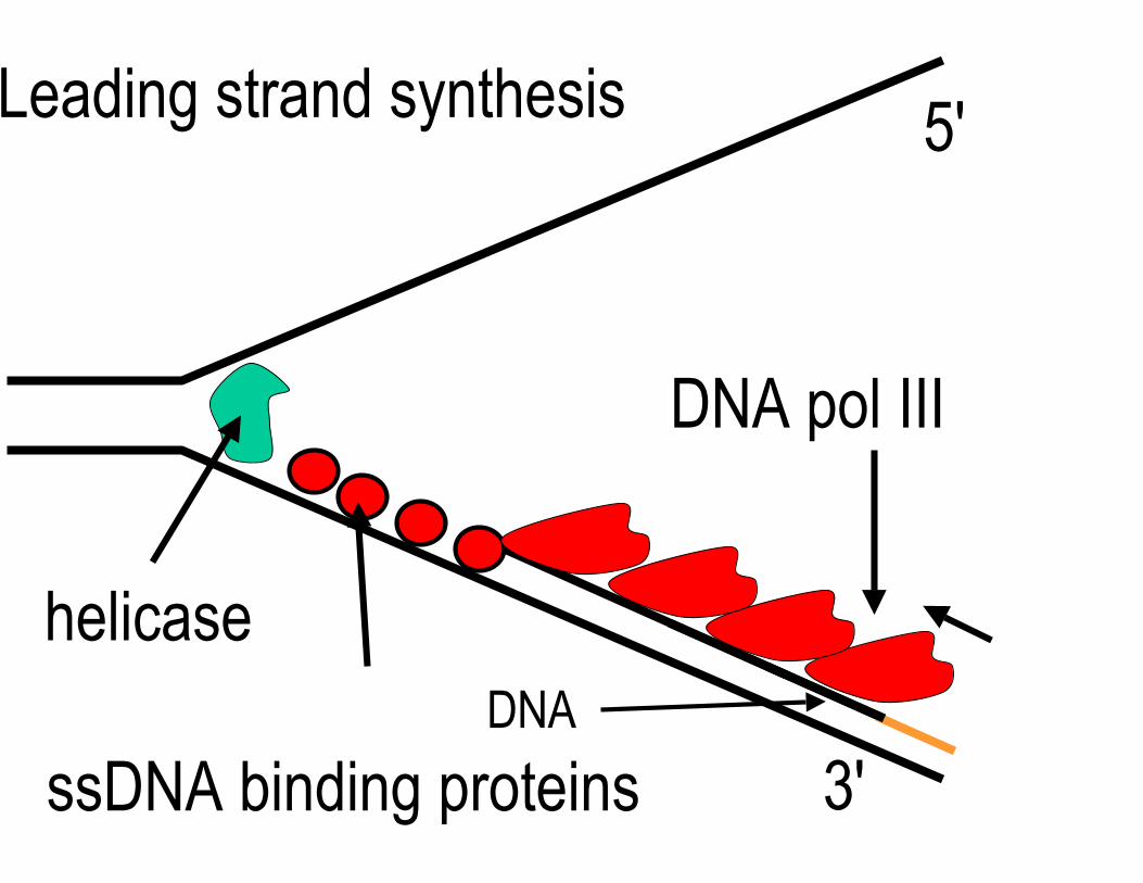
helicase
ssDNA binding proteins
DNA pol III
3'
5'Leading strand synthesis
DNA
ProofreadingPol III removes misincorporated bases using 3' to 5' exonuclease activity
This decreases the error rate to about 10-10 per base pair inserted
Diagram of DNA proofreading removed due to copyright restrictions. See Figure 7-20 in Madigan, Michael, and John Martinko. Brock Biology of Microorganisms. 11th ed. Upper Saddle River, NJ: Pearson Prentice Hall, 2006. ISBN: 0131443291.

helicase
ssDNA binding proteins
(primase) pol III
3'
Lagging strand synthesis (discontinuous) Okazaki
fragment (~1000 bases)3'
5'
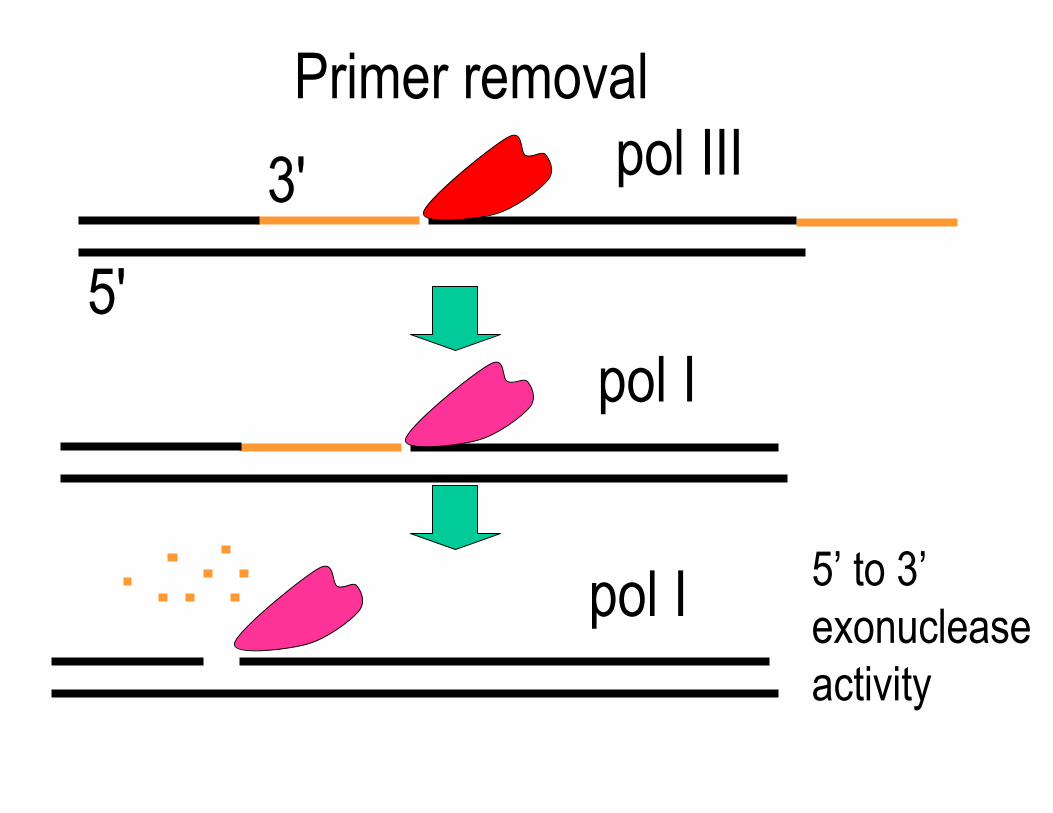
Primer removal
3' pol III
5' pol I
pol I 5’ to 3’ exonuclease activity
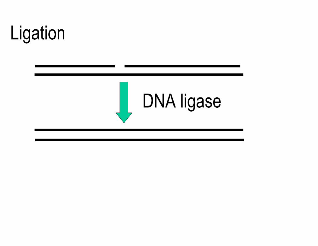
Ligation
DNA ligase
5'
3'
Free 3'-OH
Helicase
5'
5'3'
Lagging strand
Primase
RNA primer
DNA polymerase III
Leading strand
RNA primer
Single-strand binding protein
Figure by MIT OCW.
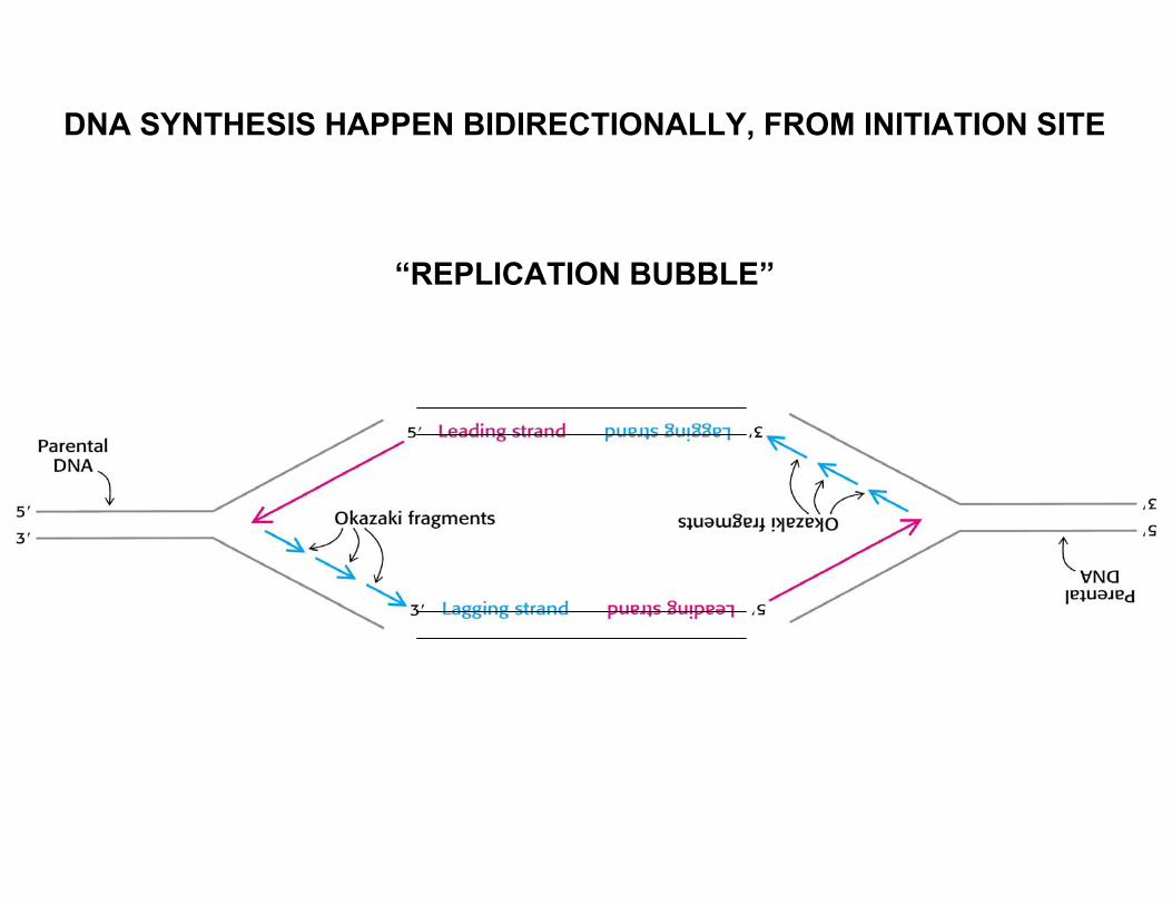
DNA SYNTHESIS HAPPEN BIDIRECTIONALLY, FROM INITIATION SITE
“REPLICATION BUBBLE”
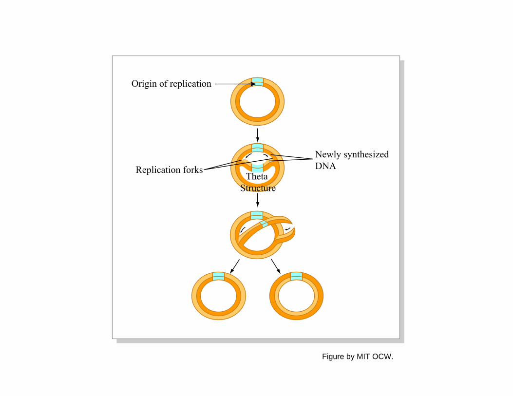
Origin of replication
Newly synthesizedDNAReplication forks
Theta Structure
Figure by MIT OCW.
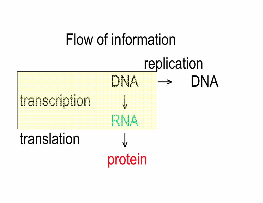
Flow of information
replication DNA
transcription ↓ RNA
→ DNA
translation ↓ protein
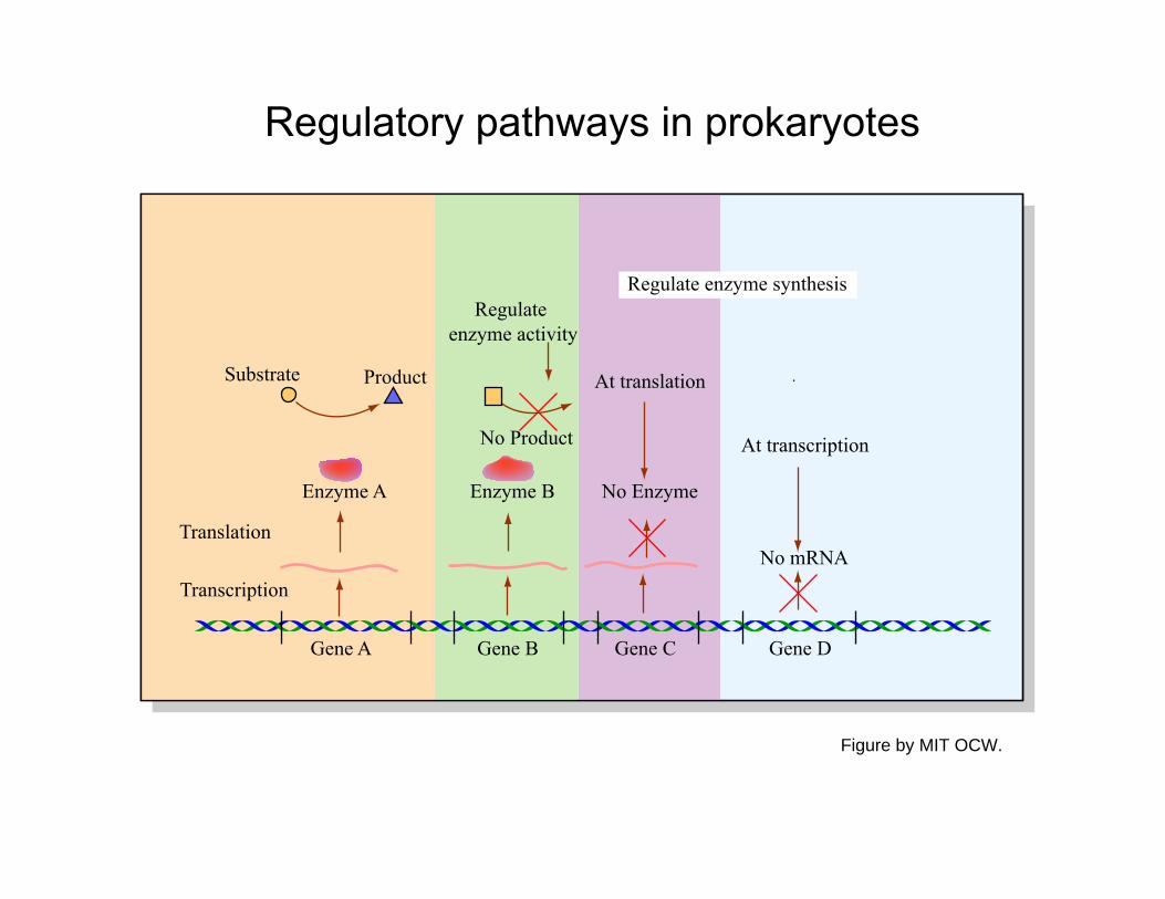
Regulatory pathways in prokaryotes
Product
No Product
Substrate
Enzyme A Enzyme B No Enzyme
No mRNATranslation
Transcription
Gene A Gene B Gene C Gene D
Regulate enzyme activity
Regulate enzyme synthesis
At translation
At transcription
Figure by MIT OCW.

Prokaryotic transcription
Transcribed regions RNA polymerase Promoters Terminators Sigma factor
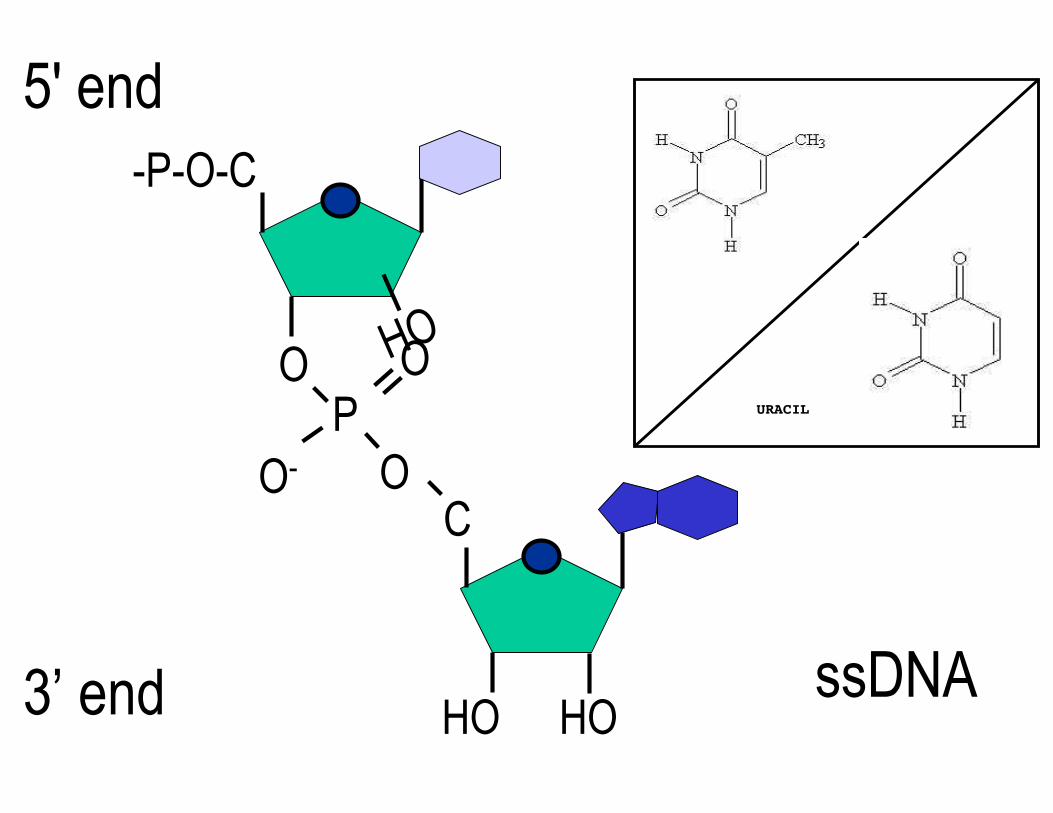
HO
URACIL
5' end -P-O-C
O OP
O- OC
3’ end HO HO ssDNA
Transcription
Diagram of RNA transcription removed due to copyright restrictions. See Figure 7-29a in Madigan, Michael, and John Martinko. Brock Biology of Microorganisms. 11th ed. Upper Saddle River, NJ: Pearson Prentice Hall, 2006. ISBN: 0131443291.
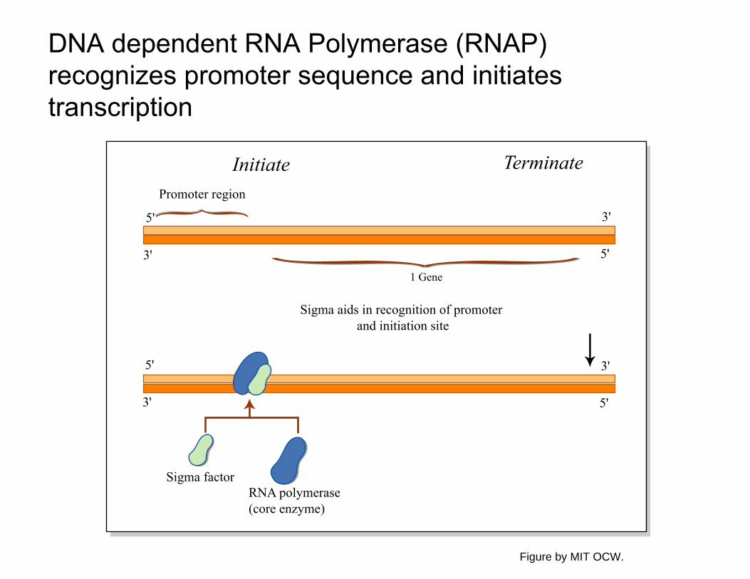
DNA dependent RNA Polymerase (RNAP) recognizes promoter sequence and initiates transcription
RNA polymerase (core enzyme)
Sigma factor
Sigma aids in recognition of promoter and initiation site
{ { {{Promoter region
Initiate Terminate
1 Gene
3'
5'
3'
5'
5'
3'
5'
3'
Figure by MIT OCW.
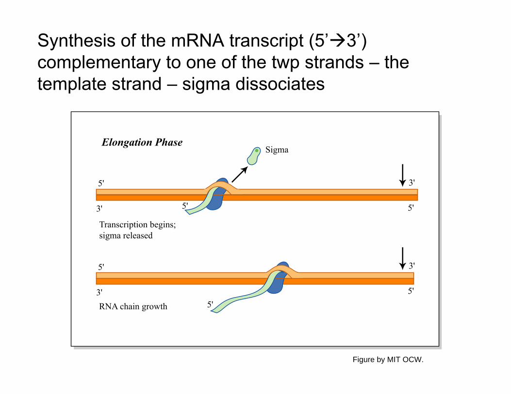
Synthesis of the mRNA transcript (5’3’) complementary to one of the twp strands – the template strand – sigma dissociates
RNA chain growth
Sigma
3'
5'
5'
3'
3'
5'
Elongation Phase
Transcription begins; sigma released
5'
5'
5'
3'
Figure by MIT OCW.
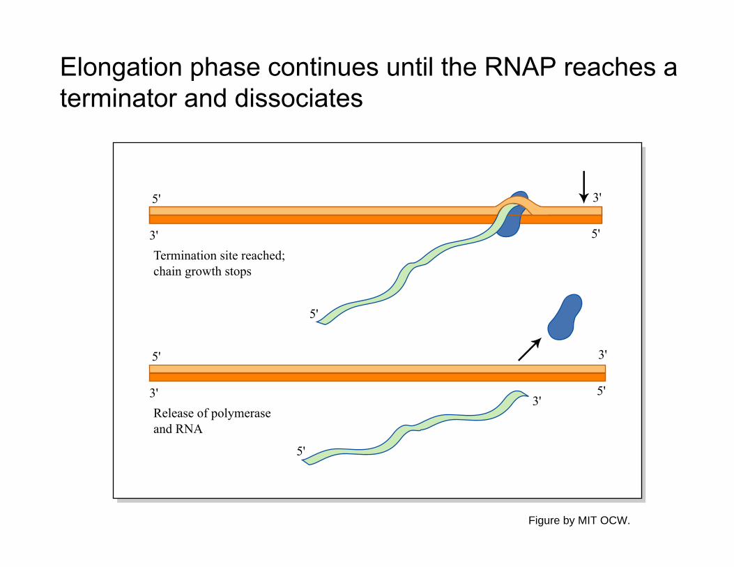
Elongation phase continues until the RNAP reaches a terminator and dissociates
Release of polymerase and RNA
3'
5'
3'
3'5'
5'
5'
5'
3'
Termination site reached; chain growth stops
5'
3'
Figure by MIT OCW.
Initiation of transcription begins with promoter binding by RNAP holoenzyme
holoenzyme = RNAP core + Sigma
Diagram of RNA polymerase and transcription removed due to copyright restrictions.
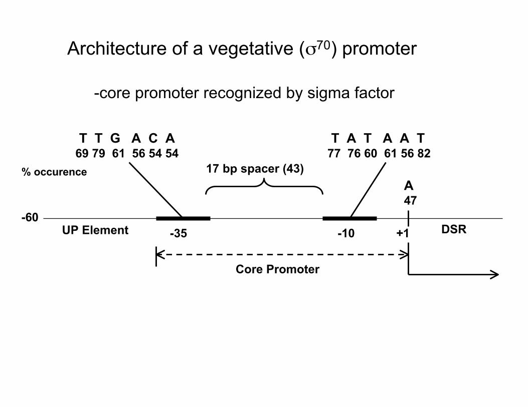
-60
Architecture of a vegetative (σ70) promoter
-core promoter recognized by sigma factor
T T G A C A T A T A A T69 79 61 56 54 54 77 76 60 61 56 82
% occurence 17 bp spacer (43) A 47
UP Element -35 -10 +1 DSR
Core Promoter
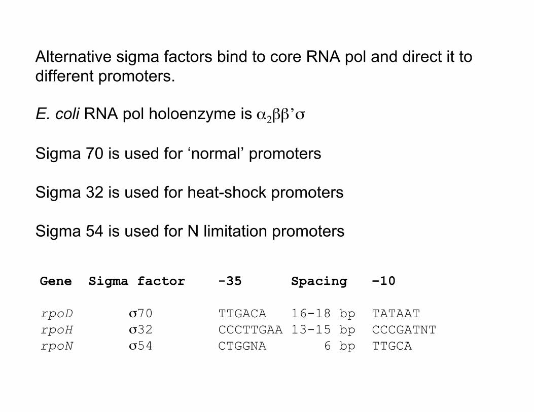
Alternative sigma factors bind to core RNA pol and direct it to different promoters.
E. coli RNA pol holoenzyme is α2ββ’σ
Sigma 70 is used for ‘normal’ promoters
Sigma 32 is used for heat-shock promoters
Sigma 54 is used for N limitation promoters
Gene Sigma factor -35 Spacing –10
rpoD σ70 TTGACA 16-18 bp TATAAT rpoH σ32 CCCTTGAA 13-15 bp CCCGATNT rpoN σ54 CTGGNA 6 bp TTGCA
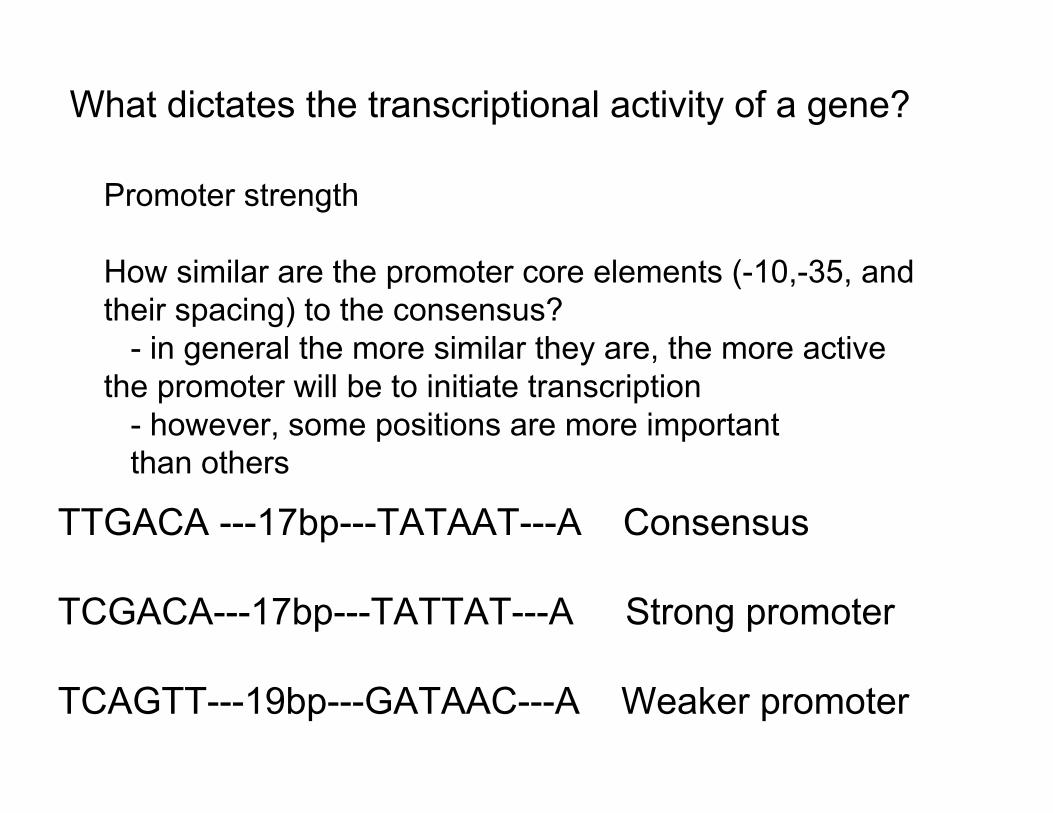
What dictates the transcriptional activity of a gene?
Promoter strength
How similar are the promoter core elements (-10,-35, and their spacing) to the consensus?
- in general the more similar they are, the more active the promoter will be to initiate transcription
- however, some positions are more important than others
TTGACA ---17bp---TATAAT---A Consensus
TCGACA---17bp---TATTAT---A Strong promoter
TCAGTT---19bp---GATAAC---A Weaker promoter
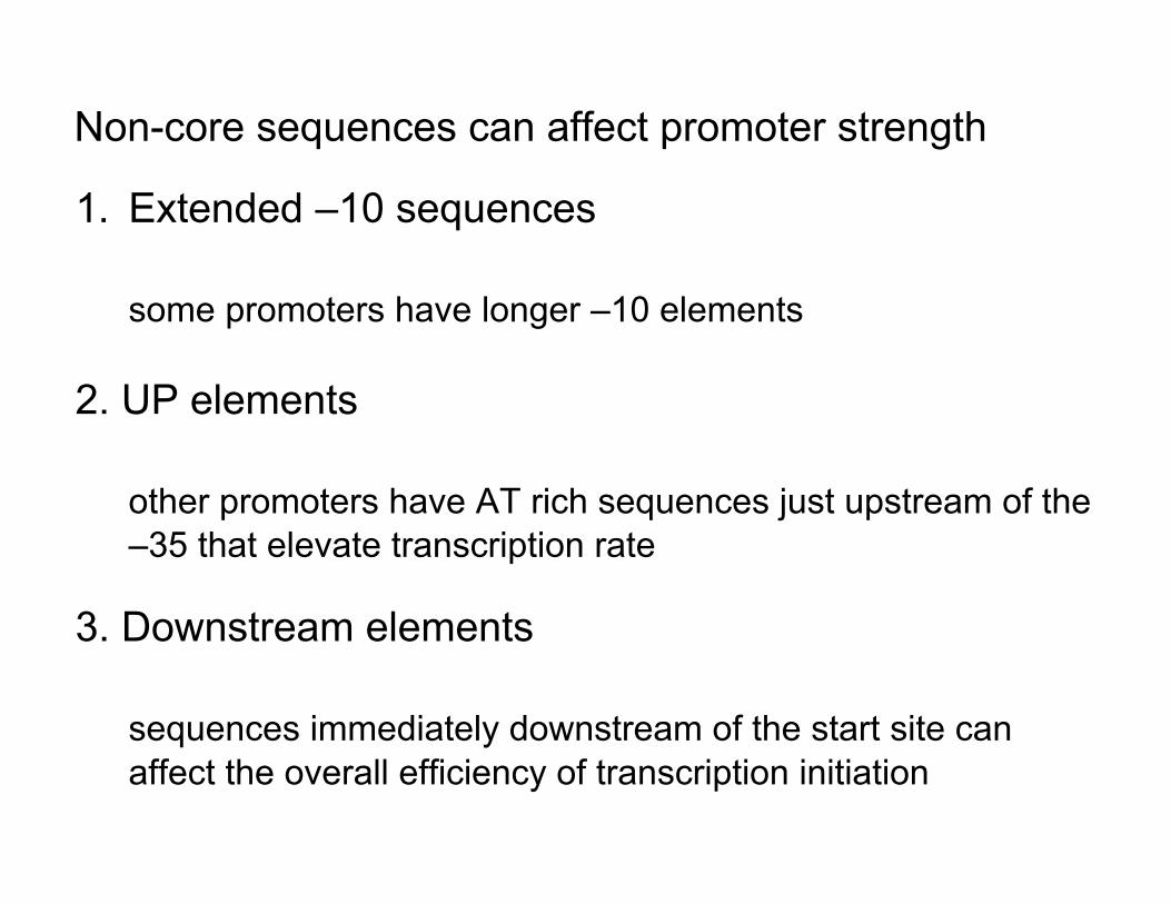
Non-core sequences can affect promoter strength
1. Extended –10 sequences
some promoters have longer –10 elements
2. UP elements
other promoters have AT rich sequences just upstream of the –35 that elevate transcription rate
3. Downstream elements
sequences immediately downstream of the start site can affect the overall efficiency of transcription initiation
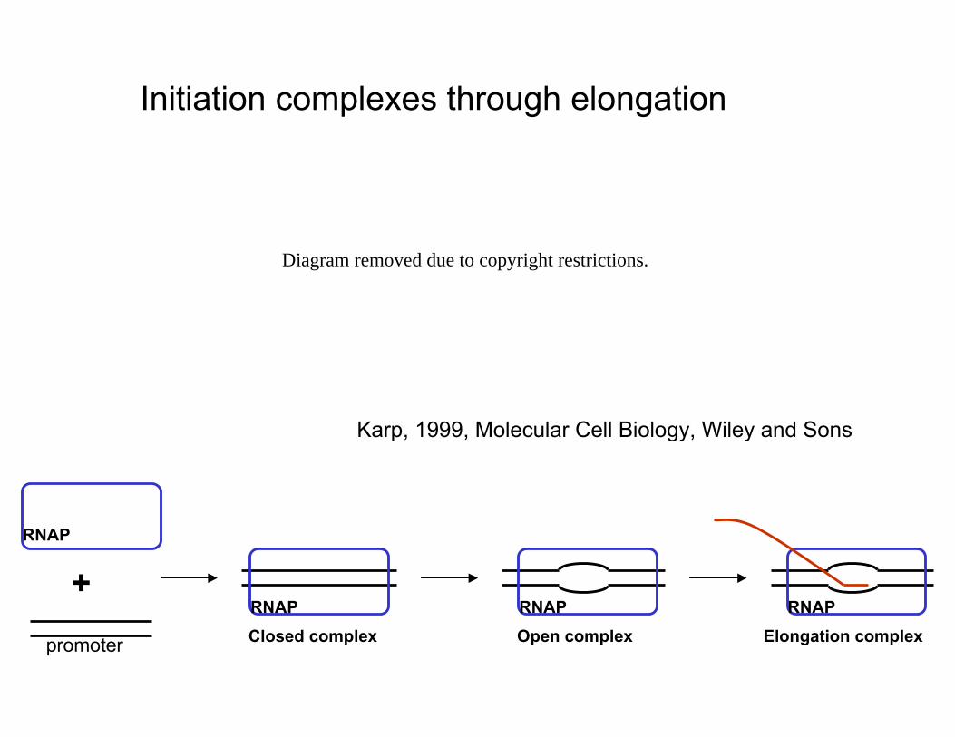
Initiation complexes through elongation
Diagram removed due to copyright restrictions.
Karp, 1999, Molecular Cell Biology, Wiley and Sons
RNAP
+ RNAP RNAP RNAP
promoter Closed complex Open complex Elongation complex
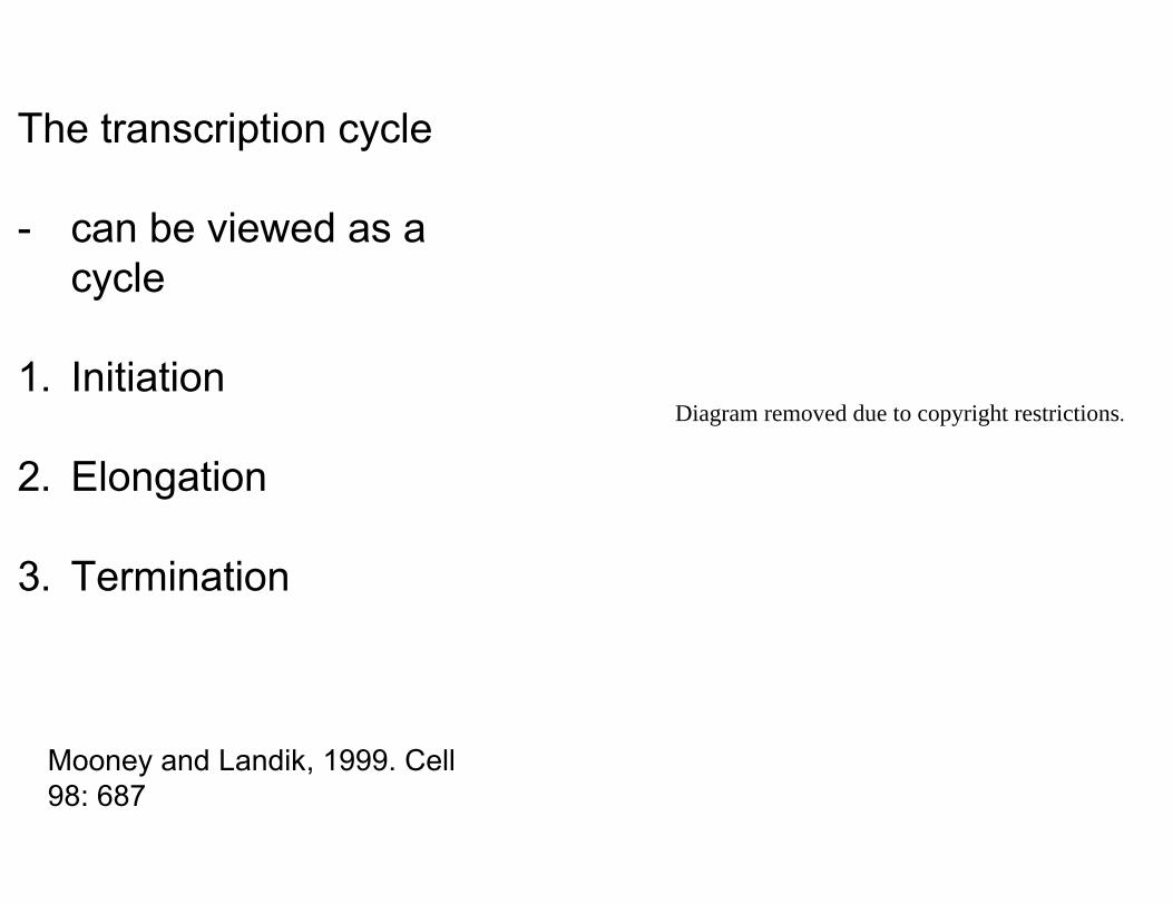
The transcription cycle
- can be viewed as a cycle
1. Initiation Diagram removed due to copyright restrictions.
2. Elongation
3. Termination
Mooney and Landik, 1999. Cell 98: 687
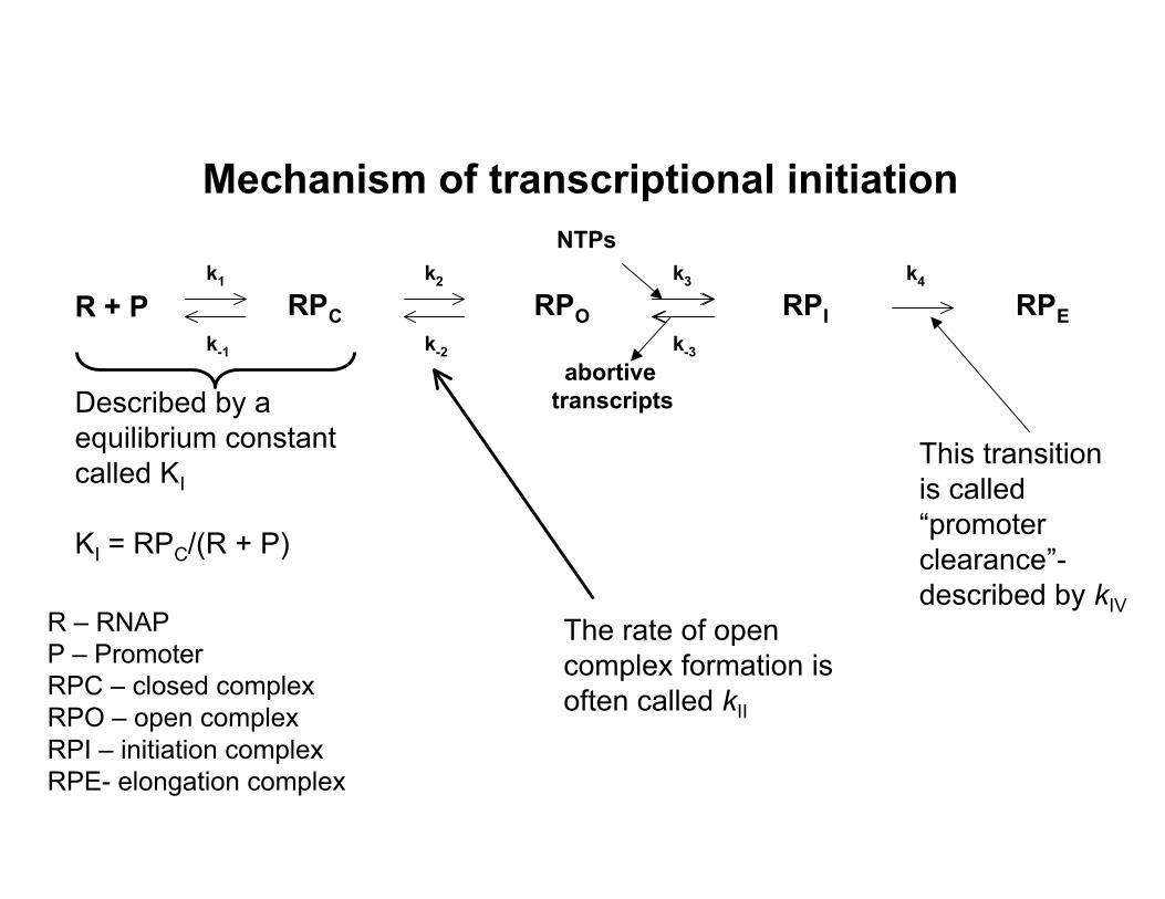
Mechanism of transcriptional initiationNTPs
k1 k2 k3 k4
RPC RPO RPI RPER + P k-1 k-2 k-3
abortiveDescribed by a transcripts
equilibrium constant This transition called KI is called “promoterKI = RPC/(R + P) clearance”-described by kIV
R – RNAP The rate of open P – Promoter complex formation is RPC – closed complex RPO – open complex often called kII
RPI – initiation complex RPE- elongation complex
Transcription termination
Diagram of transcription termination removed due to copyright restrictions. See Figure 7-32 in Madigan, Michael, and John Martinko. Brock Biology of Microorganisms. 11th ed. Upper Saddle River, NJ: Pearson Prentice Hall, 2006. ISBN: 0131443291.
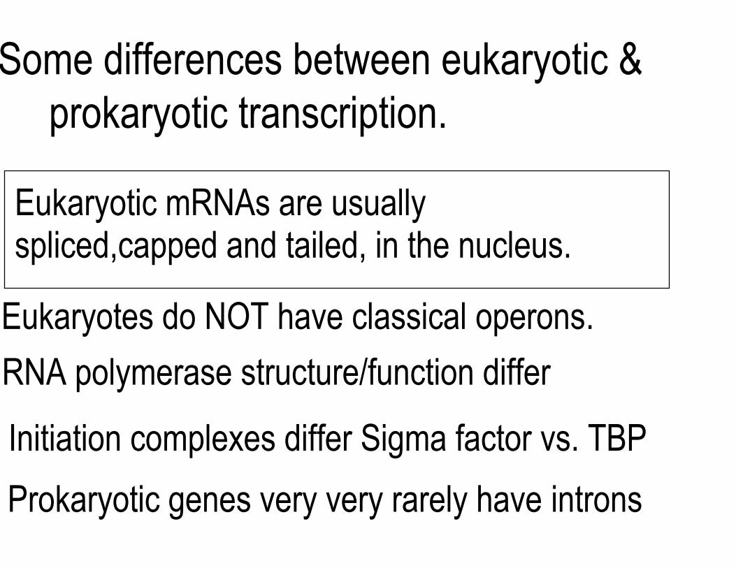
Some differences between eukaryotic & prokaryotic transcription.
Eukaryotic mRNAs are usually spliced,capped and tailed, in the nucleus.
Eukaryotes do NOT have classical operons.
RNA polymerase structure/function differ
Initiation complexes differ Sigma factor vs. TBP
Prokaryotic genes very very rarely have introns
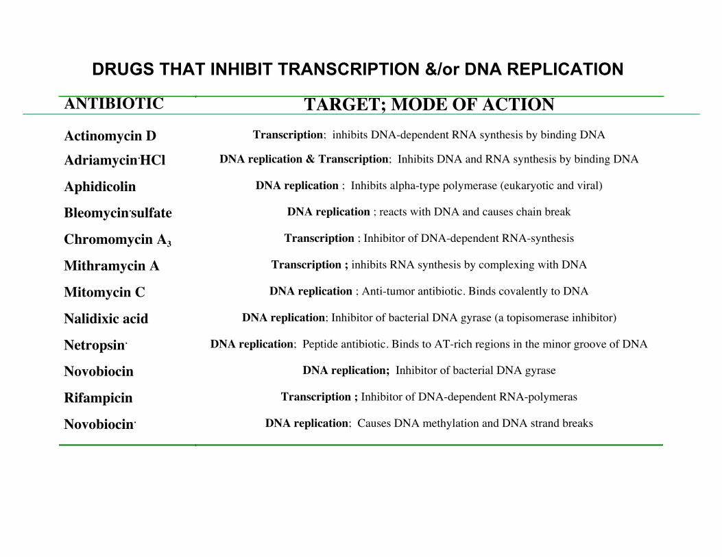
DRUGS THAT INHIBIT TRANSCRIPTION &/or DNA REPLICATION
ANTIBIOTIC TARGET; MODE OF ACTION Actinomycin D Transcription; inhibits DNA-dependent RNA synthesis by binding DNA
Adriamycin.HCl DNA replication & Transcription; Inhibits DNA and RNA synthesis by binding DNA
Aphidicolin DNA replication ; Inhibits alpha-type polymerase (eukaryotic and viral)
Bleomycin.sulfate DNA replication ; reacts with DNA and causes chain break
Chromomycin A3 Transcription ; Inhibitor of DNA-dependent RNA-synthesis
Mithramycin A Transcription ; inhibits RNA synthesis by complexing with DNA
Mitomycin C DNA replication ; Anti-tumor antibiotic. Binds covalently to DNA
Nalidixic acid DNA replication; Inhibitor of bacterial DNA gyrase (a topisomerase inhibitor)
Netropsin. DNA replication; Peptide antibiotic. Binds to AT-rich regions in the minor groove of DNA
Novobiocin DNA replication; Inhibitor of bacterial DNA gyrase
Rifampicin Transcription ; Inhibitor of DNA-dependent RNA-polymeras
Novobiocin. DNA replication; Causes DNA methylation and DNA strand breaks
TranslationCoupled transcription/translation Compartmentalization/transcript processing
Diagram of transcription and translation in prokaryotes vs. eukaryotes removed due to coypyright restrictions.
Coupled transcription/translation
Microscopic photographs of transcription and translation removed due to copyright restrictions.
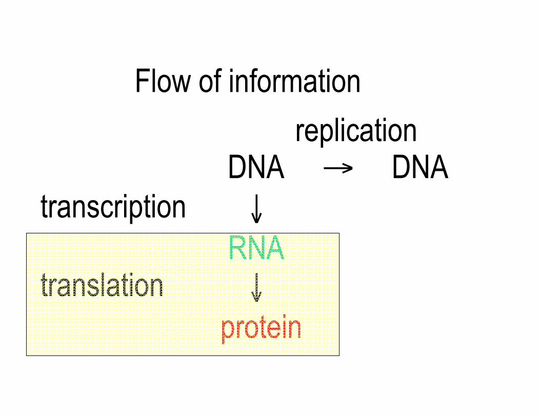
Flow of information
replication DNA → DNA
transcription ↓ RNA
translation ↓ protein
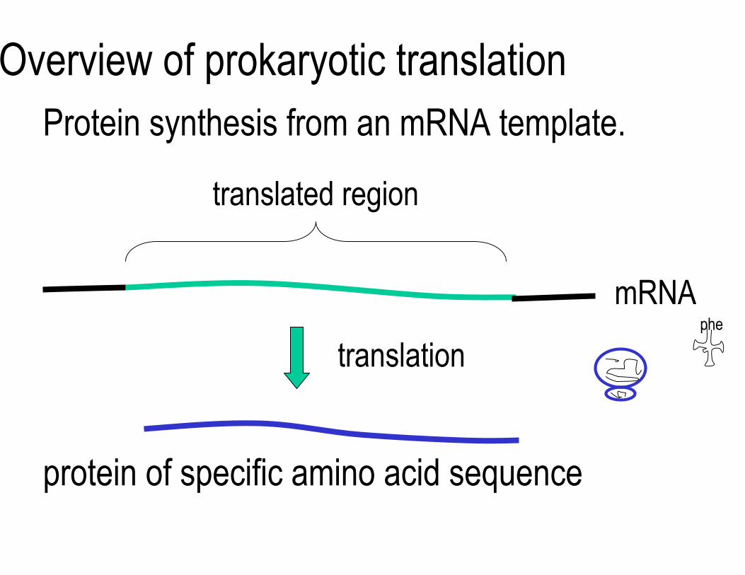
Overview of prokaryotic translation Protein synthesis from an mRNA template.
translated region
mRNA
translation phe
protein of specific amino acid sequence
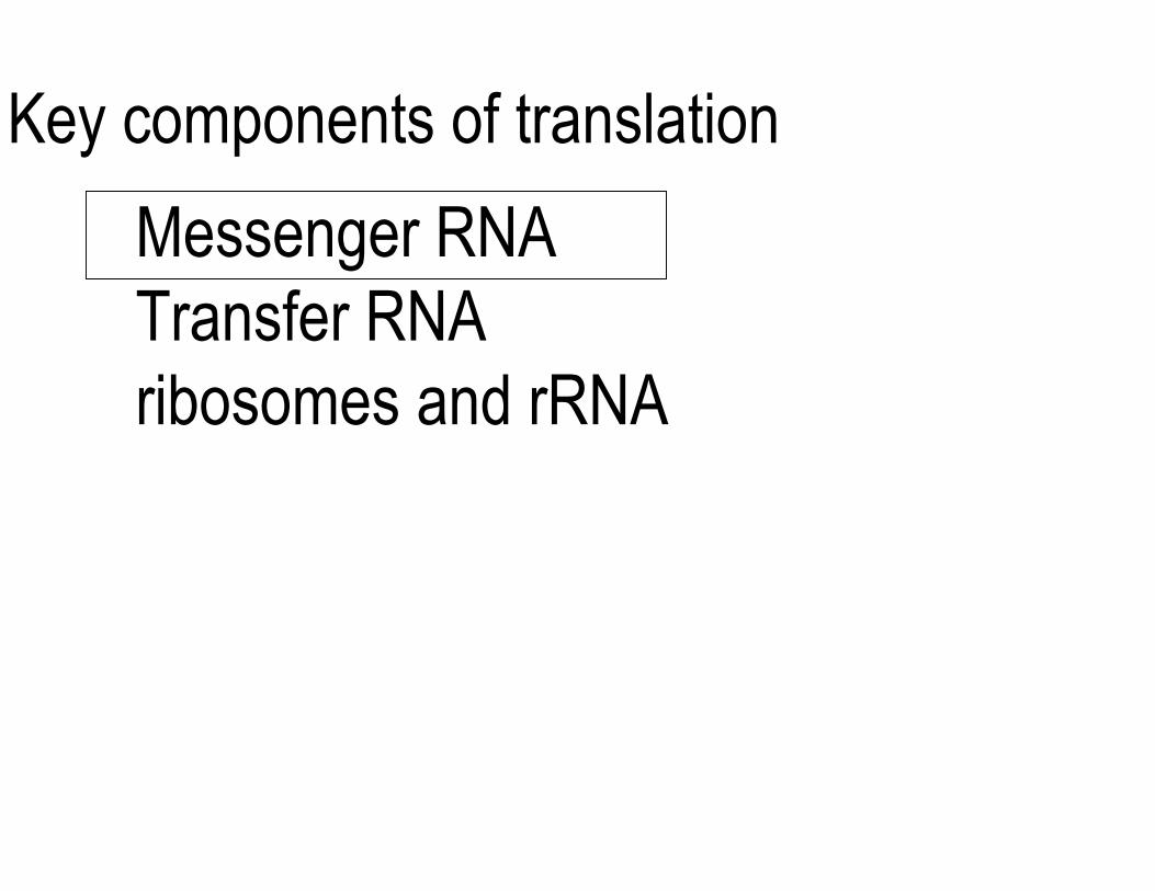
Key components of translation
Messenger RNATransfer RNA ribosomes and rRNA
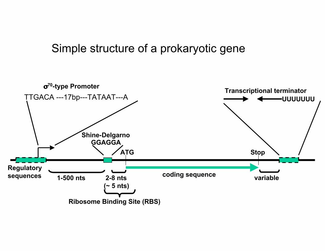
Simple structure of a prokaryotic gene
σ70-type Promoter Transcriptional terminatorTTGACA ---17bp---TATAAT---A UUUUUUU
Shine-DelgarnoGGAGGA
ATG Stop
Regulatory sequences 1-500 nts 2-8 nts coding sequence variable
(~ 5 nts)
Ribosome Binding Site (RBS)
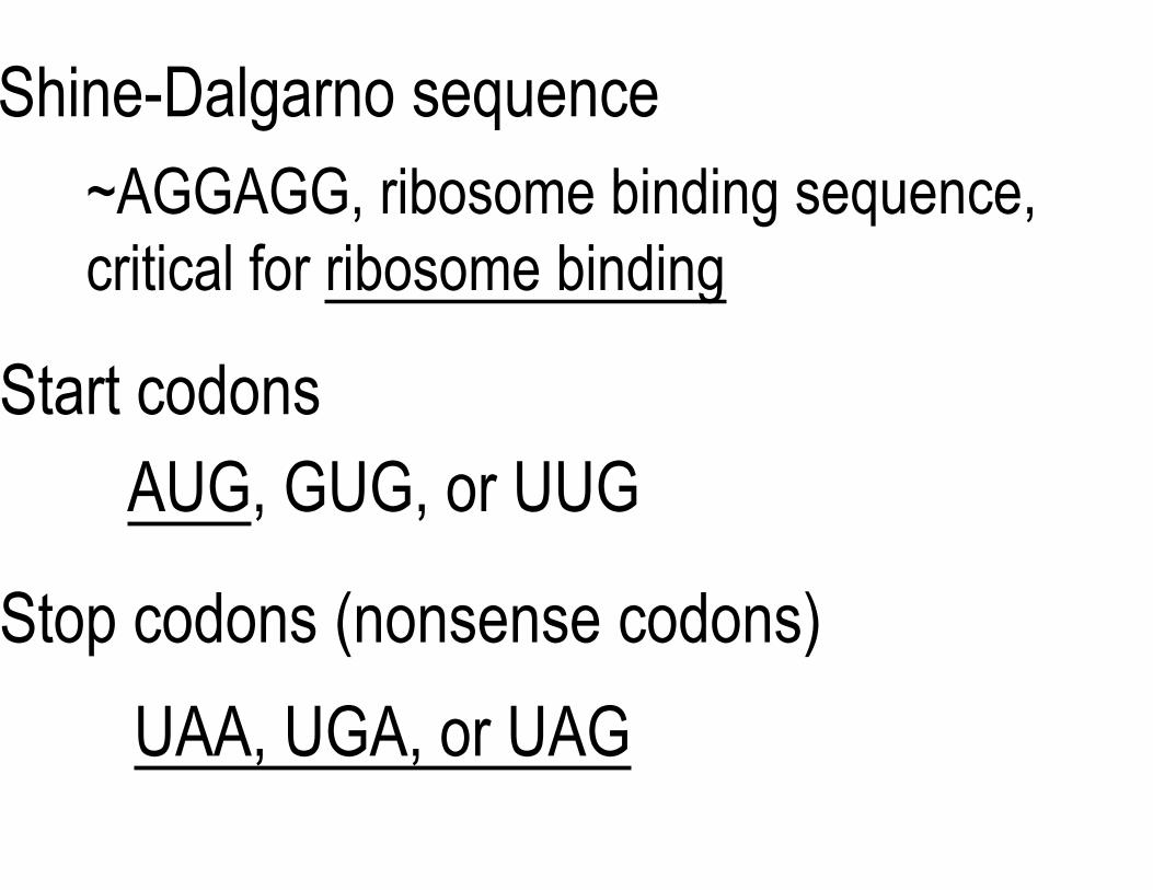
Shine-Dalgarno sequence ~AGGAGG, ribosome binding sequence, critical for ribosome binding
Start codons AUG, GUG, or UUG
Stop codons (nonsense codons)
UAA, UGA, or UAG

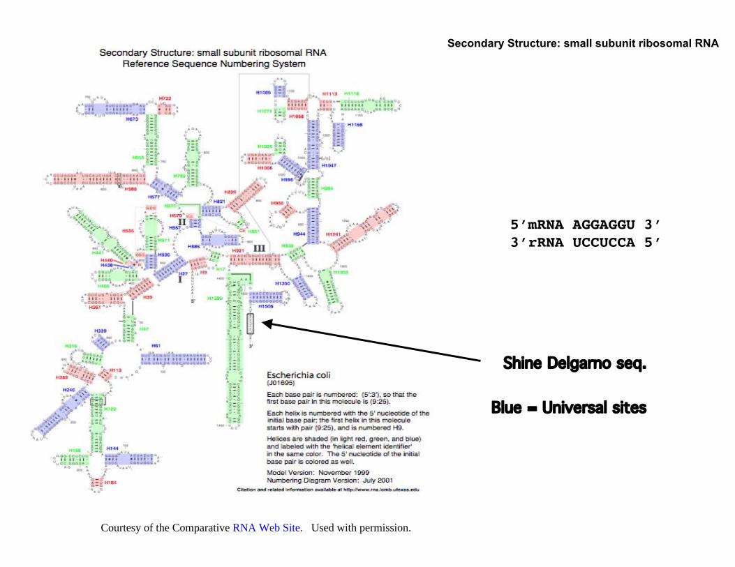
Secondary Structure: small subunit ribosomal RNA
Blue = Universal sites
Shine Delgarno seq.
5’mRNA AGGAGGU 3’ 3’rRNA UCCUCCA 5’
Courtesy of the Comparative RNA Web Site. Used with permission.
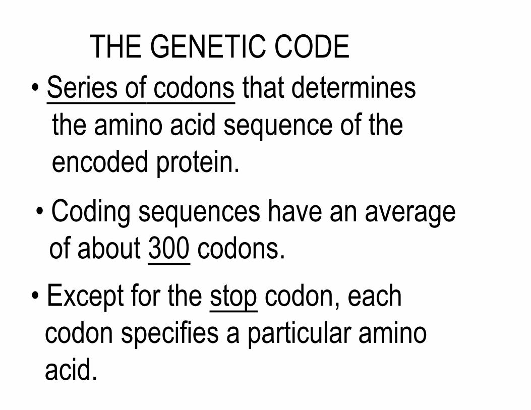
THE GENETIC CODE • Series of codons that determines
the amino acid sequence of the encoded protein.
• Coding sequences have an average of about 300 codons.
• Except for the stop codon, each codon specifies a particular amino acid.
The genetic code
Table of the genetic code removed due to copyright restrictions. See Table 7-5 in Madigan, Michael, and John Martinko. Brock Biology of Microorganisms. 11th ed. Upper Saddle River, NJ: Pearson Prentice Hall, 2006. ISBN: 0131443291.
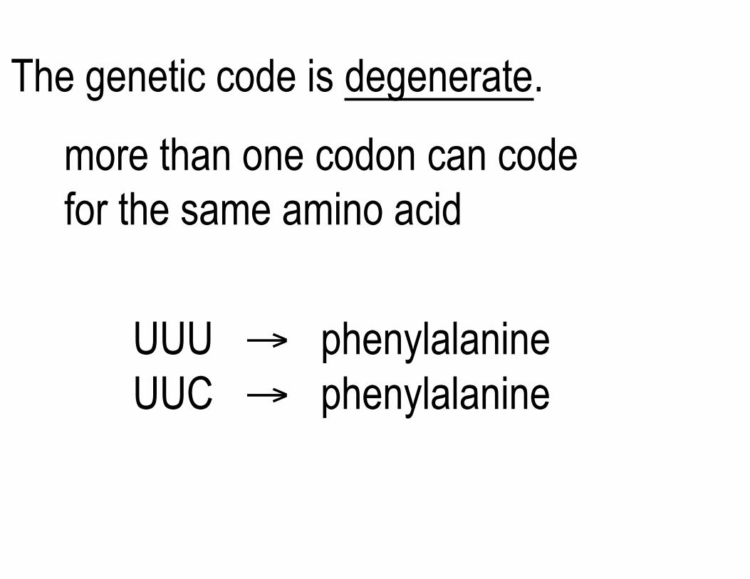
The genetic code is degenerate.
more than one codon can code for the same amino acid
UUU → phenylalanineUUC → phenylalanine

Synonyms
Different codons can code for the same amino acid
UUU → phenylalanineUUC → phenylalanine
Not all synonyms are used with equal frequency. This is called "codon usage bias".
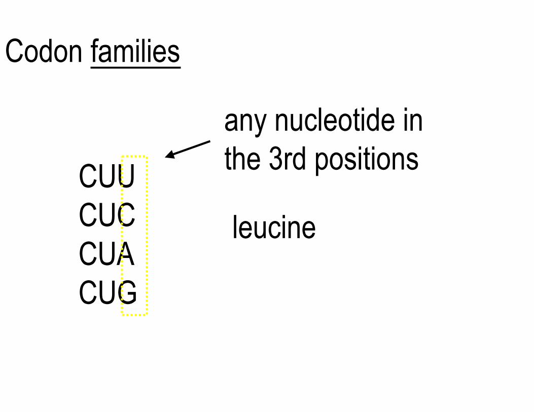
Codon families
any nucleotide in the 3rd positions
CUACUG
CUU CUC leucine
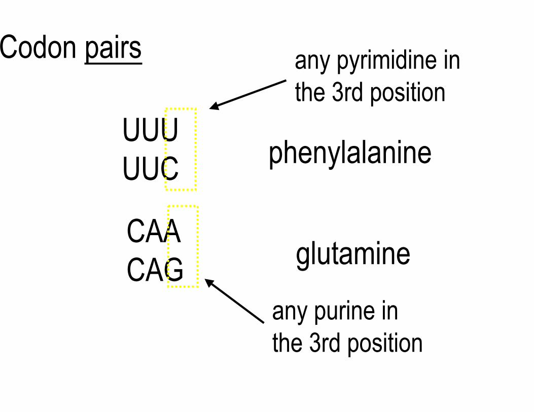
Codon pairs any pyrimidine in the 3rd position
phenylalanine
glutamine
any purine in the 3rd position
UUU UUC
CAA CAG
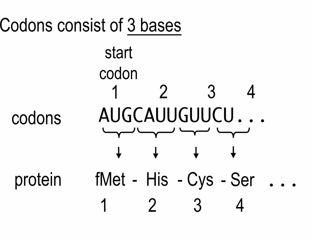
Codons consist of 3 bases
startcodon
1 2 3 4codons AUGCAUUGUUCU...
protein fMet - His - Cys - Ser ...1 2 3 4
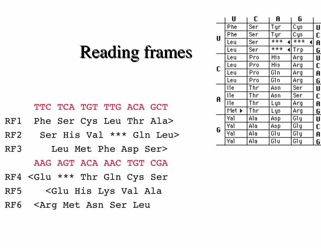
Reading framesReading frames
TTC TCA TGT TTG ACA GCT
RF1 Phe Ser Cys Leu Thr Ala>
RF2 Ser His Val *** Gln Leu>
RF3 Leu Met Phe Asp Ser>
AAG AGT ACA AAC TGT CGA
RF4 <Glu *** Thr Gln Cys Ser
RF5 <Glu His Lys Val Ala
RF6 <Arg Met Asn Ser Leu
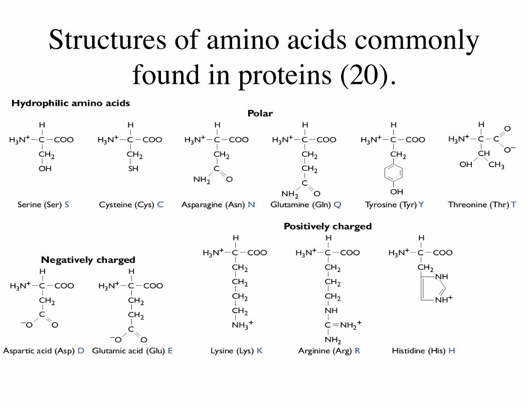
Structures of amino acids commonlyfound in proteins (20).
3
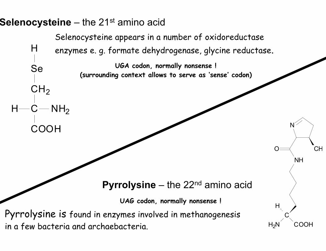
Selenocysteine – the 21st amino acid Selenocysteine appears in a number of oxidoreductase
H
in a few bacteria and archaebacteria.
CH2
CH NH2
COOH
Se
C
H2N COOH
H
NH
O
N
CH
Pyrrolysine – the 22nd amino acid
Pyrrolysine is found in enzymes involved in methanogenesis
enzymes e. g. formate dehydrogenase, glycine reductase. UGA codon, normally nonsense !
(surrounding context allows to serve as ‘sense’ codon)
UAG codon, normally nonsense !

Key components of translation
Messenger RNA Transfer RNAribosomes and rRNA
Diagrams removed due to copyright restrictions. See Figures 7-36 and 7-34 in Madigan, Michael, and John Martinko. Brock Biology of Microorganisms. 11th ed. Upper Saddle River, NJ: Pearson Prentice Hall, 2006. ISBN: 0131443291.
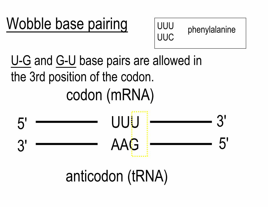
Wobble base pairing UUU phenylalanineUUC
U-G and G-U base pairs are allowed inthe 3rd position of the codon.
codon (mRNA)
3'
AAG UUU5'
5'3'
anticodon (tRNA)
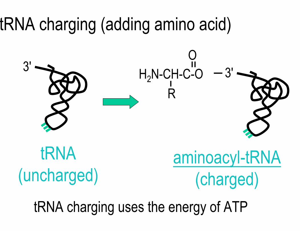
tRNA charging (adding amino acid)
O 3' 3'H2N-CH-C-O
R
tRNA aminoacyl-tRNA (uncharged) (charged)
tRNA charging uses the energy of ATP
O
OH OH
HH
H
CH2
H
OPOPOP−O
O
O− O−
O O
O−
R H C C
NH3 +
O−
O
O
OH OH
HH
H
CH2
H
OPOC
O
O−
H CR
NH2
O
Adenine
Adenine
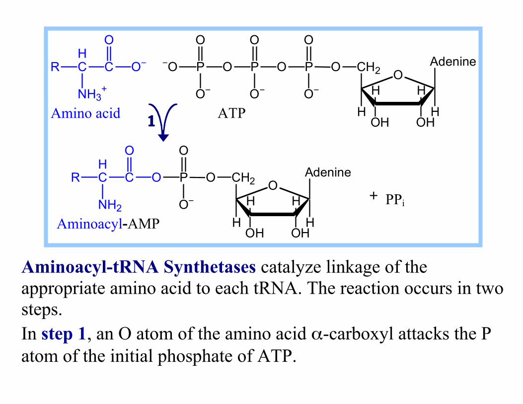
ATPAmino acid
Aminoacyl-AMP
1
+ PPi
Aminoacyl-tRNA Synthetases catalyze linkage of the appropriate amino acid to each tRNA. The reaction occurs in two steps. In step 1, an O atom of the amino acid α-carboxyl attacks the P atom of the initial phosphate of ATP.
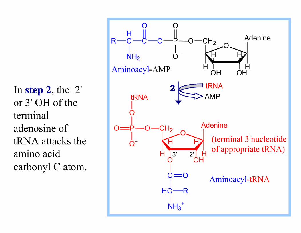
In step 2, the 2' or 3' OH of the terminal adenosine of tRNA attacks the amino acid carbonyl C atom.
O
OH OH
HH
H
CH2
H
OPOC
O
O−
H CR
NH2
O
Adenine
O
OH O
HH
H
CH2
H
OPO
O
O−
Adenine
tRNA
C
HC
O
NH3 +
R
tRNA AMP
Aminoacyl-AMP
Aminoacyl-tRNA
(terminal 3’nucleotide of appropriate tRNA)
3’ 2’
2
Aminoacyl-tRNA Synthetase
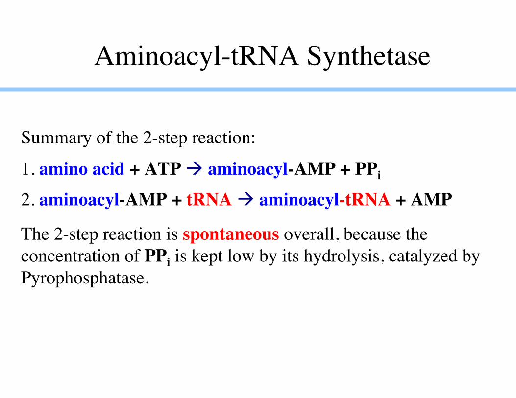
Summary of the 2-step reaction:
1. amino acid + ATP aminoacyl-AMP + PPi
2. aminoacyl-AMP + tRNA aminoacyl-tRNA + AMP
The 2-step reaction is spontaneous overall, because theconcentration of PPi is kept low by its hydrolysis, catalyzed byPyrophosphatase.
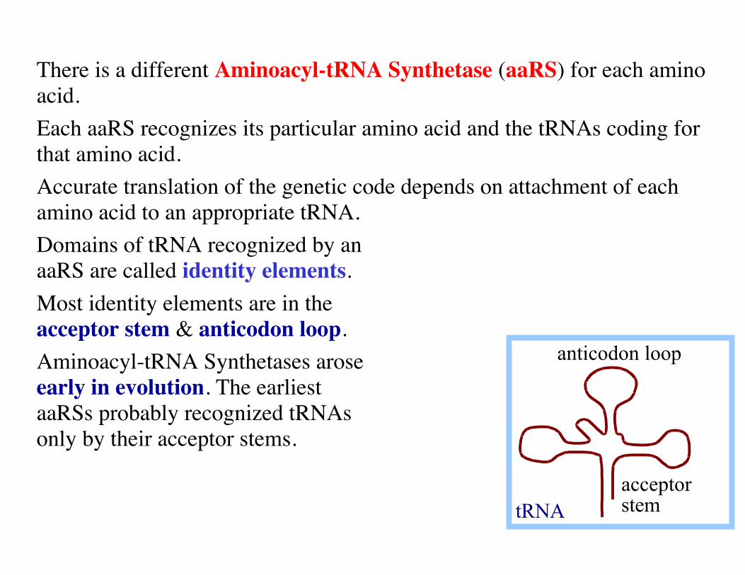
There is a different Aminoacyl-tRNA Synthetase (aaRS) for each aminoacid.Each aaRS recognizes its particular amino acid and the tRNAs coding forthat amino acid.Accurate translation of the genetic code depends on attachment of eachamino acid to an appropriate tRNA.Domains of tRNA recognized by anaaRS are called identity elements.Most identity elements are in theacceptor stem & anticodon loop. Aminoacyl-tRNA Synthetases aroseearly in evolution. The earliest aaRSs probably recognized tRNAsonly by their acceptor stems.
anticodon loop
acceptor stemtRNA
Key components of translation
Messenger RNA Transfer RNA ribosomes and rRNA
Structure of the E. coli Ribosome
Diagram of the structure of the E. coli ribosome removed due to copyright restrictions.
The cutaway view at right shows positions of tRNA (P, E sites) &mRNA (as orange beads).
Table of ribosome structure removed due to copyright restrictions. See Table 7-6 in Madigan, Michael, and John Martinko. Brock Biology of Microorganisms. 11th ed. Upper Saddle River, NJ: Pearson Prentice Hall, 2006. ISBN: 0131443291.
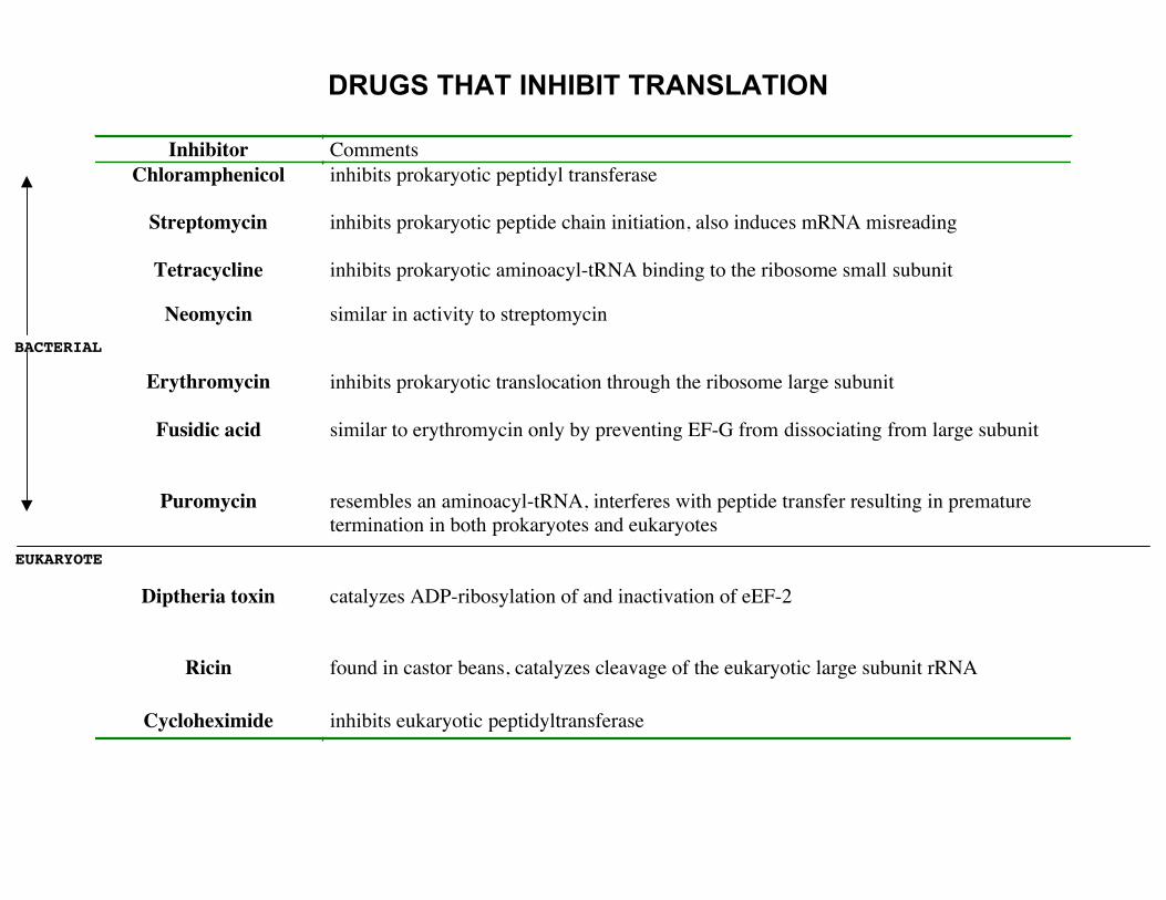
DRUGS THAT INHIBIT TRANSLATION
Inhibitor Comments Chloramphenicol inhibits prokaryotic peptidyl transferase
Streptomycin inhibits prokaryotic peptide chain initiation, also induces mRNA misreading
Tetracycline inhibits prokaryotic aminoacyl-tRNA binding to the ribosome small subunit
Neomycin similar in activity to streptomycin BACTERIAL
Erythromycin inhibits prokaryotic translocation through the ribosome large subunit
Fusidic acid similar to erythromycin only by preventing EF-G from dissociating from large subunit
Puromycin resembles an aminoacyl-tRNA, interferes with peptide transfer resulting in prematuretermination in both prokaryotes and eukaryotes
EUKARYOTE
Diptheria toxin catalyzes ADP-ribosylation of and inactivation of eEF-2
Ricin found in castor beans, catalyzes cleavage of the eukaryotic large subunit rRNA
Cycloheximide inhibits eukaryotic peptidyltransferase

Prokaryotic 70S ribosome
23s rRNA 5s rRNA 34 proteins
16s RNA 21 proteins
50ssubunit
30ssubunit
Secondary Structure: small subunit ribosomal RNA
Blue = Universal sites
Shine Delgarno seq.
5’mRNA AGGAGGU 3’ 3’rRNA UCCUCCA 5’
Courtesy of the Comparative RNA Web Site. Used with permission.
Diagram removed due to copyright restrictions. See Figure 7-38 in Madigan, Michael, and John Martinko. Brock Biology of Microorganisms. 11th ed. Upper Saddle River, NJ: Pearson Prentice Hall, 2006. ISBN: 0131443291.
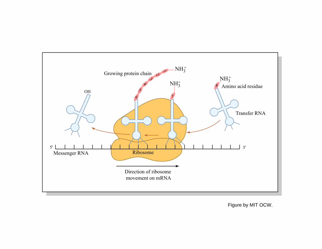
Growing protein chain
Ribosome
Direction of ribosome movement on mRNA
Messenger RNA5' 3'
Transfer RNA
OH
NH3
NH3
+
+
NH3+
Amino acid residue
1
23
4
56 7
8
Figure by MIT OCW.
Diagram removed due to copyright restrictions. See Figure 7-39 in Madigan, Michael, and John Martinko. Brock Biology of Microorganisms. 11th ed. Upper Saddle River, NJ: Pearson Prentice Hall, 2006. ISBN: 0131443291.
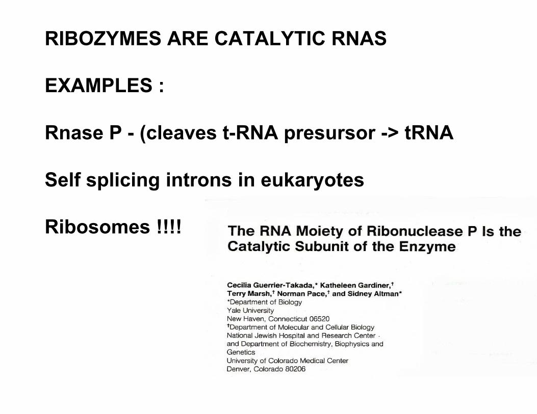
RIBOZYMES ARE CATALYTIC RNAS
EXAMPLES :
Rnase P - (cleaves t-RNA presursor -> tRNA
Self splicing introns in eukaryotes
Ribosomes !!!!


![CT3manual[1] - brock](https://img.pdfslide.us/doc/110x75/54e7e9594a7959d76d8b48c8/ct3manual1-brock.jpg)