Embed Size (px)
Citation preview
8/7/2019 Molten Globule State of Tear Lipocalin ANS Binding Restores Tertiary Interactions
http://slidepdf.com/reader/full/molten-globule-state-of-tear-lipocalin-ans-binding-restores-tertiary-interactions 1/12
Molten Globule State of Tear Lipocalin: ANS Binding Restores
Tertiary Interactions
Oktay K. Gasymov, Adil R. Abduragimov, and Ben J. Glasgow*
Departments of Pathology and Ophthalmology, UCLA School of Medicine, Jules Stein Eye Institute,100 Stein Plaza, Los Angeles CA 90095, USA
Abstract
Tear lipocalin (TL) may stabilize the lipid layer of tears through a molten globule state triggered bylow pH. EPR spectroscopy with site directed spin labeling, revealed the side chain mobility of residues on the G-strand of TL in a molten globule state; the G-strand retains β-sheet structure. Allof the side chains of G strand residues become more loosely packed, especially residues 96–99. In
contrast, the highly mobile side chain of residue 95 on the F-G loop, becomes tightly packed. ANSbinding to TL in a molten globule state reestablishes tight packing around side chains that are orientedboth inside and outside of the barrel. Unlike RBP and BLG; TL has no disulfide bond between Gand H strands. It is likely that the central β-sheet in the molten globule state of lipocalins is stabilizedby its interactions with the main α-helix, rather than the interstrand disulfide bond.
Keywords
Molten globule; Tear lipocalin; Lipocalin-1; ANS; EPR; β-lactoglobulin; Side-directed spin labeling
Introduction
Molten globule states of proteins, that have features in common with protein-foldingintermediates [1–3], are suggested to be involved in such functions as the insertion of proteinsinto membranes [4], the release of bound ligands [1,5,6] and aggregation including amyloidfibril formation [7].
Molten globule states of proteins are characterized by having native-like secondary structurethat lacks most of the specific tertiary interactions [1]. Three experimental criteria have beensuggested to confirm native-molten globule transitions in proteins. Compared to the nativestate, a protein in the molten globule state should exhibit (1) small changes in far-UV CDspectrum (intact secondary structure), (2) significant loss in near-UV CD spectrum (loss of tertiary structure) and (3) enhancement of ANS fluorescence upon binding (exposure of hydrophobic sites).
Despite the large body of data available on molten globule states of proteins, site-specificinformation is limited. Nuclear magnetic resonance (NMR) spectroscopy and a molecular
*Corresponding author: Ben J. Glasgow, Departments of Pathology and Ophthalmology, UCLA School of Medicine, Jules Stein EyeInstitute, 100 Stein Plaza, Rm# B269, Los Angeles, CA 90095, USA, (310) 825–6998, [email protected].
Publisher's Disclaimer: This is a PDF file of an unedited manuscript that has been accepted for publication. As a service to our customerswe are providing this early version of the manuscript. The manuscript will undergo copyediting, typesetting, and review of the resultingproof before it is published in its final citable form. Please note that during the production process errors may be discovered which couldaffect the content, and all legal disclaimers that apply to the journal pertain.
NIH Public AccessAuthor Manuscript Biochem Biophys Res Commun. Author manuscript; available in PMC 2007 August 24.
Published in final edited form as:
Biochem Biophys Res Commun. 2007 June 1; 357(2): 499–504.
N I H -P A A u
t h or Manus c r i pt
N I H -P A A ut h or Manus c r i pt
N I H -P A A ut h or M
anus c r i pt
8/7/2019 Molten Globule State of Tear Lipocalin ANS Binding Restores Tertiary Interactions
http://slidepdf.com/reader/full/molten-globule-state-of-tear-lipocalin-ans-binding-restores-tertiary-interactions 2/12
dynamic simulation have been employed to study molten globule states of some proteins [8–11]. The partial loss of secondary structure as well as side chain packing is site specific. Moltenglobule states of both RBP and equine BLG, which are members of lipocalin family, havesimilar features. Residues in the second β-sheet, also known as central β-sheet, (strands F, G,H and part of A) of the barrel retain native-like β-sheet structure in the molten globule state.The F,G, and H strands of BLG form hydrogen bonds early in a folding process and it has beenhypothesized that the G and H strand strands act as folding initiation sites; residues on these
strands have been catergorized as more stable with greater unfolding midpoints in urea [10].Molten globule states of these RBP and BLG also reveal that the major α-helices are packedagainst the central β-sheets as in the native structures [9–11]. Both BLG and RBP also haveinterstrand disulfide bonds that join the G and H strand, which hypothetically restrain unfoldingand facilitate retention of β structure in the lipocalins. Some lipocalins, including TL, the Gand H strands are not linked by a disulfide bridge. The molten globule state of TL provides anopportunity to obtain information on the G strand in the absence of the potentially constrainingdisulfide bond.
Tear lipocalin (TL), as a member of the lipocalin family of proteins, exhibits cup shaped ligandbinding fold within a continuously hydrogen-bonded β-barrel that is formed by eightantiparallel β-strands [12]. There is a single disulfide bridge linking the C terminal end withthe CD loop. The solution structure of TL was resolved by site directed tryptophan fluorescence
and revealed a capacious cavity that confers promiscuity in ligand binding [13]. These findingswere verified by crystallography of TL [14].
Numerous putative functions, most of which are linked to the binding of various classes of ligand, have been suggested for TL [15–23]. One postulate is that TL stabilizes and modulateslipid in the tear film through a molten globule state triggered by low pH at the aqueous-lipidinterface [5]. TL undergoes a native to molten globule transition at pH 3.0 that has beendocumented by changes in tertiary structure of the protein and ANS fluorescence enhancement[5].
There is a lack of site-specific information regarding structural consequences of ANS bindingto proteins in molten globule states. Here, based on recent developments in understandingnitroxide side chain motion in β-sheet proteins [24], site directed spin labeling (SDSL) has
been employed to extract site-specific information on dynamics of the G-strand of TL in amolten globule state with and without bound ANS. The results reported here, based on changesof dynamic modes of nitroxide side chain for G-strand residues, clearly reveal that in a moltenglobule state, the G-strand of TL retains β-sheet structure. However, packing of side chains,specifically for residues 95–96, are significantly altered. ANS binding to TL in a molten globulestate reestablish tight packing around side chains that are oriented both inside and outside of the barrel.
Materials and methods
Materials
MTSL, (1-oxyl-2,2,5,5-tetramethyl-3-pyrroline-3-methyl) methanethiosulfonate, wasobtained from Toronto Research Chemicals, Inc, Toronto, Ontario. ANS (8-anilino-1-
naphthalenesulfonic acid) was purchased from Sigma. E. Coli, BL 21 (DE3) cells and pET 20bwere obtained from Novagene. Oligonucleotide primers were obtained from Universal DNA,Inc. PCR II was obtained from Invitrogen, San Diego, CA. HiTrap columns were obtainedfrom Pharmacia Biotech Inc., Piscataway, New Jersey. Gas-permeable TPX capillaries wereobtained from Wilmad Glass Co. Inc., Buena, NJ.
Gasymov et al. Page 2
Biochem Biophys Res Commun. Author manuscript; available in PMC 2007 August 24.
N I H -P A A
ut h or Manus c r i pt
N I H -P A A ut h or Manus c r i pt
N I H -P A A ut h or
Manus c r i pt
8/7/2019 Molten Globule State of Tear Lipocalin ANS Binding Restores Tertiary Interactions
http://slidepdf.com/reader/full/molten-globule-state-of-tear-lipocalin-ans-binding-restores-tertiary-interactions 3/12
Site-Directed Mutagenesis and Plasmid Construction
The TL cDNA in PCR II previously synthesized [25], was used as a template to clone the TLgene spanning bases 115–592 of the previously published sequence [20] into pET 20b withflanking restriction sites for NdeI and BamHI as previously described [26]. For single Cysmutants of TL, C101L was chosen as the template for additional mutant cDNAs because itshowed similar structural features and binding characteristics for spin labeled lipids [27]. Eightadditional mutant cDNAs were constructed, in which corresponding amino acid residues in
the G strand of TL were substituted sequentially to cysteine. Amino acid 95 corresponds toaspartic acid, bases 397–399 of Redl [20].
Expression and Purification of Mutant Proteins
The mutant plasmids were transformed in E. Coli, BL 21 (DE3), cells were cultured, and proteinwas expressed according to the manufacturer’s protocol (Novagene), and purified as previouslydescribed [6,13].
Spin labeling of TL Mutants
Mutant and wild type TLs, 100 μM, were incubated in 5-molar excess of MTSL overnight at4°C. Unreacted spin labels were removed with a HiTrap column.
EPR Measurements and Fitting of EPR spectra
Electron paramagnetic resonance spectra were recorded at X-band with a Varian E-109spectrometer fitted with two-loop one-gap resonator [28]. For measurements, the samples of about 5μl of spin-labeled protein in various conditions (generally 50–100 μM) were loadedinto Pyrex capillaries (0.84 o.d. × 0.64 i.d.; VitroCom Inc., Mountain Lakes, NJ). Themicrowave power was 2mW incident, and the modulation amplitude was 1G. 10 mM sodiumphosphate and 30 mM sodium citrate buffers were used for pH 7.3 and pH 3.0, respectively.All measurements were conducted at room temperature. To obtain ANS –protein complexes,spin labeled mutant proteins were incubated with 3-molar excess of ANS.
EPR spectra were fit to the MOMD model of Freed and co-workers using the program NLSLas previously described for CRBP [24,29]. The apparent mean correlation time was calculatedas τ= 1/6D, where D is the rotational diffusion coefficient.
Results and Discussion
All single Cys mutant and wild-type proteins used in this study have been characterizedpreviously [27]. For G-strand residues of TL, the effects of mutations on secondary structureand binding characteristics are minimal.
The line shapes of EPR spectra reflect the dynamic modes of the nitroxide side chain that canbe modulated by protein backbone fluctuation, tertiary interaction, etc [24,30–32]. Each EPRspectrum along the sequence provides site-specific dynamic information. It has been shownthat the peak-to-peak intensity and line width of the central line of the EPR spectra correlatewith dynamic and accessibility parameters. For β-sheet proteins, the sequence pattern of thesesite specific parameters along the β-strand exhibits characteristic alternating periodicity [24,27]. The nitroxide side chain in β-sheets that is located in a tertiary interaction with a side chainof a neighboring strand gives rise to additional dynamic modes, ranging from weakly orderedto immobilized. The dynamic modes of the nitroxide side chain, which interacts with nearest-neighbor side chains oriented in parallel on the flanking strands, depend on the type of neighboring residue, whether the neighbor is hydrogen bonded, and the twisting of the β strand[24].
Gasymov et al. Page 3
Biochem Biophys Res Commun. Author manuscript; available in PMC 2007 August 24.
N I H -P A A
ut h or Manus c r i pt
N I H -P A A ut h or Manus c r i pt
N I H -P A A ut h or
Manus c r i pt
8/7/2019 Molten Globule State of Tear Lipocalin ANS Binding Restores Tertiary Interactions
http://slidepdf.com/reader/full/molten-globule-state-of-tear-lipocalin-ans-binding-restores-tertiary-interactions 4/12
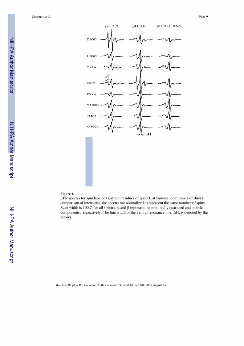
EPR spectra of spin-labeled apo-Cys mutants in various conditions are shown in Figure 1.These spectra are very similar to that of holo-forms that have been studied previously [27]. Forsome sites, two dynamic modes, indicated as α and β in Figure 1, can be easily observed. Forβ-sheets, the detection of two dynamic modes is indicative of nearest-neighbor interactions.Secondary structural content of G-strand residues can be determined from the plots of thespectral parameters, which correlate with side chain mobility and exposure, versus residuenumber (Fig. 2). The residues 96–101 exhibit the alternating periodicity, which is characteristic
of β-sheets. This feature is very consistent with the solution model and crystal structure of TLresolved by site-directed tryptophan fluorescence (SDTF) and x-ray crystallography,respectively [13,14].
TL undergoes a native to molten globule transition at pH 3.0 [5]. Comparison of EPR spectraof G-strand residues at pH 7.3 to that of pH 3.0 is very informative (Fig. 1). First, it is clearfrom the alternating periodicity that β structure persists in this strand. This is consistent withmolecular dynamic simulations for RBP that show retention in β structure in the central sheetbut disruption in strands E and F through loss of hydrogen bonds [9]. Second, nitroxide sidechain mobilities of all residues, except 95, are increased at pH 3.0, albeit to different degrees.Residues 95 and 98 show dramatic, but opposite changes. The nitroxide at position 98 becomesextremely mobile, indicative of very loose packing around this residue. Unexpectedly, thenitroxide at position 95, which is very mobile at pH 7.3, becomes more immobilized in the
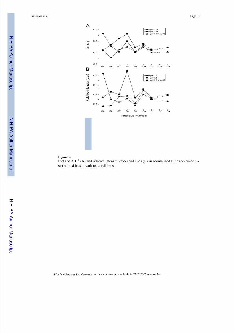
molten globule transition. At pH 3.0, this nitroxide side chain shows two dynamic modes,which arise from tertiary interactions. The spectral parameters for the G-strand (96–103) retainalternate periodicity, although the entire plot for these residues is shifted to higher valuesindicating more mobility and exposure (Fig. 2). Taken together the data indicate that the G-strand retains its β-sheet structure in a molten globule state, but tertiary interactions involvingG-strand residues are decreased, consistent with the spectral line shape (Fig. 1). Residues 95and 98 show the biggest changes in the spectral parameters (Figure 2.) in the transition frompH 7.3 to pH 3.0.
Although ANS is one of the most widely used fluorescent probes, information regarding thestructural consequences of ANS binding to proteins, particularly in molten globule state, islimited. Fluorescence enhancement upon binding to proteins can result from ion pairinginteractions, hydrophobic interactions, restricted mobility or any combination of these factors
[33–36]. EPR spectra of spin labeled G-strand mutants complexed with ANS in molten globulestate are shown in Figure 1. It is evident that ANS binding to the proteins in the molten globulestate restored tight packing for all nitroxide side chains. In some cases, the nitroxide side chains,particularly at positions 95, 96, 101, 103, become more immobile at pH 3.0 than at pH 7.3without bound ANS (Fig. 1). The entire plot of the spectral parameters for the mutant proteinsin the molten globule state complexed with ANS shifted to lower values compared to thatobserved for pH 3.0. Hence the outcome of ANS binding in influencing the dynamics of theprotein is site specific but there is clearly a global character. ANS induces more tight packingfor residues with side chains oriented internally as well as externally.
For each site on the G-strand, tertiary interactions are derived from side chains of residues fromflanked β-strands F and H. Therefore, EPR spectra of G-strand residues also reflect the statusof the F and H strands. Hence, ANS binding to the protein in molten globule state restores
tertiary interaction throughout the strand residues rather than for one or two residues (exposedor buried), that could be expected for a local binding site.
Residue I98 is located at the solvent-exposed site and most sensitive to the proteinconformational state (Fig. 1, 2). Therefore, EPR spectra of nitroxide side chain at position 98were analyzed in more detail with respect to local structural and dynamic properties. It hasbeen shown that the rotamer state of side chains in β-sheets is determined by steric interactions
Gasymov et al. Page 4
Biochem Biophys Res Commun. Author manuscript; available in PMC 2007 August 24.
N I H -P A A
ut h or Manus c r i pt
N I H -P A A ut h or Manus c r i pt
N I H -P A A ut h or
Manus c r i pt
8/7/2019 Molten Globule State of Tear Lipocalin ANS Binding Restores Tertiary Interactions
http://slidepdf.com/reader/full/molten-globule-state-of-tear-lipocalin-ans-binding-restores-tertiary-interactions 5/12
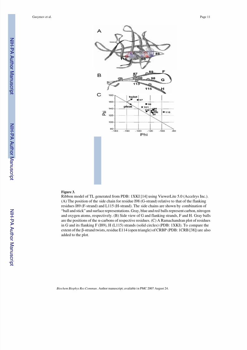
with nearest neighbors [24]. The residue I98 has two nearest neighbors, hydrogen-bonded (HB)I89 and non-hydrogen-bonded (NHB) L115, theCα-Cα distance of which are 5.0 Å and 4.6 Å,respectively (Fig 3A). The nearest neighbor interaction between the residues is modulated byextent of twisting. I98, compared to Y100, resides in a more twisted site of G-strand (Fig. 3 B,C). The EPR spectra for residue I98 reveal more asymmetric nearest neighbor interactionscompared to the spectra for Y100 (Fig. 1). Indeed, the EPR spectra for the nitroxide side chainat position 98 show two components, the mobility of which differ significantly from each other
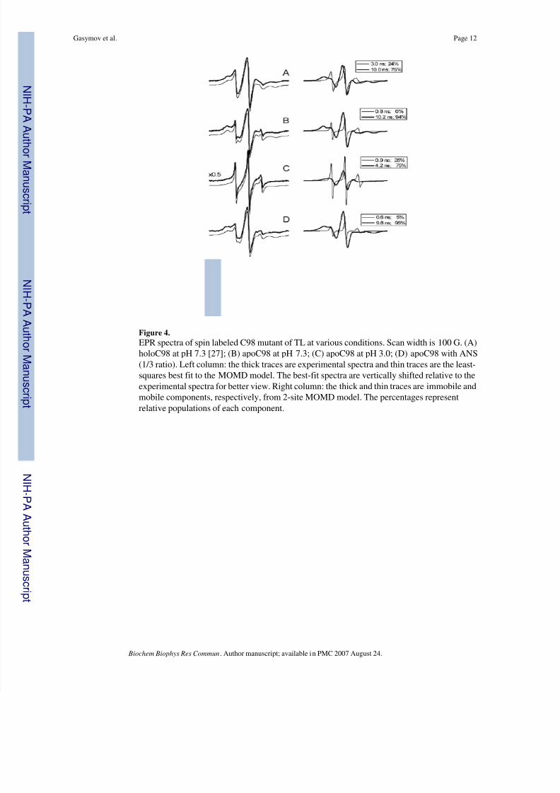
( 3.0 ns and 10.0 ns) (Fig. 4). In the apo-protein, the interaction is more asymmetric. Thecorrelation time of the first component decreases from 3.0 ns to 0.8ns. The relative populationof this component is also decreased, from 24 % to 6%. The nitroxide at position 98 in themolten globule state has significantly weaker nearest neighbor interactions compared to thatof holo-and apostate of the protein. The correlation time of the second component decreasesfrom 10.2 ns to 4.2 ns, while the relative population of the component decreases from 94% to75% (Fig. 4). The interaction of the apo-protein with ANS in the molten globule state almostcompletely restores both the population and the lifetime of the second component. It isnoteworthy that the EPR spectra of the nitroxide at position 98 is very similar to that reportedfor E114 in another β-sheet protein CRBP [24]. The residues 98 of TL and 114 of CRBP arelocated in β-strands, which have very similar twisting (Fig. 3C). The working model suggestedby others appears generally applicable for β-sheet proteins and a powerful tool for monitoringconformational changes in various environments [24].
Conclusion
SDSL enabled the acquisition of site specific dynamic information about the central β sheet inthe native-molten globule transition of TL. β structure of the G strand persisted. However,tertiary interactions were mitigated to a variable extent throughout the strand. Residue 98 inthe twisted region of the strand showed the greatest loss of tertiary interaction. Despite theabsence of a disulfide bridge joining the G-H strands in TL, the central β sheet was stabilizedprobably by the interaction with the alpha helix as suggested for RBP and BLG [10,11].
The pH driven molten globule transition is a central theme in the proposed ligand releasemechanism for TL [6,37]. The behavior of specific sites on individual strands of TL in themolten globule state has implications for understanding its mechanism of ligand release andstabilization the lipid layer of the tear film.
Acknowledgements
Supported by U.S. Public Health Service Grants NIH EY-11224 and EY00331 as well as the Edith and Lew WassermanEndowed Professorship in Ophthalmology.
References
1. Bychkova VE, Berni R, Rossi GL, Kutyshenko VP, Ptitsyn OB. Retinol-binding protein is in the moltenglobule state at low pH. Biochemistry 1992;31:7566–7571. [PubMed: 1510943]
2. Jennings PA, Wright PE. Formation of a molten globule intermediate early in the kinetic foldingpathway of apomyoglobin. Science 1993;262:892–896. [PubMed: 8235610]
3. Yao H, Takasawa R, Fukuda K, Shiokawa D, Sadanaga-Akiyoshi F, Ibayashi S, Tanuma S, UchimuraH. DNA fragmentation in ischemic core and penumbra in focal cerebral ischemia in rats. Brain ResMol Brain Res 2001;91:112–118. [PubMed: 11457498]
4. van der Goot FG, Gonzalez-Manas JM, Lakey JH, Pattus F. A ‘molten-globule’ membrane-insertionintermediate of the pore-forming domain of colicin A. Nature 1991;354:408–410. [PubMed: 1956406]
5. Gasymov OK, Abduragimov AR, Yusifov TN, Glasgow BJ. Structural changes in human tear lipocalinsassociated with lipid binding. Biochim Biophys Acta 1998;1386:145–156. [PubMed: 9675263]
Gasymov et al. Page 5
Biochem Biophys Res Commun. Author manuscript; available in PMC 2007 August 24.
N I H -P A A
ut h or Manus c r i pt
N I H -P A A ut h or Manus c r i pt
N I H -P A A ut h or
Manus c r i pt
8/7/2019 Molten Globule State of Tear Lipocalin ANS Binding Restores Tertiary Interactions
http://slidepdf.com/reader/full/molten-globule-state-of-tear-lipocalin-ans-binding-restores-tertiary-interactions 6/12
6. Gasymov OK, Abduragimov AR, Yusifov TN, Glasgow BJ. Interstrand loops CD and EF act as pH-dependent gates to regulate fatty acid ligand binding in tear lipocalin. Biochemistry 2004;43:12894–12904. [PubMed: 15461462]
7. Khurana R, Gillespie JR, Talapatra A, Minert LJ, Ionescu-Zanetti C, Millett I, Fink AL. Partially foldedintermediates as critical precursors of light chain amyloid fibrils and amorphous aggregates.Biochemistry 2001;40:3525–3535. [PubMed: 11297418]
8. Kuwajima K. The molten globule state of alpha-lactalbumin. Faseb J 1996;10:102–109. [PubMed:8566530]
9. Paci E, Greene LH, Jones RM, Smith LJ. Characterization of the molten globule state of retinol-bindingprotein using a molecular dynamics simulation approach. Febs J 2005;272:4826–4838. [PubMed:16156801]
10. Greene LH, Wijesinha-Bettoni R, Redfield C. Characterization of the molten globule of human serumretinol-binding protein using NMR spectroscopy. Biochemistry 2006;45:9475–9484. [PubMed:16878982]
11. Kobayashi T, Ikeguchi M, Sugai S. Molten globule structure of equine beta-lactoglobulin probed byhydrogen exchange. J Mol Biol 2000;299:757–770. [PubMed: 10835282]
12. Flower DR. The lipocalin protein family: structure and function. Biochem J 1996;318( Pt 1):1–14.[PubMed: 8761444]
13. Gasymov OK, Abduragimov AR, Yusifov TN, Glasgow BJ. Site-directed tryptophan fluorescencereveals the solution structure of tear lipocalin: evidence for features that confer promiscuity in ligandbinding. Biochemistry 2001;40:14754–14762. [PubMed: 11732894]
14. Breustedt DA, Korndorfer IP, Redl B, Skerra A. The 1.8-a crystal structure of human tear lipocalinreveals an extended branched cavity with capacity for multiple ligands. J Biol Chem 2005;280:484–493. [PubMed: 15489503]
15. Glasgow BJ, Marshall G, Gasymov OK, Abduragimov AR, Yusifov TN, Knobler CM. Tearlipocalins: potential lipid scavengers for the corneal surface. Invest Ophthalmol Vis Sci1999;40:3100–3107. [PubMed: 10586930]
16. Gasymov OK, Abduragimov AR, Prasher P, Yusifov TN, Glasgow BJ. Tear lipocalin: evidence fora scavenging function to remove lipids from the human corneal surface. Invest Ophthalmol Vis Sci2005;46:3589–3596. [PubMed: 16186338]
17. Selsted ME, Martinez RJ. Isolation and purification of bactericides from human tears. Exp Eye Res1982;34:305–318. [PubMed: 7067743]
18. van’t Hof W, Blankenvoorde MF, Veerman EC, Amerongen AV. The salivary lipocalin von Ebner’sgland protein is a cysteine proteinase inhibitor. J Biol Chem 1997;272:1837–1841. [PubMed:8999869]
19. Blaker M, Kock K, Ahlers C, Buck F, Schmale H. Molecular cloning of human von Ebner’s glandprotein, a member of the lipocalin superfamily highly expressed in lingual salivary glands. BiochimBiophys Acta 1993;1172:131–137. [PubMed: 7679926]
20. Redl B, Holzfeind P, Lottspeich F. cDNA cloning and sequencing reveals human tear prealbumin tobe a member of the lipophilic-ligand carrier protein superfamily. J Biol Chem 1992;267:20282–20287. [PubMed: 1400345]
21. Lechner M, Wojnar P, Redl B. Human tear lipocalin acts as an oxidative-stress-induced scavengerof potentially harmful lipid peroxidation products in a cell culture system. Biochem J 2001;356:129–135. [PubMed: 11336644]
22. Glasgow BJ, Abduragimov AR, Gassymov OK, Yusifov TN, Ruth EC, Faull KF. Vitamin E associatedwith the lipocalin fraction of human tears. Adv Exp Med Biol 2002;506:567–572. [PubMed:12613961]
23. Yusifov TN, Abduragimov AR, Gasymov OK, Glasgow BJ. Endonuclease activity in lipocalins.Biochem J 2000;347(Pt 3):815–819. [PubMed: 10769187]
24. Lietzow MA, Hubbell WL. Motion of spin label side chains in cellular retinol-binding protein:correlation with structure and nearest–neighbor interactions in an antiparallel beta-sheet.Biochemistry 2004;43:3137–3151. [PubMed: 15023065]
Gasymov et al. Page 6
Biochem Biophys Res Commun. Author manuscript; available in PMC 2007 August 24.
N I H -P A A
ut h or Manus c r i pt
N I H -P A A ut h or Manus c r i pt
N I H -P A A ut h or
Manus c r i pt
8/7/2019 Molten Globule State of Tear Lipocalin ANS Binding Restores Tertiary Interactions
http://slidepdf.com/reader/full/molten-globule-state-of-tear-lipocalin-ans-binding-restores-tertiary-interactions 7/12
25. Glasgow BJ, Heinzmann C, Kojis T, Sparkes RS, Mohandas T, Bateman JB. Assignment of tearlipocalin gene to human chromosome 9q34-9qter. Curr Eye Res 1993;12:1019–1023. [PubMed:8306712]
26. Gasymov OK, Abduragimov AR, Yusifov TN, Glasgow BJ. Solution structure by site directedtryptophan fluorescence in tear lipocalin. Biochem Biophys Res Commun 1997;239:191–196.[PubMed: 9345294]
27. Glasgow BJ, Gasymov OK, Abduragimov AR, Yusifov TN, Altenbach C, Hubbell WL. Side chainmobility and ligand interactions of the G strand of tear lipocalins by site-directed spin labeling.Biochemistry 1999;38:13707–13716. [PubMed: 10521278]
28. Hubbell WL, Froncisz W, Hyde JS. Continuous and stopped flow EPR spectrometer based on a loopgap resonator. Rev Sci Instrum 1987;58:1879–1886.
29. Budil DE, Lee S, Saxena S, Freed JH. Nonlinear-Least-Squares Analysis of Slow-Motion EPR Spectrain One and Two Dimensions Using a Modified Levenberg–Marquardt Algorithm. J Magn ResonSeries A 1996;120:155–189.
30. Columbus L, Hubbell WL. A new spin on protein dynamics. Trends Biochem Sci 2002;27:288–295.[PubMed: 12069788]
31. Hubbell WL, Cafiso DS, Altenbach C. Identifying conformational changes with site-directed spinlabeling. Nat Struct Biol 2000;7:735–739. [PubMed: 10966640]
32. McHaourab HS, Lietzow MA, Hideg K, Hubbell WL. Motion of spin-labeled side chains in T4lysozyme. Correlation with protein structure and dynamics. Biochemistry 1996;35:7692–7704.[PubMed: 8672470]
33. Collini M, D’Alfonso L, Molinari H, Ragona L, Catalano M, Baldini G. Competitive binding of fattyacids and the fluorescent probe 1-8-anilinonaphthalene sulfonate to bovine beta-lactoglobulin.Protein Sci 2003;12:1596–1603. [PubMed: 12876309]
34. Matulis D, Baumann CG, Bloomfield VA, Lovrien RE. 1-anilino-8-naphthalene sulfonate as a proteinconformational tightening agent. Biopolymers 1999;49:451–458. [PubMed: 10193192]
35. Matulis D, Lovrien R. 1-Anilino-8-naphthalene sulfonate anion-protein binding depends primarilyon ion pair formation. Biophys J 1998;74:422–429. [PubMed: 9449342]
36. Gasymov OK, Glasgow BJ. ANS Fluorescence: Potential to Augment the Identification of theExternal Binding Sites of Proteins. BBA- Proteins and Proteomics 2007;1774:403–411. [PubMed:17321809]
37. Gasymov OK, Abduragimov AR, Gasimov EO, Yusifov TN, Dooley AN, Glasgow BJ. Tear lipocalin:potential for selective delivery of rifampin. Biochim Biophys Acta 2004;1688:102–111. [PubMed:14990340]
38. Cowan SW, Newcomer ME, Jones TA. Crystallographic studies on a family of cellular lipophilictransport proteins. Refinement of P2 myelin protein and the structure determination and refinementof cellular retinol-binding protein in complex with all-trans-retinol. J Mol Biol 1993;230:1225–1246.[PubMed: 7683727]
Abbreviations
ANS 8-anilino-1-naphthalenesulfonic acid
BLG β-lactoglobulin
CRBP cellular retinol-binding protein
EPR electron paramagnetic resonance
MOMD microscopic order/macroscopic disorder
Gasymov et al. Page 7
Biochem Biophys Res Commun. Author manuscript; available in PMC 2007 August 24.
N I H -P A A
ut h or Manus c r i pt
N I H -P A A ut h or Manus c r i pt
N I H -P A A ut h or
Manus c r i pt
8/7/2019 Molten Globule State of Tear Lipocalin ANS Binding Restores Tertiary Interactions
http://slidepdf.com/reader/full/molten-globule-state-of-tear-lipocalin-ans-binding-restores-tertiary-interactions 8/12
RBP retinol-binding protein
SDSL side directed spin labeling
TL tear lipocalin
Gasymov et al. Page 8
Biochem Biophys Res Commun. Author manuscript; available in PMC 2007 August 24.
N I H -P A A
ut h or Manus c r i pt
N I H -P A A ut h or Manus c r i pt
N I H -P A A ut h or
Manus c r i pt
8/7/2019 Molten Globule State of Tear Lipocalin ANS Binding Restores Tertiary Interactions
http://slidepdf.com/reader/full/molten-globule-state-of-tear-lipocalin-ans-binding-restores-tertiary-interactions 9/12
Figure 1.
EPR spectra for spin labeled G-strand residues of apo-TL at various conditions. For directcomparison of intensities, the spectra are normalized to represent the same number of spins.Scan width is 100 G for all spectra. α and β represent the motionally restricted and mobilecomponents, respectively. The line width of the central resonance line, ΔH, is denoted by thearrows
Gasymov et al. Page 9
Biochem Biophys Res Commun. Author manuscript; available in PMC 2007 August 24.
N I H -P A A
ut h or Manus c r i pt
N I H -P A A ut h or Manus c r i pt
N I H -P A A ut h or
Manus c r i pt
8/7/2019 Molten Globule State of Tear Lipocalin ANS Binding Restores Tertiary Interactions
http://slidepdf.com/reader/full/molten-globule-state-of-tear-lipocalin-ans-binding-restores-tertiary-interactions 10/12
Figure 2.
Plots of ΔH−1 (A) and relative intensity of central lines (B) in normalized EPR spectra of G-strand residues at various conditions.
Gasymov et al. Page 10
Biochem Biophys Res Commun. Author manuscript; available in PMC 2007 August 24.
N I H -P A A
ut h or Manus c r i pt
N I H -P A A ut h or Manus c r i pt
N I H -P A A ut h or
Manus c r i pt
8/7/2019 Molten Globule State of Tear Lipocalin ANS Binding Restores Tertiary Interactions
http://slidepdf.com/reader/full/molten-globule-state-of-tear-lipocalin-ans-binding-restores-tertiary-interactions 11/12
Figure 3.
Ribbon model of TL generated from PDB: 1XKI [14] using ViewerLite 5.0 (Accelrys Inc.).(A) The position of the side chain for residue I98 (G-strand) relative to that of the flankingresidues I89 (F-strand) and L115 (H-strand). The side chains are shown by combination of “ball and stick” and surface representations. Gray, blue and red balls represent carbon, nitrogenand oxygen atoms, respectively. (B) Side view of G and flanking strands, F and H. Gray ballsare the positions of the α-carbons of respective residues. (C) A Ramachandran plot of residuesin G and its flanking F (I89), H (L115) strands (solid circles) (PDB: 1XKI). To compare theextent of the β-strand twists, residue E114 (open triangle) of CRBP (PDB: 1CRB [38]) are alsoadded to the plot.
Gasymov et al. Page 11
Biochem Biophys Res Commun. Author manuscript; available in PMC 2007 August 24.
N I H -P A A
ut h or Manus c r i pt
N I H -P A A ut h or Manus c r i pt
N I H -P A A ut h or
Manus c r i pt
8/7/2019 Molten Globule State of Tear Lipocalin ANS Binding Restores Tertiary Interactions
http://slidepdf.com/reader/full/molten-globule-state-of-tear-lipocalin-ans-binding-restores-tertiary-interactions 12/12
Figure 4.
EPR spectra of spin labeled C98 mutant of TL at various conditions. Scan width is 100 G. (A)holoC98 at pH 7.3 [27]; (B) apoC98 at pH 7.3; (C) apoC98 at pH 3.0; (D) apoC98 with ANS(1/3 ratio). Left column: the thick traces are experimental spectra and thin traces are the least-squares best fit to the MOMD model. The best-fit spectra are vertically shifted relative to theexperimental spectra for better view. Right column: the thick and thin traces are immobile andmobile components, respectively, from 2-site MOMD model. The percentages representrelative populations of each component.
Gasymov et al. Page 12
Biochem Biophys Res Commun. Author manuscript; available in PMC 2007 August 24.
N I H -P A A
ut h or Manus c r i pt
N I H -P A A ut h or Manus c r i pt
N I H -P A A ut h or
Manus c r i pt