Embed Size (px)
Citation preview
Molecular Cloning of a Polymorphic DNAEndonuclease Fragment
Associates Insulin-dependent Diabetes Mellitus with HLA-DQBirgitte Michelsen and Ake LemmarkHagedorn Research Laboratory, DK-2820 Gentofte, Denmark
Abstract
A BamHI 3.7-kilobase (kb) fragment detected by an HLA-DQ(-chain complementary DNA(cDNA) probe and negatively as-sociated with insulin-dependent diabetes mellitus (IDDM) wascloned and sequenced to localize the polymorphism to BamHIsites in intervening sequences of an HLA-DQ $-chain gene. Aprobe of the first intervening sequence (IVS 1) showed theBamHI 3.7-kb fragment in 6 of 17 HLA-DR3/4 controls but in0 of 13 DR-identical IDDM patients. All IDDMpatients (13 of13) had BamHI fragments of 12 and 4 kb, detected in 9 of 17controls (P < 0.02). The simple restriction fragment lengthpolymorphism pattern of the IVS 1 probe was exploited by com-paring 113 IDDM patients with 177 healthy controls to showincreased prevalences in IDDM of the 12-kb (P < 0.0001) and4-kb (P < 0.0001) frments. In IDDM patients younger than20 yr at onset, 98% were 12- and/or 4-kb positive, comparedwith 63% of controls (P < 0.0001), giving a relative risk of 91.8for individuals with both fragments. The 12-kb fragment waslinked to HLA-DR4, and the 4-kb fragment to HLA-DR3. Bothserologic markers were split and a non-DR3/non-DR4 IDDMpatient was 4-kb positive. HLA-DQseems therefore closer, thanHLA-DR, to an IDDM susceptibility gene.
Introduction
Type 1 (insulin-dependent) diabetes mellitus (IDDM)' affectsprimarily children and young adults and is the most commonchronic metabolic disorder in childhood (see references 1-3 forreviews). The etiology is still unknown although virus or otherenvironmental factors (1-4) have been implicated. The patho-genesis involves autoimmune phenomena, including the pres-ence of insulitis (5, 6), islet cell autoantibodies (reviewed in ref-erences 2 and 3), or other organ-specific autoimmune diseases(2, 7). In contrast to the commonview of an acute and dramaticonset, IDDM seems to evolve after immune abnormalities arepresent but long before the clinical onset (8, 9). The specificevanescence of the pancreatic # cells has yet to be clarified, butit is possible that certain autoantigens, such as an Mr 64,000protein detected by IDDM sera (10), direct the immune system
Address reprint requests to Dr. Lernmark, Hagedorn Research Labo-ratory, Niels Steensenvej 6, DK-2820 Gentofte, Denmark.
Received for publication 29 July 1986 and in revised form 25 No-vember 1986.
1. Abbreviations used in this paper: IDDM, insulin-dependent diabetesmellitus; NIDDM, non-insulin-dependent diabetes mellitus; RFLP, re-striction fiagment length polymorphism.
to the (3 cells. The disease seems to run in families, but the modeof inheritance has not been clarified. A familial hyperautoreac-tivity may be a prerequisite for IDDM to develop (1 1), and theadverse autoimmune reaction against the pancreatic ft cells maybe controlled by HLA genes in the major histocompatibilitycomplex, inasmuch as > 90%of IDDM patients are HLA-DR3and/or 4 positive (12, 13).
Although these HLA-DR specificities account for nearly allIDDMpatients, their frequencies in the background populationamount to nearly 60% (12, 13). The HLA-DR3/4 genotype isassociated with the highest risk of developing IDDM. The con-cordance rate for IDDM among monozygotic twins remains,however, below 50% (14) and the risk of an HLA-DR-identicalsibling developing IDDM is only 12%-24% (2, 13). HLA typingis therefore insufficient in predicting a development of IDDMalso because recent epidemiologic data indicate that only 13%of new IDDM patients have an affected parent or sibling (15).The question, therefore, remains whether there exist other geneslinked to HLA-DR, which would confer a greater risk for IDDMto develop. The present study describes an approach by whichto define disease susceptibility genes at the genomic level by theisolation of locus-specific genomic DNAprobes derived from apolymorphic DNAfragment.
Recently, we (16, 17) and others (18, 19) described HLA-DQ-region 3-chain DNAendonuclease fragments that differamong HLA-DR-identical healthy and IDDM individuals.These observations are important, because they suggest that DNApolymorphism may define better the susceptibility to developIDDMor other disorders found to be associated with HLA. Therestriction fragment length polymorphisms (RFLP) detected byan HLA-DQ (3-chain complementary DNA(cDNA) probe areextensive and reveal complex endonuclease fragment patterns(16-24). Because we found that HLA-DR-identical control andIDDM individuals differ with respect to a BamHI 3.7-kb frag-ment detected with an HLA-DQ (3-chain cDNAprobe (16, 17),we decided to clone this fragment from an HLA-DR4+ chro-mosomeof a healthy individual (a) to establish its origin, (b) todetermine the molecular basis of the RFLP, and (b) to testwhether a fragment of the cloned BamHI 3.7-kb fragment couldbe used to obtain a less complex fragment pattern to increasethe precision of RFLP analyses used for the detection of indi-viduals that risk developing IDDM.
Methods
Subjects. Frozen lymphocytes from HLA-DR3/4+ individuals were ob-tained from 13 patients with IDDM and 17 healthy individuals. Thesamples were kindly made available for study by Drs. A. Svejgaard andP. Platz, the Tissue Typing Laboratory, Rigshospitalet, Copenhagen andby F. Kissmeyer-Nielsen, Tissue Typing Laboratory, KommuneHos-pitalet, Aarhus, Denmark. Blood collected in EDTAwas also obtainedfrom 177 healthy blood donors at the Blood Bank, Gentofte CountyHospital, Gentofte, Denmark. There were 42 (24%) females and 131(76%) males. At routine clinical visits at the Steno Memorial Hospital,
1144 B. Michelsen and A. Lernmark
J. Clin. Invest.© The American Society for Clinical Investigation, Inc.0021-9738/87/04/1144/09 $1.00Volume 79, April 1987, 1144-1152
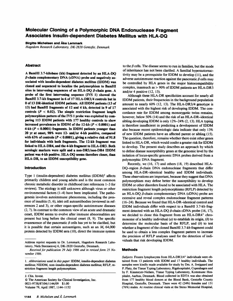
Gentofte, Denmark, 10-20 ml of blood, collected in the presence ofheparin, was obtained at random from 113 patients, 38 (34%) females,75 (66%) males, with ages varying between 2 and 66 yr at onset of IDDM(Fig. 1), and also from 46 patients with non-insulin-dependent diabetesmellitus (NIDDM). The age distribution at the time of admittance (Fig.1) was compatible with that previously reported for Denmark (25). Allthe blood samples were obtained during routine visits, and were codedand analyzed in a random fashion. The fragments detected in the RFLPanalysis were scored and recorded without the observer knowing theorigin of the sample.
Cloning of restriction fragment. In our previous family study it wasdemonstrated that the RFLPobserved after hybridization with an HLA-DQE-chain probe was linked to HLA-DR (17) and that the BamHI 3.7-kb fragment appeared on three of six HLA-DR4-containing chromo-somes in healthy individuals, but in 0 of 13 of the diabetic DR4-con-taining chromosomes (17). Weselected one informative Swedish familywithout history of either IDDM or NIDDM to clone a BamHI 3.7-kbfragment from an HLA-DR4-containing chromosome (26). The indi-vidual donating blood for cloning was a healthy HLA-DR2/4, 3.7-kbfragment-positive mother with two daughters. Her husband is HLA-DR3/7 and their two daughters are HLA-DR2/3 and 4/7, respectively.The HLA-DR4/7 daughter also has the BamHI 3.7-kb fragment, whichis therefore present on the HLA-DR4-containing chromosome of themother. Mononuclear cells were obtained from 30 ml of blood by Ficoll-Hypaque gradient centrifugation and the DNAwas extracted as describedbelow. - 50 ,g of DNAwas digested with BamHI and electrophoresedin a 1% agarose gel along with appropriate molecular weight markers.The 3.4-3.8-kb region was sliced from the gel and the DNAfragmentsrecovered by electroelution. This fraction of fragments was ligated intothe BamHI site of pUC8and used to transform Escherichia coli JM10528.Recombinant plasmids were recognized as those giving rise to whitecolonies on L-broth plates with 20 ,g/ml each of isopropyl thiogalacto-side and 5-bromo-4-chloro-3-indolyl-,8-D-galactopyranoside, and werescreened for HLA-DQ-related sequences by in situ hybridization onnitrocellulose filters with the nick-translated DQB8cDNAprobe (26).
DNApreparation. Mononuclear cells, obtained from 10 ml of bloodby Ficoll-Hypaque gradient centrifigation, were digested overnight at370C in 0.02% proteinase K and 1%b (wt/vol) SDS in 10 mMTris-HC1buffer (pH 7.4) containing 1 mMEDTA. After phenol and chloroformextractions, the DNAwas precipitated with ethanol and resuspended inTris-EDTA buffer.
Genomic blots. Lymphocyte DNA(10-20 pg) was digested with re-striction enzymes according to the suppliers (Boehringer, Mannheim,Federal Republic of Germany) specifications, electrophoresed overnightat 40 V in 196 flatbed agarose gels, and then transferred to HybondNnylon membranes (Amersham International, Buckinghamshire, UnitedKingdom) using the methods described by the supplier.
20-
16-
14-C0 Figure 1. Age variationZ 12 among the 113 IDDM0 lo- _ _ patients studied for
RFLP polymorphismE B- with the HLA-DQ f-Z 6- chain IVS 1 gene probe.
The dotted bars repre-4- rsent the 39 (35%) pa-2- tients who were positive
F1 L L----X21for the 12- and 4-kbcOr.JAd BaomHI restriction frag-
,rtoV-+'TWSments detected with theAge at onset (years ) IVS 1 gene probe.
Hybridizations. Filters were prehybridized for 2-16 h in 50% form-amide, 5 X standard saline citrate (SSC), 5 X Denharts solution, 50 mMNa2PO4 (pH 6.5), and 0.5 mg/ml denatured salmon sperm DNA. Hy-bridization was performed overnight in 50% formamide, 1 X Denhartssolution, 20 mMNa2PO4 (pH 6.5), 10% dextran sulphate, 0.2 mg/mldenatured salmon sperm DNA, and 106 dpm/ml denatured probe.Washing stringency was 0.1 X SSCat 550C.
Probes. The DQjP cDNAprobe was derived from the plasmid pII-,1 (27) by digestion with PstI and EcoRl, after which the 800-base pair(bp) fiagment was eluted from agarose melting at low temperature(BioRad Laboratories, Richmond, CA).
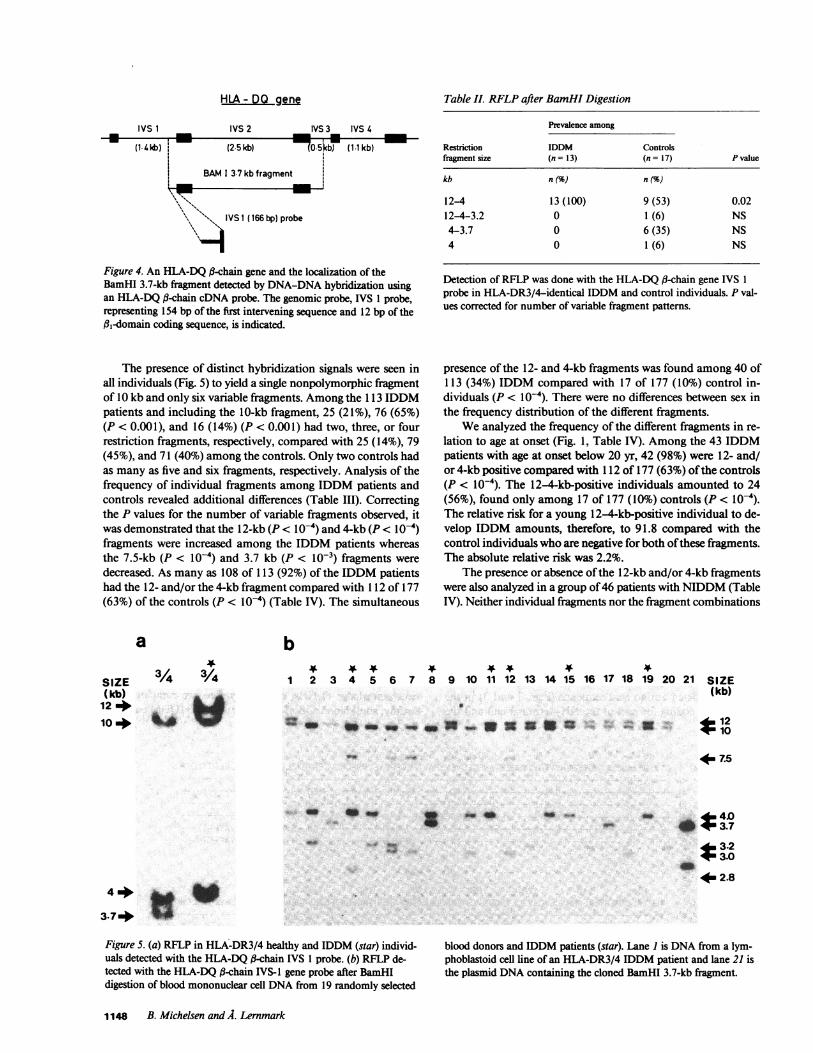
A subclone of the BamHI 3.7-kb fiagment cloned in pBR322 wasused to construct the first intervening sequence (IVS 1) probe. The re-combinant plasmid was digested with the endonuclease RSA1 to yielda fragment, which contains 154 bp of intervening sequence and 12 bpof the second exon which codes for the first domain of the HLA-DQ#-chain, as well as the portion of pBR322 that ranges from the BamHIsite in position 375 to the RSA1 site in position 2281. This 2.072-bpfragment was purified after agarose gel electrophoresis in a low-temper-ature melting-point agarose. Labeling of probes was by nick-translation,using a-[32P]deoxy CTIP (Amersham International) at 30 TBq/mmol(DQf probe) or 110 TBq/mmol (IVS 1 probe).
Construction of a series of progressive deletions for sequencing. TheBamHI 3.7-kb genomic insert was subcloned in the BamHI site of pUCl9(28). This construct was then linearized with the restriction enzymesSphl and HindII. Sphl digestion between the BamHI site in pUCl9 andthe attachment site for the reverse-sequencing primer (Amersham In-ternational) was carried out to leave a four-base 3' overhang, which isinaccessible to Exonuclease m1 (29) (Pharmacia PL Biochemicals, Upp-sala, Sweden). HindU digestion within the polylinker sequence of pUCl9,between the Sphl site and the BamHI cloning site, was used to generatea blunt end, susceptible to Exonuclease HI. There were no sites for Sphland HindU in the insert Unidirectional deletions, ranging from the primerattachment site in pUC19 to various points within the insert, were isolatedby digestion of the plasmid DNAwith Sphl and HindU, followed byphenol/chloroform extraction and ethanol precipitation. The linearizedplasmid DNAwas resuspended to 0.1 g/liter in 6.6 mMTris-HC1 (pH7.4) containing 6.6 mMMgCl2 and acubated at 370C for 5 min beforeExonuclease III was added to a final concentration of 10 U/Mi. In 15-sintervals, 35 10-Ml& aliquots were transferred to 30 ;d of 0.2 MNaClcontaining 5 mMEDTAand heated to 70'C for 10 min to inactivatethe enzyme. The DNAwas precipitated by the addition of 120 jul ofethanol and centrifuged and each pellet was resuspended in 80 ul of 0.05MNaOAc (pH 4.5) containing 0.1 MNaG, 30 mMZnSO4, and 75Vogt U/ml Si nuclease (Boehringer) to remove by incubation for 30min at room temperature the single-stranded 3'- and Y-end protutionsgenerated by Sphl and Exonuclease III, respectively. The reaction wasterminated by phenol/chloroform extraction followed by ethanol pre-cipitation. The pellets were resuspended in 10 1d 25 mMTris-HC1 (pH7.4) containing 5 mMMgC12, 5 mMdithiothreitol (DTT), 0.25 mMspermidine, I mMATP, 10 Mg/ml BSA, and 700 U/ml T4 DNAligase(Amersham), and ligated at room temperature overnight. E. coli KM109was transformed with 5 Ad of each of the ligated fractions according toHanahan (30) and the bacterial cells spread on L-broth plates containing50 Ag/ml ampicillin. About 10-20 transformants from each aliquot wereselected for characterization of deletion size by agarose gel electrophoresis.Each clone was grown overnight in a 5-ml L-broth culture in the presenceof 50 Mg/ml ampicillin. Small-scale plasmid preparations were made bythe alkaline lysis method (31), with the following modifications. Afterprecipitation with potassium acetate, supernatants were centrifuged for2 min, transferred to fresh tubes containing 0.6 vol of isopropanol, andcentrifuged for 5 min at 40C. The pellets were washed with 80% ethanoland resuspended in Tris-EDTA buffer. An equal volume of 4 MLiClwas added. After 5 min at 00C, the precipitates were collected by cen-
trifugation for 2 min. The supernatants were incubated for 30 min at371C with 25 Mg/ml RNase A, followed by phenol/chloroform extraction.The plasmid DNAwas then precipitated by incubating the samples for10 min at 00C in 2.5 vol of ethanol. After centrifugation, pellets were
HLA-DQ and Diabetes Susceptibility 1145
washed once each in 80% and 99% ethanol, air dried, and resuspendedin Tris-EDTA buffer. The deletion breakpoints were estimated by agarosegel electrophoresis of appropriate restriction digest of the plasmid DNA.This analysis made it possible to decide which clones should be furthercharacterized by sequencing.
Sequencing. Selected clones from the preparations described abovewere sequenced by the chain termination method, using double-stranded,supercoiled plasmid DNA(32). Most clones from each aliquot had dele-tion breakpoints positioned within a range of 50-100 bp. The distancebetween fractions was roughly 100-200 bp.
Statistical evaluation. The difference in frequency between controland test samples was estimated by the Fisher exact test or the x2 testwith Yate's correction. The level of significance was accepted to be P
< 0.05 after the P value was corrected for the number of variable frag-ments observed between individuals. The relative risk (RR) was calculatedfrom the formula. RR= (positive patients X negative controls)/(negativepatients X positive controls), and the absolute relative risk (ARR) fromARR = (positive patients X total controls)/(positive controls X totalpatients) X prevalence of IDDM. The prevalence of IDDM in Denmarkwas 0.38% (25).
Results
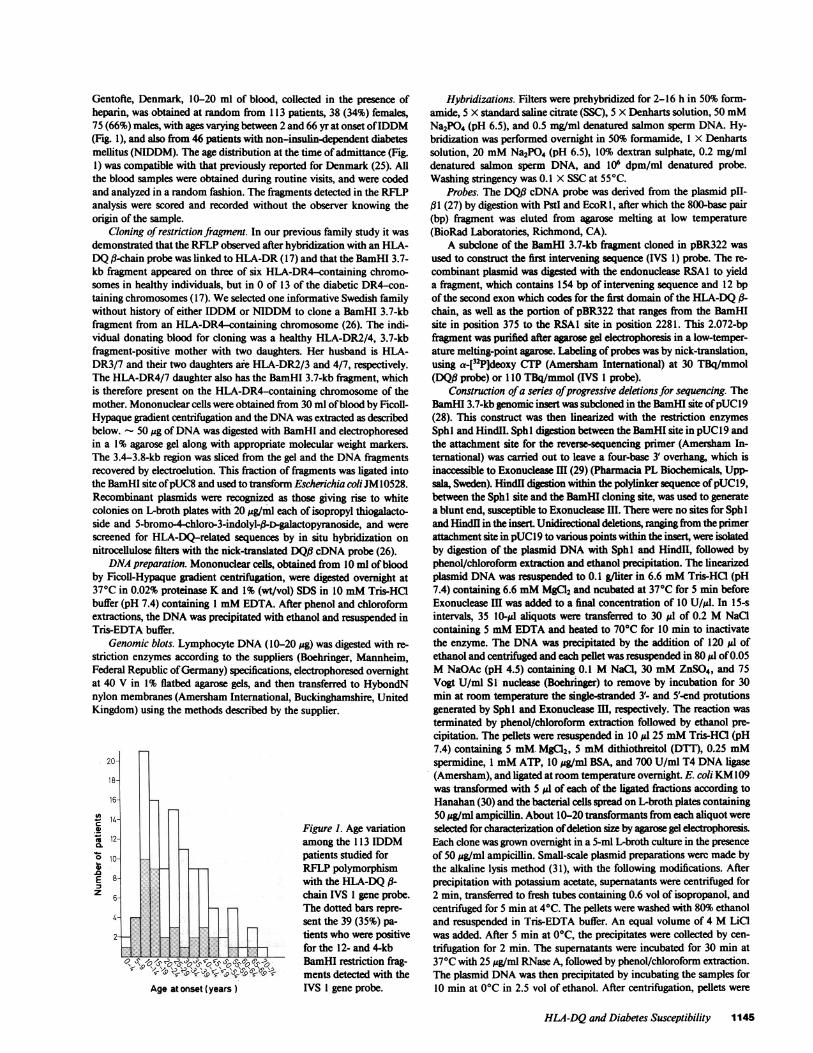
Sequence of the polymorphic BamHI 3.7-kbfragment. The entirenucleotide sequence of the cloned 3.7-kb fragment from an HLA-
Asp Phe Vai Tyr Gin Phe Lys Ala Net Cys TyrGGCCGGGCTG6GGCCGGGCCGGGGCCGGACT6ACCGGCCGGTGATTCCCC6CAGA6 6AT TTC GT6 TAC CA6 TTT AA6 6CC AT6 TGC TAC
Phe Thr Asn 61y Thr Giu Arg Val Arg Tyr Val Thr Arg Tyr Ii. Tyr Asn Arg 61u 61u Tyr Aia Arg Phe Asp SerTTC ACC AAC 666 AC6 6A6 C6C 6T6 C6T TAT 6T6 ACC AGA TAC ATC TAT AAC CA 6A6 6A6 TAC 6CA C6C TTC 6AC A6C
Asp Val 6u Val Tyr Arg Ala Val Thr Pro Lou 61y Pro Pro Asp Ala 61u Tyr Trp Asn Sir 61n Lys 61u Vai LouGAC 6TG GA6 6T6 TAC C66 6C6 6T6 ACG CC6 CT6 666 CC6 CCT 6AC 6CC 66 TAC T66 AMC A6C CA6 AA6 6M 6TC CTG
Giu Arg Thr Arg Ala 61u Lou Asp Thr Val Cys Arg His Asn Tyr Gin Lou Giu Lou Arg Thr Thr Liu Gin Arg ArgGAG AGG ACC CBS GCG GAG TTG GAC ACG GTG TGC AGA CAC AMC TAC CAG TTG GAG CTC CGC ACG ACC TTG CAG AGG CGAVB TGG BCMTC6TAUTCCCCACT6CBGTM CCCACTCTTACCTGG6CCCTASTTCTGCGGAGCTACTTTG6ACGA6GAGTTCTAATTTCTTTAACCTAGGCCGCGTTCCTGCCACCCCACTG6ACABTGGTTAMCT6CATM66TC6T666CTGTTMCATAMGTG666CATCACCTAATC6CACATCAGGCGTXACAAGTA6ACTTC6TCAACAGCCTTA TGTTTTATTMACCCTTCACCTTCCCTCTCTATCCACTTGCTTMCTTC6T6AGTMCTCTTAT6C6M6CCTCCTCMTCTTTTGCCiTT6CtAA6CA6TCCTCTCT6CCCCCAAMCT6CCCTCTTCCCCT6CCC6Cr--CC6CC6CTA6CACTGCCCCACCCA6CAA66TCCAC6T6CACTC6TC6CTCCACAM"GTCTGAGATTAACCTGTTGCTGTTAAAACTACTCCACAACT6TGCMCATGTCATCCAGCAGTTACAMTT6TCATTAAMATATGMCACTTTTCACTTCAAATTATTATTCATCGTAATTCCATTTTCTTAMGT66CTCTCATTCATAACMATGCTCMSAGGT6ACTTTTGCTAGTCATCCCAT66CCCCTACCTCACTATTCACT6CASTGTATATAACCTCATTAATCTTM TGTTAAATTAGTAGATAAAT6TSCTCAMCTGCCAATCCACTTA66CTATGTTTGTCA6ATMTATCGCASTTGCCTTGCTACTTAAATTAGCATCATTtCATATATCCTTAATTAGTACASTAMACTCTTTTMTCACATCAAMTACTACAAA6TCACT CTCCMGGTCTCCACTATAMTGGACMTGAATACTTTCAAGCAGTTGAAATTAMCTTATTMTCTSCATTTAT6ATCAT6AATTTTT CTTA PTTTC GT TACATA6AATTTATTACATTATTCTATAAAATATTGTCACCTCTTTT66CCTA66TTCTCCCCTCCCCCCATCACTATCCACTT CA TAACATTCTTAATSTTAATGGACCCATATAGTAGACACTtAMCTAAGTTCCCTTMGATGMACCATCTAT6TTATGTCAMAATCTCSATATTTCCTCTACATTAATGATTA6CATCACCACMATATATCCTMAATATTACTACAT6ATGAAACGMATGAGTCAGCCCACAC6TACTCACACCBACCTTGCCTGCGAAA6TACACAM TTAAAAGAATGAAAACACATCTCTAMAMGNXAASGCACAT6CCTAT6TTACCCASTTTTACCATM MMTCTCTTCCTAATTTGGCTCCAT6TTGATMGACAATATACMCAGTTATMGGM ATTGTTCATTCTTTATGATTATTCAATTTATGTTCCAATAGMATTCACMGACMCAGMACMATATCTTTCCATTCTGTTCACTCACATCATACTAG66CAATACTTATATTM IA MCA6TTCTAMCCATAAAAMI I ITTATCATCTTTTCTCATAAAMT6CCCTCTAI TTTITACTCCCAATCT6T6TAA6ATSAACAATCTTATAACCACATA6CT6AC'T6T6AMCA66T66ACTCCA 6BUC-AAGAACMCA6TCTTMAAu6ATSACATCTT6TAA6TC CAATBT ATT6TCABOCSACTCCACTCA66USCC66TTCAA6TCAAC--AA6CA6 AC66TATCTCTTC66AT6AT66CTCATSAStmTW.A ATT6B66T CCACCT6CT6TCCTCA6CAATCCCA6CTATAT6TATATOTCSCATTACA66CTCATTAACCTA66CT6ACCTCT6CAA6^TCTCA6AATATMTCTACA6^AG"CATACAT6ATAATATCT6ATTTTA66CAAAATAATTCTCAATA6CAA666AAT66A6TA666TA6ACA6CTA6TAATTAACTCACTtT6TBttT TAAAAATTAS66A6AA AAT6 1 6CATATATATAU G ACATTAATAMACTATA6TTTTACACTASSATAAAS6TAAATGT66CCT66 AAA6TAA6AT6AT6A^ATST6C6AAA6AT6T6TCATTTTTTTTACTAT6A6CA6CAATCT6^A^^AASTAAAAATC6A^6TtAT66CA6ACATSATSM6ATCA6T6TTAMSTTTTCAA66CCTCCTACIIII CAA~mCATTACAAC I I II SAATCACATTCTTMTTt6MGCT6TCT6TTACTA6ATCGCACATTCTSTAAA66CA666ACCAT66TAT6TTMATCM6ATTCTCAST6ATTSTCATAMATSM6TSAT6SATCTTAATCC AASACTT GGGCTCCA66TATCMCCATTCT66TTCCAA66A666NCCTTCCTACA6CA66CCT6C
aI Giu Pro Thr Val Thr Ile Ser Pro Ser Arg ThrTGTGTU6TCTCACATCTCACTCCTATATCTTTCCCTGTCTGTTACTGCCCTCAGTS GAG CCC ACA GTG ACC ATC TCC CCA TCC AGG ACA
G1u Au Lou AMn His His Asn Lou Lou Val Cys Ser Val Thr Asp Phe Tyr Pro Ala Gin Ile Lys Val Arg Trp PheGAG 6CC CTC AAC CACCAC AAC CTG CTG GTC TGC TCA GTG ACA GAT TTC TAT CCA 6CC CAG ATC AAA GTC CGG TGG m
Arg Asn Asp Gin Giu Giu Thr Thr Gly Val Val Ser Thr Pro Lou IIl Arg sn Giy Asp Trp Thr Phe Gin Ile LouCUS AMT GAC CAG GAG GAS ACA ACC GGC GTT GTG TCC ACC CCC CTT ATT AGG MCGOT GAC TGG ACC TTC CAG ATC CTG
Val Met Lou Giu Met Ser Pro Gin His Gly Asp Val Tyr Thr Cys His Val Giu His Pro Ser Lou Gin Asn Pro IleGTG ATG CTG GS ATG TCT CCC CAG CAT GGA SAC GTC TAC ACC TGC CAC GT6 GAG CAC CCC AGC CTC CAG AAC CCC ATC
Thr Val Giu TrpACC GTG GAG TUO C GTMAA666TATTAGTMCTGTTACTATGCCCCACAAGCAAAGSGCASAGCTCATTCTGACCCATCCCTCCATCTCTTATCCCTGAT6TCACTACTGAGCTG6TMCACAGAACTAGAGCACTTCTTCCTCCATGGCAASTGCATCAGAAACCTTATCTCATCACCTTTCCAGATGCTAGGGATTACTCTACATACTGMCTCTGGATCC
t00
16189
42267
68345
94423
525628731834937
1040114312461349145215551658176118641967207021732276237924822585268279128942997
1063096
1323164
1563242
1843320
18341835213558
Figure 2. The nucleotide sequence of the cloned BamHI 3.7-kb DNAendonuclease fragment from an HLA-DR4-containing chromosome of ahealthy individual.
1146 B. Michelsen and A. Lernmark
SBATCCCASSTCTSCASCSCSAGSCACGGSCCSGCSSSAACTSGAGSTCGCGCGGSCGGTTCCACAGCTCC666MG667CASSSCSSCSSCTSCSSSST
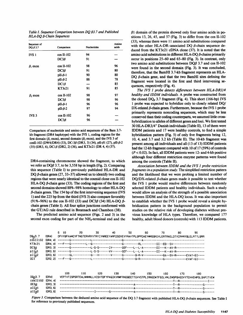
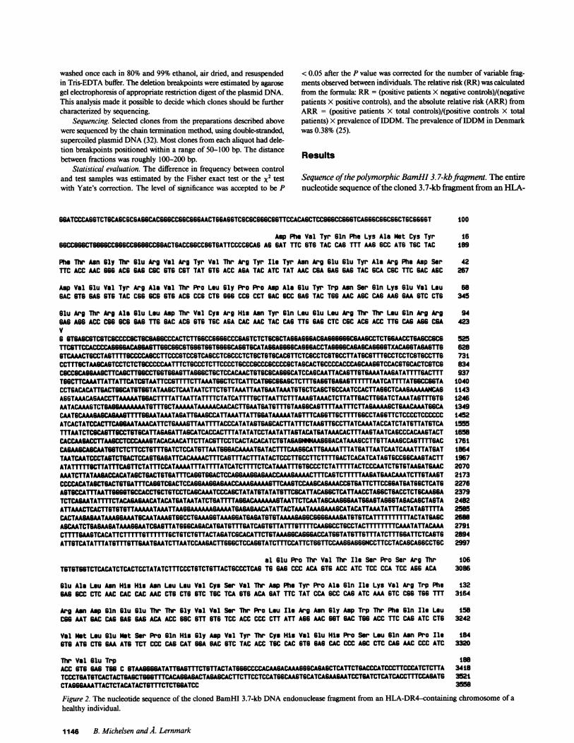
Table L Sequence Comparison between DQ133.7 and PublishedHLA-DQA-Chain Sequences
Sequence of AminoDQ, 3.7 Comparison Nucleotides acids
IVS I cos II-102 94DC3fl 91
1,3 exon cos II-102 98 96DC3ft 91 80p11-0-i 90 80pII-fl-2 88 78DC1,- 85KT3c21 91 85
32 exon cos II-102 98 97DC3fl 98 96p11-13-i 96 96pII-fl-2 97 94
IVS 3 cos II-102 96DC3fl 96
Comparison of nucleotide and amino acid sequences of the Bam 3.7-kb fragment (DR4 haplotype) with the IVS 1, coding regions for thefirst domain (AI exon), second domain (12 exon), and the IVS 3 ofcosIl- 102 (DW4/DR4) (33), DC-3fl (DR3, 3) (34), pII-ft 1 (27), pII-ft-2(35) (DR3, 6), DC1I3 (DR2, 2) (36), and KT3c2l (DR 4, 4) (37).
DR4-containing chromosome showed the fragment, to whichwe refer as DQ13 3.7, to be 3,558 bp in length (Fig. 2). Comparingthis sequence (Table I) to previously published HLA-DR andDQ#-chain genes (27, 33-37) allowed us to identify two codingregions that were nearly identical to the cosmid clone cos II-102HLA-DQ 1-chain gene (33). The coding regions of the first andsecond domains showed 88%-98% homology to other HLA-DQ,8-chain genes. The 154 bp of the first intervening sequence (IVS1) and the 225 bp from the third (IVS 3) also compare favorably(91%-96%) to the cos 11-102 (33) and DC313 (34) HLA-DQ 13-chain genes (Table I). All four splice junctions conformed withthe GT/AG rule described in Breatnach and Chambon (38).
The predicted amino acid sequence (Figs. 2 and 3) in thesecond exon coding for part of the NH2-terminal end and the
0013.7cosIIl02KT3c2IDC3gpi1otpII2DCI
(OR4)(OR4. 4)(0R4. 4)(OR3, 3)(0R3. 6)(DR3. 6)(DR2. 2)
D013.7 (DR4)cosII102 (R4. 4)
6 10
131 domain of the protein showed only four amino acids in po-sitions 13, 26, 45, and 57 (Fig. 3) to differ from the cos 11-102(33), whereas there were 11 amino acid substitutions comparedwith the other HLA-DR-associated DQ1-chain sequence de-duced from the KT3c2l cDNAclone (37). It is noted that theamino acid substitutions in different HLA-DQ13-chains primarilyoccur in positions 25-60 and 65-80 (Fig. 3). In contrast, onlytwo amino acid substitutions between DQ13 3.7 and cos II-102were found in the second domain (Fig. 3). It was concluded,therefore, that the BamHI 3.7-kb fiagment represents an HLA-DQ1-chain gene, and that the two BamHI sites defining thefiagment were located in the first and third intervening se-quences, respectively (Fig. 4).
The IVS I probe detects differences between HLA-DR3/4healthy and IDDM individuals. A probe was constructed fromthe cloned DQ#3.7 fragment (Fig. 4). This short (166-bp) IVS1 probe was expected to hybridize only to closely related DQ/DX-related 13-chain genes. Furthermore, because the IVS 1 probeprimarily represents noncoding sequences, which may be lessconserved than their coding counterparts, we assumed little cross-hybridization to alleles of different genes and loci. Wefirst tested30 HLA-DR3/4' Danish individuals (Table II), 13 of them wereIDDM patients and 17 were healthy controls, to find a simplehybridization pattern (Fig. 5) of only five fragments being 12,10, 4, and 3.7 and 3.2 kb (Table II). The 10-kb fiagment waspresent among all individuals and all (13 of 13) IDDMpatientshad the 12-kb fiagment compared with 10 of 17 (59%) of controls(P < 0.02). In fact, all IDDMpatients were 12- and 4-kb positivealthough four different restriction enzyme patterns were foundamong the controls (Table II).
Association between IDDMand the IVS 1 probe restrictionfragments in a population study. The simplified restriction patternand the likelihood that we were probing a limited number ofDQ/DX-related 1-chain genes made it possible to test whetherthe IVS 1 probe would resolve differences between randomlyselected IDDM patients and healthy individuals. Such a studywould allow an analysis of the strength of a possible associationbetween IDDM and the HLA-DQ locus. It was also importantto establish whether the IVS 1 probe would reveal a simple hy-bridization pattern in the background population to permitstudies on the relative risk of developing diabetes without pre-vious knowledge of HLA types. Therefore, we compared 177healthy, adult blood donors (controls) with 1 3 IDDMpatients.
20 30 40 50 60 70 80 90OFVYOFKAMCYFTNGTERVRYVTRYIYNREEYARFOSOVEVYRAVTPLGPPOAEYWNSOKEVLERTRAELDTVCRHNYQLELRTTLORR-------G------------L------------------G-----------A----------------------------------------F---G---------L--G------------------G--------RL---------DI--EO--SV--------------------------G------------L-S-S------IV------GEF----L--L-A--------OI---K--AV-R------------------------G------------L-S-S------VV------GEF----L--L-A--------DI---K--AV-R------------------------GL-----------G---H-------V------G-------G-R-V------------GA--SV-R------EVAY-GI-------F---G------------L------------------G-------G-R--------------G-------------EVAF-GI----
100 110 120 130 140 150 160 170 180VEPTVTISPSRTEALNHHNLLVCSVTDFYPAOIKVRWFRNOOEETTGVVSTPLIRNGOWTFOILVMLEMSPQHGDVYTCHVEHPSLONPITVEW---------------------------------------------------------------------T--R-----------------I---
0C31 (DR3.3) ---------------------------------------------A-----------------------T--R--------------S------Pl3 (DR3.6) ---------------------------------------------A-----------------------T--R--------------S------p112 (DR3.6) ---------------------I--------S--------------A-----------------------T--R--------------S------
Figure 3. Comparison between the deduced amino acid sequence of the DQ3.7 fragment with published HLA-DQ 13-chain sequences. See Table Ifor reference to previously published sequences.
HLA-DQand Diabetes Susceptibility 1147
HLA -DQ gene
WVS I
(1 4kb)
II
IVS2 IVS3 IVS4
(2 5 kb) (05b)(7 1 1 kb)
BAM I 3.7 kb fragment |_II
%\, \s I VS 1(66 bp) probe
Figure 4. An HLA-DQ a-chain gene and the localization of theBamHI 3.7-kb fragment detected by DNA-DNAhybridization usingan HLA-DQU-chain cDNAprobe. The genomic probe, IVS 1 probe,representing 154 bp of the first intervening sequence and 12 bp of the#,-domain coding sequence, is indicated.
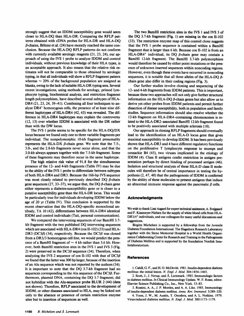
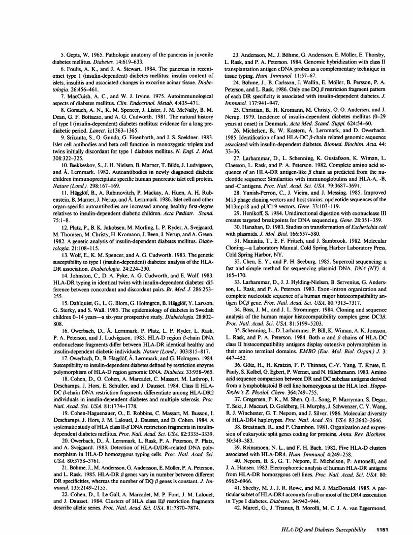
The presence of distinct hybridization signals were seen inall individuals (Fig. 5) to yield a single nonpolymorphic fragmentof 10 kb and only six variable fragments. Amongthe 1 3 IDDMpatients and including the 10-kb fragment, 25 (21%), 76 (65%)(P < 0.001), and 16 (14%) (P < 0.001) had two, three, or fourrestriction fragments, respectively, compared with 25 (14%), 79(45%), and 71 (40%) among the controls. Only two controls hadas many as five and six fragments, respectively. Analysis of thefrequency of individual fragments among IDDM patients andcontrols revealed additional differences (Table III). Correctingthe P values for the number of variable fragments observed, itwas demonstrated that the 12-kb (P < l0-4) and 4-kb (P < l0-4)fragments were increased among the IDDM patients whereasthe 7.5-kb (P < l0-4) and 3.7 kb (P < 10-3) fragments weredecreased. As many as 108 of 113 (92%) of the IDDM patientshad the 12- and/or the 4-kb fragment compared with 112 of 177(63%) of the controls (P < l0-4) (Table IV). The simultaneous
Table II. RFLPafter BamHI Digestion
Prevalence among
Restriction IDDM Controlsfragment size (n = 13) (n = 17) P value
kb n () n (%
12-4 13 (100) 9 (53) 0.02124-3.2 0 1 (6) NS4-3.7 0 6 (35) NS4 0 1(6) NS
Detection of RFLP was done with the HLA-DQ #-chain gene IVS 1probe in HLA-DR3/4-identical IDDM and control individuals. P val-ues corrected for number of variable fragment patterns.
presence of the 12- and 4-kb fragments was found among 40 of113 (34%) IDDM compared with 17 of 177 (10%) control in-dividuals (P < l0-4). There were no differences between sex inthe frequency distribution of the different fragments.
Weanalyzed the frequency of the different fragments in re-lation to age at onset (Fig. 1, Table IV). Among the 43 IDDMpatients with age at onset below 20 yr, 42 (98%) were 12- and/or 4-kb positive compared with 1 12 of 177 (63%) of the controls(P < 10-4). The 12-4-kb-positive individuals amounted to 24(56%), found only among 17 of 177 (10%) controls (P < 10-4).The relative risk for a young 12-4-kb-positive individual to de-velop IDDM amounts, therefore, to 91.8 compared with thecontrol individuals who are negative for both of these fragments.The absolute relative risk was 2.2%.
The presence or absence of the 12-kb and/or 4-kb fragmentswere also analyzed in a group of 46 patients with NIDDM(TableIV). Neither individual fiagments nor the fiagment combinations
a
SIZE 3/4 /4(kb)124a
10 4 X w
b
1 2 3 4 5 6 7 8 9 10 11 12 13 14 15 16 17 18 19 2' !o 21 SIZE(kb)
12* t10uo _ __ _nomS ." E.
4m7.5
- i .s
Figure 5. (a) RFLP in HLA-DR3/4 healthy and IDDM (star) individ-uals detected with the HLA-DQ a-chain IVS 1 probe. (b) RFLP de-tected with the HLA-DQ a-chain IVS-I gene probe after BamHIdigestion of blood mononuclear cell DNAfrom 19 randomly selected
blood donors and IDDM patients (star). Lane I is DNAfrom a lym-phoblastoid cell line of an HLA-DR3/4 IDDM patient and lane 21 isthe plasmid DNAcontaining the cloned BamHI 3.7-kb fragment.
1148 B. Michelsen and A. Lernmark
3.d
_
4 +
3.74
43.2_ +3.0
4m2.8
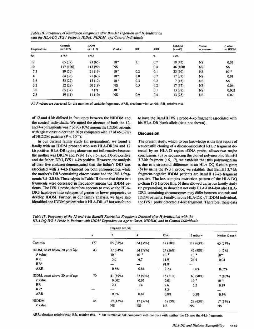
Table III. Frequency of Restriction Fragments after BamHI Digestion and Hybridizationwith the HLA-DQ IVS I Probe in IDDM, NIDDM, and Control Individuals
Controls IDDM NIDDM P value P valueFragment size (n =177) (n = 113) P value RR ARR (n = 46) vs. controls vs. IDDM
kb n(%) n (%) n(%)
12 65 (37) 73 (65) 10-4 3.1 0.7 19 (42) NS 0.0310 117 (100) 112 (99) NS 0.4 46 (100) NS NS
7.5 89 (50) 21 (19) 10-4 0.2 0.1 23 (50) NS 10-34 64 (36) 71 (63) 10-4 3.0 0.7 17 (37) NS 0.013.6 52 (29) 13 (12) 10-3 0.3 0.2 7 (15) NS NS3.2 52 (29) 20 (18) NS 0.5 0.2 17 (37) NS 0.043.0 65 (37) 7 (7) 10-3 0.1 13 (28) NS 0.0022.8 19(11) 11 (10) NS 0.9 0.4 13(28) NS 0.02
All P values are corrected for the number of variable fragments. ARR, absolute relative risk; RR, relative risk.
of 12 and 4 kb differed in frequency between the NIDDMand to have the BamHI IVS 1 probe 4-kb fiagment associated withthe control individuals. Wenoted the absence of both the 12- his HLA-DR blank allele (data not shown).and 4-kb fragments was 7 of 70 (10%) among the IDDM patientswith age at onset older than 20 yr compared with 17 of 46 (37%) Discussionof NIDDMpatients (P < l0-4).
In our current family study (in preparation), we found a The present study, which to our knowledge is the first report offamily with an IDDM proband who was HLA-DR3/4 and 12 a successful cloning of a disease-associated RFLP fragment de-kb positive. HLA-DR typing alone was not informative because tected by an HLA-D-region cDNA probe, allows two majorthe mother was DR3/4 and IVS-1 12-, 7.5-, and 3.0-kb positive conclusions: (a) by sequencing the cloned polymorphic BamHIand the father, DR3, IVS 1 4-kb positive. However, the analysis 3.7-kb fragment (16, 17), we establish that this polymorphismof their five children demonstrated that the father's DR3 was is due to a structural difference in an HLA-DQ U-chain gene;associated with a 4-kb fragment on both chromosomes while (b) by using the IVS 1 probe, we establish that BamHI 3.7-kbthe mother's DR3-containing chromosome had the IVS 1 frag- fragment-negative IDDM patients are BamHI 12-kb fragmentments 7.5-3.0 kb. The analysis in Table III shows that these two positive. The less complex restriction pattern of the HLA-DQfragments were decreased in frequency among the IDDM pa- (-chain IVS 1 probe (Fig. 5) then allowed us, in our family studytients. The IVS 1 probe therefore appears to resolve the HLA- (in preparation), to show that not only HLA-DR4-but also HLA-DR3 haplotype into subtypes of greater or lower propensity to DR3-containing chromosomes may differ between controls anddevelop IDDM. Further, in our family analysis, we have also IDDMpatients. Finally, in one HLA-DR -/7 IDDM individual,identified one IDDMpatient who is HLA-DR -/7 but was found the IVS 1 probe detected a 4-kb fragment. Therefore, these data
Table IV. Frequency of the 12 and 4-kb BamHI Restriction Fragments Detected after Hybridization with theHLA-DQ IVS I Probe in Patients with IDDMDependent on Age at Onset, NIDDM, and in Control Individuals
Fragment size (kb)
n 12 4 12-4 12 and/or 4 Neither 12 nor 4
Controls 177 65 (37%) 64 (36%) 17 (10%) 112 (63%) 65 (37%)
IDDM, onset below 20 yr of age 43 32 (74%) 34 (79%) 24 (56%) 42 (98%) 1 (2%)P value i0o 10-4 10-4 10-4 10-4RR 5.0 6.7 11.9 24.4 0.04RR* - 91.8ARR 0.8% 0.8% 2.2% 0.6% 0.02%
IDDM, onset above 20 yr of age 70 41 (59%) 37 (53%) 15 (21%) 63 (90%) 7 (10%)P value 0.002 0.02 0.01 10-4 10-4RR 2.4 1.4 2.6 5.2 0.19RR* - 8.2 -ARR 0.6% 0.6% 0.8% 0.5% 0.3%
NIDDM 46 19 (42%) 17 (37%) 6 (13%) 29 (63%) 17 (37%)P value NS NS NS NS NS
HLA-DQand Diabetes Susceptibility 1149
ARR, absolute relative risk; RR, relative risk. * RR is relative risk compared with controls with neither the 12- nor the 4-kb fragments.
strongly suggest that an IDDM susceptibility gene would seemcloser to HLA-DQ than HLA-DR. Comparing the RFLP pat-terns obtained with cDNA probes for HLA-DR and HLA-DQ(-chains, Bohme et al. (24) have recently reached the same con-clusion. Because the HLA-DQ RFLP patterns do not conformwith currently available serologic reagents (21, 23, 24), our ap-proach of using the IVS 1 probe to analyze IDDM and controlindividuals, without previous knowledge of their HLA types, isan acceptable approach. It also means that the relative risk es-timates will not be comparable to those obtained by serologictyping, in that all individuals will show a RFLP fragment patternwhereas - 20% of the background population are assigned asblanks, owing to the lack of suitable HLA-DR typing sera. Severalrecent investigations, using methods for serology, primed lym-phocyte typing, biochemical analysis, and restriction fragmentlength polymorphism, have described several subtypes of HLA-DR4 (21, 23, 24, 39-41). Combining all four techniques to an-alyze DR4' homozygous cells, the presence of at least nine dif-ferent haplotypes of HLA-DR4 (42, 43) was revealed. The dif-ference in HLA-DR4 haplotypes may explain the controversy(12, 13) over whether IDDM is associated with the DRratherthan with the DWlocus.
The IVS 1 probe seems to be specific for the HLA-DQ/DXlocus because we found only one to three variable fragments perindividual. The nonpolymorphic 10-kb fragment presumablyrepresents the HLA-DX 3-chain gene, Wenote that the 7.5-,3.0-, and the 2.8-kb fragments never occur alone, and that the3.0-kb always appears together with the 7.5-kb fragment. Someof these fragments may therefore occur in the same haplotype.
The high relative risk value of 91.8 for the simultaneouspresence of the 12- and 4-kb fragments (Table IV) may be dueto the ability of the IVS 1 probe to differentiate between subtypesof both HLA-DR4 and DR3. Because the 166-bp IVS sequencewas most closely related to previously described DQ,8-chaingene sequences (27, 33-37), we argue that, the DQ,3-chain geneeither represents a diabetes-susceptibility gene or is closer to aputative susceptibility gene than the HLA-DR locus. This wouldbe particularly true for individuals developing IDDM below theage of 20 yr (Table IV). This conclusion is supported by therecent observation that the HLA-DQ-specific monoclonal an-tibody, TA 10 (42), differentiates between HLA-DR4 identicalIDDM and control individuals (Tait, personal communication).
Wecompared the intervening sequences of our BamHI 3.7-kb fragment with the two published DQintervening sequenceswhich are associated with HLA-DR4 (cos II-102) (33) and HLA-DR3 (DC3(3) (34), respectively. Because the DC3(3 was clonedfrom a DR3/3 homozygous cell line, we would predict the pres-ence of a BamHI fragment of - 4 kb rather than 3.6 kb. How-ever, both BamHI restriction sites in the IVS 1 and IVS 3 (Fig.2) were preserved in the DC3(3 sequence (34). Therefore, whenanalyzing the IVS 2 sequence of cos II-102 with that of DC3(3we found that the latter was 300 bp larger, because of the insertionof an Alu sequence which was not reported by the authors (34).It is important to note that the DQ 3.7-kb fragment had nosequences corresponding to the Alu sequence of the DC3(3. Fur-thermore, plasmid DNA, containing the DQ3.7 fragment, didnot hybridize with the Alu-sequence probe BLUR2 (44) (datanot shown). Therefore, RFLP associated to the development ofIDDM, or other diseases associated to HLA, may be linked notonly to the absence or presence of certain restriction enzymesites but to insertion of sequences as well.
The two BamHI restriction sites in the IVS 1 and IVS 3 ofthe DQ3.7-kb fragment (Fig. 1) are missing in the cos 11-102(33). The restriction enzyme map of this cosmid clone suggeststhat the IVS 1 probe sequence is contained within a BamHIfragment that is larger than 6 kb. Because cos II- 102 is from anHLA-DR4' individual, its DQ (-chain gene may contain aBamHI 12-kb fragment. The BamHI 3.7-kb polymorphismwould therefore be caused by either point mutations or the pres-ence of unknown inserted sequences within noncoding regions.However, even though these events have occurred in noncodingsequences, it is notable that all three alleles of the HLA-DQ (3-chain gene also differ in their coding regions (Fig. 3).
Our further studies involve cloning and sequencing of the12- and 4-kb fragments from IDDMpatients. This is important,because these two approaches will not only give further structuralinformation on the HLA-DQ,8-chain genes but also allow us toderive yet other probes from IDDMpatients and permit furtherdissection of disease susceptibility, both in population and familystudies. Sequence information should also resolve whether the12-kb fragment on HLA-DR4-containing chromosomes is re-lated to the HLA-DR2-associated BamHI 12-kb fragment foundto be positively associated with multiple sclerosis (18).
Our approach in cloning RFLP fragments should eventuallylead to the identification of an HLA-D locus gene that givesmaximal susceptibility to develop IDDM. It has been previouslyshown that HLA-DR3 and 4 have different regulatory functionson the proliferative T lymphocyte response to mumps andcoxsackie B4 (45), two viruses implicated in the etiology ofIDDM (4). Class II antigens confer restriction in antigen pre-sentation perhaps by direct binding of processed antigen (46).Isolation and structural analysis of these HLA-D class II mole-cules will therefore be of central importance in testing the hy-pothesis (2, 47, 48) that the pathogenesis of IDDM is conferredby the ability of these molecules to present antigens that inducean abnormal immune response against the pancreatic (3cells.
Acknowledgments
Wewish to thank iss Aagard for expert technical assistance, A. Svejgaardand F. Kissmeyer-Nielsen for the supply of white blood cells from HLA-DR3/4+ individuals, and our colleagues for many useful discussions andsuggestions.
Birgitte Michelsen is supported by a fellowship from the JuvenileDiabetes Foundation International. The Hagedorn Research Laboratorytogether with the Steno Memorial Hospital is a World Health Organi-zation Collaborating Center for Research and Training in the Pathogenesisof Diabetes Mellitus and is supported by the foundation Nordisk Insu-linlaboratorium.
References
1. Cahill, G. F., and H. 0. McDevitt. 1981. Insulin-dependent diabetesmellitus: the initial lesion. N. Engl. J. Med. 304:1454-1465.
2. Scott, J., J. Nerup, and A. Lernmark. 1985. Immunologic factorsin diabetes mellitus. In Clinical Immunology Update. W. F. Rosse, editor.Elsevier Science Publishing Co., Inc., NewYork. 53-85.
3. Rossini, A. A., J. P. Mordes, and A. A. Like. 1985. Immunologyof insulin-dependent diabetes mellitus. Annu. Rev. Immunol. 3:289-320.
4. Yoon, J. W., M. Austin, T. Onodera, and A. L. Notkins. 1979.Virus-induced diabetes mellitus. N. Engl. J. Med. 300:1173-1179.
1150 B. Michelsen and A. Lernmark
5. Gepts, W. 1965. Pathologic anatomy of the pancreas in juvenilediabetes mellitus. Diabetes. 14:619-633.
6. Foulis, A. K., and J. A. Stewart. 1984. The pancreas in recent-onset type I (insulin-dependent) diabetes mellitus: insulin content ofislets, insulitis and associated changes in exocrine acinar tissue. Diabe-tologia. 26:456-46 1.
7. MacCuish, A. C., and W. J. Irvine. 1975. Autoimmunologicalaspects of diabetes mellitus. Clin. Endocrinol. Metab. 4:435-471.
8. Gorsuch, A. N., K. M. Spencer, J. Lister, J. M. McNally, B. M.Dean, G. F. Bottazzo, and A. G. Cudworth. 1981. The natural historyof type I (insulin-dependent) diabetes mellitus: evidence for a long pre-diabetic period. Lancet. ii: 1363-1365.
9. Srikanta, S., 0. Gunda, G. Eisenbarth, and J. S. Soeldner. 1983.Islet cell antibodies and beta cell function in monozygetic triplets andtwins initially discordant for type I diabetes mellitus. N. Engl. J. Med.308:322-325.
10. Bekkeskov, S., J. H. Nielsen, B. Marner, T. Bilde, J. Ludvigsson,and A. Lernmark. 1982. Autoantibodies in newly diagnosed diabeticchildren immunoprecipitate specific human pancreatic islet cell protein.Nature (Lond.). 298:167-169.
11. Hagglof, B., A. Rabinovitch, P. Mackay, A. Huen, A. H. Rub-enstein, B. Marner, J. Nerup, and A. Lernmark. 1986. Islet cell and otherorgan-specific autoantibodies are increased among healthy first-degreerelatives to insulin-dependent diabetic children. Acta Padiatr. Scand.75:1-8.
12. Platz, P., B. K. Jakobsen, M. Morling, L. P. Ryder, A. Svejgaard,M. Thomsen, M. Christy, H. Kromann, J. Been, J. Nerup, and A. Green.1982. A genetic analysis of insulin-dependent diabetes mellitus. Diabe-tologia. 21:108-115.
13. Wolf, E., K. M. Spencer, and A. G. Cudworth. 1983. The geneticsusceptibility to type I (insulin-dependent) diabetes: analysis of the HLA-DRassociation. Diabetologia. 24:224-230.
14. Johnston, C., D. A. Pyke, A. G. Cudworth, and E. Wolf. 1983.HLA-DR typing in identical twins with insulin-dependent diabetes: dif-ference between concordant and discordant pairs. Br. Med. J. 286:253-255.
15. Dahlquist, G., L. G. Blom, G. Holmgren, B. Hagglof, Y. Larsson,G. Sterky, and S. Wall. 1985. The epidemiology of diabetes in Swedishchildren 0-14 years-a six-year prospective study. Diabetologia. 28:802-808.
16. Owerbach, D., A. Lernmark, P. Platz, L. P. Ryder, L. Rask,P. A. Peterson, and J. Ludvigsson. 1983. HLA-D region (B-chain DNAendonuclease fragments differ between HLA-DR identical healthy andinsulin-dependent diabetic individuals. Nature (Lond.). 303:815-817.
17. Owerbach, D., B. Hdgglof, A. Lernmark, and G. Holmgren. 1984.Susceptibility to insulin-dependent diabetes defined by restriction enzymepolymorphism of HLA-D region genomic DNA. Diabetes. 33:958-965.
18. Cohen, D., 0. Cohen, A. Marcadet, C. Massart, M. Lathrop, I.Deschamps, J. Hors, E. Schuller, and J. Dausset. 1984. Class II HLA-DC(-chain DNArestriction fragments differentiate among HLA-DR2individuals in insulin-dependent diabetes and multiple sclerosis. Proc.Natl. Acad. Sci. USA. 81:1774-1778.
19. Cohen-Haguenauer, O., E. Robbins, C. Massart, M. Busson, I.Deschamps, J. Hors, J. M. Lalouel, J. Dausset, and D. Cohen. 1984. Asystematic study of HLA class II-,B DNArestriction fragments in insulin-dependent diabetes mellitus. Proc. Natl. Acad. Sci. USA. 82:3335-3339.
20. Owerbach, D., A. Lernmark, L. Rask, P. A. Peterson, P. Platz,and A. Svejgaard. 1983. Detection of HLA-D/DR-related DNApoly-morphism in HLA-D homozygous typing cells. Proc. Natl. Acad. Sci.USA. 80:3758-3761.
21. Bohme, J., M. Andersson, G. Andersson, E. Moller, P. A. Peterson,and L. Rask. 1985. HLA-DR (3 genes vary in number between differentDRspecificities, whereas the number of DQ,B genes is constant. J. Im-munol. 135:2149-2155.
22. Cohen, D., I. Le Gall, A. Marcadet, M. P. Font, J. M. Lalouel,and J. Dausset. 1984. Clusters of HLA class 11, restriction fragmentsdescribe allelic series. Proc. Nat!. Acad. Sci. USA. 81:7870-7874.
23. Andersson, M., J. Bohme, G. Andersson, E. Moller, E. Thorsby,L. Rask, and P. A. Peterson. 1984. Genomic hybridization with class IItransplantation antigen cDNAprobes as a complementary technique intissue typing. Hum. Immunol. 11:57-67.
24. Bohme, J., B. Carlsson, J. Wallin, E. Moller, B. Persson, P. A.Peterson, and L. Rask. 1986. Only one DQ( restriction fragment patternof each DRspecificity is associated with insulin-dependent diabetes. J.Immunol. 137:941-947.
25. Christian, B., H. Kromann, M. Christy, 0. 0. Andersen, and J.Nerup. 1979. Incidence of insulin-dependent diabetes mellitus (0-29years at onset) in Denmark. Acta Med. Scand. SuppL. 624:54-60.
26. Michelsen, B., W. Kastern, A. Lernmark, and D. Owerbach.1985. Identification of and HLA-DC (3-chain related genomic sequenceassociated with insulin-dependent diabetes. Biomed. Biochim. Acta. 44:33-36.
27. Larhammar, D., L. Schenning, K. Gustafsson, K. Wiman, L.Claesson, L. Rask, and P. A. Peterson. 1982. Complete amino acid se-quence of an HLA-DR antigen-like d chain as predicted from the nu-cleotide sequence: Similarities with immunoglobulins and HLA-A, -B,and -C antigens. Proc. NatL. Acad. Sci. USA. 79:3687-3691.
28. Yanish-Perron, C., J. Vieira, and J. Messing. 1985. ImprovedM13 phage cloning vectors and host strains: nucleotide sequences of theMl3mpl8 and pUCl9 vectors. Gene. 33:103-119.
29. Henikoff, S. 1984. Unidirectional digestion with exonuclease IIIcreates targeted breakpoints for DNAsequencing. Gene. 28:351-359.
30. Hanahan, D. 1983. Studies on transformation of Escherichia coliwith plasmids. J. Mol. Biol. 166:557-580.
31. Maniatis, T., E. F. Fritsch, and J. Sambrook. 1982. MolecularCloning-a Laboratory Manual. Cold Spring Harbor Laboratory Press,Cold Spring Harbor, NY.
32. Chen, E. Y., and P. H. Seeburg. 1985. Supercoil sequencing: afast and simple method for sequencing plasmid DNA. DNA (NY). 4:165-170.
33. Larhammar, D., J. J. Hylding-Nielsen, B. Servenius, G. Anders-son, L. Rask, and P. A. Peterson. 1983. Exon-intron organization andcomplete nucleotide sequence of a human major histocompatibility an-tigen DC(3 gene. Proc. Natl. Acad. Sci. USA. 80:7313-7317.
34. Boss, J. M., and J. L. Strominger. 1984. Cloning and sequenceanalysis of the human major histocompatibility complex gene DC3MB.Proc. Natl. Acad. Sci. USA. 81:5199-5203.
35. Schenning, L., D. Larhammer, P. Bill, K. Wiman, A. K. Jonsson,L. Rask, and P. A. Peterson. 1984. Both a and ,B chains of HLA-DCclass II histocompatibility antigens display extensive polymorphism intheir amino terminal domains. EMBO(Eur. Mol. Biol. Organ.) J. 3:447-452.
36. Gotz, H., H. Kratzin, F. P. Thinnes, C.-Y. Yang, T. Kruse, E.Pauly, S. Kolbel, G. Egbert, P. Wernet, and N. Hilschmann. 1983. Aminoacid sequence comparison between DRand DCsubclass antigens derivedfrom a lymphoblastoid B cell line homozygous at the HLAloci. Hoppe-Seyler's Z. Physiol. Chem. 364:749-755.
37. Gregersen, P. K., M. Shen, Q.-L. Song, P. Marryman, S. Degar,T. Seki, J. Maccari, D. Goldberg, H. Murphy, J. Schwenzer, C. Y. Wang,R. J. Winchester, G. T. Nepom, and J. Silver. 1986. Molecular diversityof HLA-DR4 haplotypes. Proc. Nat!. Acad. Sci. USA. 83:2642-2646.
38. Breatnach, R., and P. Chambon. 1981. Organization and expres-sion of eukaryotic split genes coding for proteins. Annu. Rev. Biochem.50:349-383.
39. Reinsmoen, N. L., and F. H. Bach. 1982. Five HLA-D clustersassociated with HLA-DR4. Hum. Immunol. 4:249-258.
40. Nepom, B. S., G. T. Nepom, E. Michelson, P. Antonelli, andJ. A. Hansen. 1983. Electrophoretic analysis of human HLA-DR antigensfrom HLA-DR homozygous cell lines. Proc. Nat!. Acad. Sci. USA. 80:6962-6966.
41. Sheehy, M. J., J. R. Rowe, and M. J. MacDonald. 1985. A par-ticular subset of HLA-DR4 accounts for all or most of the DR4associationin Type I diabetes. Diabetes. 34;942-944.
42. Marcel, G., J. Titanus, B. Morolli, M. C. J. A. van Eggermond,
HLA-DQand Diabetes Susceptibility 1151
G. M. T. Schreuder, R. R. P. de Vries, and M. J. Giphart. 1986. Dissectionof HLA class II haplotypes in HLA-DR4 homozygous individuals. Im-munogenetics. 23:333-340.
43. Bach, F. H., S. S. Rich, J. Barbosa, and M. Segall. 1985. Insulin-dependent diabetes-associated HLA-D region encoded determinants.Hum. Immunol. 12:59-64.
44. Deininger, P. L., D. J. Jolly, C. M. Rubin, T. Freidmann, andC. W. Schmid. 1981. Base sequence studies of 300 nucleotide renaturedrepeated human DNAclones. J. Mol. Biol. 151:17-33.
45. Bruseud, 0., J. Jervell, and E. Thorsby. 1985. HLA-DR3 and-DR4 control T-1ymphocyte responses to mumpsand Coxsackie B4 virus:
studies on patients with type 1 (insulin-dependent) diabetes and healthysubjects. Diabetologia. 28:420-426.
46. Babbitt, B. P., P. M. Allen, G. Matxueda, E. Habert, and E. R.Unanue. 1985. Binding of immunogenic peptides to Ia histocompatibilitymolecules. Nature (Lond.). 317:359-360.
47. Papadopoulos, G., and A. Lernmark. 1984. The immune responsein individuals with HLA-DR specificities conferring susceptibility to in-sulin-dependent diabetes-a hypothesis. Diabetes Res. 1:3-11.
48. Lernmark, A. 1984. Molecular biology of type 1 (insulindepen-dent diabetes) mellitus. Diabetologia. 2268:195-203.
1152 B. Michelsen and A. Lernmark