Embed Size (px)
Citation preview
Loyola University Chicago Loyola University Chicago
Loyola eCommons Loyola eCommons
Master's Theses Theses and Dissertations
1977
Restriction Endonuclease Analysis of Bacteriophage P1 DNA and Restriction Endonuclease Analysis of Bacteriophage P1 DNA and
Its Derivative Hybrid DNAs Its Derivative Hybrid DNAs
Gregory Alan Schulz Loyola University Chicago
Follow this and additional works at: https://ecommons.luc.edu/luc_theses
Part of the Medical Microbiology Commons
Recommended Citation Recommended Citation Schulz, Gregory Alan, "Restriction Endonuclease Analysis of Bacteriophage P1 DNA and Its Derivative Hybrid DNAs" (1977). Master's Theses. 2905. https://ecommons.luc.edu/luc_theses/2905
This Thesis is brought to you for free and open access by the Theses and Dissertations at Loyola eCommons. It has been accepted for inclusion in Master's Theses by an authorized administrator of Loyola eCommons. For more information, please contact [email protected].
This work is licensed under a Creative Commons Attribution-Noncommercial-No Derivative Works 3.0 License. Copyright © 1977 Gregory Alan Schulz
--
R~STRICTION ENDONUCLEASE ANALYSIS OF
BACTERIOPHAGE PJ DNA AND ITS
DERIVATIVE HYBRID DNAS
by
GreeJory Alan Sc!·~uh
A Thesis Submitted to the ~acu1t:1 0f the Gr-aduate School
of Loyc·la Ul"1ivev·sity of crdc:ago in Partial Fulfillment
of the Requirement5 for the Degr~e of
Masters uf Science
August
1977
'I I
paz
ACKNOWLEDGEMENTS
The author would like to extend sincere thanks to his
advisor, Dr. Marvin Stodolsky, for encouragement~ patience, and
advice provided during the course of this work.
The author would also like to acknowledge the students
and faculty of the Department of Microbiology fat~ advice~ in
struction, and helpful discussion throughout his entire graduate
studies.
i i
--
VITA
The author, Gregory Alan Schulz_ is the son of Robert
William Schulz, Sr., and Stella (Haas) Schulz. He was born
July 24, 1951, in Alton, Illinois.
His elementary education was obta·ined at St. Mary 1 s Grade
School, Alton, Illinois, and h·is secondary education was obtained
at Marquette High School, also in Alton, Illinois.
In September. 1969, he entered Loyola University of
Chicago, and in June, 1973~ received the degrer of Bachelor of
Science with a major in biology.
In \June, 1974, he joined the Department of Microbiology
of Loyola University. He preS('ntly holds the position of Re
search Assistant at Rush-Presbyteri<'ln-St. Lukc 1 s Medical Center,
Chicago, Illinois.
Mr. Schulz is co-author· oii one pub1icat·ion: Integration
Sites of Foreign Genes in the Chromosome of Coliphage Pl: A
Finer Resolution. 1976. Virology }]:299-302.
..
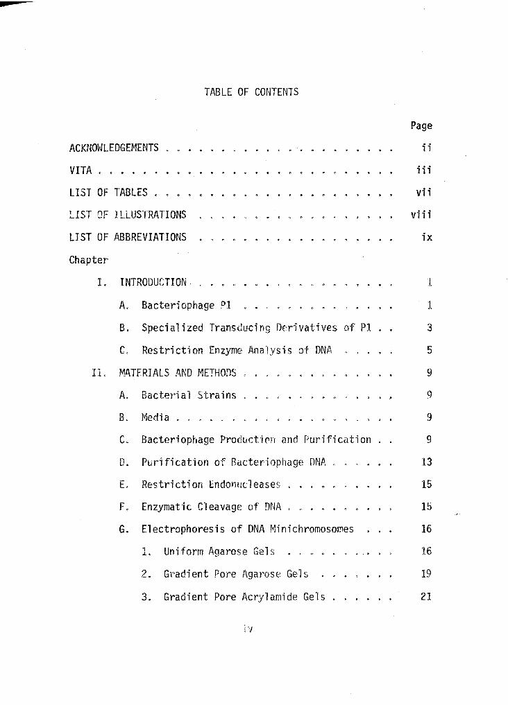
TABLE OF CONTENTS
ACKNmJLEDGEMENTS
VITA • • • • •
LIST OF TABLES •
LIST OF ILLUSTRATIONS
LIST OF ABBREVIATIONS
Chapter,
L
IL
INTRODUCTION . .
A. Bacteriophage Pl
B. Specialized Transducing Derivatives of Pl .
C. Restriction Enzyme J\na·!ysis of DNft,
MATERIALS AND f~ETHODS
A. Bacterial Strains
B. Media ..... .
Page
ii
iii
vii
viii
ix
1
1
3
5
9
9
9
C. Bacteriophage Producti0n and Purification 9
D. Purification of Bacteriophage ONA 13
L
F.
G.
Restriction Endonucleases
Enzymat·i c Cleavage of DNA
Electrophoresis of DNA Minichromosomes
1. Uniform Agarose Gels
2. Gradient Pore Agarose Gels
3. Gradient Pore Acryla~ide Gels
;y
15
15
16
16
19
21
...
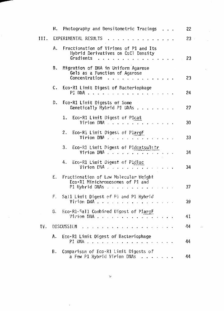
H. Photography and Densitometric TraC"i ngs 22
III. EXPERH1ENTAL RESULTS . . . . . . . . . . 23
A. Fractionation of Virions of Pl and Its Hybrid Derivatives on CsCl Density Gradients . . . . . . . < . . 23
B. Nigration of DNA in Uniform Agarose Gels as a Function of Agarose Concentration . . < . . . . . . . . . 23
c. EcooRI Limit Digest of Bacteriophage Pl DNA . . . . 0 . < . . . ,, . 24
D. Eco•Rl Limit Digests of Some Genetically !lybr·id Pl DNAs . 0 . . ' . 27
1. Eco·Rl limit Digest of Pleat Vil'·i on DNJ.I. ' . . . . . . . . C' 0 . 30
2. Eco•Rl limit Digest of P1_~!11E. Virion DNA . . . . . 33
3. Eco•Rl Limit D·lgest of Pldcatsuls tr Virion DNA ' . . . . . ' . . 34
4. Eco·Rl limit 0·1 gest of Pldlac Virion eN A " . . . 34
L Fractionation of Low Molecular Weight Eco•Rl Minichromosomes of PI and Pl Hybrid DNAs 0 . . . .. . ' 37
F. Sail Limit Digest nf P1 and Pl Hybr·id Vir'i on DNA . . . . . ' . . 0 39
G. Eco·Rl-Sall Combined Digest of PlaroF ... ~-~--
Vi 1'1 on DNA . 41
IV. DISCUSSICN ,, ,, . . 0 ' . . . . ' 44
A. Eco·Rl Limit Digest of Bacteriophage Pl DNA . . . . 0 ' . . . ' . . 44
B. Comparison of ECO"Rl L'imit Digests of a Few Pl Hybrid Virion DNAs . . . . 44
pt
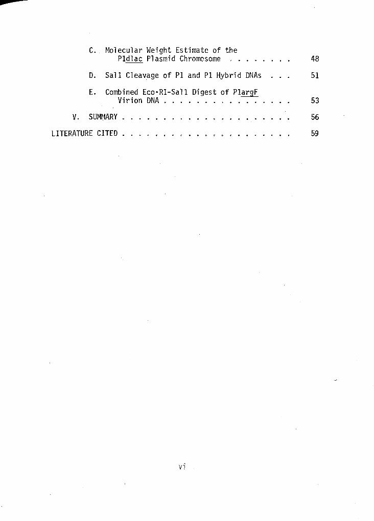
C. Molecular Weight Estimate of the Pldlac Plasmid Chromosome , .
D. Sall Cleavage of PI and Pl Hybrid DNAs
E. Combined Eco·Rl-Sall Digest of PlargF Virion DNA .
V. SUMr:1ARY
LITERATURE CITED
vi
48
51
53
56
59
....
LIST OF TABLES
Table
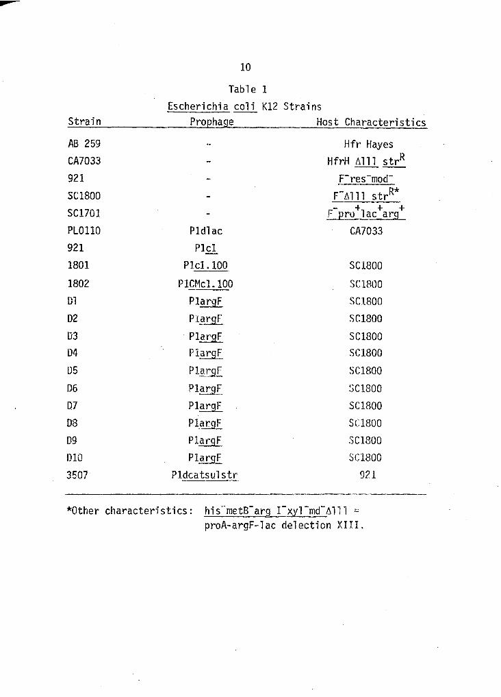
1. Escherichia coli K12 Strains
2. Restriction Endonucleases ..
vii
Page
10
17
p
LIST OF ILLUSTRATIONS
Figure Page
1. Relative Mobility versus Molecular Weight of DNA as a Function of Agarose Concentration . . 25
2. Eco·Rl Pl DNA Limit Digest . . . . . . 28
3. Comparison of the Eco·Rl Cleavage Patterns of Pl and Derivative Hybrid DNAs in Agarose Gels . . . . . . 31
4. Agarose Gel of the Eco•Rl Limit Digest of Three Pl.argF Hybrid DNAs ' . . 35
5. Detection of Low Molecular Weight Minichromosomes from Eco•Rl Digests of Pl and Derivative Hybrid DNAs in Gradient Pore Acrylamide Gels . ,
' ' . 38
6. Gradient Pore Agarose Gel of the Sall Limit Digests of Pl and Derivative Hybrid ONAs . . . . . 40
7. Agarose Gel of a Combined Eco·Rl-Sall Digest of P1~ V·lrion DNA . 43
8. PlargF-PJ Q.lE.c_ DNA Hetel'Odup·l ex . . . ' . ~9
p
cat
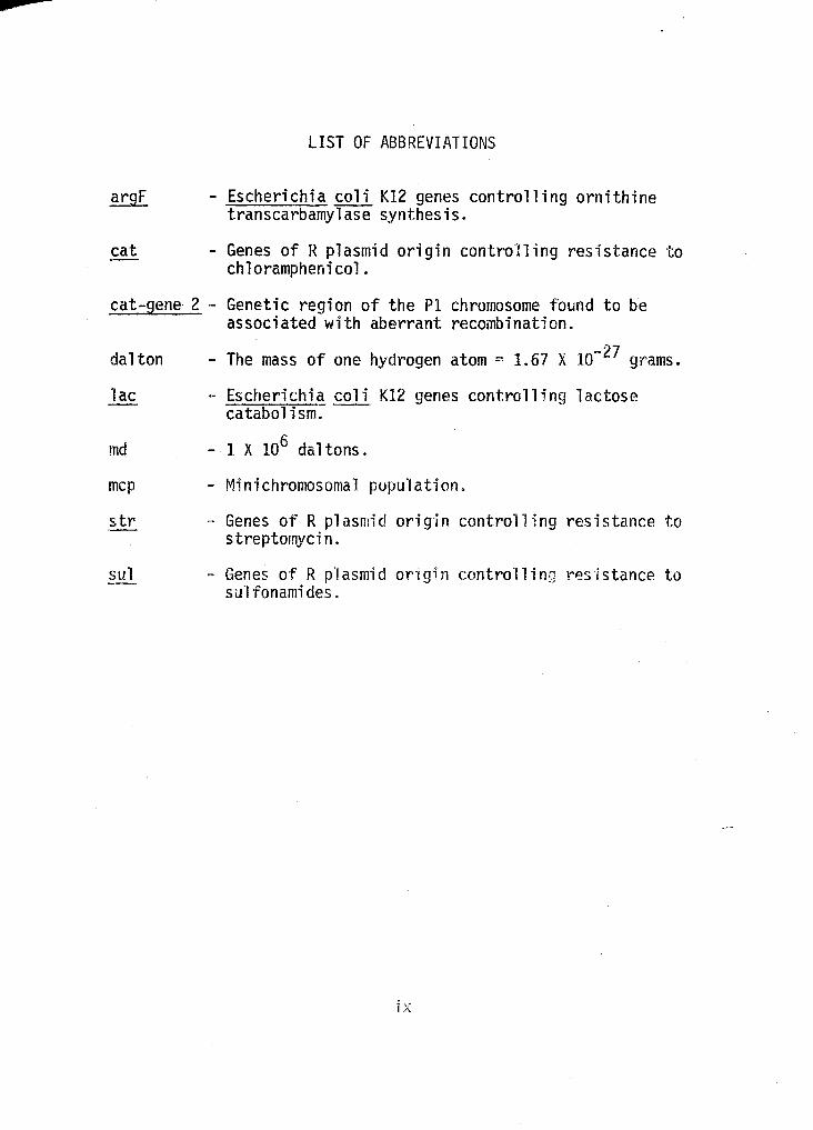
LIST OF ABBREVIATIONS
- Escherichia coli K12 genes controlling ornithine transcarbamylase synthesis.
- Genes of R plasmid origin controlling resistance to chloramphenicol.
cat-gene· 2 - Genetic region of the PI chromosome found to be associated with aberrant recombination.
dalton
lac
md
mcp
str
sul
- The mass of one hydrogen atom = 1.67 X lo- 27 grams.
- Escherichia coli K12 genes controlling lactose catabolism.
- 1 X 10⁶ daltons.
- Minichromosomal population.
- Genes of R plasmid origin controlling resistance to streptomycin.
- Genes of R plasmid on gin controlling resistance to sulfonamides.
ix
•
CHAPTER I
INTRODUCTION
A. Bacteriophage Pl.
Pl is a temperate bacteriophage which lysogenizes a few
coliform bacterial species. Since its discovery and isolation
{4), bacteriophage Pl has proven to be a valuable tool to the
bacterial geneticist because it mediates generalized transduction
(15) ·in which the transducing particles carry a defined length of
deoxyribonucleic acid (DNA) (11). This characteristic has en
abled the use of Pl to define linkage groups within the bacterial
chromosome (12) , 1 ocate ch romosoma 1 I oci of' integra ted prophage
(21), and observe the effect that the presence of foreign genes
has upon replication of the bacterial chromosomes (3).
Most Pl virions (PlB) produced following lytic infection
of the host contain 1inear, duplex DNA having a molecular weight
of 66 X 106 (29). In the iyso9enic state, PJ exhibits several
interesting characteristics not seen in other coliphage, such as
those of the lambdoid group< It exists ·in the cytoplasm of its
host as a circular, duplex DNA molecule of 52 X 106 daltons {11).
There is only one copy of the plasmid per host chromosome, and no
covalent association bet~veen the prophage PI and lts host chromo-
some has been shown (11). The lytic cyc·le of Pl is believed to
i nvo 1 ve the formation of concatomers (end to end ~1enomi c
1
.....
2
oligomers) perhaps proceeding through a rolling circle mechanism
of replication and/or recombination of monomeres. Encapsidation
of phage DNA proceeds through the action of a precessive, volume
dependent, headful packaging system on concatomers. A randomly
permuted collection of phage DNA molecu1es are sequentially en
capsidated into phage head:., each containing more than one unit
phage genome of DNA- This packaging system was first hypothe
sized for bacteriophage T4 by Streisenger et ill· {25) from
purely genetic data. A recombination between the redundant ends
of the virion DNA is believed responsible for converting the
linear, infecting DNA, into circular Pl plasmid DNA. This con··
comitant elimination of genetic redundancy accounts for the lower
molecular weight of the plasmid DNA.
Within a lysate of Pl virions~ there also are a certain
percentage of Pl small capsid morphology variants (PlS). The PlS
comprise about 20% of the total v1rion population, and are w~ll
separated from Pll3 in isopycnic CsCJ density gracdents (respec
tive densities 1.42 and 1.47) (27).
The PI genetic map. constructed through bacter·iophage
crosses, is linear, as opposed to a circular map one would expect
from a purely random, permuted population of virion ONAs (22).
This suggests that the ends of the Pl genetic map are due to the
existence of a high frequency recombinationa1 region known as a
hot spot. The above observations suggest that other systems are
functioning in conjunction with a vo·l ume dependent, headflll
3
chromosome packaging system.
B. Specialized transducing derivatives of Pl.
Another characteristic of bacteriophage Pl is fts
ability to carry out specialized transduction of some genes of
Escherichia coli (I. coli) and R factors. A number of these
specialized transducing derivatives have been isolated. PlCM
(Pleat) was isolated by Kondo and Mitsuhashi (14) during studies
of the ability of phage Pl to transduce genes from a drug resist
ance plasmid. Pleat carries chloramphenicol resistance genes
(cr.1R) s and expresses all normal Pl functions.
The isolation of specialized argF transducing bacterio
phage has been accomplished i~ three laboratories (8, 13, 24}.
These argF derivatives carry a segment of E. coli genes which
control ornithine transcarbamylase synthesis.. The lambdoid and
P22 derivatives are dE:ficient in some bacter·l.ophage functions
and must accordingly be propagated with the assistance of helper
phage. The bacteriophage PlE.t.9£ derivatives were ·isolated
through a one-step transduct·lon procedure: a bacted ophage Pl
mediated low frequency transduction from E. coli K12
argi+pro+arg+lac+ to Plcl.lOO lysogenic I· coli argi.
proA-argF-lac deletion XIII (24), As in the case of Pleat, the
PlargF derivatives thus far iso·lated show no defects in normal
Pl functions. Thus, PlargF is a stable hybrid of bacteriophage
Pl DNA and E. co_1 i _arg~. genes> without a Hering any Pl functions.
4
lida et ~· (10) have been analyzing a family of Pl
derivatives with drug resistance genes from R plasmids and pro
vided us with the lysogen 3507, Pldcatsulstr (PlE~). This
specialized transducing derivative is a hybrid of Pl and genes
from an R plasmid controlling antibiotic resistance to chloram
phenicol, the sulfonamides, and streptomycin.
Of the few specialized transducing derivatives of Pl thus
far isolated, Pldlac has been most extensively investigated.
Luria and co-workers first detected Pldlac upon tr·ansduction of
Shigella dysenteriae lac- with virions produced ·in;_. coli lac+
( 16). The frequency of occurrence of these Pl.QJ ac tt·ansductants
is over one hundred fold less than those produced by simple lac
integration. Once formed, Pldlac. promotes specialized transduc-·
tion of the lactose catabolizing genes at a high frequency.
Further investigation of Pldlac by Rae and Stodolsky (20) pro---...... .
vided genetic evidence that thi) Pldlac_ chromosomes can be too
large to be encapsidated genetically intact. A t~st of this
deduction is included in this thesis.
Mapping data on some Pl specialized trdnsducing deriva-
tives has been analyzed in order to locate the position on the Pl
chromosome where foreign genetic materia 1 is i ntegr·ated. Genetic
crosses have previously shown that the _cat-gen~ region of the
Pl chromosome ·is involved in the aberrant recombinational events
underlying the de .~avo forrrmticn of the qeneti,:ally hybrid,
specialized transducing deY'ivatlVes (26). In each case studied,
5
at least one of the union sites between Pl and foreign gene
sequences is in the cat-gene 2 region.
C. Restriction enzyme analysis of DNA.
The primary analytical tool used was electrophoretic
fractionation of minichromosomal populations (mcp) produced by
restriction endonucleases. This procedure is knmvn as s;leav~
analysis and has enabled the identification of chromosomal alter
ations of many hybrid derivatives of Pl. The isolation and sub
sequent exploitation of restriction enzymes, as an analytical
tool in molecular biology research, has a lengthy history. Ob··
servations made in the early 1950's by Luria and Bertani led to
the development of the primary a:1alytical system used here
(17, 5). They found that systems exist for the destruction of
foreign chromosomes absorbed by bacteria. Basically, these
initial observations showed that the host range of a given bac
teriophage depended in part on t~e bacterial strain on which the
phage had last propagated. This affect was called host con
trolled variation and was shown to be a purely host function,
that is exerted on foreign DNA which has gained entrance to the
bacterial cell. This system is now known as host restriction
modification (R-M) and has been detected in many microbial
species (18).
Restriction-modification systems consist of two related
enzymes. One enzyme, the restrict"ion enzyme, c1eaves foreign,

6
unmodified DNA at specific nucleotide sequences which then enable
non-specific, cellular exonucleases to further digest the foreign
DNA. The second enzyme, a modification enzyme, protects host DNA
from attack by its own restriction enzyme by modifying the host
DNA, often by methylation of specific bases at susceptible DNA
sequences. The site specificity of the two enzymes is identical.
Many sites are 4-6 base pairs 'long and possess two-fold rota-
tional symmetry. For example, the cleavage site for the restric-
t . d l E Rl f E 1 .. 5'-GfA-A-T-T-C (c.). 1on en onuc ease co· , rom..::..· <:.Q___l 1s C-T-T-A-AIG-S' u
Once such enzymes were identified and pur"ified in
quantity, .:!.!!_vitro chromosomal analysis was revolutionized (19).
These endonucleases produce genetically defined fragments;
herein called .minicDromosomes, which are unique to a particular
genome both in number and molecular weight. Minichromosomes can
be fractionated by a number of analytical techniques.
Analytical agarose gel electro~horesis has been the chief
too1 used in the fractionation of mcp produced by restriction
enzymes (23). Uniform agar-ose gels pro vi de an easy~ inexpens ·ive,
and relatively accurate method for determining the molecular
weights of a mcp of DNA molecules produced through the use of
restriction endonucleases. For a given concentration of agarose,
DNA, in general, will migrate with ct velocity inverse·ly propor-
tional to its logarithmic molecular weight. A plot of the
distance migrated versus the lrg molecular weight is thu~ linear.
Given a DNA population of standard molecular weight
7
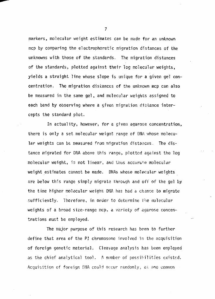
markers, molecular weight estimates can be made for an unknm•m
mcp by comparing the electrophoretic migration distances of the
unknowns with those of the standards. The migration distances
of the standards, plotted against their log molecular weights,
yields a straight line whose slope is unique for a given gel con
centration. The migration distances of the unknown mcp can also
be measured in the same gel, and molecular weights assigned to
each band by observing where a given migration distance inter
cepts the standard plot.
In actuality, however, for a given agarose concentration,
there is only a set molecular weight range of DNA whose molecu
lar weights can be measured from migration distances. The dis
tance migrated for DNA above this l~ange, plotted against the log
molecu'lar weight, is not linear, and thus accurate molecular
weight estimates cannot be made. DNAs whose molecular weights
are below this range simp1y migr·ate through and off of the gel by
the time higher molecular weight DNA has had a ch1nce to migrate
sufficiently. Therefore~ in order to determine the molecular
weights of a broad size-range mcp, a variety of agarose concen
trations must be employed.
The major purpose of this research has been to further
define that area of the Pl chromosome involved in the acquisition
of foreign genetic material. Cleavage analysis has been employed
as the chief analytical tool. fl., number of possH>ilHies existed.
Acquisition of foreign DNA could occur randomly. ~t one common
......
8
site, or at a few unique sites on the Pl chromosome. Such
chromosomal regions would be considered hot spots for recombina
tion. As compared with a Pl control, differences in electro
phoretic patterns among different hybrid mcp reveal how the in
tact chromosome is altered by the presence of foreign genes.
CHAPTER II
MATERIALS AND METHODS
A. Bacterial strains.
Strains of Escherichia coli lysogenic for bacteriophage
Pl wet~e obtained from the collection of M. Stodolsky and other
Pl investigators and are listed in Table 1. All lysogenic
strains used for bacteriophage production contained a temperature
sensitive, thermally inducible Pl prophage.
B. r·1edia.
Luria broth {1 broth: 10 g Bacto-tryptone, 5 g Bacto
yeast extract) and 5 g NaCl per l) pH 7.2) was used for propaga-
tion of bacterial lysogens and bactP.riophage production,
C. Bacteriophage production and purification.
For lal'ge scale virus production, the method of Yamamoto
and Alberts was used with slight modification (28). Overnight
starter cultures of !;_. coli lysogens were gr·own in L broth at
room temperature with no agitation for approximately 16 h. A
1:20 dilution was made from the starter cultures into a 2-1 flask
containing 500 ml of L broth prewarmed to 30 C. Freshly inocu
lated cultures appeared slightly turbid, and never exceeded 5
Klett units (at 540 nanometers (nm) using a Klett-Summerson
Photoelectric Colorimeter).
10
Table 1 Escherichia coli K12 Strains
Strain Prophage Host Characteristics
AB 259
CA7033 921
SC1800
SC1701 PLOllO 921 1801
1802 D1 D2
D3
04
05
06
D7
D8
09
010
3507
Pldlac P1cl
Plcl.lOO
PlCMcL 100 PlargF
P!ifrgr P1argF Plarg[
Pl~_r9r
Pl.~.!1lf.
P1argF P1_argF PlargF
P1.~rgF
Pldcatsul stt~
------------ ·-------·
Hfr Hayes HfrH ~111 strR ----
F-res-mod-F-~ 111 strR* - + + + F Ero lac arg
CA7033
SC1800
SC1800 SC1800 SC1800
SC1800 SC1800
SC1800
SC1800 SC1800 SC1800 SC1800 SC1800
921
*Other characteristics: his-metB-arg r-xyl-md-~111 = proA-argF-lac delection XIII.
11
Culture incubation proceeded at 30 C on a rotary shaker,
and growth was follm'led by Klett readings. Thermal induction was
initiated when, in logarithmic phase, cultures reached 40-50
Klett units (corresponding to about 2 X 10⁸ I· coli per ml).
Cultures were removed from the shaker and heated with swirling
over an open flame, to rapidly raise the temperature to 40 C.
Cultures were then transferred to a 40 C gyratory waterbath
shaker (New Brunswick Scientif·ic Co.), and brisk'ly agitated to
assure sufficient aeration throu9hout induction. If excessive
foaming occurred, anti-foam spray (D0\'1-Corning) was used to
eliminate this problem. Induction proceeded for 20 min at 40 C,
after which time cultures were transferred back to the 30 C ro-
tary shaker, where growth and subsequent bacterial lysis was
measured spectrophotometri ca'lly. During this period, t-1gC1 2 was
added to cultures to obtain a final concentration of 0.01 M in
order to stabilize the mature virions.
Following induction, and cell lysis, who.le cells and eel-
lular debris were pelleted at 4 C in 500-ml centrifuge bottles,
using the Sorva 1 GS-3 rotor at 9, 000 rpm (13, 700 g) fOl~ 20 min
(Sorval RC2-B refrigerated centrifuge). The supernatant contain-
ing the mature virions was decanted back into 2-1 flasks and made
0.5 M in NaCl and 10% (w/v) in Polyethylene Glycol 6000 (PEG
6000) in the cold. Lysates were left at 4 C overnight.
Culture volumes used in phage production were dictated
mainly by convenience in concentrating the phage ir Pxisting
12
1 aboratory equipment. When larger culture vo·J umes caul d not be
conveniently handled by centrifugation, the following changes
were made. For induction, cultures were pooled into a large
flask and a sparger was inserted for aeration. Warm, sterile l
broth (65 C) was added to the culture to quickly raise the tem
perature to 40 C. The flask was then submerged into a 40 C
waterbath. Following a 20 min induction period, 500-ml culture
volumes were aliquoted from the pooled cultures. Growth and sub-.
sequent lysis occurred in the 30 C l~otary shaker. Following
lysis~ cultures were filtered through 1 inch Hyflo-Super Cell
(Fisher Scientific) using a Buchner funnel and a large suction
flask. Filtrate containing the mature virions was then made up
to 0.5 M NaCl and 10% (wfv) PEG 6000 as described above. The
remaining procedures for phage purification \'lere ~Jdentical.
Following overnight storage at 4 C the phage-PEG 6000
aggregate of the treated lysates was pelleted at 4 C in 500-ml
centrifuge bottles~ using the Sorval GS-3 rotor at 9,000 rpm
(13,700 g) for 20 min (Sorval RC2-B refrigerated centrifuge).
The pellets were suspended in phage buffer (P buffer: 0.1 M
Tris-HCl, pH 7.2, 0.01 M MgC1 2) whose volume never exceeded 5 ml
per a 500-ml starting culture volume.
The fina1 step in phage purification employed CsCl step
gradients. CsCl gradients were prepared in 12 ml polyallomer
centrifuge tubes (14.5 X 96 mm: Internat·lona1 EqLdpment Co.) by
1ayedng 2 ml of a 1.6·-densHy CsC1 solution in r• buffer
13
underneath 5 ml of a 1.4-density CsCl solution, also in P buffer.
After gently layering the suspended phage-PEG aggregate on top
of the gradient, mineral oil was used to fill the tubes. Equi
librium centrifugation was carried out at 4 C in an International
SB 283 rotor at 35,000 rpm (180,000 g) for approximately 16 h, in
an International B 35 or B 60 preparative ultracentrifuge. Sharp
bands were clearly visible following centrifugation, and phage
Net'e collected by using a needle and syringe either from the top
of the tube, or by piercing the side.
Phage were immediately put into prepar·ed dialysis tubing.
Preparation of dialysis tubing involved boiling 1n 1% Na2co3 for
1 h, rinsing thoroughly with distilled water, and storing in 70%
ethanol in the cold. Phage were dialyzed against 5 one-1 changes
of P buffer which effectively removed all cesium chloride. Phage
were stored at 4 C.
D. Purification of bacteriophage DNA.
Purification of phage DNA involved a detergent disr~ption
of phage with sodi urn dodecyl sulfate ( SDS), foll olf:ed by a phenol
extraction of the disrupted phage proteins. All purified bac
teriophage were diluted to an opti ca 1 density of 20 at 260 nm.
For more dilute phage, DNA extl~action was perfon11ed at the exist
ing concentration. The entire extraction procedure was most
easily performed in 10-ml Co rex centrHuge tubes. Phage sus
pended in disruption buffer (D buffer: 0.1 I~ soJium phosphate,
14
pH 7. 2, 25 mt1 EDTA) were heated at 65 C in a pre\'larmed waterbath
for 10 min and then chilled on ice. Solutions were then made 0.3
M KCl and put on ice to precipitate the SDS. A low speed centri
fugation was performed (3,000 g for 10 min) to pel"let the SDS.
The viscous supernatant was then decanted into another 10-ml
Corex tube where phenol extraction of the disrupted phage pro
teins was performed.
Redistilled phenol was used in all extractions. An
appropriate volume of phenol was saturated with an equal volume
of D buffer, and the organic phase was made 0.08% (w/v) ahydroxyquinol-ine sulfate (Schwartz-Mann) in order to inactivate
any residual peroxides in the phenol. A11 preparations were ex
tracted 3 times with an equal volume of phenol. Each extrac
tion was performed at room temperature by gentle mixing for 15
min. After each extraction a low speed centrifugation was per
formed (3,000 g for 10 min) in order to separate aqueous and
or·gani c 1 ayers. The interface 1\las extracted t~>Ji ce ~ but removed
before the final extraction. rollowing this final extraction.
DNA was gently pipetted into dialysis tubing to avoid shear
breakage. Extensive dialysis was performed in order to remove
all traces of phenol. Phenol absorbs at 270 nm, so the dialysis
buffer (0.01 M Tris-HCl, pH 7.'t., 1 m~1 EDTA) was monitored and
changed until its absorbance at 270 nm was under an optical
density of 0.05. This usually required 5 to 6 one-1 changes of
dialysis buffer. DNA was assayerl for purity and concentration
15
spectrophotometrically at 260 and 280 nm, respectively. A
260:280 ratio of approximately two was required. DNA concentra
tions were computed based on the standard that at 260 nm, one
optical density unit corresponds to a DNA concentration of 50 ug ·
per ml.
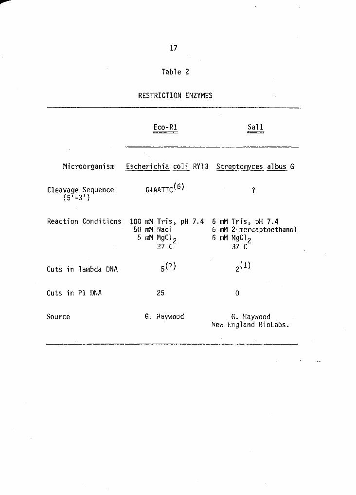
E. Restriction endonucleases.
The majority of restriction enzymes were obtained from
Or. G. Haywood, University of Chicago. Some enzymes were also
purchased from New England Biolabs, Beverly~ MA. All restric
tion enzymes used~ along with their appropriate r~action condi
tions, are listed in Table 2.
F. Enzymatic cleavage of DNA.
For each enzyme used, it was empirically determined the
quantity needed to give a limit digest of a given amount of DNA.
This was done by digesting a known quantity of DNA with succes~·
sive 2-fold dilutions of enzyme until a partial d~gRst was ob
tained. Digestion was assayed through agarose gel electro
phoresis as described below. Hamilton syringes (Hamilton Co.~
Reno, Nev.) were used in all manipulations of DNA, enzyme, and
reaction mixtures.
DNA to be cleaved was placed in a .small disposable tube,
heated at 65 C for 5 min in order to inactivate any residual
nucleases which may have escaped phenol extraction, and then
chilled on ice. The appropriau~ arnotmt of 10 X reC~ci ion buffer
16
and enzyme (Table 2) were then added, and slight vortexing was
used to assure proper mixing. Reactions usually proceeded for
2 h and were terminated by heating at 65 C for 5 min. If DNA
was not immediately used, it was stored at -40 C with no dele
terious effects.
G. Electrophoresis of DNA minichromosomes.
Electrophoresis was carried out in either agarose and/or
acrylamide gels. Uniform agarose gels were most commonly used.
Gradient pore a.garose and acry<lamide 9e<ls were employed for high
resolution, and to simultaneously observe a wide molecular weight
range of restriction products.
1. <Uniform agarose gels. UnHorm agarose gels were
prepared by the method of Sharp e~ -~· with modifications made
necessary because of the electrophoresis apparatus used (23). An
EC 490 electrophoresis apparatus was used for all uniform agarose
gels (EC Apparatus Corp.~ Jacksonville, Fla.). Slab gels run in
this apparatus are 17 X 13 em X 3 or 6 mm. A 10% (w/v) acryla~
mide plug was used to seal the bottom of the gel chamber. This
plug was formed by using a 20% (w/v) solut·ion of Cyanogum-4.1
(EC Apparatus Corp.) in electrophoresis buffer (E buffer: 0.09
M Tris, pH 8.3, 0.09 M boric acid:, 2.8 mM EDTA) containing 0.1%
(v/v) NNWN' tetramethylenediamine (H1ED). Po.lymerization was
promoted with 0.1% (w/v) ammonium persu1fate. A 50-ml plug was
used for a 6-mm thick gel, and a :3o .. m·1 plug was usrd for a 3~mM
17
Table 2
RESTRICTION ENZYMES
Eco-R1 Sall
Microorganism Escherichia coli RY13 Str~~t~ces. albu~ G
Cleavage Sequence (5'-3')
Reaction Conditions
Cuts in lambda DNA
Cuts in Pl DNA
Source
100 mM Tris, pH 7.4 50 mM Nacl 5 mM MgC1 2
37 c
5(7)
25
G. Haywood
?
6 m~1 Tr-i s, pH 7. 4 6 mM 2-mercaptoethanol 6 mM MgC1 2
37 c
2(1)
0
G. Haywood New England Biolabs.
-----.-------~-
18
thick gel. Agarose concentrations employed varied from 0.35%
(w/v) - 1.0% (w/v), depending upon the size of the fragments
being resolved. Agarose (Seakem) was dissolved in E buffer by
boiling for 5-10 min, and then cooled to 50 C before pouring into
the gel chamber. A comb-like slot former, previously soaked in
Photo-flo (Kodak Chemical Co.) was inserted into the molten aga
rose to form sample slots. The ge·l was allowed to cool and so-·
lidify completely, at which time the slot former was carefully
removed. The buffer chambers were then filled with E buffer, and
a recirculating buffer pump was activated. Gels were prerun at
50 V for one h in the cold using cold tap water as a coolant for
the cooling plates of the apparatus. Following the prerun)
samples were introduced into the preformed slots us·ing a long
stem pasteur pipette. Before addition to sample slots for elec
trophoresis, the appropriate volume of cleaved DNA was heated at
65 C for 5 min in order to separate any fragments whose cohesive
ends may have associated during coo"!ing and storage, Bromphenol
blue in 60% (w/v) sucrose was added sparingly to each sample; the
dye set~ving as a tracking substance and the sucrosr: increas·ing
the density of the samples so that they seeded under buffer in
the sample slots. Samples were initially pulled into the gel at
150 V for 10 min after which the running voltage was established.
Running voltage of the gels varied from 20 to 60 V and gels were
run overnight for approximately 16 to 24 hr or until the tracking
dye had rc.>ached the aga rose-acry"l ::Jmi de interface Ge 1 s were
19
carefully removed and stained in the dark with 0.5 ug per ml
ethidium bromide (Sigma Chemical Co., St. Louis, Mo.) in E buffer.
2. Gradient pore agarose gels. A 3-mm thick agarose gel
was used for all gradient gels and a 10% (w/v) acrylamide plug
was also employed as previously described.
A gradient former was constructed from two 100-ml
graduated Nalgene cylinders. In one cy'iinder, two holes of ap
proximately 3 mm in diameter, diametrically opposing one another,
were drilled into the side of the cylinder as close to the base
as possible. In the second cylinder, one 3 mm diameter hole was
also drilled into the side as close to the base as possible.
Three glass tubes, 5 mm long and 3 mm in outside diameter were
cemented into the holes, being careful not to allow them to pro
trude into the cylinders. These glass fittings served as connec
tions for rubber tubing. A piece of Nalgene tubing about 1 inch
long was cemented to two glass fittings connecting the bases of
both cylinders. The two cylinders wer·e then permanently mounted
on a square of plexiglass and the final glass connection was
fitted with a long piece of rubber tubing, ending in a Y joint,
hooked to 2 small bore plastic tubes. This allowed for two out
let ports for the molten agarose. Each cyl"inder had a magnetic
stir bar placed in the bottom.
Two agarose solutions wete prepared corresponding to the
'lower and upper gradient limits of agarose concentration. These
solutions were also made 10 and 10% (w/v) sucrose, respectively.
20
Both sucrose and agarose were disso·i ved in E buffer by boiling
for 5 min. Eighty ml of each solution (160 ml total) was suffi
cient to fill the gel chamber. The lower limit concentration of
agarose employed was 0.35% (w/v) and the upper limit was 1.7%
(w/v). All gradients were made between these two concentrations.
The gradient former was placed ·in a 4-l Maker and lead weights
were used to anchor it. The beaker was placed on a stirring
plate and filled with warm water (50 C), The tube connecting the
base of the two cylinders was clamped off and the cylinders were
then filled with the two molten agarose solutions; the hiQiher ron·~
centration in the rear cylinder·. Theelectrophoresis apparatus
was prewarmed to 50 C by running wa1~m tap water through the cool
ing coils. This was done in order to allow the gradient to
stabilize before solidifying. The gel chamber· was set in aver
tical position and E buffer was poured into the bottom of the
chamber in order to provide some back pressure to inhibit leak
age of the molten agarose past the acrylamide plug. The two stir
bars were set in vigorous motion, and the two outlet hoses were
put into the bottom of the gel chambe1~ just above the acrylamide
plug. The gradient was then formed from bottom to top in the
follovling manner. Agarose was forced out of the front (1 ight)
chamber by air pressure before the clamp between the two cylin
ders was released. Care was then taken to release the clamp in
such a manner that there was no back flow of agarose from the
front chamber to the rear chambe~. The flow rate into the
21
electrophoresis chamber was adjusted by a clamp on the outlet
hose to an approximate total pouring time of 15 min. The molten
agarose gradient was a 11 owed to stabi 1 i ze for another 15 min by
keeping warm water flowing through the cooling coils. Solidifi-·
cation was then achieved by forcing coo1 tap water {25 C) through
the cooling coils, displacing the warm water.
After complete solidification. a third agarose solution
was prepared in E buffer with a concentration of agarose slightly
below that of the lower gradient concentration. This solution
was prepared by bo-iling in the same manner as the other solu
tions. This solution was poured boiling onto the top of the gel
chamber in order to affect a seal with the upper portion of the
gradient gel. This proved necessary so that the gel sections
would not separate during prolonged electrophoresis. This gel
was allowed to r.ool to 45 C, and then a slot former was intra-~
duced in the same manner as described earlier for uniform per
centage agarose gels. After solidification of this upper gel,
the slot former was removed and the gel was p}Aepared for electro-
phoresis as previously described. Gradient pore agarose gels
were usually run at a voltage slightly higher than straight per
centage gels. Gels were stained as previously described.
3. _Gradient _P-ore acry·lamicie__y_els. Gradient pore acryla-·
mide slab gels were purchased from Pharmacia Fine Chemicals
(Uppsula, Sweden) and ranged in concentration from 5 to 30% (w/v)
acry1amide. These gels were r!m o•-, a Phannacia siab c;e'l
22
electrophoresis apparatus, and all methods concerning the use of
these gels were consistent with the above methods for agarose
gels.
H. Photography and densitometric tracings.
Fluroescence of DNA-ethidium bromide complexes were
viewed under long wavelength ultraviolet light (Ultraviolet
Products, San Gabriel, Calif.). All photographs were taken with
a Polaroid r~P-3 Land camera using a yellow filter (Kodak No. 9
Wratten gelatin filter). Polaroid Type 57 high speed film was
used for the majority of prints. Kodak Tri-X Pan film was used
for the majority of negatives. Polaroid Type 55 P/N film \'las
used for both prints and large negatives. Some of these large
negatives were used for densitometric tracings (,Joyce~L.oebel
microdensitometer) to determine relative molar quant'ities of DNA
in resolved bands.
-
CHAPTER III
RESULTS
A. Fractionation of virions of Pl and Pl hybrid derivatives on
cesium chloride density gradients.
Some qualitative information has been obtained from the
visual comparison of virion band thickness after fractionation of
Pl and Pl hybrid lysates on isopycnic CsCl densit.v gradients.
Cesium chloride grad·ients were used in the final step of phage
purifications following initial co-precipitation with PEG-6000,
as described in Materials and Methods. At the last step of
purification virions are visible as bands, PlB and PIS. Visual
comparisons of four popu·lations of genetically hybrid Pl virions
revealed some interesting facts with respect to relative distri-
bution PlB and PlS, when compared to the Pl control.
Pleat and PlargF are indistinguishable from the Pl
contt~ol. In contrast, Pldcss and Pld1ac have un '~xcess of small
capsid morphology variants (PlSL Pld1ac has an <'<pproximate 3:1
ratio of PlB to PlS. Pldcss has about equal tlr1ckness of small
and normal virion bands. Pldcss and Pldlac, therefore, both have
mutations in virion capsid size distribution.
B. Migration of DNA in uniform agarose gels as a function of
agarose concentration.
:, .... r...j
I
" 24
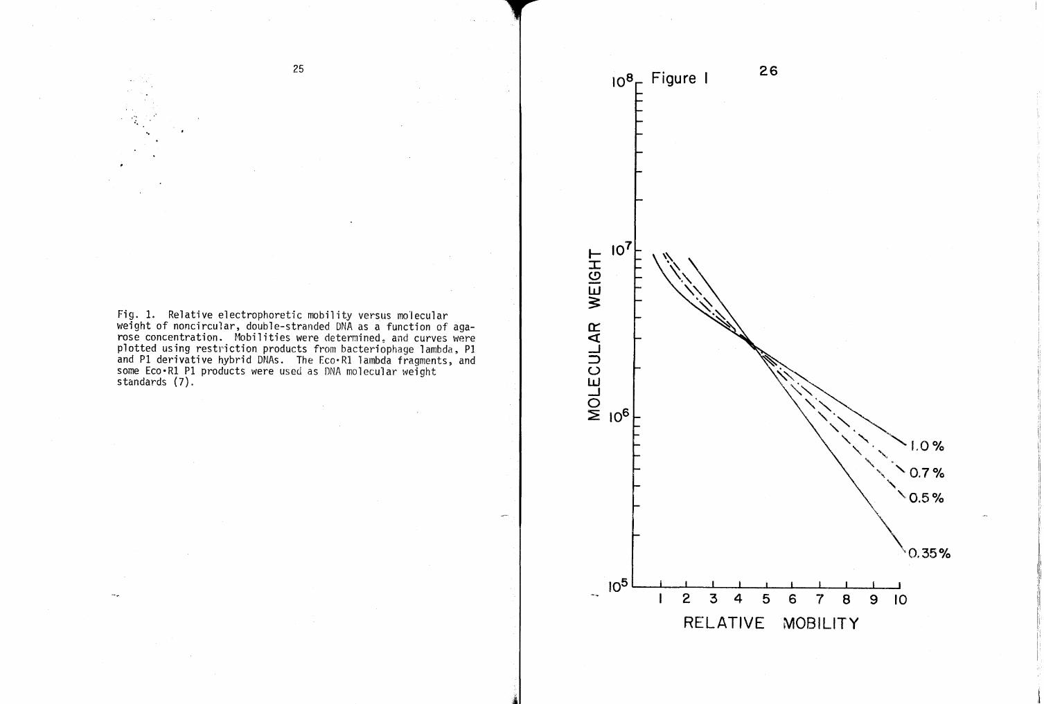
Figure 1 is semilogarithmic graph of relative migration
distance versus molecular weight of DNA over a range of agarose
concentrations. Helling et ~· have constructed a similar plot
using Eco·Rl restriction products from a variety of DNAs (7).
Figure 1 was constructed using Eco·Rl restriction products from
bacteriophage lambda, Pl and Pl hybrid DNAs. Figure 1 clearly
shows how the use of differing percentages of agarose allows for
molecular weight estimations of a much broader molecular weight
range of DNA. Relatively large molecular weight DNA, 10 X 106
daltons (10 md), is best resolved on low percentage gelss and
relatively small molecular weight DNA, one md, is best resolved
on higher percentage gels. Figure 1 also shows that for a given
agarose concentration5 the plot of relative mobility versus log
molecular weight is linear only up to a given molecular weight
of DNA.
C. The Eco·Rl limit digest of bacteriophage P1 DNA.
The 1imit digest of p·l virion DNJ\ by the restriction
endonuc·lease Eco·Rl produced 17 minichromosomes ranging in
molecular weight fro~ 9.6 to 0.56 md.
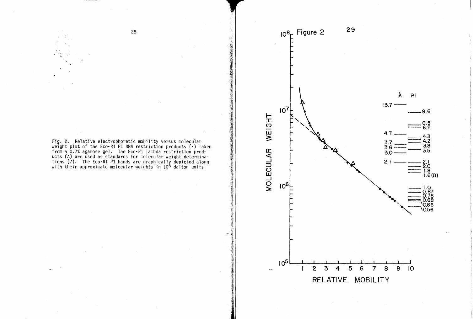
Figure 2 graphically depicts the complete Eco·Rl cleavage
pattern of bacteriophage Pl and lambda DNAs. The lambda DNA
Eco·Rl bands serve as standards for determining molecular weights
or other cleavage products run on the same gel. Figure 2 also
iriustrates the log molecular weight versus mobility p1ot of Pl
and lambda Eco·Rl minichromosomes run on a 0.7% (w/v) agarose
.·· .. ..
25
Fig. 1. Relative electrophoretic mobility versus mo·lecular weight of noncircular, double-stranded DNA as a function of agarose concentration. r~obil iti es were determined~ and curves were plotted using restriction products from bacteriophage lambda, PI and Pl derivative hybrid DNAs. The Eco·Rl lambda fragments, and some Eco•Rl Pl products were used as DNA molecular weight standards (7).
._ :r: (.!) -w s
Figure I 26
2 3 4 5 6 7 8 9 10
RELATIVE MOBILITY
1.0°/o
27
gel. Some of the molecular weights are taken from the first
description of an Eco·Rl digest of P1 DNA (7). All molecular
weights of minichromosomes depicted in Figure 2 were assessed by
employing a variety of gel concentrations~ ranging from 0.35 to
1.0% agarose, along with differing amounts of DNA ranging from
one to 10 ug. Larger amounts of DNA were used with higher per
centage gels to resolve small, quickly migrating minichromosomes.
Sma'Jler quantities of DNA were used with lower percentage gels to
resolve large, slowly migrating minichromosomes. Note in Fig
ure 2 that the log molecular weight versus mobility plot is
linear for only a given molecular weight range of minichromo
somes, thus necessitating the use of differing percentage gels
for complete assessment of the population.
Analysis of many gels has revealed the 1.6-md P1 band to
be either an unresolved doublet, or to contain DNA fragments from
a chromosomal duplication. This observation has subsequently
been confirmed (2). The combined mo1ecu1ar \';eights of the 17 P1
Eco·R1 mini chromosomes, 0. 56 md or· above (assuming that the
1.6-md band is a doublet) is approximately 52 md. Eight lower
molecular weight minichromosomes have also been detected using
gradient pore acrylamide gels, as will be presented later.
D. Eco·Rl limit digests of some gene-cica'lly hybrid Pl DNAs.
With an established Eco·Rl cleavage pattern for bacterio
phage Pl DNA, comparisons can now be made with the Eco·R1
.. .. ..
28
Fig. 2. Relative electrophoretic mobility versus molecular weight plot of the Eco•Rl PI DNA restriction products (·) taken from a 0.7% agarose gel. The Eco·Rl lambda restriction products (~) are used as standards for molecular weight determinations (7). The Eco·Rl Pl bands are graphically depicted along with their approximate molecular weights in 106 dalton units.
0:: <( ....J ::::> (.) w ....J 0 ~
Figure 2 29
A PI
f3.7---9.6
==6.5 6.2 4.7~ 43
==. 37 _4.2 3.6:::::: 3.8 3·.0- 3.5
2.1- 2.1 --2.0 _1.8
1.6(0)
--1.0 --0.87 --0.78
--"~su
I~ I 2 3 4 5 6 7 8 9 10
RELATIVE MOBILITY
30
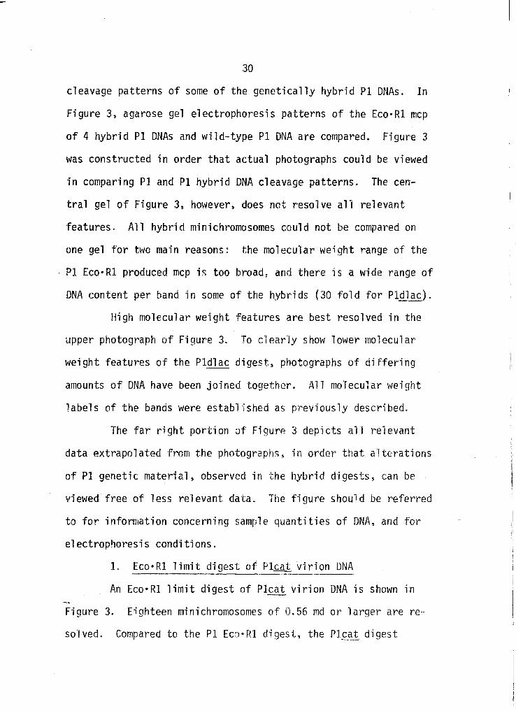
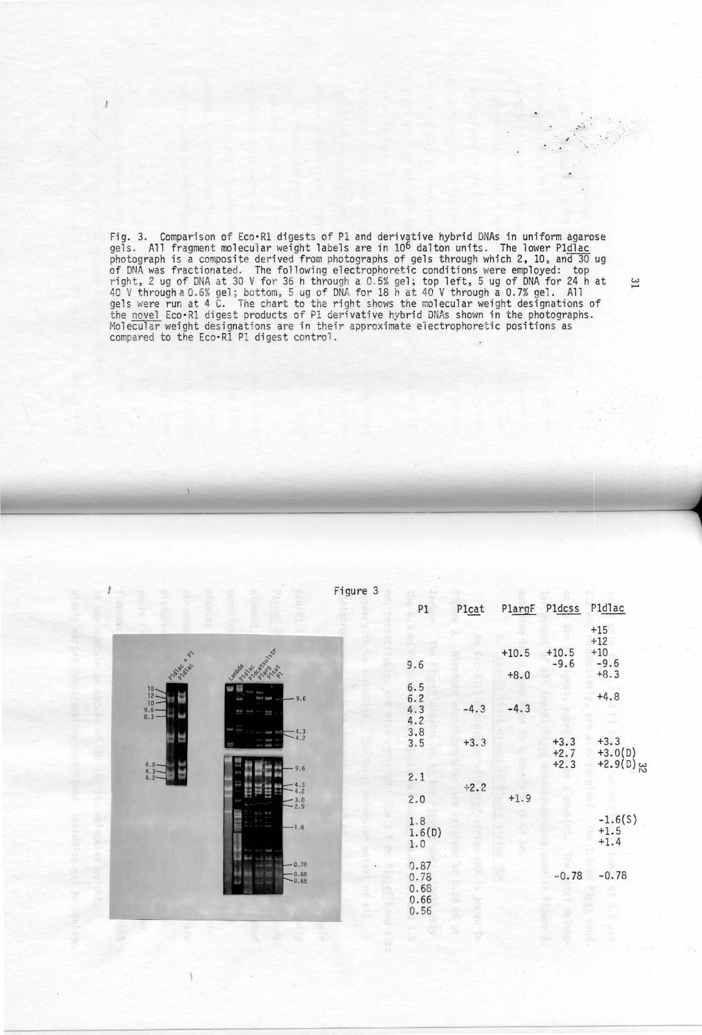
cleavage patterns of some of the genetically hybrid Pl DNAs. In
Figure 3, agarose gel electrophoresis patterns of the Eco·Rl mcp
of 4 hybrid Pl DNAs and wild-type Pl DNA are compared. Figure 3
was constructed in order that actual photographs could be viewed
in comparing Pl and Pl hybrid DNA cleavage patterns. The cen
tral gel of Figure 3, however, does not resolve all relevant
features. All hybrid minichromosomes could not be compared on
one gel for two main reasons: the molecular weight range of the
PI Eco•Rl produced mcp is too broad~ and there is a wide range of
DNA content per band in some of the hybrids (30 fold for Pldlac).
High molecular weight features are best resolved in the
upper photograph of Figure 3. To c1ear·ly show lower mo1ecular
weight features of the Pldlac digest, photographs of differing
amounts of DNA have been joined together. All molecular weight
labels of the bands were established as previously described.
The far right portion of Figure 3 depicts all relevant
data extrapolated from the photographs, in or·der that alterations
of Pl genetic material, observed in the hybrid digestss can be
viewed free of less relevant data. The figure should be referred
to for information concerning sample quantities of DNA, and for
electrophoresis conditions.
1. Eco·Rl limit digest of Pleat virion DNA
An Eco·Rl limit digest of Pleat_ virion DNA is shown in
Figure 3. Eighteen minichromosomes of 0.56 md or larger are re
so·lved. Compared to the Pl Eco·Rl digest, the Pl~at digest
j
.• . .: ' .
./
Fig. 3. Compa r is on of Eco·Rl di gests of Pl and deri vat ive hybrid DNAs i n un i form agarose gels. All f ragment molecu l ar wei ght labe ls are in 106 da l ton units. The lower Pldlac pho tog raph i s a composite der ived from photographs of gel s through whi ch 2, 10 , and 30 ug of DNA was fractionated. The fo l l owing el ectrophoreti c condit i ons were employed : top r ight , 2 ug of DNA at 30 V for 36 h thro ugh a 0. 5% gel ; t op l eft , 5 ug of DNA fo r 24 hat 40 V th rough a0 . 6% gel ; bottom , 5 ug of DNA for 18 h at 40 V th rough a 0. 7% gel . All gels were run at 4 C. The chart to the r igh t shows the molecular we ight designati ons of t he novel Eco• Rl di gest products of Pl deri vat i ve hybrid DNAs shown in the photographs . Molecular weight designations are in th eir approximat e el ectrophoreti c positions as compared to t he Eco• Rl Pl di gest con t ro l.
I Figure 3
Pl Pleat Pla rgF Pldcss Pldlac
+15 +12
~' -6 +10.5 +10.5 +10 ...,_., X
~ (, "'"'.:) ...... 'be, 'be, 9. 6 -9.6 -9.6 ~~ ~t> ...,_'ti e,'~> ,o, 'b
~"' ~"' ,f ...,_l> ...,_lS~,'b~,e,~, +8 . 0 +8. 3 v ~ ~
6.5 6. 2 +4.8 4.3 -4. 3 -4. 3 4.2 3.8 3. 5 +3.3 +3.3 +3. 3
w .......
+2 . 7 +3 . 0(0) +2.3 +2. 9( D) w
N 2. 1
+2. 2 2.0 +L 9
1.8 - 1.6(S) 1. 6(0) +1. 5 1. 0 +1. 4
0. 87 0.78 - 0.78 - 0. 78 0. 68 0. 66 0.56
33
lacks only the 4.3-md Pl band. Two additional bands of 3.3 and
2.2 md, respectively, are also resolved. The 1.6-md Pleat band,
as in the Pl digest, appears to be a doublet. The total molecu
lar weight of all Eco•Rl Pleat minichromosomes seen in Figure 3,
assuming the 1.6-md band is a doublet, is 54 md.
2. Eco•Rl limit digest of Pl£IQ[ virion DNA
An Eco•Rl limit digest of Plarg~ virion DNA is shown in
Figure 3. Nineteen minichromosomes are resolved of 0.56 md or
larger, The PlargF digest5 as in the Plea! digest, lacks only
the 4.3-md Pl band. Three additional bandss 10.5s 8.0~ and 1.9
md, respectively, are also resolved. THe 1.6-md PlargF band also
appears to be a doublet. The total molecular weight of all
minichromosomes shown is approximately 68 md.
Ten PlargF hybrid DNAs have been charactet"i zed by tl1ei r
Eco•Rl limit digests. The digest shown in Figure 3 is the only
PlargF Eco·Rl digest which has revealed an overlarge chromosomal
element, such that one unit phage genome cannot be encapsidated
genetically intact. The other digests characterized are all
missing the 4.3-md Pl bands and show only one additional mini-
chromosome band of 1.9 md. Digests of two of these hybrids are
pictured in Figure 4. The only other difference in these di-
gests from the one pictured in Figure 3, is that the observed
fluorescent intensity of the 9.6-md PlargF bands indicates that -it may contain an excess of DNA fo1~ its molecular \'Ieight.
Widely varied fractionation conditions, including gel percentag~
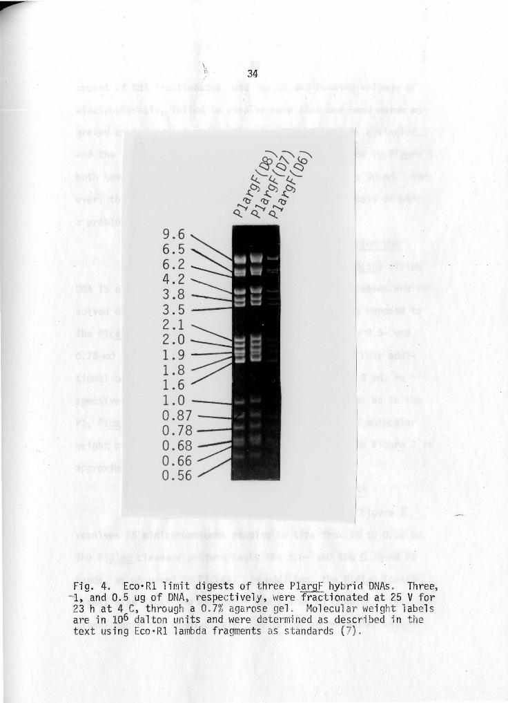
9.6 6.5 6.2 4.2 3.8 3.5 2 .. 1 2.0 1.9 1.8 1.6 1.0--0.87--0.78--0.68 0.66 0.56
34
Fig. 4. Eco· Rl 1 irnit digests of t hree PlargF hybrid DNAs . Three, 1, and 0.5 ug of DNA , respectively, were frac t ionat ed at 25 V for 23 hat 4 C, through a 0. 7% agarose ge l . Molecular we ight l abels are in 106 da lton uni ts and were determined as descr i bed i n the text us ing Eco •Rl l ambda fragments as standards (7) .
35
amount of DNA fractionated, and length and running voltage of
electrophoresis, failed to resolve more than one band which mi
grated as 9.6 md. Assuming that the 9.6-md band is a singlet,
and the 1.6-md band is a doublet, the Plarg[ hybrids in Figure 4
both have a total molecular weight of approximately 50 md. How
ever, the 9.6-md band is believed to contain an excess of DNA;
a problem which will be taken up later.
3. Eco·R1 limit digest of Pldcatsulstr virion DNA
An Eco·R1 limit digest of Pldcatsulstr (Pldcss) virion
DNA is also shown in Figure 3. Nineteen minichromosomes are re
solved of 0.56 md or larger. The Pl_gcss digest, as opposed to
the Pleat and PlargF Eco·Rl digests, lacks both the 9.6- and
0.78-md Pl bands, but retains the 4.3-md Pl band. Four addi
tional bands are resolved of 10.5, 3.3, 2.7, and 2.3 md, re
spectively. The 1.6-md band appears to be a doublet as in the
Pl, Pleat, and Pl_argF Eco·Rl digests. The combined molecular
weight of all the Pldcss minichromosomes pictured in Figure 3 is
approximately 70 md.
4. Eco·Rl limit digest of Pldlac virion DNA
The Pldlac Eco·Rl limit digest pictured in Figure 3
resolves 25 minichromosomes ranging in size from 15 to 0.56 md.
The Pldlac cleavage pattern lacks the 9.6- and the 0.78-md Pl
bands, as observed in Pldcss. In addition, the Pldlac Eco•Rl
-digest also lacks one member of the L6-md Pl doublet. Ten addi
tional bands are resolved with the following molecu·lar vJeights:
36
15, 12, 10, 8.3, 4.8, 3.3, 3.0, 2.9, 1.5, and 1.4 md. The 3.0-
and 2.9-md bands are both doublets. To unambiguously establish
that there is no Pldlac fragment at the 9.6-md position, Pld1ac and
Pld1ac plus Pl digests are compared in the upper left photograph of
Figure 3. The appearance of the 9.6-md fragment in the combined
digest shows that the Pldlac fragment with the 8.3-md designation
is indeed nove 1 .
When fragments of a substrate chromosome are present in
equimolar quantities, there is a uniform decrease in the DNA con
tent of resolved bands with the molecular weight of the fragments.
While this trend has been evident in the Pl digest, and the di
gests of the Pl hybrid derivatives thus far showns the Pldlac
Eco·Rl digest presents a striking contrast. The partially re
solved 15-, ·12-, and 10-md Pldlac triplet contains less DNA than
expected for its aggregate molecular weight, as observed from
fluorescent intensity of these bands. The 4.8-md Pldlac band is
also DNA poor. Densitometric tracing made from negatives of
photographed gels have confirmed these visual observations. This
point is mentioned here so as not to complicate later interpreta
tion of these results. A meaningful estimate of the size of the
Pldlac chromosome cannot be made from the Eco.Rl digest shown
here~ without a more detai'Jed, genet·ic description of the Pldlac
chromosome. A molecular weight estimate of the Pldlac chromosome
~ill be reserved for the Discussion.
,, 37
E. Fractionation of low molecular weight Eco•Rl minichromosomes
of Pl and derivative hybrid DNAs.
Gradient pore acrylamide gels (Pharmacia) have been
employed to detect low molecular weight minichromosomes produced
through Eco·Rl digests of Pl and Pl hybrid DNAs. These low
molecular weight minichromosomes could not adequately be resolved
on agarose gels.
In order to detect and resolve low molecular weight
minichromosomes, gradient pore acrylamide ge1s were employed.
Acrylamide is the favored material for low porosity gels. Acryl
amide can be used at much higher concentrations than agarose, and
thus enables the resolution of much lower mo1ecular weight DNA
molecules than agarose. Gradient pore acrylamide gels provide
even greater resolution of low mo·lecular weight DNA molecules be
cause of the added self-sharpening quality of the der.reasing
paras i ty ge 1 "
Figure 5 is a gradient por·e acrylamide r~el ranging from
5 to 30% (\.'1/v) acr-ylamide. Low molecular weight L:'co·Rl m·ini
chromosomes of Pl, Pleat·~ PliirgF~ Pl_dcss s and Plcll ac DNA have
been resolved. One ug of DNA was used for fractionation of all
digests except Pldlac where L5 ug was employed. ~1olecular
weight estimates of minichromosomes are not given because of
lack of low-md references for molecular weight extrapolations.
The arrow on either side of the photograph in Figure 5 indicates
the position of the 0.56-md minichromosome present in all PI and
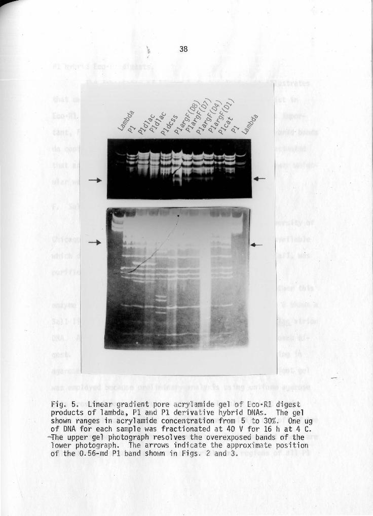
Fig. 5. Linear grad·ien t pore acry1amide ge l of E o·Rl di ges t products of lambda, Pl and Pl der ivative hybrid DNAs. The g~l shown ranges in acrylamide concentration from 5 t o 30%. One ug of DNA for each sample was frac tionated at 40 V for 16 h at 4 C.
-The upper gel photograph resolves the overexposed bands of the lower photograph. The arrows indi cat e the approxi mate position of the 0.56-md Pl band shown i n Figs . 2 and 3.
39
Pl hybrid Eco•Rl digests.
The importance of Figure 5 is two-fold: it illustrates
that many lower molecular weight minichromosomes do exist in
Eco•Rl limit digests of Pl and Pl hybrid DNAs and, most impor
tant, Figure 5 shows that all lower molecular weight hybrid bands
do contain the low molecular weight Pl bands. It is estimated
that approximately 2 md of DNA is contained in these lower molec
ular weight bands for each digest.
F. Sall limit digests of Pl and derivative hybrid DNAs.
Through communication with lk. G. Haywood~ University of
Chicagos it was learned that a restrict·ion enzyme was available
which did not put any breaks in Pl DNA. This enz:{me, Sall, was
purified from StreRtomyces ~1 bus G (1).
The primary concern with Sall was to assess whether this
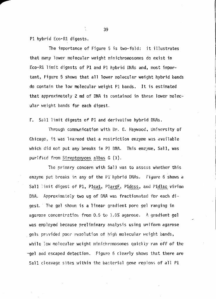
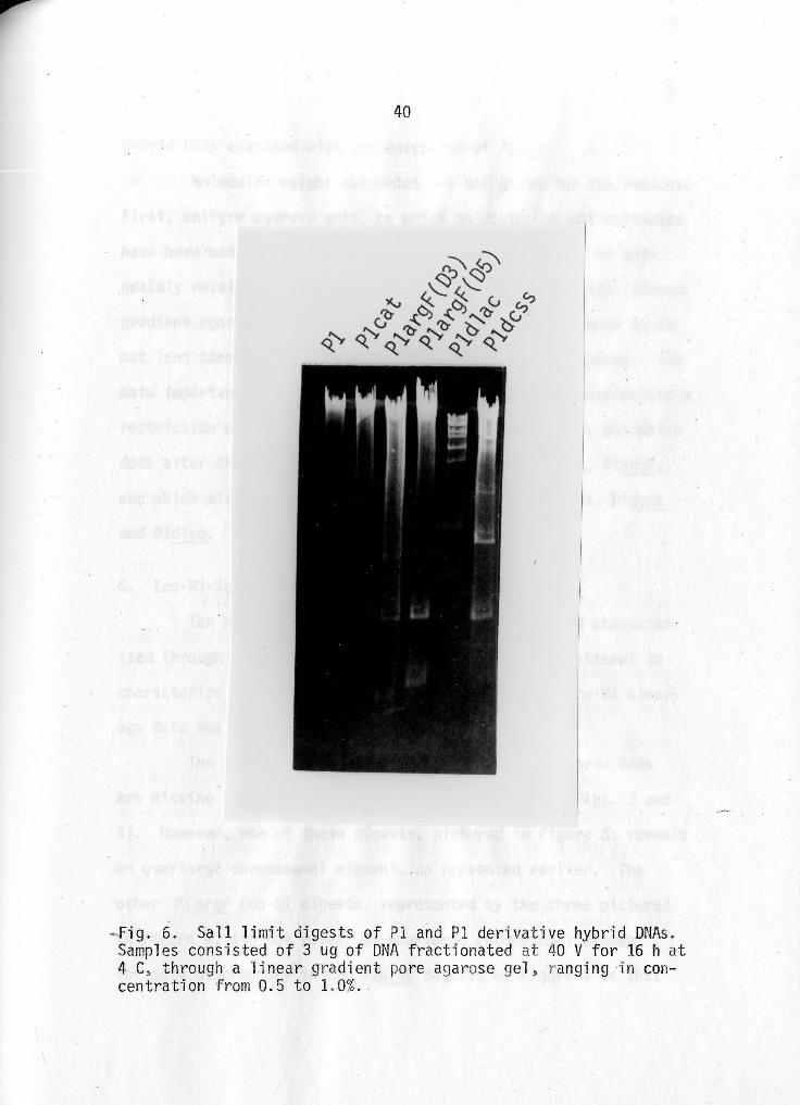
enzyme put br~eaks in any of the Pl hybrid DNAs. Figtwe 6 shows a
Sall 1imit digest of Pl, Pleats Plargf_~ Pldcss_, and Pldlac virion
DNA. Approximately two ug of DNA \'las fractionated for each di-·
gest. The gel shown is a linear gradient pore gel ranging in
agarose concentration from 0. 5 to J. 0% agarose. P, gradient gel
was employed because preliminary analysis using uniform agarose
gels provided poor resolution of high molecular weight bands,
while low molecular weight minichromosomes quickly r·an off of the
-gel and escaped detection. Figure 6 c·l early shows that there are
Sall cleavage sites within the bacterial gene regions of all Pl
40
-Fig. 6. Sall limit digests of Pl and Pl derivati ve hybrid DNAs . Samples consisted of 3 ug of DNA fractionated at 40 V for 16 h at 4 C, through a li near gradient po re agarose gel, ranging in concentration from 0.5 to 1. 0%.
41
hybrid DNAs examined with the exception of Pleat.
Molecular weight estimates are not given for two reasons.
First, uniform agarose gels, on which molecular weight estimates
have been made from other restriction digests, failed to ade
quately retain or resolve the Sall restriction products. Second,
gradient agarose gels, such as the one pictured in Figure 6, do
not lend themse1ves to accurate molecu·lar \'Ieight estimates. The
main importance of Figure 6 remains~ however, that there exists a
restriction enzyme~ Sall, which fails to alter Pl DNA, but which
does alter the DNA of one of its nondefective hybrids, PlargFs
and which also alters the DNA of two defective hybrids, Pldcss
and Pldlac.
G. Eco•Rl-Sqll combined digest of PlE_!:]f_ virion DNA.
Ten nondefective Pl~r~F hybrid DNAs have been character
ized through their Eco·Rl cleavage patterns. In an attempt to
characterize these PlargF hybrids as a family, the Eco•Rl cleav
age data has presented some problems.
The Eco·Rl limit digest of an ten PlargF hybr-id DNAs
are missing the 4.3-md Pl band as shown previously (Figs. 3 and
4). However, one of these digests, pictured in Figure 3, reveals
an over·large chromosomal element, as presented earlier. The
other PlargF Eco·Rl digests, represented by the three pictured
-in Figure 4, did not reveal an overlarge chromosomal element.
The only discrepancy in the PlargF digests of Figure 4 is that
42
the 9.6-md bands appear to contain an excess of DNA for their
molecular weight.
An Eco·Rl-Sall co-digest of the two PlargF hybrids shown
in Figure 4 .was performed in order to resolve whether the major
ity of bacterial gene DNAwas hidden in the 9.6-md EcoeRl band.
Sall was chosen for the co-digest because it does not put any
breaks in Pl DNA, but does have cleavage sites in the bacterial
gene region of the PlargF hybrid DNAs. Therefore, any additional
band(s) appearing in the co-digest, with a molecular weight(s)
greater than 1.9 md (the only additional band observed in the
Eco•Rl digests of these two PlErgF hybrid DNAs) would confirm the
presence of more bacterial gene DNA than observed in the Eco•Rl •
digests.
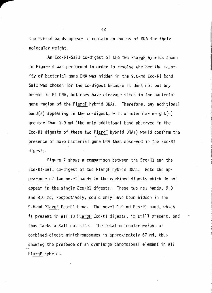
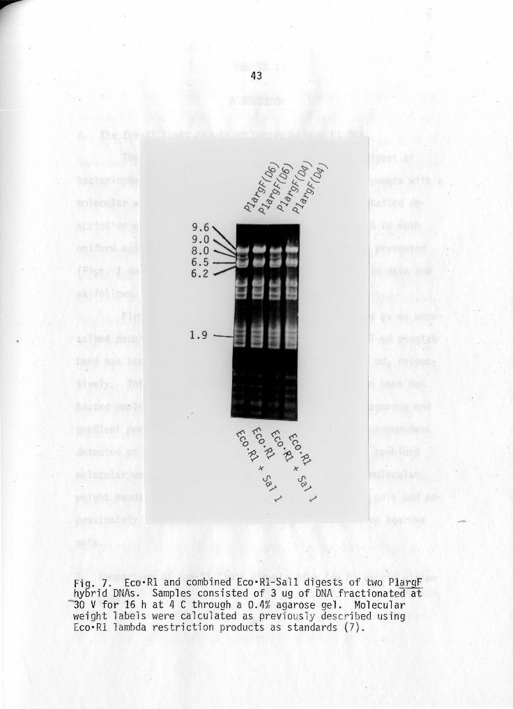
Figure 7 shows a comparison between the Eco•Rl and the
Eco•Rl-Sall co-digest of two Pl_argF hybrid DNAs. Note the ap
pearance of two novel bands in the combined digests which do not
appear in the single Eco•Rl digests. These two new bands, 9.0
and 8.0 md, respectively, could only have been hidden in the
9.6-md Plarg_[ Eco·Rl band. The novel L9-md Eco·Rl band, wh·ich
is present in all 10 PlargF Eco·Rl digests, is stil"l present, and
thus lacks a Sall cut site. The total molecular weight of
combined-digest minichromosomes is approximately 67 md, thus
showing the presence of an overlarge chromosomal element in all
PlargF hybrids.
9.6 9.0 8.0 6.5 6.2
1.9
43
Fig. 7. Eco·Rl and combined Eco·Rl-Sall digests of bw PlargF hybrid DNAs. Samples consisted of 3 ug of DNA f ractionated at
- 30 V for 16 hat 4 C through a 0.4% agarose gel. Molecular weight labels were calculated as previ ously described using Eco•Rl lambda res t riction products as standards (7).
CHAPTER IV
DISCUSSION
A. The Eco•R1 limit digest of bacteriophage P1 DNA.
The first fractionation of an Eco•Rl limit digest of
bacteriophage P1 DNA revealed the presence of 15 fragments with a
molecular weight gl~eater than 0.6 md {7). A more detailed de
scription of the Eco•Rl limit digest of Pl virion DNA in both
uniform agarose and gradient pore acrylamide gels is presented
(Figs. 1 and 3). Amendments to the original published data are
as follows.
First, the 1.6-md P1 band has been recognized as an unre-
solved doublet. Second, the originally reported 0.67-md singlet . band has been resolved as a doublet of 0.68 and 0.65 rnd, respec
tively. Thirds lower molecular weight fragments have been de
tected employing fractionation through both un·iform agarose and
gradient pore acrylamide gels (Figs. 3 and 4}. Minichrornosornes
detected on agar~ose gels, 0. 56 md or greater, have a combined
molecular weight of approximately 53 md. The lower molecular
weight bands as detected on gradient pore acrylam·ide gels add ap-
proximately 2 md to the 17 minichromosomcs detecter:l on agarose
gels.
B. Comparison of Eco•Rl limit digests of a few Pl hybrid virion
DNAs.
44
45
P1 virion DNAs differ from their counterpart plasmid DNAs
in being linear, permuted, and terminally redundant (11). Since
all of the digests shown in Figure 3 are those of virion DNAs,
the question arises whether cleavage patterns of a population of
virion DNAs are an accurate assessment of the chromosomal struc
ture of their plasmid DNAs. In order to resolve this question,
an Eco•Rl digest of Pldlac plasmid DNA, supplied by J~ Bornhoeft
and M. Stodolsky~ was performed. A"l'! plasmid digest bands de
tected are present in the virion DNA digests and vice versa.
Thus, we are assuming for Pl_dlac:~ Pl) and the other (less defec
tive) hybrids, that cleavage patter·ns of a population of virion
DNAs are indeed indicative of plasmid DNA digests. This assump
tion has subsequently been confirmed (2). By the same reasoning,
absences or additions of Eco·Rl produced minichromosomes in Pl
hybrid virion DNA digests, as compar·ed to the Pl control, do re
flect alterations in the corresponding plasmid chromosome.
Deletions of minichromosome(s) ·in hybrid digests, as
compared to Pl are subject to the following interpretation. 1\b
sence of a sing·le minichromosomc band in hybrid digests imp"lies
the insertion of foreign genetic mater·ial somewhere in the cor
responding PI minichromosome. Absence of 2 or more minichromo~
somes in hybrid digests implies the loss of some Pl genes, with
the subsequent insertion of the foreign genetic element somewhere
in the genetic region defined b_y tho missing Pl minichromosomes.
The following is a summary of deletions of Eco·Rl
46
produced Pl minichromosomes observed in the Pl hybrid DNA Eco·Rl
cleavage patterns shovm in Figure 3. Pleat and PlargF 1 ack
solely the 4.3-md Pl band. Pldcss lacks the 9.6- and the 0.78-md
Pl bands. Pldlac lacks the 9.6-, the 0.78- and one member of the
1.6-md Pl doublet.
An interpretation of these results is as follows: cat
and argF elements are encoded in the 4.3-md Pl segment. The css
element is encoded in the Pl genetic region defined by the 9.6-
and 0.78-md bands, and lac is encoded in the Pl genetic region
defined by the 9.6-, 0.78··) and one L6-md Pl segment"
As stated earlier, mapping data obtained through bacte
riophage crosses has shown that the ca_t-gene 2 region of the Pl
chromosome, without exception, is involved in the aberrant re-
combinational events underlying the_<;!~ .novo formation of the
genetically hybrid, specialized transducing derivative of Pl
(26). At least one of the sites of union of Pl and foreign
genetic sequences is in the cat-·gen~_?_ genetic region. Hereto
fore, there has been no fine structura·l information on the cat-
gene 2 region. Gene 2 function has been shown to be altered in
Pldlacs (20L but there has been no simple assay for the presence
or absence of the cat insertion locus in Pldlac. Results pre
sented here show that there is an Eco•Rl cut site(s) separating
loci of cat and aDl£ from that of cs~ and .·lac. Cat and ar_gf are
encoded to the 1 eft of this site, and .c~~- and l_ac to the right.
An Eco·Rl cleavage map has been constructed and the order of
47
these Pl DNA Eco•Rl segments is 4.3, 9.6, 0.78 and 1.6 (2). Css
presumably has termini in the 9.6- and 0.78-md segments, and lac
in the 9.6- and 1.6-md segments.
The cat-gene 2 region of the Pl chromosome has been shown
to be a hot spot for aberrant recombination (26). Based on the
Eco·Rl cleavage data presented here, the following finer resolu
tion of the cat-gene 2 genetic region has been established.
First, a unique recombinational hot spot does not exist. Aside
from this fact, the following possibilities do exist: either
there are at least two unique hot spots on the Pl chromosome
where foreign genetic elements are inserted; or there is an ex
tended region of the Pl chromosome considered to be a hot spot
for aberrant recombinational events.
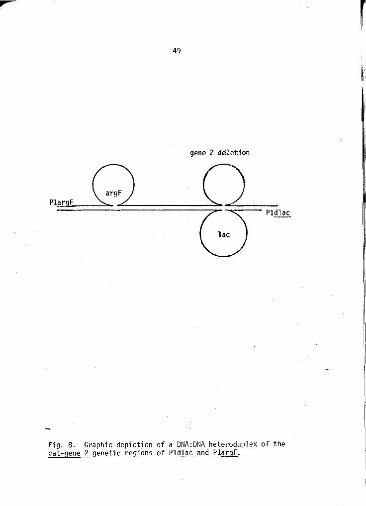
For continuing studies of Pl aberrant recombination, this
finer resolution of the cat-gene 2 genetic region provides great
utility in Pl hybrid DNA heteroduplex studies. For instance, a
heteroduplex of PlargF and Pldlac DNA shou'ld have the following
structure: argF insertion ·roop- dup'lex DNA region - lac inser
tion loop/corresponding gene 2 deletion loop. This theoretical
heteroduplex is depicted in Figure 8. Similar heteroduplexes
could be constructed between Pleat and Pldla~, Pleat and Pldcs~,
and PlargF and Pldcss. Electron microscopic measurements would
reveal. the size of the two foreign genetic e1ements, the distance
between their sites of integration, and the corresponding size of
the Pl gene deletion.
48
C. Molecular weight estimate of the Pldlac plasmid.
As stated in Results, there are complications in esti
mating the size of the Pldlac plasmid chromosome from the Eco·Rl
limit digest of a population of Pldlac virion DNAs shown in
Figure 3.
When fragments of a substrate chromosome are present in
equimolar quantities, there is a uniform decrease in the DNA
content of resolved bands with the molecular weight of the fr·ag
ments. While this trend is clearly evident in most of the re
striction enzyme digests presented hel"e, the Pld.iac Eco·Rl digest
pre~ents some complications.
The additive molecular weight of all Pl_dlac, Eco•Rl frag
ments is about 110 md. However~ the 15··, 12- ~ 10-, and 4. 8-md
Pldlac Eco·Rl digest bands do not appear to be present in equi
molar quantities as assessed from ge·l photographs. To use this
information in formulating an estimate of the si:ze of the Pl_c[L9c
plasmid chromosome, recent data on the ·lac region of the E. coli
chromosome plays an important role.
Recent electron microscopic analysis of the lac re9ion of
the E. coli chromosome has revea'led a 47-md region containing
genetic regions on either size of lac which are inverted repeats
of one another. This region has been designated a3B3~lac-B5a5
where a3s3 and ~5a5 are inverted repeats of one another (9). If
this region is encoded in the bacterial region of Pldlac,. the
anomalous DNA content of the aforementioned Pldlac Eco•Rl bands
49
gene 2 delet_ion
Pldlac
-Fig. 8. Graphic depiction of a DNA:DNA heteroduplex of the cat-gene 2 genetic regions of Pldla~ and PlargF.
r
50
could be explained as follows.
A heterogeneous population of Pldlac plasmids could arise
from the process of recombinational inversion, whereby one-half
of the Pldlac plasmid population would contain the above region
of DNA in the order a3e3-lac-B5a5, and the other half would con
tain this genetic region in the order of a5e5-1ac~e3a3. In the
Eco·Rl digest of virion DNA obtained from this heterogeneous
population of plasmid DNA, each of the aS sequences would be rep
resented on two distinct Eco•Rl minichromosomes. However~ these
fragments would only be present in one-half molar quantities with
respect to the other Pldl ac Eco • Rl m·i ni chromosomes. Thus, the
presence of an invertable repeat may be responsible for the
a noma 1 ous DNA content of the 15-, 12··, 10-, and 4. 8-md Pldl ac
Eco•Rl minichromosomes.
Two assumptions are thus being made concerning the
structure of the Pldlac plasmid chromosome~ based on its Eco•Rl
limit digest cleavage pattern. One, 'Lhe a3s3-lac-a5a5 E. coli_
chromosome segment is encoded ·in the bacteria 1 gene segment of
the Pldlac plasmid. Two, this segment is represented by four
minichromosomes of 15~ 12, 10 and 4.8 nid, respectivelys each
present in one-half molar quantity. A ~i~imal molecular weight
estimate of the Pldlac plasmid can be made by assuming that the
15- and 4.8-md segments are present in the inverted recombinant.
Thus, if the total molecular weight of all Eco·Rl minichromosomes
is 110 md, a minimal size estimate of the Pldlac plasmid is
51
88 md (110-(12+10)).
In estimating the size of the Pl and bacterial gene
segments of the Pldlac plasmid, it is not knovm whether or not the
missing Eco•Rl Pl bands, totaling 12 md, are present in novel
Pldlac bands~ · Therefore, the possible molecular weight range of
the Pl and bacterial segments of the Pld1ac plasmid are 54-42 mds
and 34-46 md, respectively.
It is apparent that the data confirm deductions of Rae
and Stodolsky (20) that the Pldlac p1asmid chromosome is too
large to be transduced genetically intact, and that even the bac
terial segment of the Pldlac plasmid must frequently be split
during scission of the Pldlac chromosome into virion chromosomes.
D. Sall cleavage of Pl and Pl hybrid DNA.
A restriction enzyme from the bacterial species Strepto
myces albus G, Sall, has been shown to leave normal Pl DNA vir·
tually unaltered. The nondefective Pl hybrid derivat·ive, Pleat,
has also been shown not to possess any Sall susceptible genetic
sequences. Three specialized transducing derivatives of Pls the
nondefective PlargF, and the defective hybrids Pless and Pldlag_,
have been shown to possess genetic sequences which are suscep
tible to cleavage by Sall. These results have been pr·esented in
Figure 5.
- Two important facts emerge from these Sall digests.
First, it is indeed striking that a DNA molecule the size of Pl
•

r 52
plasmid DNA (53 md) does not contain any Sall susceptible
cleavage sites. Second, it is equally as important that a non
defective hybrid derivative of Pl, PlargF, does contain Sall sus
ceptible sites in its bacterial gene region. This second point
requires some expanded explanation.
As presented earlier, PlargF lysogens yield a population
of virions which are indistinguishable from Pl in all normal
functions, including net virion yield and relative production of
normal and small capsid morphology variants. Therefore, Sall can
be used to convert a circular PlargF plasmid into a linear mole
cule with all Pl genes left unaltered. This is essentially the
same as putting a single, site specific break in Pl plasmid DNA,
converting it from a circular to a "linear molecule, without al
tering any Pl functions. This fact ·is of enormous potent·la1 im
portance and some valuable information concerning the plasmid
will aid in an explanation.
Pldlac has been shown here, by analytical procedures~ and
previously by purely genetic data (20) ~ to be over·large with
respect to the size of Pl plasmid. One unit phage genome of
Pldlac DNA cannot be encapsidated 9enetical1y ·intact. However,
the Pldlac p'lasmid has been shown to be extreme·ly stab1e~ and to
replicate faithfully in conjunction with the host chromosome.
Therefore, the Pl plasmid is certainly capable of maintaining and
replicating excess foreign DNA which has been inserted into its
-genome. The problem with Pldlac l·ies in the fact that some Pl
genes are missing as a result of~~- ·insertion, and important
53
Pl functions are thus either altered or ~issing.
Now that methods are available for putting a single break
in Pl DNA, through the use of Sall and PlargF, foreign genetic
material can be inserted into Pl DNA, sealed with ligase~ and put
back into a permissive host with subsequent maintenance and
faithful replication. In essence, any DNA can be inserted into
the PI genome and faithfully passed to successive generations.
E. Combined Eco·Rl-Sall digests characterizing al1 Pl51_r_g_F
hybrids as a family.
Ten PlargF hybrid DNAs have been characterized by their
Eco·Rl cleavage patterns. In an attempt to classify these
hybrids as a PlargF family, the Eco•Rl cleavage data have pre~
sented some problems. All Eco·Rl Pl.~r.g[ DNA digests revealed the
absence of the same PI genetic material of 4.3 md. Howevers all
digests were not consistent in the re·lative amounts of bactedal
gene DNA resolved.
Figure 3 contains an Eco•Rl "l"imit digest c'leavage pattern
of DNA from virions of one PlargF hybrid lysate. Nineteen mini
chromosomes with a combined molecular weight of approximately
70 md are resolved .. Thus, for this PlargF hybrid, the Eco·Rl
data reveals an overlarge chromosoma·l element, when compared to
the Pl control, meaning the corresponding PlargF plasmid contains
-too much DNA to be transduced genet·ically intact. However, for
the other 9 PlargF hybrids characterized, represented by the
54
three Eco•Rl limit digests in Figure 45 the Eco·Rl data resolves
no overlarge chromosomal elements. Thus, these PlargF hybrid
derivatives initially appeared to contain a small enough amount
of DNA to be transduced genetically intact.
One difference, however, is evident when the Eco•Rl
digests of the PlargF hybrids pictured in Figure 4 are compared
to the Pl control. The 9.6-md PlargF band appears~ by eye5 to
contain an excess of DNA. Some P~argF bacterial gene DNA could
possibly be hidden in this 9.6-md band, although differing frac-
tionation conditions were unable to resolve more than one band.
To resolve this possibility an Eco•Rl--San combined di-
gest was performed. Sall was used, as stated earlier, because it
had been shown not to put any breaks in Pl DNA, but to have
cleavage sites in the ErgF bacterial gene region. Thus, if the
combined digest revealed more DNA than represented by the 1. 9-md
PlargF band present in all hybrid digests, some bacterial gene
DNA must have been hidden in some of the normal Pl bands.
The combined Eco•Rl-Sall digest of the two PlargF hybrid
DNAs whose single Eco·Rl digests are depicted in Figure 4, are
shown in Figure 7. Excess DNA was revealed by the combined di
gests amounting to approximately 17 md, not revealed in the
single Eco•Rl digests. Two new bands were resolved of 9.0 and
8.0 md, respectively. This DNA could only have been hidden in
the 9.6-rnd Eco·Rl digest band. The combined molecular weight of
all co-digest minichromosornes is approximately 68 rnd.
55
All 10 PlargF hybrids can thus be characterized as a
family. All are nondefective in Pl functions, and each hybrid
chromosome is overlarge with respect to the Pl control. There
fore, their entire chromosome cannot be transduced genetically
intact.
---
CHAPTER V
SUMMARY
There exists a region on the Pl chromosome which has been
shown, without exception, to be involved in the aberrant recombi-
national processes underlying the de novo formation of the genet----ically hybrid Pl derivatives. This genetic region has been named
cat-gene 2. In an attempt to further define this genetic region,
on a purely analytical basis, the technique of cleavage analysis
has been employed. Agarose and acrylamide gel ele:::trophoresis
have been the chief tools used in ·identifying genetic differences
between respective Pl hybrid minichromosomal popu1ations produced
by restriction enzymes. Some qua'litative information has also
been obtained through fractionation of Pl and its derivative
virions on cesium chloride density gradients.
Four Pl hybrid derivatives were analyzed by equilibdum
density centrifugation with respect tothe r·elative distribution
of small (PIS) and normal size (P1B) capsid virions. Pleat and
PlargF lysogens are identical to Pl in virion capsid size distri
bution. In contrast, both Pldcss and Pl dl_2~ lysogens show ex
cessive production of small capsid morphology variants. Thus,
both Pldcss and Pldlac lysogens have mutations affecting virion
capsid size. - The cleavage analysis of these four Pl hybrids has mainly
56
57
involved the use of the restriction enzyme Eco·Rl. Eco·Rl limit
digests of Pl, Pleat, PlargF, Pldcss, and Pldlac virion DNAs were
fractionated on both agarose and acrylamide gels. Comparison of
the hybrid digests with that of the Pl control revealed that
there exists an EcoeRl susceptible site separating the loci of
integration of argF and cat, from that of lac and css. There
fares there does not exist a unique site in the ~at-gene 2
region of the Pl chromosome where aberrant recombinational
processes occur.
Molecular weight estimates \-Jere a'lso made in agarose gels
from Eco·Rl 1imit digests of Pl and "its hybrid DNAs. The molecu
lar weight estimates revealed that PlargF, Pldcss, and Pldlac
plasmid DNAs are overlarge with respect to the Pl control, such
that one unit phage genome of DNA cannot be transduced geneti
cally intact. P~cat plasmid DNA is not overlarge and one unit
phage genome can be transduced genetically intact.
In addition to Eco•Rl, the restr·Jction enzyme Sall was
also used for cleavage analysis. sa·ll does not put any breaks in
normal Pl DNAo Sall limit digests of Pl and derivative hybrid
OtiAs were performed. All hybrid DNAs were shown to possess Sall
susceptible cleavage sites with the exception of Pleat DNA. Of
greatest importance here is the fact that a nondefective Pl
hybrid, PlargF, possesses Sall cleavage sites in its bacterial
gene region. Thus, Sall and PlargF. plasmid DNA can affectively
be used to put a single break in Pl plasmid DNA, thus allowing
58
for the insertion of foreign DNA into the Pl plasmid chromosome.
An Eco•Rl-Sall combined digest was performed on some
PlargF hybrid DNAs whose single Eco·Rl digests failed to ade
quately resolve any excess of DNA as compared to the Pl control.
The combined digests did reveal an excess of DNA which allowed
all ten PlargF hybrids used to be classified as a family.

LITERATURE CITED
1. Arrand, J. R., P. A. Myers, and R. J. Roberts. Unpublished observations.
2. Bachi, B., and W. Arber. Virology (in press).
3. Berg, C. M., and L. Caro. 1967. Chromosome replication in Escherichia coli. I. Lack of influence of the integrated R factor. J. Nol. Biol. 29:419··431.
4. Bertani, G. 1951. Studies on lysogenesis. I. The mode of phage liberation by lysogenic Escherichia coli. J. BacteriaL 62:293-300. ----~-· -~
5. Bertani, G., and J. J. Weigle. 1953. Host controlled variation in bacterial viruses. J. Bacterial. 65:113-121.
6. Hedgpeth, J. ~ H. Goodman, and H. Boyer. 1972. DNA nucleotide sequence restricted by the R1 endonuclease. Proc. Nat. Acad. Sci. 69:3448-3452.
7. Helling~ R., H. Goodman, and H. Boyer. 1974~ Analysis of endonuclease R.EcoRl fragments of DNA from lambdoid bacteriophage and other viruses by agarose gel electrophoresis. J. Viral. 14:1235-1244.
8. Hoppes l., and J. Roth. 1973. Specialized transducing phages derived from Salmonella phage P22. Genetics 76: 633-654.
9. Hu, S., E. Ohtsubo, and N. Davidson. 1975. Electron microscope heteroduplex studies of sequence relations among plasmids of Escherichia co1i: Structure of Fl3 andrelated F-primes. J. Bacterial. 122:749-763.
10. Iida, S.~ B. Bachi, and W. Arber. In progress.
11. Ikeda, H., and J. Tomizawa. mosomal replicator unit. Bio. 33:791-798.
196B. Phage Pl, an extrachroCold Spring Harbor Symp. Quant.
12. Jacob, F. 1955. Transduction of lysogeny in Escherichia - coli. Virology _!_:207-220.
59
60
13. Kikuchi, A., D. Esleviers, and L. Gorini. 1975. Isolation and characterization of lambda transducing bacteriophage for argF, argi and adjacent genes. J. Bacterial. 122: 727-742.
14. Kondo, E., and S. Mitsuhashi. 1964. Drug resistance of enteric bacteria. IV. Active transducing bacteriophage P1CM produced by the combination of R factor with bacteriophage Pl. J. Bacterial. 88:1266-1276.
15. Lennox, E. S. 1955. Transduction by bacteriophage Pl. Virology 1:190-206.
16. Luria, S. E., J. N. Adams, and R. C. Ting. 1960. Transduction of lactose-utilizing ability among strains of I· coli and~- dysenteriae and the properties of the transducing phage particles. Virology j£:348-390.
17. Luria, S. E., and M. L. Human. 1952. A nonhereditary, host-induced variation of bacterial viruses. J. Bacterial. 64:557-569.
18. Meselson, M., R. Yuan, and J. Heywood. 1972. Restriction and modification of DNA. Ann. Rev. B·iochema 41:447-466.
19. Nathans, D. s and H~ 0. Smith. 1975. Restriction endonucleases in the analysis and restructuring of DNA molecules. Ann. Rev. Biochem. 44:273-293.
20. Raes f-1. E., and M. Stodolsky. 1974. Chromosome breakage, fusing and reconstruction during Pldlac transduction. Virology 58:32-54.
21. Rothman, J. L. 1965. Transduction studies on the relation between prophage and host chromosome. tl. Mol. Biol. 12: 892-912.
22. Scott, J. R. 1968. Genetic studies on bacteriophage Pl. Virology 36:564-574.
23. Sharp, P. A., B. Sugden, and J. Sambrook. 19730 Detection of two endonuclease activities in Haemo~hilus parainf1u'" enzae using analytical agarose-ethidium bromide electrophoresis. Biochem. 12:3055-3063.
-24. Stodolsky, M. phage PI.
1973a. Bacterial gene acquisition by proAbstracts, Amer. Soc. for Microbial. V151.
61
25. Streisenger, G., J. Emrich, and M. t1. Stahl. 1967. Chromosome structure in phage T4. III. Terminal redundancy and length determination. Proc. Nat. Acad. Sci. 57:292-295.
26. Walker, D. H., and J. T. Walker. 1975. Genetic studies of coliphage Pl. I. Mapping by use of prophage deletion. J. Viral. 16:525-534.
27. Walker, D. H., and T. F. Anderson. 1970. Morphological variants of coliphage Pl. J. Viral. 5:765-782.
28. Yamamoto, K., and B. M. Alberts. 1970. Rapid bacteriophage sedimentation in the presence of polyethylene glycol and its application to large scale virus production. Virology 40:734-744,
29. Yun, T., and D. Vapnek. 1976. Electron microscopic analysis of bacteriophage Pl, PlCM, and P?: Determi na-· tion of genome sizes, sequence homology, and location of antibiotic resistance determinants. Virology (in press).
--
\ ,.~ 62
APPROVAL SHEET
The thesis submitted by Gregory A. Schulz has been read and approved by the following committee:
Dr. Marvin Stodolsky, Director Associate Professor, Microbiology, Loyola
Dr. Tadayo Hashimoto Professor, MicrobiologyJ Loyola
Dr. Stelios Aktipis Professor, Biochemistry, Loyola
Dr. Eugene 0. Major Assistant Professor, ~1i crobio logy, Loyo 1 a
The final copies have been examined by the director of the thesis and the signature which appears below verifies the fact that any necessary changes have been incorporated and that the thesis is now given final approval by the Conmittee with reference to content and form.
The thesis is therefore accepted in partial fulfillment of the requirements for the degree of Master of Science.
-#f/y-/17/ Date Director's