Embed Size (px)
Citation preview

Molecular Cell Biology
Microtubules and Intermediate Filaments
Cooper

Microtubules and their Motors
• Intro• Vesicle Trafficking• Cilia• Mitosis

Microtubule Structure
• Cross-‐sec@on– Hollow tube– 24 nm wide– 13-‐15 protofilaments
• Helical structure• Polar
– Plus ends generally distal– Minus ends generally proximal (at MTOC)
• Composed of Tubulin α/β Heterodimer

Microtubule Structure & Assembly
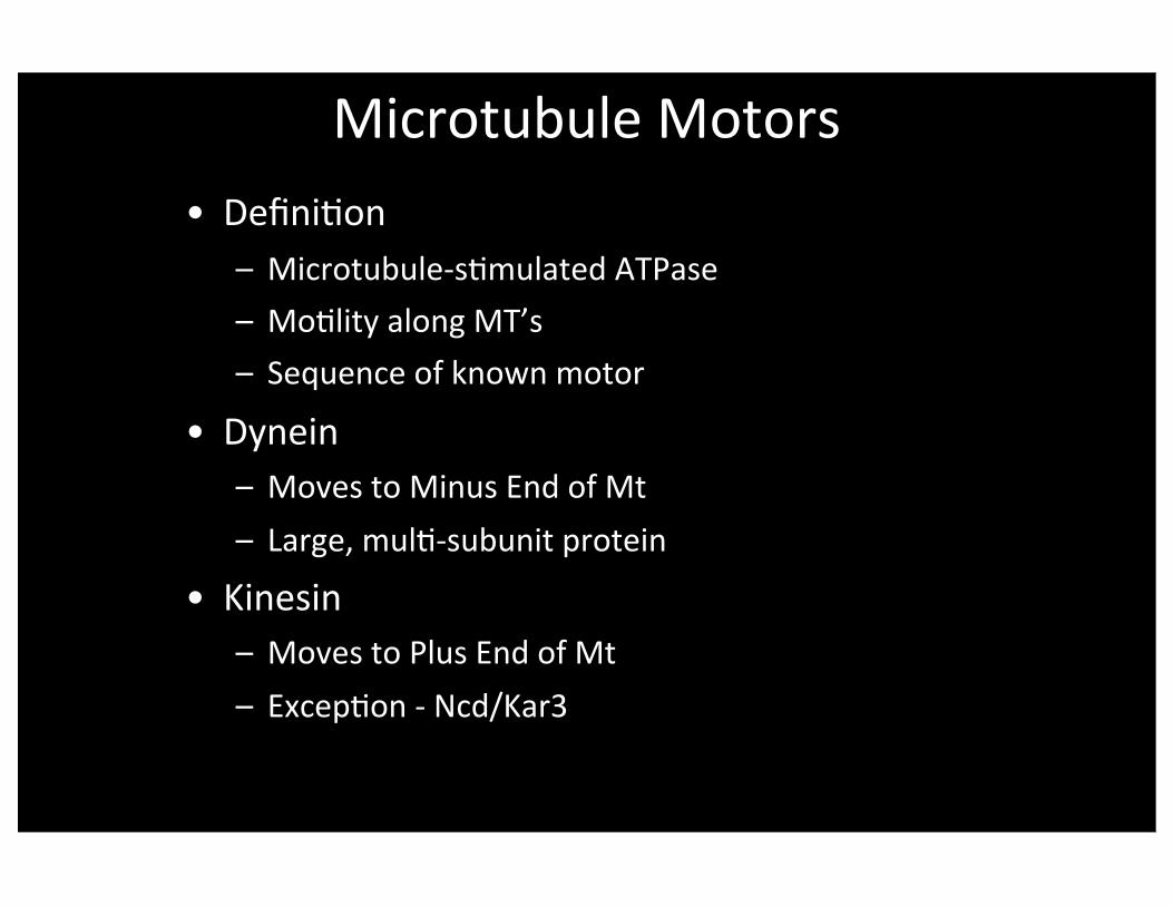
Microtubule Motors• Defini@on
– Microtubule-‐s@mulated ATPase– Mo@lity along MT’s– Sequence of known motor
• Dynein– Moves to Minus End of Mt– Large, mul@-‐subunit protein
• Kinesin– Moves to Plus End of Mt– Excep@on -‐ Ncd/Kar3

Discovery of Kinesin
• Search for Motor for Axonal Transport– Development of Video-‐enhanced DIC Imaging
• Movement Requires ATP
• AMPPNP Freezes Par@cles
• Microtubule Affinity Chromatography– Bind in AMPPNP, Release in ATP

Kinesin Structure

Kinesin Movement and Processivity

Kinesin Superfamily Structures

Kinesin Superfamily Phylogene@c Tree

Cytoplasmic Dynein
• Discovered Biochemically• Minus End Motor for Vesicle Transport• Requires Dynac@n Complex for Func@on• Moves the Mito@c Spindle

Dynein and Kinesin Motor
Domain Structures

Dynein Motor Subunit Architecture

Model for Interac@ons between Dynein, Dynac@n Complex, Microtubules, and Cargo

Membrane Trafficking -‐ ER and Golgi
• Posi@oning ER & Golgi– Golgi near MTOC
• Minus Ends are at MTOC• Golgi Posi@on Requires Dynein
– ER• Tubular network spread about the cell
• Kinesin moves the tubules peripherally

Microtubules (Red) and ER (Green)

Vesicle Traffic: Trans-‐Golgi to Plasma Membrane
• Kinesin -‐ “KIF13A”– Discovered by sequencing
– Plus-‐end Directed, fast (0.3 µm/s)
– Binds AP-‐1 (affinity chromatography) and mannose 6-‐P receptor
– Inhibit func@on (express tail as dominant nega@ve) -‐> less M6PR at cell surface

Xenopus MelanophorePigment GranuleMovement
• Vesicle Move Along Microtubules
• Vesicles Carry Dynein, Kinesin & Myosin-‐V
• Regula@on of the motors accounts for the dispersion / aggrega@on
Inward Mo@on(Movie Loops)

Xenopus MelanophorePigment GranuleMovement
• Vesicle Move Along Microtubules
• Vesicles Carry Dynein, Kinesin & Myosin-‐V
• Regula@on of the motors accounts for the dispersion / aggrega@on
Outward Mo@on(Movie Loops)

Cilia in Ac@on

Chlamydomonas Cilia Sperm Flagellum

Cilia on Surface of Epithelial Cells
Pseudostratied ciliated epithelium in trachea. Human.
Mallory Azan High magnication.

Structure of Axoneme: Cross-‐sec@on

Axonemes are Anchored at theirBase in Basal Bodies

Conversion of Sliding to Bendingto Wave Forma@on
• Slide on only side of axoneme
• Propagate down the long axis
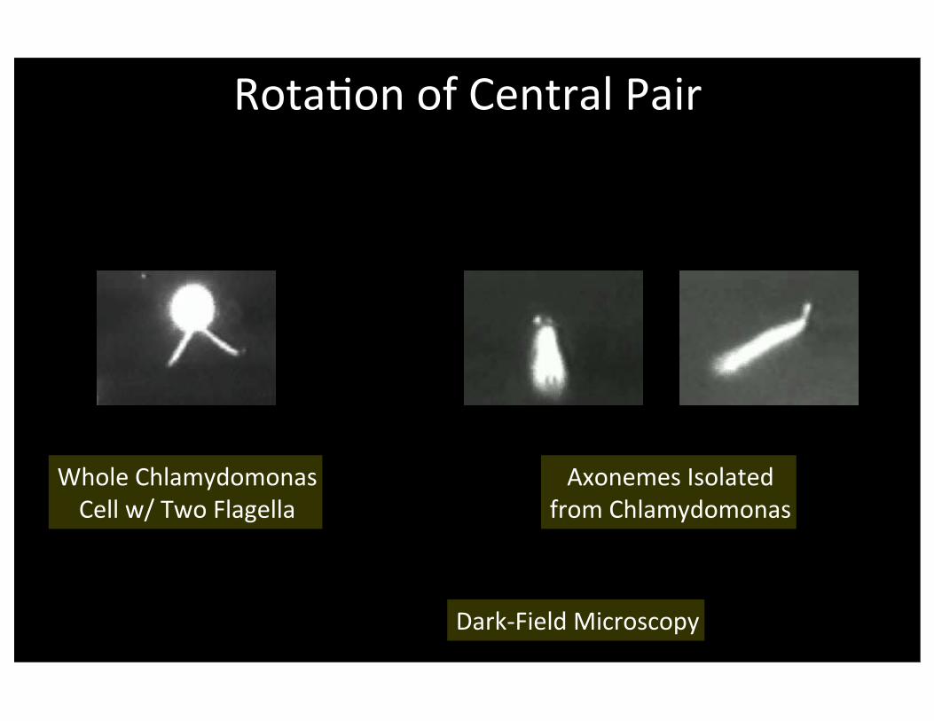
Rota@on of Central Pair
Whole ChlamydomonasCell w/ Two Flagella
Axonemes Isolated from Chlamydomonas
Dark-‐Field Microscopy

Experimental Approaches to Study Cilia in Chlamydomonas
• Axoneme 2-‐D gel -‐ 250 polypep@des!
• Mutants -‐ Collect & Characterize
• What Structures and Polypep@des Missing?

Missing Structures in Mutant

Missing Polypep@des in Mutant

Primary Cilium
• Kidney Tubule Epithelium• Defec@ve in Polycys@c Kidney Disease – 4th most common cause of kidney failure
– Autosomal Dominant
• How does loss of the cilium cause the disease?

Mitosis Background
• Names of Stages: Interphase, prophase, metaphase, anaphase, telophase
• Interphase MTs disassemble then reassembly as Spindle MTs
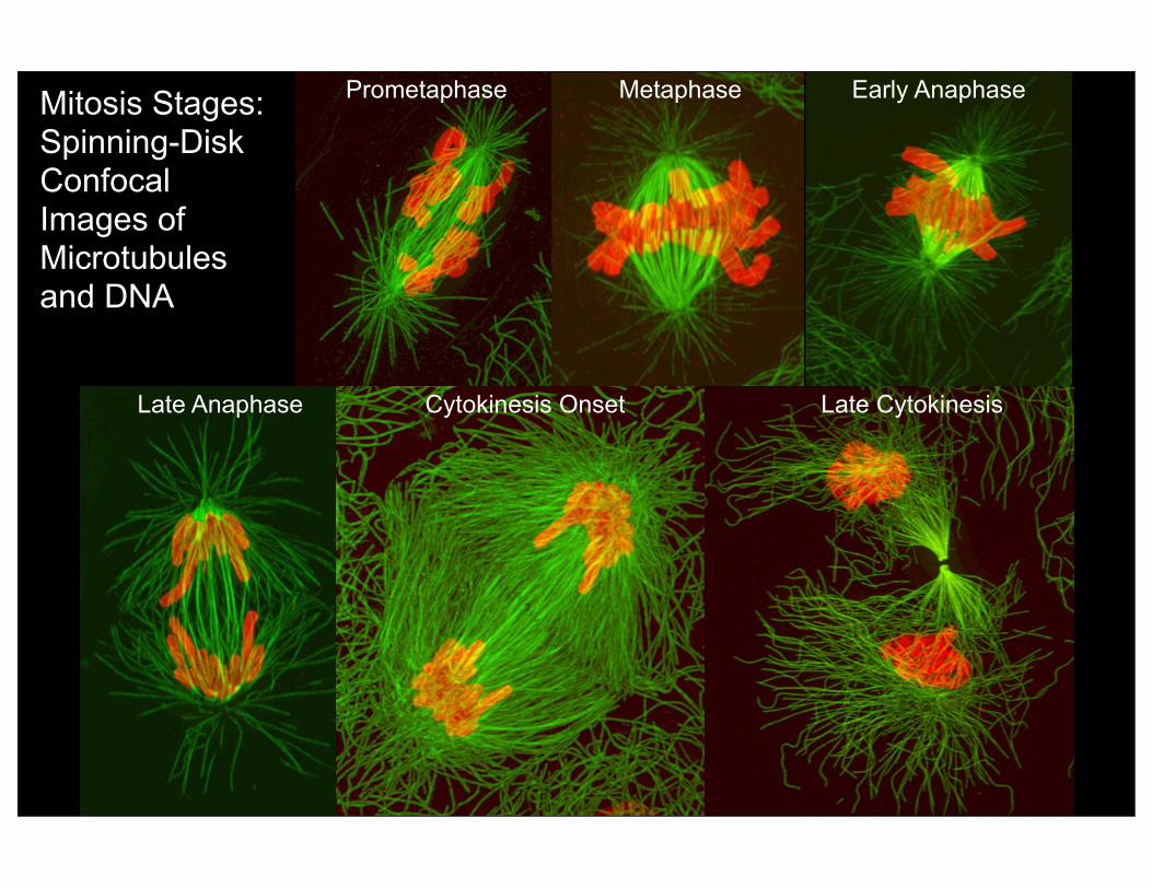
Mitosis Stages: Spinning-Disk Confocal Images of Microtubules and DNA
Early Anaphase
Late Anaphase
MetaphasePrometaphase
Cytokinesis Onset Late Cytokinesis

Boveri: Centrosome and Centriole

Centrosomes
• Animals: Centriole Pair in Amorphous Cloud
• Ends of MT’s in Cloud.No Rela@onship to Centrioles. Different from Rela@onship of Basal Body and Axoneme MT’s.
• Flowering Plants: Lack Centrioles
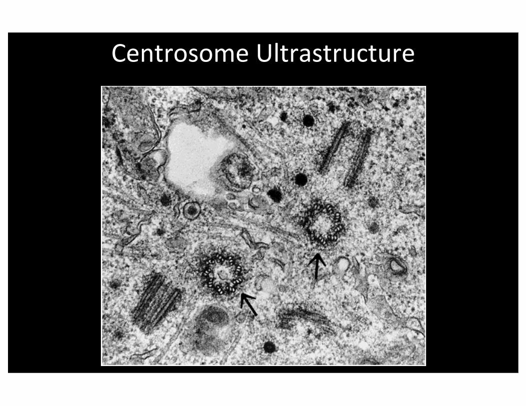
Centrosome Ultrastructure

Centriole Fine Structure

Mito@c Spindle Assembly
• Centrosome duplicates and separates
• Nuclear envelope breakdown in animals
• MT’s rearrange via dynamic instability

Spindle MT’s

Dynac@n RNAi
Control
Mito@c Spindle Rota@on in C. elegans Embryo

Chromosome Congression to Metaphase Plate
• Kinetochores capture MT’s
• Chromosome pulled to Pole– Force at Kinetochore
• Chromosome pushed away from Pole– Forces on arms– Force at Kinetochore
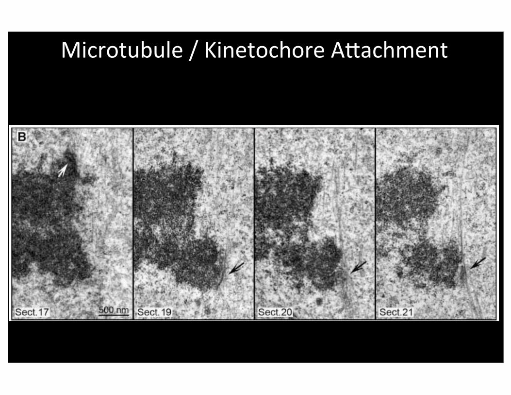
Microtubule / Kinetochore Amachment

Metaphase Normal

Types of Mt / Kc Amachment
Amphitelic
Monotelic
Syntelic
Merotelic

Metaphase -‐ Merotelic Chrom

Metaphase to Anaphase
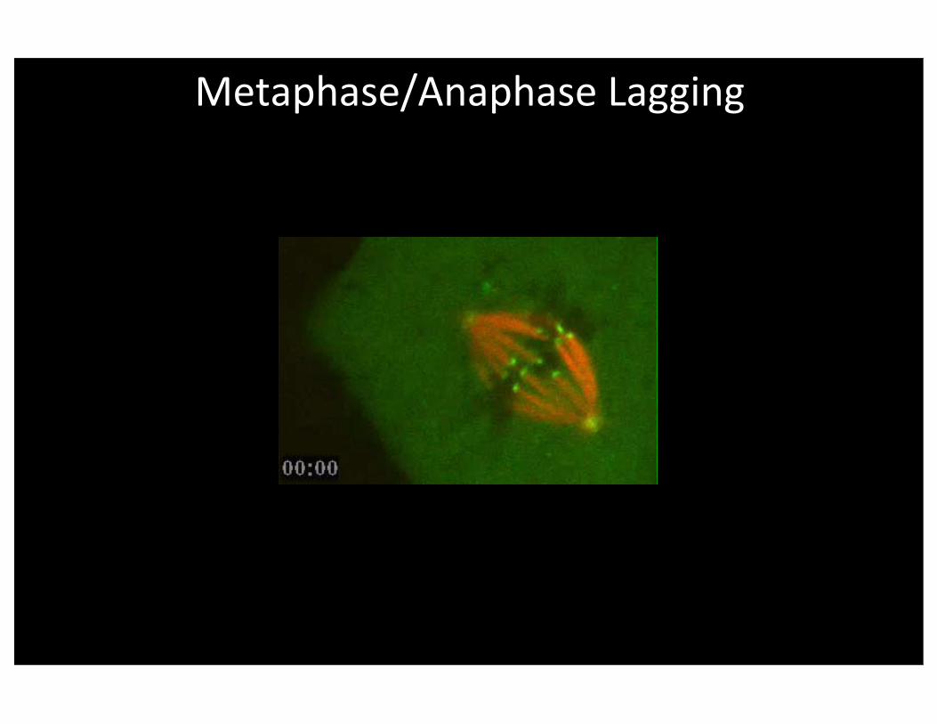
Metaphase/Anaphase Lagging

Anaphase

• Centromere splits and Chromosomes Move
Anaphase A: Chromosome to Pole
GFP-‐labeledCentromeres

Models for Chromosomes Moving to the Pole
• Treadmilling?
– Depolymeriza@on at Pole
• Depolymeriza@on at Kinetochore
– How remain bound while end shrinks?
• Motors at Kinetochore or Pole

Pac-‐Man and Poleward Flux Models for Anaphase A

Poleward Tubulin Flux in Anaphase A
Movement to Pole...
Blue: Photobleach Mark, 0.7 µm/min
Yellow: Edge of Chromosome, 1.2 µm/min

Kinetochore as a slip-‐clutch mechanism
High tension:Switch to polymeriza@on to prevent detachment
Low tension: Depolymeriza@on generates
force and movement

Anaphase B
Pole -‐ Pole Separa@on

Intermediate Filaments

Introduc@on
• Filaments 10 nm wide => “intermediate”• Present in Metazoa / Animals
– i.e. not Plants or Unicellular Organisms
• Complex Gene Superfamily– 70 in Human Genome
• Specific Expression at Different Times and Places

Intermediate Filament Biochemical Proper@es In Vitro
• Very stable. Limle subunit exchange.• Very strong. Filaments do not break.
– MT’s strong but brimle– Ac@n weak

Intermediate FilamentPoten@al Func@ons In Vivo
• Mechanical Strength of Cytoplasm
• Help a Layer of Epithelial Cells Resist Shear Stress -‐ Filaments Connect to Cell-‐cell Junc@ons
• Hold Nucleus in Center of Cell

IntermediateFilamentStructure &Assembly

Intermediate Filaments by EM:Filament Unraveling

Classes of Intermediate Filaments
Class Name CellsNumber ofIsoforms
Size(kD) Polymers
I Acidic Keratin Epithelia ~15 40-60 Obligate HeteropolymersII Basic Keratin Epithelia ~15 50-70 One acidic + one basic
III Vimentin Mesenchymal 1 53III Desmin Muscle 1 52 Homopolymers (singleIII Glial Fibrillary Glia 1 51 type of subunit) or
Acidic Protein (GFAP) co-polymers w/ eachIII Peripherin Neurons >1 58 other at varied ratios
IV Neurofilament H Neurons 1 135-150IV Neurofilament M Neurons 1 105-110 H & M each requireIV Neurofilament L Neurons 1 60-70 L for polymerIV Nestin Glial scars, Early
neurons & muscle1 240
V Lamin A All 1 60-75 Homopolymers orV Lamin B All 1 60-75 Heteropolymer

Regula@on of IF Assembly
• Notoriously Stable– No Nucleo@de
• Filaments Move Limle– Precursors Move More
• Disassemble Somewhat during Mitosis– Phosphoryla@on by Cyclin-‐depen Kinase

Vimen@n Filaments in a Cultured Cell

Vimen@n
• All Cells in Early Development• Cage Around Nucleus• Interacts with Mt’s• Vimen@n Knockout Mouse
– Ini@ally normal at gross inspec@on– Cultured cells have altered proper@es of uncertain significance

FRAP of Vimen@n vs. Kera@n in One Cell
Left: Vimentin (Green)Right: Keratin (Red)
10-min time intervals

Dynamics of Kera@n Par@cles in Periphery
11 micrometersover 10 minutes
18 micrometersover 10 minutes
Desmin
• Expressed in Muscle• Elas@c Elements to Prevent Over-‐stretching• Connects / Aligns Z lines• Knockout Mouse -‐ Deranged Myofibril Architecture

Kera@ns
• Expressed in Epithelia• Kera@n Filaments Connect to Desmosome and Hemidesmosomes
• Differen@a@on of Epidermis includes Produc@on of Massive Amounts of Kera@n
• Provides Outer Protec@on of Skin• Composes Hair, Nails, Feathers, etc.

Density of Kera@n Filaments in Outer Epidermis Layers

Kera@n Muta@ons are Basis forHuman Epidermal Diseases
• Structure/Func@on Analysis of Kera@n Assembly
• Point Muta@on in Terminal Domain Fails to Assemble
• Mutant is Dominant, even in Low Amounts, in Cultured Cells and Mice

Epidermolysis Bullosa Simplex
Wild-type Mutant

Kera@ns and EBS

Neurons
• Neurofilament H, M, L Copolymer
• Prevent Axon Breakage
• Diseases with Clumps of Neurofilaments– Superoxide dismutase model for ALS
– Clumps are secondary, not causa@ve
Neurofilament Transport in Axons
Photobleached Zone in the Middle

Neurofilament Transport in Axons
Photobleached Zone in the Middle

Lamins
• Square Larce on Inner Surface of Nuclear Membrane
• Present in Metazoans (Animals, not Plants or unicellular organisms)
• Mitosis Breakdown– Phosphoryla@on of A & C by Cyclin-‐depen Kinase
– B remains with Membrane
• Muta@ons Cause Accelerated Aging Diseases– Progerias -‐ Dominant Muta@ons

EM of Nuclear LaminaNuclear Pores

End