Embed Size (px)
Citation preview

325
Address correspondence to Egil Fosslien, M.D., Departmentof Pathology (M/C 847), College of Medicine, University ofIllinois at Chicago, 1819 West Polk Street, Chicago, IL 60612,USA; tel 312 996 7323; fax 312 996 7586; e-mail:[email protected].
Review: Molecular Pathology of Cyclooxygenase-2in Cancer-induced Angiogenesis
Egil FosslienDepartment of Pathology, College of Medicine, University of Illinois at Chicago, Chicago, Illinois
Abstract. Cancer-induced angiogenesis is the result of increased expression of angiogenic factors, ordecreased expression of anti-angiogenic factors, or a combination of both events. For instance, in coloncancer, the malignant cells, the stromal fibroblasts, and the endothelial cells all exhibit strong stainingfor cyclooxygenase-2 (COX-2), the rate-controlling enzyme in prostaglandin (PG) synthesis. In variouscancer tissues, vascular endothelial growth factor (VEGF) and transforming growth factor β (TGF- β)co-localize with COX-2. Strong COX-2 and VEGF expression is highly correlated with increased tumormicrovascular density (MCD); new vessels proliferate in areas of the tumor that express COX-2. Moreover,high MVD is a predictor of poor prognosis in breast and cervical cancers. COX-2 and VEGF expressionare elevated in breast and prostate cancer tissues and their cell-lines. In vitro, PGE2 induces VEGF.Supernatants of cultured cells from breast, prostate, and squamous cell cancers contain angiogenic proteinssuch as COX-2 and VEGF that induce in vitro angiogenesis. A selective COX-2 inhibitor, NS-398,restores tumor cell apoptosis, reduces microvascular density, and reduces tumor growth of PC-3 prostatecarcinoma cells xenografted into nude mice. The COX-2 produced by a malignant tumor and COX-2produced by the surrounding host tissue both contribute to new vessel formation, which explains howselective COX-2 inhibition reduces tumor growth where the tumor COX-2 gene has been silenced bymethylation. (received 6 June 2001, accepted 8 July 2001)
Keywords: cancer, angiogenesis, microvascular density, COX-2, PGE2, VEGF, TGF-β, iNOS, TSP-1,p53, hypoxia, NSAIDs, celecoxib, rofecoxib
Introduction
Angiogenesis, the formation of new vessels fromexisting vessels, is an important feature ofembryogenesis, inflammation, and cancer growthand metastasis [1]. Chronic inflammation is a riskfactor for cancer [2], but the exact reason why isunknown. At the site of inflammation, cyclo-oxygenase-2 (COX-2) is the rate-limiting enzymein the synthesis of pro-inflammatory and angiogenicprostaglandins (PG) such as PGE2, which inducesmetalloproteinases (MMP) and vascular endothelialgrowth factor (VEGF) [3,4]. The antiphlogistic andanalgesic effects of nonsteroidal anti-inflammatory
drugs (NSAIDs) are largely due to their inhibitionof COX-2 [5].
A variety of human malignancies overexpressCOX-2 and prostaglandin [6] along with VEGF andtransforming growth factor-beta (TGF-β). Forinstance, in colorectal carcinoma strong COX-2expression, as evidenced by immunostaining, ishighly correlated with tumor microvascular density:a large number of small vessels form around the areasthat express COX-2 [7]. Furthermore, the expressionof COX-2, TGF-β, and VEGF in the same areas ofthe tumor suggests their coordinated expression inthe cancer-induced angiogenesis.
Tumor invasion into the local tissue and tumorgrowth at the site of metastasis are preceded bytumor-induced proliferation of an abundantlyvascular stroma, for instance in breast cancer [8].For such tumors, anti-angiogenesis therapy is anencouraging new approach. COX-2, the angiogenic
0091-7370/01/0400/0325, $6.00; © 2001 by the Association of Clinical Scientists, Inc.
Annals of Clinical & Laboratory Science, vol. 31, no. 4, 2001

326
molecules associated with it, and their interrelatedsignaling pathways in malignant tumors are thereforeof major interest in understanding the tumor-induced angiogenesis. Studying the expression ofCOX-2 in cancer tissues and its role in the growthof malignant tumors is important because NSAIDsmight help to prevent cancer [9]. Furthermore,selective COX-2 inhibitors are available that blockthe effects of COX-2 expression but spare theexpression of COX-1 [5,10].
Vasculogenesis and Angiogenesis
A. The yolk sac
During implantation, trophoblast invasion,
vasculogenesis, and angiogenesis are essential for theformation of the placenta. The human trophoblastsecretes metalloproteinases, and metalloproteinaseinhibitors can abort in vitro invasion. In contrast tothe invasion by a malignant tumor, the invasion bythe trophoblast is stringently controlled. However,the controlling factors are unknown [11]. Manyangiogenic factors that are highly expressed inmalignant tumors are essential for blastocystimplantation and yolk sac vasculogenesis, for form-ation of new vessels from stem cells, and for yolk sacangiogenesis (Fig. 1) [12,13]. Vasculogenesis requiresthe development of the hemangioblast, the precursorfor hematopoesis and vessel development, into theendothelial cell. A multitude of endothelial cells form
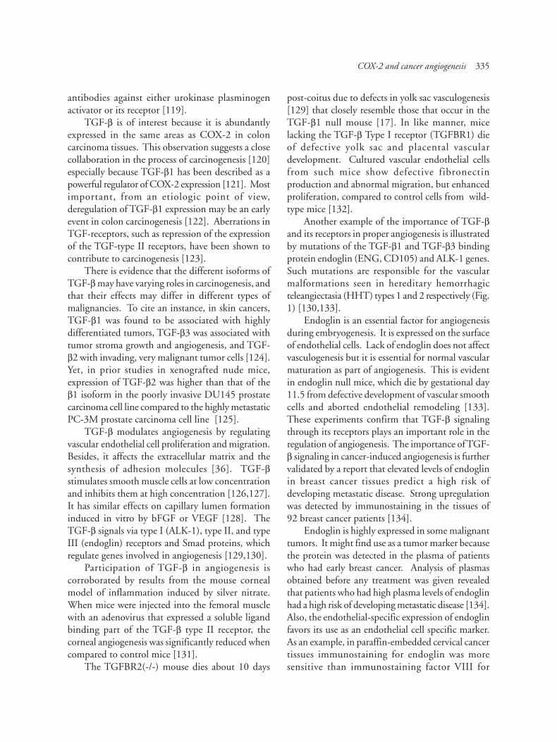
Fig. 1. Cyclooxygenase (COX-2), prostaglandin (PG), and transforming growth factor (TGF)-β are involved in vasculardevelopment and vascular pathology from vasculogenesis and angiogenesis, to atherosclerosis and cancer. Lack ofmaternal COX-2 or its inhibition by a nonsteroidal anti-inflammatory drug (NSAID) prevents implantation. Lack ofTGF-β1 is lethal due to faulty vasculogenesis and angiogenesis during embryogenesis with a penetration of about 50%.Lack of the TGF-β receptor component endoglin (ENG) prevents proper angiogenesis. Disruption of the PGE2receptor EP4 gene prevents closure of the ductus arteriosus. COX-2 and TGF-β co-localize in atherosclerotic lesionsand in carcinogen-induced colon carcinomas, suggesting coordinated expression. In the cancer, COX-2 induces newvessel formation via PGE2 that induces vascular endothelial growth factor (VEGF). In some poorly differentiatedmalignancies, vasculogenesis is also induced. A human cancer tissue may overexpress one or more of the proteinsindicated in the highlighted boxes. Text in italics indicates an agent used in vitro.
Annals of Clinical & Laboratory Science

327
the capillaries, which mature into veins and arteriesduring angiogenesis.
Expression of COX-2 and TGF-β and itsreceptors is fundamental for blastocyst implantation[14] and yolk sac vascularization. Gene knockoutexperiments have shown that expression of COX-2, prostaglandin and prostaglandin receptors, andTGF-β and TGF-β receptors are essential forembryonic vasculogenesis and angiogenesis.
For successful implantation, the blastocyst mustinduce the expression of maternal COX-2 in theuterus [15]. Studies of the COX-2 null mouse provethe vital importance of the COX-2 expression;complete failure of implantation renders the micesterile. Half of the COX-2 knockout mice developcardiac fibrosis and all develop renal dysplasia [16].
In the mouse, disruption of the gene for TGF-β1, a strong inducer of COX-2, causes defective vesselformation and may cause death. However, there aredosage and mouse-strain effects [17,18] thatmodulate the severity of this gene disruption. One-half of TGF-β1(-/-) and one quarter of TGF-β(+/-)mice died at about 1.5 wk post-conception becauseof failing endothelial cell differentiation [17].
VEGF can regulate vascular permeability and isan important mediator of vasculogenesis andangiogenesis [19]. VEGF signals are conveyed viathe VEGF receptors Fli-1 and Flk-1. Fli-1 isexpressed in the hemangioblast. Disruption of theFlk-1 gene is lethal in mice at about day 9 ofgestation, due to lack of hematopoesis andvasculogenesis. Normally, Fli-1 is strongly expressedduring physiological vasculogenesis and angiogenesisin embryos and is re-expressed during tumor-inducedangiogenesis [20].
B. Vascular remodeling
COX-2 and prostaglandins are evidently involvedin vascular remodeling. For example, rapid andselective physiological vascular remodeling occursduring closure of the ductus arteriosus and there maybe a difference in response of the prenatal andpostnatal ductus.
Firstly, a lack of the PGE2 receptor EP4 in theEP4-null mice causes the ductus to remain open.By comparison, indomethacin administered duringpregnancy may cause premature closure of the ductus
arteriosus. In fetal lambs, the COX-2 is expressedin the endothelium of the ductus arteriosis, whileCOX-1 is expressed in both the endothelium andthe muscular layers [21].
Secondly, the inhibition of prostaglandinsynthesis by indomethacin constricts the fetal ductusarteriosus in normal mice, but not in EP4(-/-) mice,which do not express the prostaglandin EP4 receptor.Such knockout mice generally die shortly after birth.They (as well as the few that survive for a longertime) have a patent ductus arteriosus (Fig. 1) [22].
In mice, a lack of either COX-1 or COX-2 orboth does not cause premature closure of the ductusarteriosus in utero. Furthermore, lack of COX-1does not affect postnatal life. In contrast, about onethird of COX-2 null mice and almost 80% of COX-2(-/-) + COX-1(+/-) mice die within 48 hr afterbirth. Importantly, the absence of both COXisoforms causes postnatal death within 12 hr [23].This suggests that maternal prostaglandins alone canmaintain intrauterine patency of the ductusarteriosus. During closure of the ductus arteriosus,the expression of the 3 TGF-β isoforms increasessignificantly. Over the 10 postnatal days, it increasesfrom the very low levels before birth. The highestlevels are seen in the neo-intima and the outer musclemedia, but TGF-β is not detected in vascularendothelial cells [24].
With advancing age, there is diffuse intimalthickening (DIT) of the elastic arteries (Fig. 1)[25,26] and there is evidence that TGF-β is involvedin vascular remodeling. A comparison of 30-mo-old versus 6- mo-old Fisher rats revealed significantincreases of TGF-β and MMP-2 in the intima, whichwas 5-fold thicker in the older rats [27].
C. Inflammation
Chronic inflammation is a risk factor for a variety ofhuman malignancies [2]; many of the angiogenicproteins expressed in cancer tissues are the same asthose expressed at sites of inflammation [5]. In vitrostudies and animal models of inflammatoryangiogenesis show that COX-2 plays a pivotal rolein new vessel formation at sites of inflammation. Forinstance, COX-2 is strongly expressed and producespro-inflammatory prostaglandins such as PGE2.Besides, PGE2 can induce VEGF, for instance in
COX-2 and cancer angiogenesis

328
synovial fibroblasts in vitro. The signaling occursvia the prostaglandin EP2 receptor and protein kinaseA pathways [4].
Angiogenesis is an essential part of the formationof granulation tissue [28]. In an animal model ofwound healing, application of neutralizing anti-VEGF antibody to a post-surgical wound demon-strated that VEGF is an essential angiogenic signaltransducer in the formation of wound granulationtissue. Neutralizing anti-VEGF antibody reducedgranulation tissue VEGF levels and new vesselformation [29].
COX-2 and VEGF are both expressed in ratsponge-implant-angiogenesis [30]. COX-2 is locatedin the endothelial cells of the new vessels that areformed in the sponge-granuloma tissue [31]. Theexperimental angiogenesis can be blocked by eithera non-selective NSAID, indomethacin, or byselective COX-2 inhibitors, NS-398 or JTE-522, aswell as by VEGF anti-sense oligonucleotides [30].VEGF-induced angiogenesis in a mouse corneamodel of angiogenesis can be inhibited by NS-398or by cyclosporin-A administered systematically.Importantly, PGE2 restores corneal angiogenesiseven in the presence of these inhibitors [32].
In a rat model of pleurisy, carrageenan-treatedrats express elevated levels of COX-2 in the lung.Treatment of rats with ip melatonin prior to thecarrageenan administration inhibits the COX-2expression [33]. High dosages of melantonin wereused in this study, ranging from 12.5 to 50 mg/kg.However, it is remarkable that melatonin can inhibitCOX-2 expression.
The Malignant Tumor
A. Cancer-induced angiogenesis
The pathological growth of human cancers resemblesin several ways the physiological growth of theimplanting blastocyst. Both need to evade theimmune defenses of the host and secure a vascularsupply in order to grow [34,35]. To induce newvessel formation, the cancer must induce theexpression of angiogenic growth factors and theirreceptors and regulate the expression of proteolyticenzymes and adhesion molecules [36]. In a mannersimilar to the blastocyst, the malignant tumor forces
the host tissue to grant it a supply of oxygen andnutrients that enables the tumor expansion, whichotherwise is confined to 1-2 mm by the maximumoxygen diffusion limit. Progression from limitedtumor growth to invasion and metastases requiresdegradation of the extracellular matrix by proteolysisby metalloproteases (MMP, matrixins), induction ofangiogenesis, tumor cell migration, and modificationof cell adhesion [37,38].
COX-2 is important because it inducesmetalloproteinase and strongly induces VEGF, thecommon angiogenic factor, which thereupon directstumor-induced angiogenesis [39]. Together, thesefactors contribute to tumor growth, invasion, andmetastasis. Magnetic resonance imaging (MRI)shows that tumor-induced angiogenesis follows acommon pattern, leading to a well-perfused tumorperiphery but a residual necrotic core. Host vesselsenter the periphery of the tumor, and the perfusionfront moves toward the center of the tumor [40].
B. COX-2 expression in cancer tissues
Enhanced COX-2 expression has been demonstratedin numerous types of cancer cells and tissues, suchas colorectal cancer [5]. For example, COX-2expression was increased in all but 2 of 20 lungcancers, 14 of 20 colon adenocarcinomas, and 11 of20 breast tumors, but not in epithelial cells of thecorresponding non-tumor tissue. Expression ofCOX-2 was minimal in the stromal cells of thetumors and in normal tissues [41]. Furthermore,immunohistochemical expression of COX-2 wassignificantly increased in malignant cells of all but 1of 13 cervical carcinomas, compared to non-tumorcervical tissue [42].
High expression levels of COX-2 may foretell apoor prognosis; for instance, in 24 patients withcervical carcinoma who had been treated withradiation, increased expression of COX-2 wasassociated with shorter survival [43]. In patients withgastric cancer, the tissue COX-2 levels weresignificantly higher in invasive versus non-invasivetumors [44]. A study of 63 patients with colorectalcancer showed that high COX-2 expressioncorrelated with tumor recurrence, and particularlywith hematogenous spread of the tumors [45]. Thefollow-up period in this study (6 to 98 mo, average
Annals of Clinical & Laboratory Science

329
60 mo) was significantly longer than in earlier studiesthat did not show correlation between COX-2expression and survival. The Kaplan-Meyer curvesclearly showed longer survival for patients with lowexpression of COX-2 in their tumor tissues [45].
Prostatic tissue cancer cells showed significantlyelevated, mainly intracytoplasmic, expression ofCOX-2 compared to the COX-2 expressed along thecell membrane in benign prostatic hyperplasia. Theintensity of staining of COX-2 was significantlyhigher in poorly-differentiated versus well-differentiated prostate cancers and correlated withthe Gleason grade [46]. Other studies corroboratethese findings. For instance, significant elevationsof COX-2 protein and mRNA were demonstratedin prostate cancer cells by immunostaining and RT-PCR in a series of 28 carcinomas, compared tocontrols (8 cases of benign prostatic hyperplasia and8 prostate samples that were free of tumor) [47]. Inanother series of prostatic carcinoma, all but 4 of 31cancers showed intense and uniform immuno-staining for COX-2 [48]. COX-2 was expressed inall but 2 of 28 primary skin melanomas (16 showingmoderate to strong expression), but not in 4 benignnevi [49]. All except 1 of 29 retinoblastomas over-expressed COX-2 [50].
In a study of 100 patients with colon cancer,those with COX-2-expressing tumors hadsignificantly shorter survival time than those withCOX-2-negative tumors; moreover, correlation wasnoted between COX-2 expression and microvasculardensity [7]. Immunostaining for CD-34 to showthe microvascular density of the tumors revealed thata large number of small vessels had formed aroundareas that expressed COX-2 [7]. In addition, a studyof paraffin-embedded tumor tissue from 120 patientswith breast cancer showed that a high tumor MVDcorrelated with the histological grade and with poorsurvival [51]. In another study of patients with breastcancer, detecting increased MVD of axillary lymphnodes by iv digital subtraction angiography predictedlymph node metastases [52], suggesting that MVDmay be reliably determined using non-invasivetechniques.
These findings in malignant tumors suggest thathigh COX-2 expression is associated with tumor-induced angiogenesis, invasion, and metastases.
C. VEGF in cancer tissues
Enhanced expression of VEGF has beendemonstrated in, for instance, ovarian [53], breast[54], renal [55,56], and esophageal [57] cancers andin malignant melanomas [58]. The malignant cellsin breast cancers showed variable expression ofVEGF, but no VEGF expression was found inepithelial cells in areas of breasts that were free oftumor [54]. VEGF expression was increased in 55prostate cancers compared to 5 cases of prostaticadenomas and 20 normal prostate samples; elevationof VEGF correlated with tumor MVD, tumor grade,and stage [59]. In 85 patients that underwentsurgical resection of stage I non-small cell lungcancers, high tumor MVD and poor prognosiscorrelated with high levels of VEGF expression [60].
Immunostaining of VEGF might be an indicatorof aggressiveness in serous ovarian tumors. Firstly,VEGF immunostaining of tumor tissue wassignificantly greater in 32 invasive tumors, comparedto16 borderline and 10 benign ovarian tumors.Secondly, there was significant correlation betweenVEGF elevation and metalloproteinase-2 (MMP-2)elevation [53]. Moreover, in 44 cases of hepato-cellular carcinoma, the microvascular densitycorrelated significantly with the expression levels ofVEGF mRNA. Poorly encapsulated tumors showedhigher levels than well-encapsulated tumors.Interestingly, serum VEGF concentration wassignificantly higher in patients with tumors,compared to patients with benign liver disease [61].
However, high MVD does not always indicatea poor prognosis, particularly in cases with extensivetumor necrosis. For instance, in a series of 49localized renal cell carcinomas, MVD was inverselycorrelated with the magnitude of tumor necrosis; lowMVD and extensive necrosis predicted a pooroutcome [62]. And, in a study of 69 patients withstage I-II non-small-cell lung carcinomas, the cancercells, stromal fibroblasts, and endothelial cells in thetumors all exhibited strong VEGF staining.However, the level of tumor microvascular densityby CD34 immunostaining in these cases correlatedneither with the tumor levels of VEGF and itsreceptors, nor with the patients’ survival [63].Determining the levels of VEGF and its receptorsflt-1 and KDR/flk-1 by immunostaining in 35
COX-2 and cancer angiogenesis

330
adenocarcinomas, 6 papillary serous carcinomas, and6 carcinosarcomas was ineffective in predictingmetastases, recurrence, or survival [64]. On the otherhand, immunostaining for VEGF may be of use todifferentiate malignant melanomas from benign nevi.Cytoplasmic VEGF was found in almost half of 45malignant melanomas and the intensity ofimmunostaining was related to the Clark level. Incontrast, VEGF was not detected in benign nevi [58].
D. Haptoglobin
It has been reported that the multifunctionalhaptoglobin molecule is angiogenic (for a review see[5]). Haptoglobin expression is regulated bydexamethasone and by IL-6, which is induced byPGE2 [65]. Whether haptoglobin induces VEGFis unknown. Haptoglobin expression has beendetected in human breast carcinoma tissue extractsand cell lines [66,67]. Furthermore, in vitro celllines established from squamous cell and ovariancancers and from embryonic lung secrete a multi-protein complex that has subunits with sequencehomology to the β chain of haptoglobin [68].
E. Vascular patterns in malignant tumors
Although tumor-induced angiogenesis resembles theangiogenesis observed at sites of inflammation, thereare important differences between physiologicalangiogenesis and cancer-induced angiogenesis. Inaddition to increased microvascular density in andaround many tumors, the pattern of neoplasia-induced vascularization differs from normal vascularpatterns. Measuring and evaluating this differencemight have significant diagnostic and prognosticvalue. To illustrate, in uveal melanomas, the presenceof parallel vessels with cross-linking indicates a poorprognosis [69]. Moreover, the fractal character oftumor-induced new vessel proliferation reflects ahigher degree of irregular branching and vesseldistribution, compared to physiological angiogenesis.This difference is probably caused by more variablelocal distribution of vascular growth factors withinthe tumor, owing to the inherent genetic instabilityand progressive molecular diversity of malignant cells[70]. Caution is advised when extensive necrosis ispresent. In the renal carcinoma cases mentioned
above, the microvascular complexity as evidenced bythe fractal dimension of vessels visualized with CD34staining in the tumors correlated inversely with theextent of tumor necrosis, which predicted a pooroutcome [62].
F. Cancer cell supernatants
Supernatants of cultured cells from several types ofcancer contain angiogenic factors, such as fibroblastgrowth factor-2 (FGF-2) in prostate cancer [71] andTGF-β, PGE2, and VEGF in squamous cell cancer[72]. Supernatants of cultured cells from head andneck squamous cell cancer, breast cancer, and livercancer secrete PGE2, VEGF, and TGF-β and inducein vitro angiogenesis [72-74]. VEGF expression iselevated in the cancer tissue and cell lines derivedfrom breast tumors [54,75]. Prostaglandin E2induces VEGF in a variety of cell lines and VEGFexpression strongly stimulates angiogenesis in vitro.As examples, PGE2 induces VEGF in cell linesderived from the rat osteoblasts [76] and Müller cells[3], as well as human rheumatoid synovial fibroblasts[4] and monocytes [77].
Metalloproteases play an important role intumor cell invasion by proteolysis of the extracellularmatrix. As a case in point, cultured prostate cancercells release proMMP-2, proMMP-9, and MMP-9into the medium and invade through Matrigel. Thefactor release and tumor cell invasion can be inhibitedby a PLA2 inhibitor, 4-bromophenacyl bromide, andby a selective COX-2 inhibitor, NS-398. Inhibitionof lipoxygenase with esculetin does not affect MMPsecretion. On the other hand, the tumor cell invasioncan be restored by PGE2 supplementation. Theseexperiments indicate that cycloxygenase-2 inducesmetalloproteinases (MMP) via PGE2 [78].
G. Tumor xenograft-induced angiogenesis
Experiments have shown that COX-2 and VEGFare essential angiogenic proteins that determine thegrowth of various xenografted human cancer cells.In addition, such experiments have corroborated thesequential propagation of the angiogenic signal fromCOX-2 via PGE2 to VEGF. The tumor-inducedCOX-2 expression in the host tissue located adjacentto the malignant tumor adds to the angiogenesis
Annals of Clinical & Laboratory Science

331
induced by the COX-2 produced by the cancer cells,and both enhance tumor growth.
The significance of host COX-2 expression incancer growth is illustrated by the proliferation ofLewis lung cancer cells xenografted into the COX-2(-/-) mouse. The xenograft growth is significantlyreduced compared to that in the wild-type mouse.Expression of VEGF by host COX-2 null fibroblastsis reduced >90% and is comparable to the reducedVEGF expression by wild-type murine fibroblastsfollowing treatment with a selective COX-2 inhibitor[79].
The importance of VEGF in tumor-inducedangiogenesis is evidenced by the effect of neutralizinganti-VEGF antibody in nude mice xenografted withtumor. The antibody treatment completely inhibitsthe angiogenesis by xenografted cell lines derivedfrom human rhabdomyosarcoma and prostatecancer. The tumors become dormant after the initialangiogenesis-independent growth phase [80], thusindicating the critical importance of tumor-inducednew vessel formation in the tumor growth.
Molecular Pathology
A. Angiogenic factors
1. Cyclooxygenase and carcinogenesisTransgenic overexpression of either COX-1 or COX-2 can transform cells [81,82]. No sequenceaberration of the COX-2 gene has been reported inmalignant cells. A great variety of cancer cells andtissues overexpress COX-2 and prostaglandins thatreduce cancer cell apoptosis [6] and induceangiogenesis via VEGF. NSAIDs inhibit the growthof such cancers and the anti-neoplastic effects of theNSAIDs are partly due to COX-2 inhibition [5].
On the other hand, neoplastic transformationappears to be independent of cyclooxygenase. Thus,Ha-ras or SV40 cause in vitro transformation offibroblasts that lack COX-1 or COX-2, or both. Bycomparison, when the genes for either or bothenzyme isoforms are present, the correspondingprotein is highly expressed in the transformed cells.These results suggest that cyclooxygenases play a rolein tumorigenesis at a later step [83].
Several oncogenes and growth factors can induceCOX-2 expression. In cultured CaCo-2 colon
carcinoma cells, COX-2 is upregulated by insulin-like growth factor (IGF)-II via the IGF-I receptor[84]. Besides, TGF-α, epidermal growth factor(EGF), TGF-β, and hepatocyte growth factor (HGF)can all induce COX-2 [5,6].
2. Prostaglandin and carcinogenesisProstaglandin E2 induces VEGF and basic fibroblastgrowth factor (bFGF) [3]. There is evidence thatbFGF might contribute to cancer-induced new vesselformation. For example, when co-cultured withbovine aortic endothelial cells, 4 human glioma celllines that expressed high levels of bFGF mRNAinduced the endothelial cells to form tubes. Thetube formation was blocked by supplementationwith anti-bFGF antibody. Moreover, 3 humanglioma cell lines with low expression of bFGF failedto induce tube formation [85].
PGE2 induces the expression of metalloprotein-ases by a multistep process involving NF-kappaB. Itwas recently suggested that PGE2 might enhancethe invasive potential of colorectal carcinomathrough activation of the phosphatidylinositol 3-kinase (PI3K)/protein kinase B (Akt/PKB) pathway.PGE2 treatment of LS-174 human colorectalcarcinoma cells increased their motility and alteredtheir shape [86]. Prostaglandin E2 suppresses bothcellular and humoral immunity [2,87] (reviewed in[6]). Inhibition of cyclooxygenase with NSAIDsrestores the immune reactivity [5,88].
3. VEGF and carcinogenesisVascular endothelial cell growth factor (VEGF) isthe primary member of a family of growth factors.VEGF and its homologues can convey regulatorysignals via receptors KDR/Flk-1, Flt-1, and Flt-4 [89](Fig.2). However, VEGF and its homologues havedifferent effects on different cells. For instance, theorf paracoxvirus encodes the VEGF-E homologue[90] that causes orf disease in sheep and is associatedwith vascular proliferation at the site of infection inhuman erythema multiforme [91,92].
The VEGF gene is located at chromosomeregion 6p12 [93]. A 600 bp genomic fragment wasmapped to chromosome region 6p12 and was highlyamplified in almost half of a series of human non-small cell lung cancer; however, it lacked sequencehomology to any known protein [94].
COX-2 and cancer angiogenesis
332
VEGF stimulates the activation of cPLA2 andthe release of arachidonic acid by human umbilicalvein endothelial cells in vitro [95]. VEGF inducesCOX-1 but not COX-2 in cultured endothelial cells;it has been suggested that COX-1 has a physiologicalrole in maintenance of vascular structures [96]. Sucha feedback loop might help to maintain a basal levelof VEGF expression.
4. Aging and carcinogenesisWith advancing age, both angiogenic and anti-angiogenic changes have been observed. In vitrostudies suggest that as the generation of reactiveoxygen species (ROS) increases with advancing age,there is increased expression of COX-2. The ROSreduces IkappaB expression and enhances theexpression of NF-kappaB, a strong COX-2 inducer[97]. On the other hand, as the oxidative stressincreases with age, the mitochondrial function maydecline [98,99], and there is some evidence that
decline of entothelial energy metabolism may inhibitangiogenesis [100].
B. Anti-angiogenic proteins
Physiological angiogenesis is the result of a dynamicbalance between angiogenic and anti-angiogenicfactors [101]. Orderly angiogenesis is essential forthe endometrial cycle, for inflammation, and for thegranulation tissue of wound repair [29]. Derangedexpression of anti-angiogenic factors caused byviruses or by loss of function of tumor suppressorgenes has been linked to various humanmalignancies. Because they can counterbalance theangiogenic effects of COX-2, a few anti-angiogenicfactors are discussed below.
1. Thrombospondin-1 (TSP-1)Reports on the role of the glycoprotein TSP-1 incarcinogenesis are controversial. Some reports
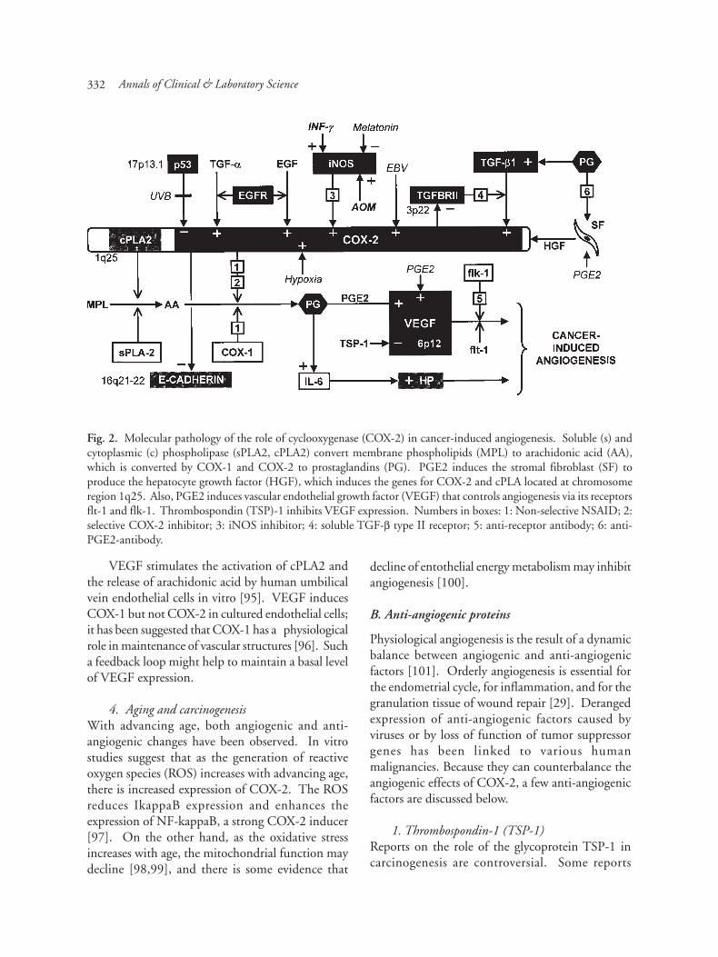
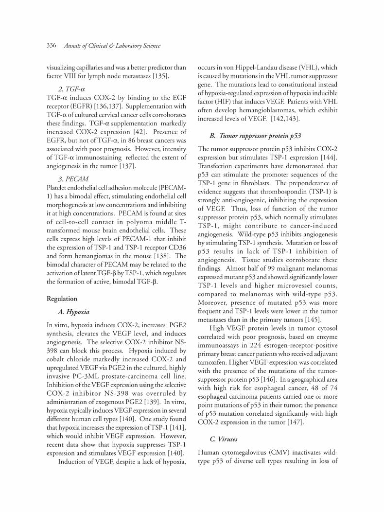
Fig. 2. Molecular pathology of the role of cyclooxygenase (COX-2) in cancer-induced angiogenesis. Soluble (s) andcytoplasmic (c) phospholipase (sPLA2, cPLA2) convert membrane phospholipids (MPL) to arachidonic acid (AA),which is converted by COX-1 and COX-2 to prostaglandins (PG). PGE2 induces the stromal fibroblast (SF) toproduce the hepatocyte growth factor (HGF), which induces the genes for COX-2 and cPLA located at chromosomeregion 1q25. Also, PGE2 induces vascular endothelial growth factor (VEGF) that controls angiogenesis via its receptorsflt-1 and flk-1. Thrombospondin (TSP)-1 inhibits VEGF expression. Numbers in boxes: 1: Non-selective NSAID; 2:selective COX-2 inhibitor; 3: iNOS inhibitor; 4: soluble TGF-β type II receptor; 5: anti-receptor antibody; 6: anti-PGE2-antibody.
Annals of Clinical & Laboratory Science
333
indicate that thrombospondin inhibits angiogenesisin vitro, while other reports state the opposite. Somestudies find elevated levels of TSP-1 in cancer tissues,while other studies report decreased levels. However,as explained below, the strongest evidence suggeststhat TSP-1 is a major anti-angiogenic protein. Mostimportantly, transgenic expression of TSP-1 can limittumor angiogenesis and growth. For instance,injection of an adenovirus containing TSP-1 plasmidinto pre-established tumors reduces tumor-inducedangiogenesis, tumor growth, and metastatic spread.The overexpression of TSP-1 in squamous carcinomacell lines inhibits their in vivo growth. Dependingupon the cell-line used, overexpression of TSP-1either prevents xenograft growth or, when a tumordevelops, it exhibits marked central necrosis [102].
Cholangiocarcinoma is a tumor with lowvascularity compared to hepatocellular carcinoma.When tumor tissues were compared to the matchingnon-tumor tissue, there were significant differencesin the ratio of TSP-1 to VEGF expression in thesetwo types of tumor. Cholangiocarcinomas showedmuch higher TSP-1 and lower VEGF expression thanhepatocellular carcinomas [103]. These findingsseem to corroborate that TSP-1 can inhibit VEGFexpression and reduce tumor-induced angiogenesis.On the other hand, TSP-1 was strongly expressedand the expression levels were associated with highmicrovascular density in 87 of 98 pancreaticcarcinomas. It was proposed that TSP-1 expressionplayed a principal role in the new vessel formationand the spread of these tumors [104]. However, invitro, thrombospondin-1 inhibits endothelial cellmigration and tube formation. The inhibitory signalis transmitted through CD36, an endothelial celltransmembrane glycoprotein and is completelydependent on its expression [105]. As discussedbelow, both TSP-1 and CD36 are downregulatedby the platelet endothelial cell adhesion molecule(PECAM) [106].
In vitro assays and tissue culture experimentsindicate that TSP-1 may also inhibit angiogenesisby inhibiting MMP activity [107]. Almost half of99 malignant melanomas expressed mutant p53 andsignificantly lower TSP-1 levels and highermicrovessel counts than the tumors with the wild-type p53. Besides, the presence of mutated p53
was significantly more frequent and the TSP-1 levelssignificantly lower in the tumor metastases than inthe primary tumors [108]. In contrast,immunostaining for p53 and TSP-1 in a series of 65colon cancer tissues showed accumulation of p53 in42 cases with reciprocal expression of TSP-1 in theareas that expressed p53, suggesting that p53 cansuppress TSP-1 in vivo [109].
Surprisingly, a TSP-1 plasmid expression vectorinserted into a human prostate-carcinoma cell-linefailed to reduce the growth of the cancer cells in vitro.However, when the cells were transfected along witha liposomal agent and then xenografted into nudemice, there was reduced microvessel density andextensive necrosis in the tumors compared toxenografted but non-transfected tumor cells [101].
Mevalonate induces the expression of TSP-1(Fig. 3). As a case in point, incubation of culturedhuman vascular smooth muscle cells for 24 hr withlovastatin at concentrations as low as 1 µM/Lsignificantly inhibited TSP-1 expression. Theexpression of TSP-1 was restored by co-incubationwith mevalonate [110].
2. Metalloproteinase inhibitorsIntroducing a recombinant adenovirus containingthe matrix metalloproteinase inhibitor TIMP-2 intotumor cells xenografted into mice inhibits tumordevelopment, but the degree of inhibition is tumorcell dependent. In the case of MDA-MB231 humanbreast cancer cells, transfection completely inhibitstumor development from the xenografted cells. Incontrast, treatment of murine LLC lung cancer andC51 colon cancer cells with the recombinant virusreduces the tumor establishment by about one half.Furthermore, injection of the adenovirus into pre-established tumors reduces tumor-induced angio-genesis, tumor growth, and metastatic spread [111].
3. AngiostatinAngiostatin can diminish vascular density andbranching. For instance, in the quail chorioallantoicmembrane assay, it reduced angiogenesis by almost70% compared to the normal rate of developmentalangiogenesis [112]. There is evidence thatangiostatin affects angiogenesis by inhibiting thesurface F0F1-ATPase activity of endothelial cells,thereby reducing endothelial cell metabolism [100].
COX-2 and cancer angiogenesis

334
C. Biphasic growth factors
Biphasic growth factors are characterized by an abilityto induce tubulogenesis (ie, the formation of tubularstructures) in vitro. Several are morphogens thatare intimately involved in vasculogenesis andangiogenesis. The present author speculates that abiphasic growth factor is involved in establishing thecurvature of a blood vessel wall and therebydetermines the vessel diameter [113]. Severalbiphasic morphogens that are expressed duringembryogenesis are re-expressed during inflammationand repair, and cancer growth, invasion, andmetastasis. The important biphasic morphogens inconnection with COX-2 and cancer-inducedangiogenesis are TGF-β, TGF-α, and PECAM,
1. TGF-βTransforming growth factor β (TGF-β) inducesCOX-2 via the TGF-β type 2 receptor (TGFBR2)
[5,6]. It is secreted as a latent protein and convertedto a 25 kDa active form. There is some evidencethat TSP-1 can bring about this activation in vitro[114]. Other investigators found no evidence of suchactivation in vitro [115]. However, supernatantsfrom glioma cell lines contain high levels of TSP-1that can activate TGF-β [116].
Because TGF-β is a bimodal growth factor, aprotein that activates it would appear to have bimodalproperties as well. Assuming that TSP-1 can activatelatent TGF-β in certain cells, this might explain theobservation that TSP-1 at low concentrationsstimulated the growth of cultured bovine endothelialcells while it inhibited their growth at higherconcentrations [117]. In addition, activation ofTGF-β might explain how TSP-1 enhances tumorinvasion by breast cancer cells in vitro [118]. Thisimpression is supported by in vitro experiments usingpancreatic tumor cells; supplementation by eitherTSP-1 or TGF-β produced upregulation of tumorcell invasion that was completely reversed by
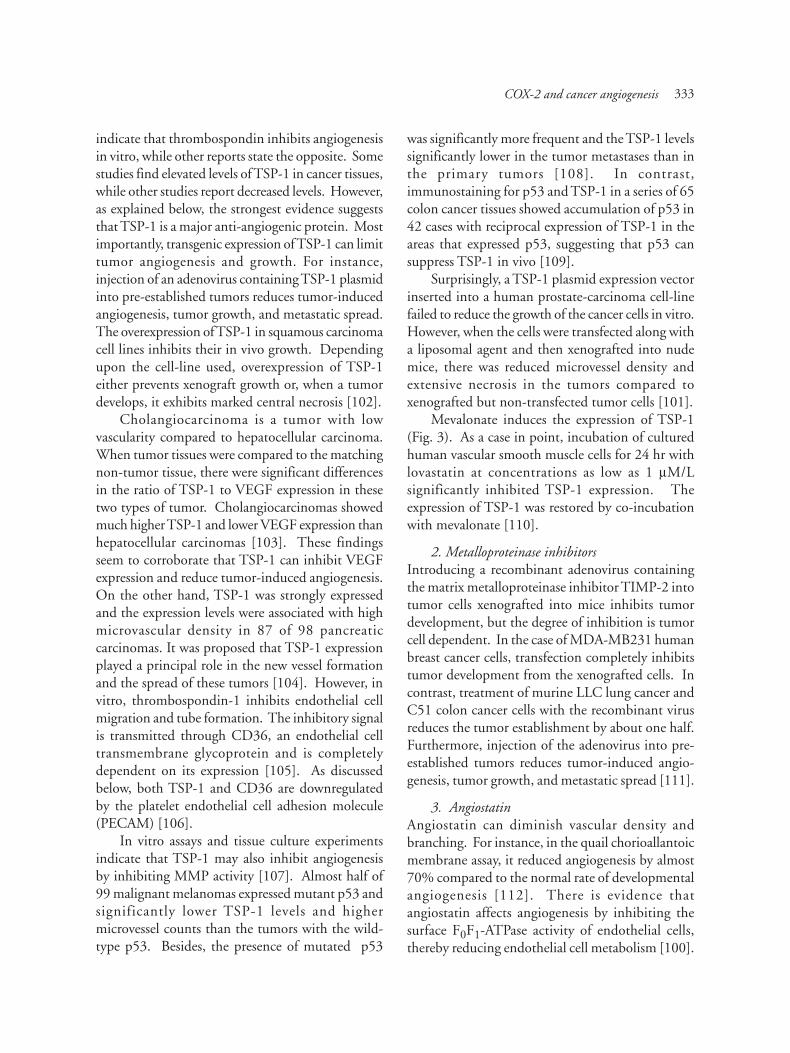
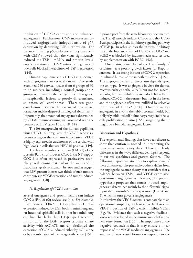
Fig. 3. Thrombospondin (TSP-1) activates the transforming growth factor beta (TGF-β). The statin-type of cholesterollowering drugs lower the synthesis of mevalonate, which reduces the stimulation of TSP-1 synthesis and therebyincreases angiogenesis by reducing the inhibition of VEGF. The reduction in mevalonate synthesis also decreases thesynthesis of coenzyme (Co) Q10, which reduces mitochondrial synthesis of adenosine triphosphate (ATP) that reducesendothelial cell proliferation.
Annals of Clinical & Laboratory Science
335
antibodies against either urokinase plasminogenactivator or its receptor [119].
TGF-β is of interest because it is abundantlyexpressed in the same areas as COX-2 in coloncarcinoma tissues. This observation suggests a closecollaboration in the process of carcinogenesis [120]especially because TGF-β1 has been described as apowerful regulator of COX-2 expression [121]. Mostimportant, from an etiologic point of view,deregulation of TGF-β1 expression may be an earlyevent in colon carcinogenesis [122]. Aberrations inTGF-receptors, such as repression of the expressionof the TGF-type II receptors, have been shown tocontribute to carcinogenesis [123].
There is evidence that the different isoforms ofTGF-β may have varying roles in carcinogenesis, andthat their effects may differ in different types ofmalignancies. To cite an instance, in skin cancers,TGF-β1 was found to be associated with highlydifferentiated tumors, TGF-β3 was associated withtumor stroma growth and angiogenesis, and TGF-β2 with invading, very malignant tumor cells [124].Yet, in prior studies in xenografted nude mice,expression of TGF-β2 was higher than that of theβ1 isoform in the poorly invasive DU145 prostatecarcinoma cell line compared to the highly metastaticPC-3M prostate carcinoma cell line [125].
TGF-β modulates angiogenesis by regulatingvascular endothelial cell proliferation and migration.Besides, it affects the extracellular matrix and thesynthesis of adhesion molecules [36]. TGF-βstimulates smooth muscle cells at low concentrationand inhibits them at high concentration [126,127].It has similar effects on capillary lumen formationinduced in vitro by bFGF or VEGF [128]. TheTGF-β signals via type I (ALK-1), type II, and typeIII (endoglin) receptors and Smad proteins, whichregulate genes involved in angiogenesis [129,130].
Participation of TGF-β in angiogenesis iscorroborated by results from the mouse cornealmodel of inflammation induced by silver nitrate.When mice were injected into the femoral musclewith an adenovirus that expressed a soluble ligandbinding part of the TGF-β type II receptor, thecorneal angiogenesis was significantly reduced whencompared to control mice [131].
The TGFBR2(-/-) mouse dies about 10 days
post-coitus due to defects in yolk sac vasculogenesis[129] that closely resemble those that occur in theTGF-β1 null mouse [17]. In like manner, micelacking the TGF-β Type I receptor (TGFBR1) dieof defective yolk sac and placental vasculardevelopment. Cultured vascular endothelial cellsfrom such mice show defective fibronectinproduction and abnormal migration, but enhancedproliferation, compared to control cells from wild-type mice [132].
Another example of the importance of TGF-βand its receptors in proper angiogenesis is illustratedby mutations of the TGF-β1 and TGF-β3 bindingprotein endoglin (ENG, CD105) and ALK-1 genes.Such mutations are responsible for the vascularmalformations seen in hereditary hemorrhagicteleangiectasia (HHT) types 1 and 2 respectively (Fig.1) [130,133].
Endoglin is an essential factor for angiogenesisduring embryogenesis. It is expressed on the surfaceof endothelial cells. Lack of endoglin does not affectvasculogenesis but it is essential for normal vascularmaturation as part of angiogenesis. This is evidentin endoglin null mice, which die by gestational day11.5 from defective development of vascular smoothcells and aborted endothelial remodeling [133].These experiments confirm that TGF-β signalingthrough its receptors plays an important role in theregulation of angiogenesis. The importance of TGF-β signaling in cancer-induced angiogenesis is furthervalidated by a report that elevated levels of endoglinin breast cancer tissues predict a high risk ofdeveloping metastatic disease. Strong upregulationwas detected by immunostaining in the tissues of92 breast cancer patients [134].
Endoglin is highly expressed in some malignanttumors. It might find use as a tumor marker becausethe protein was detected in the plasma of patientswho had early breast cancer. Analysis of plasmasobtained before any treatment was given revealedthat patients who had high plasma levels of endoglinhad a high risk of developing metastatic disease [134].Also, the endothelial-specific expression of endoglinfavors its use as an endothelial cell specific marker.As an example, in paraffin-embedded cervical cancertissues immunostaining for endoglin was moresensitive than immunostaining factor VIII for
COX-2 and cancer angiogenesis
336
visualizing capillaries and was a better predictor thanfactor VIII for lymph node metastases [135].
2. TGF-αTGF-α induces COX-2 by binding to the EGFreceptor (EGFR) [136,137]. Supplementation withTGF-α of cultured cervical cancer cells corroboratesthese findings. TGF-α supplementation markedlyincreased COX-2 expression [42]. Presence ofEGFR, but not of TGF-α, in 86 breast cancers wasassociated with poor prognosis. However, intensityof TGF-α immunostaining reflected the extent ofangiogenesis in the tumor [137].
3. PECAMPlatelet endothelial cell adhesion molecule (PECAM-1) has a bimodal effect, stimulating endothelial cellmorphogenesis at low concentrations and inhibitingit at high concentrations. PECAM is found at sitesof cell-to-cell contact in polyoma middle T-transformed mouse brain endothelial cells. Thesecells express high levels of PECAM-1 that inhibitthe expression of TSP-1 and TSP-1 receptor CD36and form hemangiomas in the mouse [138]. Thebimodal character of PECAM may be related to theactivation of latent TGF-β by TSP-1, which regulatesthe formation of active, bimodal TGF-β.
Regulation
A. Hypoxia
In vitro, hypoxia induces COX-2, increases PGE2synthesis, elevates the VEGF level, and inducesangiogenesis. The selective COX-2 inhibitor NS-398 can block this process. Hypoxia induced bycobalt chloride markedly increased COX-2 andupregulated VEGF via PGE2 in the cultured, highlyinvasive PC-3ML prostate-carcinoma cell line.Inhibition of the VEGF expression using the selectiveCOX-2 inhibitor NS-398 was overruled byadministration of exogenous PGE2 [139]. In vitro,hypoxia typically induces VEGF expression in severaldifferent human cell types [140]. One study foundthat hypoxia increases the expression of TSP-1 [141],which would inhibit VEGF expression. However,recent data show that hypoxia suppresses TSP-1expression and stimulates VEGF expression [140].
Induction of VEGF, despite a lack of hypoxia,
occurs in von Hippel-Landau disease (VHL), whichis caused by mutations in the VHL tumor suppressorgene. The mutations lead to constitutional insteadof hypoxia-regulated expression of hypoxia induciblefactor (HIF) that induces VEGF. Patients with VHLoften develop hemangioblastomas, which exhibitincreased levels of VEGF. [142,143].
B. Tumor suppressor protein p53
The tumor suppressor protein p53 inhibits COX-2expression but stimulates TSP-1 expression [144].Transfection experiments have demonstrated thatp53 can stimulate the promoter sequences of theTSP-1 gene in fibroblasts. The preponderance ofevidence suggests that thrombospondin (TSP-1) isstrongly anti-angiogenic, inhibiting the expressionof VEGF. Thus, loss of function of the tumorsuppressor protein p53, which normally stimulatesTSP-1, might contribute to cancer-inducedangiogenesis. Wild-type p53 inhibits angiogenesisby stimulating TSP-1 synthesis. Mutation or loss ofp53 results in lack of TSP-1 inhibition ofangiogenesis. Tissue studies corroborate thesefindings. Almost half of 99 malignant melanomasexpressed mutant p53 and showed significantly lowerTSP-1 levels and higher microvessel counts,compared to melanomas with wild-type p53.Moreover, presence of mutated p53 was morefrequent and TSP-1 levels were lower in the tumormetastases than in the primary tumors [145].
High VEGF protein levels in tumor cytosolcorrelated with poor prognosis, based on enzymeimmunoassays in 224 estrogen-receptor-positiveprimary breast cancer patients who received adjuvanttamoxifen. Higher VEGF expression was correlatedwith the presence of the mutations of the tumor-suppressor protein p53 [146]. In a geographical areawith high risk for esophageal cancer, 48 of 74esophageal carcinoma patients carried one or morepoint mutations of p53 in their tumor; the presenceof p53 mutation correlated significantly with highCOX-2 expression in the tumor [147].
C. Viruses
Human cytomegalovirus (CMV) inactivates wild-type p53 of diverse cell types resulting in loss of
Annals of Clinical & Laboratory Science
337
inhibition of COX-2 expression and enhancedangiogenesis. Furthermore, CMV increases tumor-induced angiogenesis independently of p53expression by depressing TSP-1 expression. Forinstance, infecting p53-defective astrocytoma cellswith CMV showed that the virus significantlyreduced the TSP-1 mRNA and protein levels.Supplementation with CMV anti-sense oligonucleo-tides fully blocked the abolition of TSP-1 expression[144].
Human papilloma virus (HPV) is associatedwith angiogenesis in cervical cancer. One studyexamined 230 cervical tissues from 6 groups of 31to 43 subjects, including a control group and 5groups with tumors that ranged from low grade,intraepithelial lesions to poorly differentiatedsquamous cell carcinomas. There was goodcorrelation between the extent of new vesselformation and the degree of histological abnormality.Importantly, the amount of angiogenesis determinedby CD34 immunostaining was associated with thepresence of HPV types 16 and 18 [148].
The E6 oncoprotein of the human papillomavirus (HPV)-16 upregulates the VEGF gene via apromoter region that contains 4 Sp-1 sites. VEGFis highly expressed in carcinomas of the cervix, withhigh levels in cells that are HPV-16 positive [149].
The latent membrane protein (LMP-1) of theEpstein-Barr virus induces COX-2 via NF-kappB.COX-2 is often expressed in preinvasive naso-pharyngeal lesions that harbor the virus and innasopharyngeal carcinomas. In vitro studies suggestthat EBV, present in over two-thirds of such tumors,contributes to VEGF expression and tumor-inducedangiogenesis [150].
D. Regulation of COX-2 expression
Several oncogenes and growth factors can induceCOX-2 (Fig. 2) (for review, see [6]). For example,EGF induces COX-2. TGF-β enhances COX-2expression induced by EGF both in mink lung andrat intestinal epithelial cells but not in a mink lungcell line that lacks the TGF-β type I receptor.Inhibition of the EGF receptor tyrosine kinaseactivity with AG1478 entirely abolished theexpression of COX-2 induced either by EGF aloneor by a combination of the two growth factors [151].
A prior report from the same laboratory documentedthat TGF-β strongly induces COX-2 and that COX-2 can participate in the inhibitory signaling pathwayof TGF-β. In other studies the in vitro inhibitorypart of the biphasic effects of TGF-β via COX-2 andPGE2 was blocked by indomethacin and restoredby supplementation with PGE2 [152].
Oncostatin, a member of the IL-6 family ofcytokines, is a potent growth factor for Kaposi'ssarcoma. It is a strong inducer of COX-2 expressionin cultured human aortic smooth muscle cells [153].The angiogenic effect of oncostatin depends uponthe cell type. It was angiogenic in vitro for dermalmicrovascular endothelial cells but not for macro-vascular, human umbilical vein endothelial cells. Itinduced COX-2 in the former, but not in the latter,and the angiogenic effect was nullified by selectiveinhibition of COX-2 [154]. Oncostatin wasangiogenic in vivo in the rabbit corneal model, butit slightly inhibited calf pulmonary artery endothelialcells proliferation in vitro [155], suggesting that itmight be a bimodal angiogenic factor.
Discussion and Hypothesis
The experimental findings that have been discussedshow that caution is needed in interpreting thesometimes contradictory data. There are clearlydifferences in the ways different cell types respondto various cytokines and growth factors. Thefollowing hypothesis attempts to explain some ofthese differences. The present hypothesis differs fromthe angiogenic balance theory that considers that abalance between TSP-1 and VEGF expressiondetermines angiogenesis. Rather, the presenthypothesis proposes that cancer-induced angio-genesis is determined mainly by the differential signalinput that controls VEGF expression (Figs. 4 and5), which in turn governs angiogenesis.In this view, the VEGF system is comparable to anoperational amplifier, with negative feedback viaVEGF induction of TSP-1, which inhibits VEGF,(Fig. 5). Evidence that such a negative feedback-loop exists was found in the murine model of retinalnew vessel formation [156]. The importance of thenegative feedback is that it may ensure a linearresponse of the VEGF-mediated angiogenesis. Theamount of new vessel formation responds to the
COX-2 and cancer angiogenesis

338
magnitude of the differential input to COX-2 andTSP-1. All angiogenic and anti-angiogenic signalsthat regulate the VEGF system are important, butonly the difference between the stimulating andinhibiting signals determines the amount of VEGFexpression and angiogenesis.
TSP-1 may paradoxically behave as anangiogenic agonist. Located predominantly at thetumor invasion front, TSP-1 mRNA was stronglyexpressed in all but 11 of a series of 98 pancreaticcarcinomas. TSP-1 levels correlated with the tumormicrovascular density. It was concluded that TSP-1induces angiogenesis [104]. However, the malignanttumor might over-express TSP-1 and provide astrongly inhibitory signal to VEGF, but might beoverruled by a stronger angiogenic signal to VEGF
– for example, caused by powerful overexpression ofCOX-2. In the cited experiments, COX-2 expressionwas not analyzed, so this suggestion appearsreasonable, but speculative.
Furthermore, this hypothesis may explain whyloss of the tumor-suppressor protein p53 functionis so significant to tumor growth. Such loss not onlyenhances COX-2 but in addition reduces TSP-1expression, which together greatly increase VEGFprotein expression, angiogenesis, tumor invasion,and metastasis.
In most cases of cervical cancer, p53 is notmutated, but rather is elevated [148]. One mightspeculate that p53 expression could be induced invivo in response to a malignant tumor, to defendagainst tumor-induced angiogenesis in an attempt
Fig. 4. Evidence-based and hypothetical illustration of regulation and cancer-induced deregulation of angiogenesis.The cancer distorts the physiological balance between angiogenic and anti-angiogenic factors that regulate the expressionof vascular endothelial growth factor (VEGF). COX-2 increases VEGF and metalloproteinases (MMP) andthrombospondin (TSP-1) inhibits VEGF and MMP expression. Overexpression of COX-2 by cancer cells increasesVEGF and MMP expression, inhibits apoptosis, and promotes angiogenesis, invasion, and metastatic spread. Loss offunction of the tumor suppressor protein p53 increases tumor-induced angiogenesis by reducing TSP-1 and increasingCOX-2 expression, resulting in reduced inhibition and increased stimulation of VEGF by TSP-1 and COX-2 respectively.Ctomegalovirus (CMV) inhibits TSP-1 expression, which decreases the inhibition of VEGF expression. Humanpapilloma virus (HVP) contains a VEGF-like segment. Both viruses induce angiogenesis in the cervical cancer.
Annals of Clinical & Laboratory Science

339
to combat tumor growth, invasion, and metastasis.A better understanding of the pathophysiologyinvolved in the growth of a specific tumor mightbe obtained by determining the levels of bothangiogenic and anti-angiogenic factors that affectVEGF expression, and not assaying only VEGF.
Finally, the role of interference of energymetabolism in the inhibition of angiogenesis is mostinteresting. Angiostatin inhibits the surface F0F1-ATPase activity of endothelial cells [100]. Incomparison, the cholesterol-lowering statin drugsinhibit coenzyme Q10 synthesis and therebymitochondrial oxidative phosphorylation and ATPgeneration [99]. Such statins might thereby inhibitangiogenesis,, and at the same time enhanceangiogenesis by reducing TSP-1 expression.
Diagnosis and Therapy
A. Tumor markers
Several proteins that are angiogenesis regulators havebeen detected in blood and some are elevated inpatients with cancer. However, their potential useto detect or monitor post-operative patients fortumor recurrence needs clarification [157,158].
Tumor vascular density is a prognostic factor fora variety of cancers. It reflects the importance of thevascular supply to the tumor growth. Furthermore,the fractal character of tumor-induced vesselformation exhibits differences compared to the fractalcharacter of physiological new vessel formation.Tumor-induced vessels follow a more irregularcourse, probably due to inherent regional geneticdifferences in the tumor cell population. Magneticresonance imaging can detect alterations in vesselpermeability and permits in vivo evaluation ofexperimental tumor-induced angiogenesis. In thefuture it might be used in clinical studies to followthe reduction in cancer-induced angiogenesis that isachieved by COX-2 inhibition [159,160].
B. Anticancer therapy with NSAIDs
1. Non-selective NSAIDsMany epidemiological studies, a large number of invitro and animal experiments, and several clinicalstudies have demonstrated that non-selectiveNSAIDs can restrain the development and growthof different types of cancer [5]. Non-selectiveNSAIDs (nsNSAIDs) differ in their ability to inhibitthe two known isoforms of cyclooxygenase (COX)
Fig. 5. Simplified diagram of the angiogenesis control system. Triangles represent enzymes or growth factors. Resistorssymbolize receptors or drugs. Imputs A-D control angiogensis via COX-2 (stimulation) or TSP-1 (inhibition) ofVEGF, the output of which induces its stimultor TSP-1, which via its receptor CD36 inhibits VEGF, thus forming anegative feedback loop for VEGF. Nonsteroidal inflammatory drugs (NSAIDs) inhibit the stimulatory signal (PGE2).Examples of signal imputs: A: mevalonate, p53; B: CMV; C: p53; D: TGF-β; EBV.
COX-2 and cancer angiogenesis

340
[5], but most of them significantly inhibit COX-1in platelets. Especially after prolonged use, they cancause bleeding and gastrointestinal ulceration anduncouple mitochondria [161].
Some anti-neoplastic effects of NSAIDs mightbe independent of cyclooxygenase inhibition. Forinstance, NSAIDs suppress colony formation on softagar by transformed fibroblasts that either possessor lack COX-1 or COX-2 [83]. These anti-neoplastic effects might be due, in part, to themitochondrial uncoupling that is common to manynon-selective NSAIDs.
2. Selective COX-2 inhibitorsNSAIDs that inhibit COX-2 restore tumor cellapoptosis in vitro and reduce in vivo COX-2-tumor-induced angiogenesis. Furthermore, COX-2inhibition lowers the synthesis of metalloproteinasesand reduces matrix proteolysis. Also, as noted above,by inhibiting the COX-2 synthesis induced by thetumor in the adjacent host tissue, such agentsobstruct the growth of xenografted tumors in whichCOX-2 has been silenced by methylation [79,162].These selective COX-2 inhibitors might have greatpotential for treatment of human cancer. Incomparison to nonselective NSAIDs, selective COX-2 inhibitors have fewer gastrointestinal side effects,do not interfere with platelet function, and do notuncouple mitochondria. Selective COX-2 inhibitorshave shown significant anti-cancer effects in a varietyof animal tumor xenograft models. They inhibit thegrowth and spread of the malignant cells [5,6].
The US Food and Drug Administration (FDA)has approved celecoxib and rofecoxib for treatmentof rheumatoid arthritis and osteoarthritis respectively.A review of >50 clinical studies involving 13,000patients and lasting 12 wk to 2 yr concluded thatthese drugs are well-tolerated [163]. Twice dailydoses of celecoxib at 200 mg [164] and 400 mg [165]have been well tolerated by arthritis patients. It isencouraging that selective COX-2 inhibitors mightprevent the development of cancer in patients withpremalignant conditions such as adenomatouspolyposis of the colon. A 6-mo clinical trialdemonstrated that celecoxib, 400 mg twice daily (30patients), but not 100 mg twice a day (32 patients)significantly reduced the number of polyps,compared to 15 patients given placebo [166].
The patient's physician should carefullysupervise any selective COX-2 therapy. For example,it has been advised that while gastrointestinalbleeding is not a problem in the use of the selectiveCOX-2 inhibitors, the possible development ofdyspepsia and renal problems should be carefullymonitored [167]. Renal side effects might appeardue to inhibition of COX-2 in the macula densaand medullary interstitial cells [168]. Second,because of the role COX-2 plays in implantation,COX-2 inhibition is not advisable in women whodesire to conceive. Pregnant women should not useNSAIDs that inhibit COX-2 because the enzymeprevents the premature closure of the fetal ductusarteriosus [169]. Third, because the statin type ofcholesterol-lowering drugs also lowers the synthesisof mevalonate and thereby the synthesis of the anti-angiogenic protein TSP-1, it might be reasonable tore-evaluate the need for statin therapy in patientswith cancer. Fourth, analysis of 3 studies involving>17,000 rheumatoid arthritis patients showed anincrease of thromboembolic events (eg, myocardialinfarction , stroke) in patients who took rofecoxib50 mg daily, compared to those who took celecoxib400 mg twice daily plus low dose aspirin [170].
Conclusions
High expression levels of cyclooxygenase (COX-2),vascular endothelial growth factor (VEGF), andmetalloproteinase (MMP), and low thrombospondin(TSP-1) expression in cancer tissues is associated withhigh tumor vascular density (MVD), a prognosticfactor in human malignancies that predicts aggressivegrowth, invasion, and metastases. The human cancerderails the normal balance between angiogenic(COX-2) and anti-angiogenic factors (TSP-1) thatcontrol the expression of VEGF, the pivotal mediatorof new vessel formation. By expressing COX-2,which via PGE2 induces VEGF, the solid cancerforces its host to provide a vascular supply, whichenables the tumor to grow beyond 1-2 mm.
The importance of COX-2 in angiogenesis isevidenced by its role in implantation-induced newvessel formation. The trophoblast of the implantingblastocyst induces maternal COX-2 expression,which is essential for the placental vasculogenesis andangiogenesis that are required for successful
Annals of Clinical & Laboratory Science

341
implantation. In granulation tissue at the sites ofinflammation, and in a variety of solid tumors, COX-2 is re-expressed.
Chronic inflammation is a risk factor for cancer.The exact reason is not known, but in vitro,transgenic overexpression of COX-2 is capable oftransforming cells. On the other hand, COX-2negative cells can also be transformed. No geneabnormality of COX-2 in human cancer has beenreported, but COX-2 can be induced by a variety ofoncogenes, cytokines, growth factors, andcarcinogens – for example, benzo[a]pyrene intobacco smoke. In a large variety of human cancers,COX-2 is upregulated, producing eicosanoids suchas PGE2, which induces VEGF and therebyangiogenesis. By inhibiting COX-2 in experimentalcancers that overexpress this enzyme, the balancebetween angiogenic and anti-angiogenic signals,which control VEGF expression and angiogenesis,can be restored, causing tumor necrosis and tumorregression to a small, dormant state.
References
1. Suh DY. Understanding angiogenesis and its clinicalapplications. Ann Clin Lab Sci 2000;30:227-238.
2. Eschwege P, de Ledinghen V, Camilli T, Kulkarni S,Dalbagni G, Droupy S, Jardin A, Benoit G, WekslerBB. Arachidonic acid and prostaglandins, inflam-mation and oncology. Presse Med 2001;30: 508-510.
3. Cheng T, Cao W, Wen R, Steinberg RH, LaVail MM.Prostaglandin E2 induces vascular endothelial growthfactor and basic fibroblast growth factor mRNAexpression in cultured rat Müller cells. InvestOphthalmol Vis Sci 1998;39:581-591.
4. Ben-Av P, Crofford LJ, Wilder RL, Hla T. Inductionof vascular endothelial growth factor expression insynovial fibroblasts by prostaglandin E and inter-leukin-1: a potential mechanism for inflammatoryangiogenesis. FEBS Lett 1995;372:83-87.
5. Fosslien E. Biochemistry of cyclooxygenase (COX)-2 inhibitors and molecular pathology of COX-2 inneoplasia. Crit Rev Clin Lab Sci 2000;37:431-502.
6. Fosslien E. Molecular pathology of cyclooxygenase-2 in neoplasia. Ann Clin Lab Sci 2000;30:3-21.
7. Masunaga R, Kohno H, Dhar DK, Ohno S,Shibakita M, Kinugasa S, Yoshimura H, TachibanaM, Kubota H, Nagasue N. Cyclooxygenase-2expression correlates with tumor neovascularization
and prognosis in human colorectal carcinomapatients. Clin Cancer Res 2000;6:4064-4068.
8. Brown LF, Guidi AJ, Schnitt SJ, Van De Water L,Iruela-Arispe ML, Yeo TK, Tognazzi K, Dvorak HF.Vascular stroma formation in carcinoma in situ,invasive carcinoma, and metastatic carcinoma of thebreast. Clin Cancer Res 1999;5:1041-1056.
9. Williams CS, Mann M, DuBois RN. The role ofcyclooxygenases in inflammation, cancer, anddevelopment. Oncogene 1999;18:7908-7916.
10. Gately S. The contributions of cyclooxygenase-2 totumor angiogenesis. Cancer Metastasis Rev 2000;19:19-27.
11. Bischof P, Campana A. A putative role for oncogenesin trophoblast invasion? Hum Reprod 2000;15(Suppl 6):51-58.
12. Murray MJ, Lessey BA. Embryo implantation andtumor metastasis: common pathways of invasion andangiogenesis. Semin Reprod Endocrinol 1999;17:275-290.
13. Zoltowska A, Stepinski J, Lewko B, Zamorska B,Roszkiewicz A, Serkies K, Kruszewski WJ.Malformations of angiogenesis in the lowdifferentiated human carcinomas. immunohisto-chemical study. Arch Immunol Ther Exp (Warsz)2001;49:59-61.
14. Smith SK. Angiogenesis and implantation. Ann SurgOncol 2001;8:72-79.
15. Fazleabas AT, Kim JJ, Srinivasan S, Donnelly KM,Brudney A, Jaffe RC. Implantation in the baboon:endometrial responses. Semin Reprod Endocrinol1999;17:257-265.
16. Dinchuk JE, Car BD, Focht RJ, Johnston JJ, JaffeeBD, Covington MB, Contel NR, Eng VM, CollinsRJ, Czerniak PM, et al. Renal abnormalities and analtered inflammatory response in mice lackingcyclooxygenase II. Nature 1995;378:406-409.
17. Dickson MC, Martin JS, Cousins FM, Kulkarni AB,Karlsson S, Akhurst RJ. Defective haematopoiesis andvasculogenesis in transforming growth factor-beta 1knock out mice. Development 1995;121:1845-1854.
18. Kallapur S, Ormsby I, Doetschman T. Straindependency of TGFbeta function during embryo-genesis. Mol Reprod Dev 1999;52:341-349.
19. Chung IB, Yelian FD, Zaher FM, Gonik B, EvansMI, Diamond MP, Svinarich DM. Expression andregulation of vascular endothelial growth factor in afirst trimester trophoblast cell line. Placenta 2000;21:320-324.
20. Shalaby F, Rossant J, Yamaguchi TP, Gertsenstein M,Wu XF, Breitman ML, Schuh AC. Failure of blood-island formation and vasculogenesis in Flk-1-
COX-2 and cancer angiogenesis

342
deficient mice. Nature 1995;376:62-66.21. Clyman RI, Hardy P, Waleh N, Chen YQ, Mauray
F, Fouron JC, Chemtob S. Cyclooxygenase-2 plays asignificant role in regulating the tone of the fetal lambductus arteriosus. Am J Physiol 1999;276 :R913-921.
22. Segi E. A study for functions of prostaglandin Ereceptor EP4 subtype by analysing knockout mice.Yakugaku Zasshi 2001;121:35-45.
23. Loftin CD, Trivedi DB, Tiano HF, Clark JA, LeeCA, Epstein JA, Morham SG, Breyer MD, NguyenM, Hawkins BM, Goulet JL, Smithies O, Koller BH,Langenbach R. Failure of ductus arteriosus closureand remodeling in neonatal mice deficient incyclooxygenase-1 and cyclooxygenase-2. Proc NatlAcad Sci USA 2001;98:1059-1064.
24. Tannenbaum JE, Waleh NS, Mauray F, Gold L,Perkett EA, Clyman RI. Transforming growth factor-beta protein and messenger RNA expression isincreased in the closing ductus arteriosus. Pediatr Res1996;39:427-434.
25. Song J, Stastny J, Fosslien E, Robertson AL. Effectof aging on human protein composition. I. One-dimensional polyacrylamide gel electrophoreticanalysis of tissue extracts. Exp Mol Path 1985;43:233-241.
26. Song J, Stastny J, Fosslien E, Robertson, AL. Effectof aging on human aortic protein composition. II.Two-dimensional polyacrylamide gel electrophoreticanalysis of tissue extracts. Exp Mol Path 1985;43:297-304.
27. Li Z, Froehlich J, Galis ZS, Lakatta EG. Increasedexpression of matrix metalloproteinase-2 in thethickened intima of aged rats. Hypertension 1999;33:116-123.
28. Ghosh AK, Hirasawa N, Niki H, Ohuchi K.Cyclooxygenase-2-mediated angiogenesis incarrageenin-induced granulation tissue in rats. JPharmacol Exp Ther 2000;295:802-809.
29. Howdieshell TR, Callaway D, Webb WL, GainesMD, Procter CD Jr, Sathyanarayana, Pollock JS,Brock TL, McNeil PL. Antibody neutralization ofvascular endothelial growth factor inhibits woundgranulation tissue formation. J Surg Res 2001;96:173-182.
30. Majima M, Hayashi I, Muramatsu M, Katada J,Yamashina S, Katori M. Cyclooxygenase-2 enhancesbasic fibroblast growth factor-induced angiogenesisthrough induction of vascular endothelial growthfactor in rat sponge implants. Br J Pharmacol 2000;130:641-649.
31. Majima M, Isono M, Ikeda Y, Hayashi I, HatanakaK, Harada Y, Katsumata O, Yamashina S, Katori M,
Yamamoto S. Significant roles of induciblecyclooxygenase (COX)-2 in angiogenesis in ratsponge implants. Jpn J Pharmacol 1997;75:105-114.
32. Hernandez GL, Volpert OV, Iniguez MA, LorenzoE, Martinez-Martinez S, Grau R, Fresno M, RedondoJM. Selective inhibition of vascular endothelialgrowth factor-mediated angiogenesis by cyclosporinA: roles of the nuclear factor of activated T cells andcyclooxygenase 2. J Exp Med 2001;193:607-620.
33. Cuzzocrea S, Costantino G, Mazzon E, Caputi AP.Regulation of prostaglandin production in carra-geenan-induced pleurisy by melatonin. J Pineal Res1999;27:9-14.
34. Fosslien E. Escape from immunological surveillancein blastocyst implantation and cancer. Ann Clin LabSci 2000;30:111-112.
35. Saaristo A, Karpanen T, Alitalo K. Mechanisms ofangiogenesis and their use in the inhibition of tumorgrowth and metastasis. Oncogene 2000;19:6122-6129.
36. Li C, Guo B, Bernabeu C, Kumar S. Angiogenesisin breast cancer: the role of transforming growthfactor beta and CD105. Microsc Res Tech 2001;52:437-449.
37. Price JT, Bonovich MT, Kohn EC. The biochemistryof cancer dissemination. Crit Rev Biochem Mol Biol1997;32:175-253.
38. John A, Tuszynski G The role of matrix metallopr-oteinases in tumor angiogenesis and tumor metas-tasis. Pathol Oncol Res 2001;7:14-23.
39. Detmar M. Tumor angiogenesis. J Investig DermatolSymp Proc 2000;5:20-23.
40. Craciunescu OI, Das SK, Clegg ST. Dynamiccontrast-enhanced MRI and fractal characteristics ofpercolation clusters in two-dimensional tumor bloodperfusion. J Biomech Eng 1999;121:486.
41. Soslow RA, Dannenberg AJ, Rush D, Woerner BM,Khan KN, Masferrer J, Koki AT. COX-2 is expressedin human pulmonary, colonic, and mammarytumors. Cancer 2000;89:2637-2645.
42. Kulkarni S, Rader JS, Zhang F, Liapis H, Koki AT,Masferrer JL, Subbaramaiah K, Dannenberg AJ.Cyclooxygenase-2 is overexpressed in human cervicalcancer. Clin Cancer Res 2001;7:429-434.
43. Gaffney DK, Holden J, Davis M, Zemmpolich K,Murphy KJ, Dodson M. Elevated cyclooxygenase-2expression correlates with diminished survival incarcinoma of the cervix treated with radiotherapy.Int J Radiat Oncol Biol Phys 2001;49:1213-1217.
44. Ohno R, Yoshinaga K, Fujita T, Hasegawa K, IsekiH, Tsunozaki H, Ichikawa W, Nihei Z, Sugihara K.Depth of invasion parallels increased cyclooxygenase-
Annals of Clinical & Laboratory Science

343
2 levels in patients with gastric carcinoma. Cancer2001;91:1876-1881.
45. Tomozawa S, Tsuno NH, Sunami E, Hatano K,Kitayama J, Osada T, Saito S, Tsuruo T, Shibata Y,Nagawa H. Cyclooxygenase-2 overexpressioncorrelates with tumour recurrence, especially haem-atogenous metastasis, of colorectal cancer. Br J Cancer2000;83:324-328.
46. Madaan S, Abel PD, Chaudhary KS, Hewitt R, StottMA, Stamp GW, Lalani EN. Cytoplasmic inductionand over-expression of cyclooxygenase-2 in humanprostate cancer: implications for prevention andtreatment. BJU Int 2000;86:736-741.
47. Yoshimura R, Sano H, Masuda C, Kawamura M,Tsubouchi Y, Chargui J, Yoshimura N, Hla T, WadaS. Expression of cyclooxygenase-2 in prostate carcin-oma. Cancer 2000;89:589-596.
48. Kirschenbaum A, Klausner AP, Lee R, Unger P, YaoS, Liu XH, Levine AC. Expression of cyclooxygenase-1 and cyclooxygenase-2 in the human prostate.Urology 2000;56:671-676.
49. Denkert C, Kobel M, Berger S, Siegert A, Leclere A,Trefzer U, Hauptmann S. Expression of cyclo-oxygenase 2 in human malignant melanoma. CancerRes 2001;61:303-308.
50. Karim MM, Hayashi Y, Inoue M, Imai Y, Ito H,Yamamoto M. Cox-2 expression in retinoblastoma.Am J Ophthalmol 2000;129:398-401.
51. Tas F, Yavuz E, Aydiner A, Saip P, Disci R, Iplikci A,Topuz E. Angiogenesis and p53 protein expressionin breast cancer: prognostic roles and interrelation-ships. Am J Clin Oncol 2000;23:546-553.
52. Shimizu T, Hino K, Tauchi K, Ansai Y, Tsukada K.Predication of axillary lymph node metastasis byintravenous digital subtraction angiography in breastcancer, its correlation with microvascular density.Breast Cancer Res Treat 2000;61:261-269.
53. Garzetti GG, Ciavattini A, Lucarini G, PugnaloniA, De Nictolis M, Amati S, Romanini C, Biagini G.Expression of vascular endothelial growth factorrelated to 72-kilodalton metalloproteinase immuno-staining in patients with serous ovarian tumors.Cancer 1999;85:2219-2225.
54. Yoshiji H, Gomez DE, Shibuya M, Thorgeirsson UP.Expression of vascular endothelial growth factor, itsreceptor, and other angiogenic factors in humanbreast cancer. Cancer Res 1996;56:2013-2016.
55. Paradis V, Lagha NB, Zeimoura L, Blanchet P,Eschwege P, Ba N, Benoit G, Jardin A, Bedossa P.Expression of vascular endothelial growth factor inrenal cell carcinomas. Virchows Arch 2000;436:351-356.
56. Slaton JW, Inoue K, Perrotte P, El-Naggar AK,Swanson DA, Fidler IJ, Dinney CP. Expression levelsof genes that regulate metastasis and angiogenesiscorrelate with advanced pathological stage of renalcell carcinoma. Am J Pathol 2001;158:735-743.
57. Ogata Y, Harada Y, Fujii T, Yamana H, Fujita H,Shirouzu K. Immunohistochemical localization ofvascular endothelial growth factor in esophagealcancer. Kurume Med J 1996;43:157-163.
58. Bayer-Garner IB, Hough AJ Jr, Smoller BR. Vascularendothelial growth factor expression in malignantmelanoma: prognostic versus diagnostic usefulness.Mod Pathol 1999;12:770-774.
59. Strohmeyer D, Rossing C, Bauerfeind A, KaufmannO, Schlechte H, Bartsch G, Loening S. Vascularendothelial growth factor and its correlation withangiogenesis and p53 expression in prostate cancer.Prostate 2000;45:216-224.
60. Han H, Silverman JF, Santucci TS, Macherey RS,d'Amato TA, Tung MY, Weyant RJ, Landreneau RJ.Vascular endothelial growth factor expression in stageI non-small cell lung cancer correlates with neoangio-genesis and a poor prognosis. Ann Surg Oncol 2001;8:72-79.
61. Li XM, Tang ZY, Qin LX, Zhou J, Sun HC. Serumvascular endothelial growth factor is a predictor ofinvasion and metastasis in hepatocellular carcinoma.J Exp Clin Cancer Res 1999;18:511-517.
62. Sabo E, Boltenko A, Sova Y, Stein A, Kleinhaus S,Resnick MB. Microscopic analysis and significanceof vascular architectural complexity in renal cellcarcinoma. Clin Cancer Res 2001;7:533-537.
63. Decaussin M, Sartelet H, Robert C, Moro D, ClarazC, Brambilla C, Brambilla E. Expression of vascularendothelial growth factor (VEGF) and its tworeceptors (VEGF-R1-Flt1 and VEGF-R2-Flk1/KDR) in non-small cell lung carcinomas (NSCLCs):correlation with angiogenesis and survival. J Pathol1999;188:369-377.
64. Fine BA, Valente PT, Feinstein GI, Dey T. VEGF,flt-1, and KDR/flk-1 as prognostic indicators inendometrial carcinoma. Gynecol Oncol 2000;76:33-39.
65. Marinkovic S, Baumann H. Structure, hormonalregulation, and identification of the interleukin-6-and dexamethasone-responsive element of the rathaptoglobin gene. Mol Cell Biol 1990;10:1573-1583.
66. Stastny J, Prasad R, Fosslien E. Tissue proteins inbreast cancer, as studied by use of two-dimensionalelectrophoresis. Clin Chem 1984;30:1914-1918.
COX-2 and cancer angiogenesis

344
67. Nayak SK, Kakati S, Harvey SR, Malone CC,Cornforth AN, Dillman RO. Characterization ofcancer cell lines established from two humanmetastatic breast cancers. In Vitro Cell Dev BiolAnim 2000;36:188-193.
68. Harvey SR, Nayak SK, Markus G, OuhammouchM, Hemperly JJ, Dillman RO, Doyle DJ. Cancercells release a covalent complex containing disulfide-linked domains from urinary plasminogen activator,neural cell adhesion molecule, and haptoglobin alphaand beta chains. Arch Biochem Biophys 1997;345:289-298.
69. Mehaffey MG, Folberg R, Meyer M, Bentler SE,Hwang T, Woolson R, Moore KC. Relativeimportance of quantifying area and vascular patternsin uveal melanomas. Am J Ophthalmol 1997;123:798-809.
70. Gazit Y, Baish JW, Safabakhsh N, Leunig M, BaxterLT, Jain RK. Fractal characteristics of tumor vasculararchitecture during tumor growth and regression.Microcirculation 1997;4:395-402.
71. Hepburn PJ, Griffiths K, Harper ME. Angiogenicfactors expressed by human prostatic cell lines: effecton endothelial cell growth in vitro. Prostate 1997;33:123-132.
72. Benefield J, Petruzzelli GJ, Fowler S, Taitz A, KalkanisJ, Young MR. Regulation of the steps of angiogenesisby human head and neck squamous cell carcinomas.Invasion Metastasis 1996;16:291-301.
73. Relf M, LeJeune S, Scott PA, Fox S, Smith K, LeekR, Moghaddam A, Whitehouse R, Bicknell R, HarrisAL. Expression of the angiogenic factors vascularendothelial cell growth factor, acidic and basicfibroblast growth factor, tumor growth factor beta-1, platelet-derived endothelial cell growth factor,placenta growth factor, and pleiotrophin in humanprimary breast cancer and its relation to angiogenesis.Cancer Res 1997;57:963-969.
74. Yamaguchi R, Yano H, Iemura A, Ogasawara S,Haramaki M, Kojiro M. Expression of vascularendothelial growth factor in human hepatocellularcarcinoma. Hepatology 1998;28:68-77.
75. Harris AL, Zhang H, Moghaddam A, Fox S, Scott P,Pattison A, Gatter K, Stratford I, Bicknell R. Breastcancer angiogenesis--new approaches to therapy viaantiangiogenesis, hypoxic activated drugs, andvascular targeting. Breast Cancer Res Treat 1996;38:97-108.
76. Harada S, Nagy JA, Sullivan KA, Thomas KA, EndoN, Rodan GA, Rodan SB. Induction of vascularendothelial growth factor expression by prostaglandin
E2 and E1 in osteoblasts. J Clin Invest 1994;93:2490-2496.
77. Hoper MM, Voelkel NF, Bates TO, Allard JD, HoranM, Shepherd D, Tuder RM. Prostaglandins inducevascular endothelial growth factor in a humanmonocytic cell line and rat lungs via cAMP. Am JRespir Cell Mol Biol 1997;17:748-756.
78. Attiga FA, Fernandez PM, Weeraratna AT, ManyakMJ, Patierno SR. Inhibitors of prostaglandinsynthesis inhibit human prostate tumor cellinvasiveness and reduce the release of matrixmetalloproteinases. Cancer Res 2000;60:4629-4637.
79. Williams CS, Tsujii M, Reese J, Dey SK, DuBoisRN. Host cyclooxygenase-2 modulates carcinomagrowth. J Clin Invest 2000;105:1589-1594.
80. Borgstrom P, Bourdon MA, Hillan KJ, SriramaraoP, Ferrara N. Neutralizing anti-vascular endothelialgrowth factor antibody completely inhibits angio-genesis and growth of human prostate carcinomamicro tumors in vivo. Prostate 1998;35:1-10.
81. Narko K, Ristimaki A, MacPhee M, Smith E,Haudenschild CC, Hla T. Tumorigenic transform-ation of immortalized ECV endothelial cells bycyclooxygenase-1 overexpression. J Biol Chem 1997;272:21455-21460.
82. Liu CH, Chang SH, Narko K, Trifan OC, Wu MT,Smith E, Haudenschild C, Lane TF, Hla T.Overexpression of cyclooxygenase-2 is sufficient toinduce tumorigenesis in transgenic mice. J BiolChem 2001;276:18563-18569.
83. Zhang X, Morham SG, Langenbach R, Young DA.Malignant transformation and antineoplastic actionsof nonsteroidal antiinflammatory drugs (NSAIDs)on cyclooxygenase-null embryo fibroblasts. J ExpMed 1999;190:451-459.
84. Di Popolo A, Memoli A, Apicella A, Tuccillo C, diPalma A, Ricchi P, Acquaviva AM, Zarrilli R. IGF-II/IGF-I receptor pathway up-regulates COX-2mRNA expression and PGE2 synthesis in Caco-2human colon carcinoma cells. Oncogene 2000;19:5517-5524.
85. Abe T, Okamura K, Ono M, Kohno K, Mori T, HoriS, Kuwano M. Induction of vascular endothelialtubular morphogenesis by human glioma cells. Amodel system for tumor angiogenesis. J Clin Invest1993;92:54-61.
86. Sheng H, Shao J, Washington MK, DuBois RN.Prostaglandin E2 increases growth and motility ofcolorectal carcinoma cells. J Biol Chem 2001;276:18075-18081.
87. Plescia OJ, Smith AH, Grinwich K. Subversion ofimmune system by tumor cells and role of
Annals of Clinical & Laboratory Science

345
prostaglandins. Proc Natl Acad Sci USA 1975;72:1848-1851.
88. Grinwich KD, Plescia OJ. Tumor-mediatedimmunosuppression: prevention by inhibitors ofprostaglandin synthesis. Prostaglandins 1977;14:1175-1182.
89. Shibuya M. Structure and function of VEGF/VEGF-receptor system involved in angiogenesis. Cell StructFunct 2001;26:25-35.
90. Clauss M. Molecular biology of the VEGF and theVEGF receptor family. Semin Thromb Hemost2000;26:561-569.
91. Mourtada I, Le Tourneur M, Chevrant-Breton J, LeGall F. Human orf and erythema multiforme. AnnDermatol Venereol 2000;127:397-399
92. Savory LJ, Stacker SA, Fleming SB, Niven BE, MercerAA. Viral vascular endothelial growth factor plays acritical role in orf virus infection. J Virol 2000;74:10699-10706.
93. Wei MH, Popescu NC, Lerman MI, Merrill MJ,Zimonjic DB. Localization of the human vascularendothelial growth factor gene, VEGF, atchromosome 6p12. Hum Genet 1996;97:794-797.
94. De Juan C, Iniesta P, Cruces J, Sanchez A, MassaMJ, Gonzalez-Quevedo R, Torres AJ, Balibrea JL,Benito M. DNA amplification on chromosome 6p12in non small cell lung cancer detected by arbitrarilyprimed polymerase chain reaction. Int J Cancer 1999;84:344-349.
95. Wheeler-Jones C, Abu-Ghazaleh R, Cospedal R,Houliston RA, Martin J, Zachary I. Vascular endo-thelial growth factor stimulates prostacyclin prod-uction and activation of cytosolic phospholipase A2in endothelial cells via p42/p44 mitogen-activatedprotein kinase. FEBS Lett 1997;420:28-32.
96. Bryant CE, Appleton I, Mitchell JA. Vascularendothelial growth factor upregulates constitutivecyclooxygenase 1 in primary bovine and humanendothelial cells. Life Sci 1998;62:2195-2201.
97. Kim HJ, Kim KW, Yu BP, Chung HY. The effect ofage on cyclooxygenase-2 gene expression: NF-kappaB activation and IkappaBalpha degradation.Free Radic Biol Med 2000;28:683-692.
98. Knight JA. The biochemistry of aging. Adv ClinChem 2000;35:1-62.
99. Fosslien E. Mitochondrial medicine–molecularpathology of defective oxidative phosphorylation.Ann Clin Lab Sci 2001;31:25-67.
100. Moser TL, Kenan DJ, Ashley TA, Roy JA, GoodmanMD, Misra UK, Cheek DJ, Pizzo SV. Endothelialcell surface F1-F0 ATP synthase is active in ATPsynthesis and is inhibited by angiostatin. Proc Natl
Acad Sci USA 2001;98:6656-6661.101. Jin RJ, Kwak C, Lee SG, Lee CH, Soo CG, Park
MS, Lee E, Lee SE. The application of an anti-angiogenic gene (thrombospondin-1) in the treat-ment of human prostate cancer xenografts. CancerGene Ther 2000;7:1537-1542.
102. Streit M, Velasco P, Brown LF, Skobe M, Richard L,Riccardi L, Lawler J, Detmar M. Overexpression ofthrombospondin-1 decreases angiogenesis andinhibits the growth of human cutaneous squamouscell carcinomas. Am J Pathol 1999;155:441-452.
103. Kawahara N, Ono M, Taguchi K, Okamoto M,Shimada M, Takenaka K, Hayashi K, Mosher DF,Sugimachi K, Tsuneyoshi M, Kuwano M. Enhancedexpression of thrombospondin-1 and hypovascul-arity in human cholangiocarcinoma. Hepatology1998;28:1512-1517.
104. Kasper HU, Ebert M, Malfertheiner P, Roessner A,Kirkpatrick CJ, Wolf HK. Expression of thrombo-spondin-1 in pancreatic carcinoma: correlation withmicrovessel density. Virchows Arch 2001;438:116-120.
105. Dawson DW, Pearce SF, Zhong R, Silverstein RL,Frazier WA, Bouck NP. CD36 mediates the in vitroinhibitory effects of thrombospondin-1 onendothelial cells. J Cell Biol 1997;138:707-717.
106. Sheibani N, Frazier WA. Thrombospondin-1,PECAM-1, and regulation of angiogenesis. HistolHistopathol 1999;14:285-294.
107. Bein K, Simons M. Thrombospondin type 1 repeatsinteract with matrix metalloproteinase 2. Regulationof metalloproteinase activity. J Biol Chem 2000;275:32167-32173.
108. Grant SW, Kyshtoobayeva AS, Kurosaki T, JakowatzJ, Fruehauf JP. Mutant p53 correlates with reducedexpression of thrombospondin-1, increased angio-genesis, and metastatic progression in melanoma.Cancer Detect Prev 1998;22:185-194.
109. Tokunaga T, Nakamura M, Oshika Y, Tsuchida T,Kazuno M, Fukushima Y, Kawai K, Abe Y, KijimaH, Yamazaki H, Tamaoki N, Ueyama Y. Alterationsin tumour suppressor gene p53 correlate with inhib-ition of thrombospondin-1 gene expression in coloncancer cells. Virchows Arch 1998;433:415-418.
110. Riessen R, Axel DI, Fenchel M, Herzog UU,Rossmann H, Karsch KR. Effect of HMG-CoAreductase inhibitors on extracellular matrixexpression in human vascular smooth muscle cells.Basic Res Cardiol 1999;94:322-332.
111. Li H, Lindenmeyer F, Grenet C, Opolon P, MenashiS, Soria C, Yeh P, Perricaudet M, Lu H. AdTIMP-2 inhibits tumor growth, angiogenesis, and
COX-2 and cancer angiogenesis

346
metastasis, and prolongs survival in mice. HumGene Ther 2001;12:515-526.
112. Parsons-Wingerter P, Lwai B, Yang MC, Elliott KE,Milaninia A, Redlitz A, Clark JI, Sage EH. A novelassay of angiogenesis in the quail chorioallantoicmembrane: stimulation by bFGF and inhibition byangiostatin according to fractal dimension and gridintersection. Microvasc Res 1998;55:201-214.
113. Fosslien E. The establishment and maintenance ofcurvature: a morphodynamic hypothesis. MedHypothesis (submitted).
114. Murphy-Ullrich JE, Poczatek M. Activation of latentTGF-beta by thrombospondin-1: mechanisms andphysiology. Cytokine Growth Factor Rev 2000;11:59-69.
115. Grainger DJ, Frow EK. Thrombospondin 1 does notactivate transforming growth factor beta-1 in achemically defined system or in smooth-muscle-cellcultures. Biochem J 2000;350:291-298.
116. Kawataki T, Naganuma H, Sasaki A, Yoshikawa H,Tasaka K, Nukui H. Correlation of thrombo-spondin-1 and transforming growth factor-betaexpression with malignancy of glioma.Neuropathology 2000;20:161-169.
117. Qian X, Wang TN, Rothman VL, Nicosia RF,Tuszynski GP. Thrombospondin-1 modulatesangiogenesis in vitro by up-regulation of matrixmetalloproteinase-9 in endothelial cells. Exp Cell Res1997;235:403-412.
118. Wang TN, Qian X, Granick MS, Solomon MP,Rothman VL, Berger DH, Tuszynski GP.Thrombospondin-1 (TSP-1) promotes the invasiveproperties of human breast cancer. J Surg Res 1996;63:39-43.
119. Bleuel K, Popp S, Fusenig NE, Stanbridge EJ,Boukamp P. Tumor suppression in human skincarcinoma cells by chromosome 15 transfer orthrombospondin-1 overexpression through haltedtumor vascularization. Proc Natl Acad Sci USA1999;96:2065-2070.
120. Shao J, Sheng H, Aramandla R, Pereira MA, LubetRA, Hawk E, Grogan L, Kirsch IR, Washington MK,Beauchamp RD, DuBois RN. Coordinate regulationof cyclooxygenase-2 and TGF-beta1 in replicationerror-positive colon cancer and azoxymethane-induced rat colonic tumors. Carcinogenesis 1999;20:185-191.
121. O'Mahony CA, Beauchamp RD, Albo D, Tsujii M,Sheng HM, Shao J, Dubois RN, Berger DH.Cyclooxygenase-2 alters transforming growth factor-beta 1 response during intestinal tumorigenesis.Surgery 1999;126:364-370.
122. Cardillo MR, Yap E. TGF-beta1 in colonic neoplasia:a genetic molecular and immunohistochemicalstudy. J Exp Clin Cancer Res 1997;16:281-288.
123. Kim SJ, Im YH, Markowitz SD, Bang YJ. Molecularmechanisms of inactivation of TGF-beta receptorsduring carcinogenesis. Cytokine Growth Factor Rev2000;11:159-168.
124. Gold LI, Jussila T, Fusenig NE, Stenback F. TGF-beta isoforms are differentially expressed in increasingmalignant grades of HaCaT keratinocytes, suggest-ing separate roles in skin carcinogenesis. J Pathol2000;190:579-588.
125. Connolly JM, Rose DP. Angiogenesis in two humanprostate cancer cell lines with differing metastaticpotential when growing as solid tumors in nudemice. J Urol 1998;160:932-936.
126. Qiu L. Regulatory role of hormones and growthfactors in the pathogenesis of human uterineleiomyomas (PhD Dissertation in Pathology).Graduate College, Health Sciences Center,University of Illinois, Chicago, 1995.
127. Fosslien E, Lin Q, Yamashiroya H. Human uterineleiomyoma cells: Growth responses to epidermalgrowth factor and transforming growth factor beta.Ann Clin Lab Sci 1997;7:318-319.
128. Pepper MS, Vassalli JD, Orci L, Montesano R.Biphasic effect of transforming growth factor-beta1 on in vitro angiogenesis. Exp Cell Res 1993;204:356-363.
129. Oshima M, Oshima H, Taketo MM. TGF-betareceptor type II deficiency results in defects of yolksac hematopoiesis and vasculogenesis. Dev Biol1996;179:297-302.
130. Azuma H. Genetic and molecular pathogenesis ofhereditary hemorrhagic telangiectasia. J Med Invest2000;47:81-90.
131. Sakamoto T, Ueno H, Sonoda K, Hisatomi T,Shimizu K, Ohashi H, Inomata H. Blockade ofTGF-beta by in vivo gene transfer of a soluble TGF-beta type II receptor in the muscle inhibits cornealopacification, edema and angiogenesis. Gene Ther2000;7:1915-1924.
132. Larsson J, Goumans MJ, Sjostrand LJ, van RooijenMA, Ward D, Leveen P, Xu X, ten Dijke P,Mummery CL, Karlsson S. Abnormal angiogenesisbut intact hematopoietic potential in TGF-beta typeI receptor-deficient mice. EMBO J 2001;20:1663-1673.
133. Li DY, Sorensen LK, Brooke BS, Urness LD, DavisEC, Taylor DG, Boak BB, Wendel DP. Defectiveangiogenesis in mice lacking endoglin. Science 1999;284:1534-1537.
Annals of Clinical & Laboratory Science

347
134. Li C, Guo B, Wilson PB, Stewart A, Byrne G,Bundred N, Kumar S. Plasma levels of solubleCD105 correlate with metastasis in patients withbreast cancer. Int J Cancer 2000;89:122-126.
135. Brewer CA, Setterdahl JJ, Li MJ, Johnston JM,Mann JL, McAsey ME. Endoglin expression as ameasure of microvessel density in cervical cancer.Obstet Gynecol 2000;96:224-228.
136. DuBois RN, Awad J, Morrow J, Roberts LJ 2nd,Bishop PR. Regulation of eicosanoid production andmitogenesis in rat intestinal epithelial cells bytransforming growth factor-alpha and phorbol ester.J Clin Invest 1994;93:493-498.
137. Soares R, Pereira MB, Silva C, Amendoeira I, WagnerR, Ferro J, Schmitt FC. Expression of TGF-alphaand EGFR in breast cancer and its relation toangiogenesis. Breast J 2000;6:171-177.
138. Sheibani N, Frazier WA. Down-regulation of plateletendothelial cell adhesion molecule-1 results inthrombospondin-1 expression and concertedregulation of endothelial cell phenotype. Mol BiolCell 1998;9:701-713.
139. Liu XH, Kirschenbaum A, Yao S, Stearns ME,Holland JF, Claffey K, Levine AC. Upregulation ofvascular endothelial growth factor by cobalt chloride-simulated hypoxia is mediated by persistent induc-tion of cyclooxygenase-2 in a metastatic humanprostate cancer cell line. Clin Exp Metastasis 1999;17:687-694
140. Laderoute KR, Alarcon RM, Brody MD, CalaoaganJM, Chen EY, Knapp AM, Yun Z, Denko NC,Giaccia AJ. Opposing effects of hypoxia onexpression of the angiogenic inhibitor thrombo-spondin 1 and the angiogenic inducer vascularendothelial growth factor. Clin Cancer Res 2000;6:2941-2950.
141. Phelan MW, Forman LW, Perrine SP, Faller DV.Hypoxia increases thrombospondin-1 transcript andprotein in cultured endothelial cells. J Lab Clin Med1998;132:519-529.
142. Harris AL. von Hippel-Lindau syndrome: target foranti-vascular endothelial growth factor (VEGF)receptor therapy. Oncol 2000;5(Suppl 1):32-36.
143. Krieg M, Haas R, Brauch H, Acker T, Flamme I,Plate KH. Up-regulation of hypoxia-induciblefactors HIF-1alpha and HIF-2alpha under normoxicconditions in renal carcinoma cells by von Hippel-Lindau tumor suppressor gene loss of function.Oncogene 2000;19:5435-5443.
144. Cinatl J Jr, Kotchetkov R, Scholz M, Cinatl J, VogelJU, Driever PH, Doerr HW. Human cytomegalo-virus infection decreases expression of thrombo-
spondin-1 independent of the tumor suppressorprotein p53. Am J Pathol 1999;155:285-292.
145. Dameron KM, Volpert OV, Tainsky MA, Bouck N.Control of angiogenesis in fibroblasts by p53 regul-ation of thrombospondin-1. Science 1994;265:1582-1584.
146. Linderholm BK, Lindahl T, Holmberg L, Klaar S,Lennerstrand J, Henriksson R, Bergh J. Theexpression of vascular endothelial growth factorcorrelates with mutant p53 and poor prognosis inhuman breast cancer. Cancer Res 2001;61:2256-2260.
147. Biramijamal F, Allameh A, Mirbod P, Groene HJ,Koomagi R, Hollstein M. Unusual profile and highprevalence of p53 mutations in esophageal squamouscell carcinomas from northern Iran. Cancer Res2001;61:3119-3123.
148. Nair P, Gangadevi T, Jayaprakash PG, Nair MB, NairMK, Pillai MR. Increased angiogenesis in the uterinecervix associated with human papillomavirusinfection. Pathol Res Pract 1999;195:163-169.
149. Lopez-Ocejo O, Viloria-Petit A, Bequet-Romero M,Mukhopadhyay D, Rak J, Kerbel RS. Oncogenesand tumor angiogenesis: the HPV-16 E6 onco-protein activates the vascular endothelial growthfactor (VEGF) gene promoter in a p53 independentmanner. Oncogene 2000;19:4611-4620.
150. Murono S, Inoue H, Tanabe T, Joab I, Yoshizaki T,Furukawa M, Pagano JS. Induction of cyclo-oxygenase-2 by Epstein-Barr virus latent membraneprotein 1 is involved in vascular endothelial growthfactor production in nasopharyngeal carcinoma cells.Proc Natl Acad Sci USA 2001;98:6905-6910.
151. Saha D, Datta PK, Sheng H, Morrow JD, Wada M,Moses HL, Beauchamp RD. Synergistic inductionof cyclooxygenase-2 by transforming growth factor-beta1 and epidermal growth factor inhibits apoptosisin epithelial cells. Neoplasia 1999;1:508-517.
152. Shinar DM, Rodan GA. Biphasic effects oftransforming growth factor-beta on the productionof osteoclast-like cells in mouse bone marrowcultures: the role of prostaglandins in the generationof these cells. Endocrinology.1990;126:3153-3158.
153. Bernard C, Merval R, Lebret M, Delerive P,Dusanter-Fourt I, Lehoux S, Creminon C, Staels B,Maclouf J, Tedgui A. Oncostatin M inducesinterleukin-6 and cyclooxygenase-2 expression inhuman vascular smooth muscle cells : synergy withinterleukin-1beta. Circ Res 1999;85:1124-1131.
154. Pourtau J, Mirshahi F, Li H, Muraine M, VincentL, Tedgui A, Vannier JP, Soria J, Vasse M, Soria C.Cyclooxygenase-2 activity is necessary for the
COX-2 and cancer angiogenesis
348
angiogenic properties of oncostatin M. FEBS Lett.1999:459:453-457.
155. Vasse M, Pourtau J, Trochon V, Muraine M, VannierJP, Lu H, Soria J, Soria C. Oncostatin M inducesangiogenesis in vitro and in vivo. ArteriosclerThromb Vasc Biol 1999;19:1835-1842.
156. Suzuma K, Takagi H, Otani A, Oh H, Honda Y.Expression of thrombospondin-1 in ischemia-induced retinal neovascularization. Am J Pathol1999;154:343-354.
157. Morelli D, Lazzerini D, Cazzaniga S, Squicciarini P,Bignami P, Maier JA, Sfondrini L, Menard S,Colnaghi MI, Balsari A. Evaluation of the balancebetween angiogenic and antiangiogenic circulatingfactors in patients with breast and gastrointestinalcancers. Clin Cancer Res 1998;4:1221-1225.
158. Kuroi K, Toi M. Circulating angiogenesis regulatorsin cancer patients. Int J Biol Markers 2001;16:5-26.
159. Daldrup H, Shames DM, Wendland M, OkuhataY, Link TM, Rosenau W, Lu Y, Brasch RC.Correlation of dynamic contrast-enhanced MRimaging with histologic tumor grade: comparisonof macromolecular and small-molecular contrastmedia. Am J Roentgenol 1998;171:941-949.
160. Turetschek K, Huber S, Floyd E, Helbich T, RobertsTP, Shames DM, Tarlo KS, Wendland MF, BraschRC. MR imaging characterization of microvesselsin experimental breast tumors by using a particulatecontrast agent with histopathologic correlation.Radiology 2001;218:562-569.
161. Fosslien E. Adverse effects of nonsteroidal anti-inflammatory drugs on the gastrointestinal system.Ann Clin Lab Sci 1998;28:67-81.
162. Toyota M, Shen L, Ohe-Toyota M, Hamilton SR,Sinicrope FA, Issa JP. Aberrant methylation of the
cyclooxygenase 2 CpG island in colorectal tumors.Cancer Res 2000;60:4044-4048.
163. Whelton A, Maurath CJ, Verburg KM, Geis GS.Renal safety and tolerability of celecoxib, a novelcyclooxygenase-2 inhibitor. Am J Ther 2000;7:159-175.
164. Goldstein JL, Correa P, Zhao WW, Burr AM,Hubbard RC, Verburg KM, Geis GS. Reducedincidence of gastroduodenal ulcers with celecoxib, anovel cyclooxygenase-2 inhibitor, compared tonaproxen in patients with arthritis. Am J Gastro-enterol 2001;96:1019-1027.
165. Silverstein FE, Faich G, Goldstein JL, Simon LS,Pincus T, Whelton A, Makuch R, Eisen G, AgrawalNM, Stenson WF, Burr AM, Zhao WW, Kent JD,Lefkowith JB, Verburg KM, Geis GS. Gastro-intestinal toxicity with celecoxib vs nonsteroidal anti-inflammatory drugs for osteoarthritis and rheum-atoid arthritis: the CLASS study: A randomizedcontrolled trial. (Celecoxib Long-term ArthritisSafety Study). JAMA 2000;284:1247-1255.
166. Steinbach G, Lynch PM, Phillips RK, Wallace MH,Hawk E, Gordon GB, Wakabayashi N, Saunders B,Shen Y, Fujimura T, Su LK, Levin B. The effect ofcelecoxib, a cyclooxygenase-2 inhibitor, in familialadenomatous polyposis. N Engl J Med 2000;29;342:1946-1952.
167. Emery P. Cyclooxygenase-2: a major therapeuticadvance? Am J Med 2001;110:42S-45S.
168. Breyer MD, Harris RC. Cyclooxygenase 2 and thekidney. Curr Opin Nephrol Hypertens 2001;10:89-98.
169.Takahashi Y, Roman C, Chemtob S, Tse MM, LinE, Heymann MA, Clyman RI. Cyclooxygenase-2inhibitors constrict the fetal lamb ductus arteriosusboth in vitro and in vivo. Am J Physiol Regul IntegrComp Physiol 2000;278:R1496-1505.
.
.
Annals of Clinical & Laboratory Science