Embed Size (px)
Citation preview
Contents lists available at ScienceDirect
Annals of Diagnostic Pathology
journal homepage: www.elsevier.com/locate/anndiagpath
Composition of the immune microenvironment differs between carcinomasmetastatic to the lungs and primary lung carcinomas
Wijendra Senarathnea, Semir Vranicb, Joanne Xiua, Inga Rosea, Peggy Gatesa, Zoran Gatalicaa,⁎
a Caris Life Sciences, Phoenix, AZ, USAb College of Medicine, Qatar University, Doha, Qatar
A R T I C L E I N F O
Keywords:PD-L1PD-1ImmunohistochemistrySequencingMutational loadPrimary lungMetastasis
A B S T R A C T
Lungs are among the most common sites for development of both primary and metastatic carcinomas. Tumorcells expression (TC) of PD-L1 is an important predictor of the of response to immune check-point inhibition inNSCLC, while the composition of the immune cells (IC) in the tumor microenvironment including PD-L1+ cellsis believed to predict responses in tumors of some other primary sites. Total mutational load (TML) and mi-crosatellite instability (MSI) also play a role in response to the immune checkpoint blockade. We investigatedimmune microenvironment characteristics (PD-1, PD-L1, CD8) of 257 lung biopsies including 81 primary(NSCLC) and 176 metastatic tumors to the lungs. TML and MSI were calculated from massively parallel se-quencing (592-gene panel). TC expression of PD-L1 was more common in NSCLC than in metastatic carcinomas(28% vs. 10%, p = 0.009), while PD-L1-positive IC were present at relevant percentages (1–5%) exclusively inmetastatic carcinomas (31% IC positive vs. 0%, p < 0.001). Metastatic carcinomas carried significantly lowerTML in comparison with the NSCLCs (6.6 mutations on average vs. 10, p = 0.01). All primary NSCLC weremicrosatellite stable, and only 2 metastatic carcinomas exhibited MSI-H status. The number of PD-1+ andCD8+ tumor infiltrating lymphocytes did not differ significantly between the primary and metastatic carci-nomas. Our study revealed significant differences in tumor immune microenvironment (PD-L1 in IC and TC), andits relationship to TML between NSCLC and metastatic cancers. These differences could determine the choice of apredictive biomarker test and subsequently effect(s) of the immune therapy treatments in various advancedcancers.
1. Introduction
Immune checkpoint inhibitors have improved cancer treatment inthe recent years, with significant survival benefits in advanced malig-nancies of diverse lineages (e.g. melanoma, non-small cell lung cancer[NSCLC], renal cell carcinoma, bladder carcinoma, classical Hodgkinlymphoma). Tumor expression of CD274 (programmed cell death 1 li-gand 1 or PD-L1) is the most commonly used predictive biomarker forselection of patients for immune check point inhibition, but it is still inneed of refinement, particularly differentiating its expression on cancercells and in the immune cells of the tumor environment [1].
Suppression of the programmed cell death 1 (PD1 encoded byPDCD1gene), expressed on activated T-lymphocytes by its ligands PD-L1 and PD-L2 (CD273, PDCD1LG2) represent a major im-munosuppressive mechanism in the tumor microenvironment [2].Blockade of that inhibition may reactivate T-cell function and inducetheir antineoplastic activity [2,3]. PD-L1 expression, measured by im-munohistochemistry, can be found in both tumor (cancer) cells (TC)
and inflammatory/reactive “immune” cells (IC), and TC of PD-L1hasbeen successfully used to select patients for immune check point in-hibitors [4-6]. However, a subset of PD-L1 TC-negative tumors may stillrespond to the PD-1/PD-L1 blockade while failure to the therapy hasbeen observed in some PD-L1 TC-positive cancers [1]. Therefore, sub-stantial efforts have been invested in refining existing and identifyingadditional biomarkers that would predict patients' responses to theimmune checkpoint inhibition. Consequently, in recurrent and meta-static bladder (urothelial) carcinomas, expression of PD-L1 on immunecells (IC) had been described as a better predictive biomarker to ate-zolizumab [3,7]. Among other potential predictive biomarkers, in-creased CD8+ T-cell density and PD-1 overexpression on T-cells, havebeen investigated [1,2,8-10]. Most recently, an increased expression ofthe cancer neoantigens and measurement of tumor mutational load andmicrosatellite instability have emerged as the potent predictors of theresponse to the immune check point blockade therapies [11,12].
While PD-L1 expression in cancer cells (TC) of the NSCLC has beenparticularly well characterized, PD-L1 in cancers metastatic to lungs
https://doi.org/10.1016/j.anndiagpath.2017.12.004
⁎ Corresponding author at: Caris Life Sciences, 4610 South, 44th Place, Phoenix, AZ 85040, USA.E-mail address: [email protected] (Z. Gatalica).
Annals of Diagnostic Pathology 33 (2018) 62–68
1092-9134/ © 2017 The Authors. Published by Elsevier Inc. This is an open access article under the CC BY-NC-ND license (http://creativecommons.org/licenses/BY-NC-ND/4.0/).
T

(the most common site of dissemination for numerous malignancies)was not. We comparatively analyzed distribution of PD-L1 along withPD-1 and CD8 in neoplastic (TC) and immune cells (IC) of the tumormicroenvironment between primary (NSCLC) and metastatic tumors tothe lung (carcinomas, sarcomas, melanomas) in order to gain insight intheir differences which could lead to improved selection and treatmentoutcomes for both primary lung carcinomas and for a wide variety ofdisseminated malignancies.
2. Materials and methods
2.1. Samples
Two-hundred fifty seven formalin-fixed paraffin-embedded tissuesamples (81 NSCLC and 176 metastatic tumors to the lung) were pro-filed at the CLIA/CAP/ISO-certified laboratory (Caris Life Sciences,Phoenix, AZ). Histologic diagnosis for all cases was confirmed by aboard certified pathologist (Z.G.) and appropriate slides were used formolecular profiling.
Caris Life Sciences maintains a de-identified database that housescommercial laboratory results stripped of identifiers. The tumor pro-filing data for this study was obtained from this de-identified database.This analysis was retrospective and only consisted of results that werealready stored in the database. This research was compliant with 45CFR 46.101(b). Therefore, the project was deemed exempt from IRBoversight and consent requirements were waived.
2.2. Immunohistochemistry
The samples were evaluated for PD-L1 (SP142 antibody), PD-1(NAT105 antibody), and CD8 expression (SP57 antibody) using auto-mated immunohistochemical (IHC) staining methods. Expression of 4mismatch repair proteins (MMRP) was tested in selected cases (equi-vocal microsatellite result in NGS analysis) by IHC (MLH1, M1 anti-body; MSH2, G2191129 antibody; MSH6, 44 antibody; PMS2, EPR3947antibody).
PD-L1 positivity was defined as expression of membranous stainingat ≥5% cells in TCs or ICs as suggested earlier [13-16]. Due to theobserved low PD-L1 expression in IC (none of the tumors had PD-L1positivity in ICs exceeding 5%), when IC was statistically analyzedalone we dichotomized PD-L1 IC variable into two categories (< 1%= negative and ≥1% = positive).
PD-1 and CD8 expressions were investigated in the IC (T-lympho-cytes, histiocytes and dendritic cells) component. Whenever possible,ten consecutive tumor fields were microscopically reviewed under 40×objective (high-power field, hpf) and the total number of PD-1+ andCD8+ cells was recorded. In case of small biopsies, the whole slideswere evaluated for both markers. Mean cohort values for both variableswere used for dichotomization in the statistical analysis.
All cases were further stratified into 4 categories based on thepresence or absence of PD-L1 expression on TC or ICs (tumor micro-environment, TME, Table 4) [17].
All cases were evaluated by 2 investigators (W.S. and board-certi-fied pathologist Z.G.); discordances in interpretations were resolved atthe double headed microscope evaluation.
2.3. Next-generation sequencing (NGS)
Tumor mutational load (TML) was calculated using the massivelyparallel (next-generation) sequencing (Illumina NextSeq platform).Only missense mutations that were not previously reported as germlinevariants were used for TML estimation. NGS panel included 592 genes(list of the genes is available here: http://www.carismolecularintelligence.com/solid_tumors_international).
The TML variable was categorized as follows: Low TML (≤ 6); in-termediate (7-16) and high TML (≥17). This categorization was
previously validated, based on the microsatellite instability (MSI) andNGS data comparisons (available here: http://www.carislifesciences.com/platforms/cmi-overview/total-mutational-load-tml/).Microsatellite instability (MSI) status was determined by sequenceanalysis of microsatellite repeat tracts in 7317 target loci in the 592-gene panel.
2.4. Statistical methods
The two-tailed Fisher exact test and χ2 test were applied for thecorrelation between the variables (p≤ 0.05).
3. Results
3.1. Patients and tumor sample characteristics
The study included the samples from 120 male and 137 femalepatients (mean age: 62.4 for male and 62.6 for female patients; ranges:12–90 years for male and 7–95 years for female patients).
The histologic subtypes of primary NSCLC included 15 squamouscell carcinomas, 61 adenocarcinomas and 5 other NSCLCs (two ade-nosquamous, 2 large cell carcinomas and one NSCLC not further spe-cified). Metastatic tumors to the lung, most commonly included carci-nomas (n = 126), including colon (n = 51), gynecologic (n = 22),breast (n = 21), head and neck (n = 15), pancreas (n = 10) andkidney (n = 7) primary sites; the remaining 50 metastatic tumors in-cluded 15 soft tissue sarcomas, 11 malignant melanomas and 24 casesof miscellaneous histologic types of solid cancers.
Types of specimens submitted for evaluation included 169 small(needle) biopsies (51 NSCLC and 118 metastatic tumors) and 88 sur-gically resected samples (30 NSCLC and 58 metastatic tumors)(p = 0.57).
3.2. PD-L1 expression in primary (NSCLC) and metastatic tumors to thelung
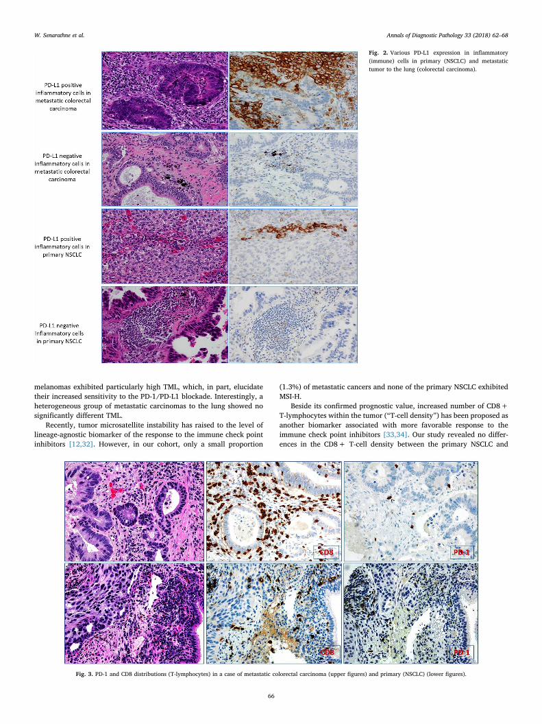
The results of PD-L1 expression in TC and IC are summarized inTables 1–3 and illustrative cases of primary NSCLC and metastaticcolorectal carcinoma are shown on Figs. 1–2. Specimen type (small vs.surgical biopsy) had no influence on frequency of PD-L1 expression inTCs and ICs (p = 0.23 and 0.86, respectively).
Overall, TC PD-L1 positivity in primary NSCLC was observed in 23of 81 cases (28%) and in 24 of 176 metastatic tumors (14%)(p = 0.009, Fisher's exact test). Among the 24 positive metastatic tu-mors, 13 were carcinomas (Table 1). Interestingly, all three PD-L1+breast carcinomas were triple-negative (ER-/PR-/Her2-) carcinomaswhile 5 out of six head and neck carcinomas were squamous cell car-cinomas. In non-carcinomatous metastases, PD-L1 expression was alsoobserved in 4/11 (36%) metastatic melanomas and 3/12 (20%) softtissue sarcomas (Table 1).
In adjacent normal lung tissue, PD-L1 expression was observed inalveolar macrophages (positive internal control cell type). However,PD-L1 expression in intratumoral IC was generally low in both cohorts(none of the tumors had IC PD-L1 above 5%).
However, when> 1% IC threshold for positivity was applied, asignificantly higher proportion of IC PD-L1staining was observed inmetastatic carcinomas than in primary NSCLCs (31% vs. 0%)(p < 0.001) (Tables 1). Notably, other histologic types of metastatictumors (e.g. melanomas and sarcomas) also showed significantly higherIC PD-L1 expression (13–36% of cases) than NSCLCs. Consequently,tumor immune microenvironment (TME) categories differed sig-nificantly between the primary NSCLC and metastatic carcinomas to thelung (Table 3) (see Fig. 3).
No significant difference in TC PD-L1 expression was observedwithin the two major primary NSCLC subgroups (adenocarcinoma vs.squamous cell carcinoma, p = 0.19, Table 1) whereas significant
W. Senarathne et al. Annals of Diagnostic Pathology 33 (2018) 62–68
63
differences in TC PD-L1 expression were seen among the metastaticcarcinomas based on their lineages (from 0% positivity in pancreatic to40% positivity in head and neck carcinomas, Table 1).
3.3. Relationship between the PD-L1 expression and tumor mutational load(TML)
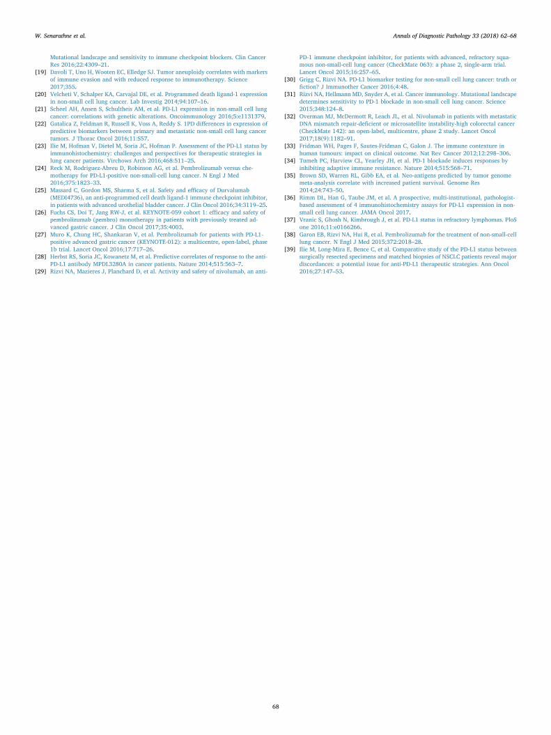
Tumor mutational load (TML) was analyzed in 229 samples (76 NSCLCand 153 metastatic tumors). The NSCLC cases exhibited significantly higherTML in comparison with the metastatic carcinomas (10 mutations onaverage vs. 6.6, p < 0.001, Table 4). Also, PD-L1 TC positive NSCLCs hadhigher TML compared with the PD-L1 negative primary tumors (p = 0.05).
MSI status was evaluated in 256 cases total; only two metastatictumors (1.3%) to the lung (one endometrial carcinoma and one ade-nocarcinoma of presumably intestinal origin) exhibited MSI-H status.None of the NSCLC (0%) had MSI-H (three equivocal cases by NGS hadintact MMRP expression by IHC).
No significant differences in TML were observed between the var-ious histologic types in the metastatic carcinomas group. However,metastatic melanomas (n = 10) had particularly high TML with anaverage of 32.7 mutations (range, 1–130 mutations) (Table 4).
3.4. PD-1 and CD8 expression in primary (NSCLC) and metastatic tumorsto the lung
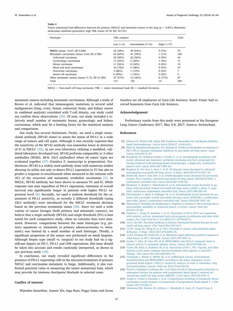
The mean number of CD8+ IC cells per 10 hpf/whole slides (smallbiopsies) was 388 (range, 3 to> 3000) while the mean number of PD-1positive cells was 33.7 (range, 0 to 280). The average number of PD-1+and CD8+ ICs did not differ significantly between the primary andmetastatic tumors to the lung (p = 1.0 and 0.13, respectively)(Table 2). A good correlation between PD-1+ and CD8+ cells (T-
Table 1PD-L1 expression in tumor cells was significantly higher in NSCLC compared with themetastatic carcinomas (p=0.003) while IC within metastatic tumors exhibited sig-nificantly higher PD-L1 expression (p < 0.001).
Histotype PD-L1 expression in tumor cells Total
[< 5%] [≥5%]
NSCLC 58 (72%) 23 (28%) 81- Adenocarcinoma 44 (72%) 17 (28%) 61- Squamous cell carcinoma 11 (73%) 4 (27%) 15- Other NSCLC 3 (60%) 2 (40%) 5
Metastatic carcinomas 113 (90%) 13 (10%) 126- Colorectal carcinoma 49 (96%) 2 (4%) 51- Gynecologic carcinomas 21 (95%) 1 (5%) 22- Breast carcinoma 18 (86%) 3 (14%) 21- Head and neck carcinomas 9 (60%) 6 (40%) 15- Pancreatic carcinoma 10 (100%) 0 (0%) 10- Renal cell carcinoma 6 (86%) 1 (14%) 7
Other metastatic tumors 39 (78%) 11 (22%) 50- Soft tissue tumors 12 (80%) 3 (20%) 15- Malignant melanoma 7 (64%) 4 (36%) 11- Other cancers 20 (83%) 4 (17%) 24
Total 210 47 257
Histotype PD-L1 expression in inflammatory cells Total
[< 1%] [≥1%]
NSCLC 81 (100%) 0 (0%) 81- Adenocarcinoma 61 (100%) 0 (0%) 61- Squamous cell carcinoma 15 (100%) 0 (0%) 15- Other NSCLC 5 (100%) 0 (0%) 5
Metastatic carcinomas 87 (69%) 39 (31%) 126- Colorectal carcinoma 32 (63%) 19 (37%) 51- Gynecologic carcinomas 16 (73%) 6 (27%) 22- Breast carcinomas 13 (62%) 8 (38%) 21- Head and neck carcinomas 12 (80%) 3 (20%) 15- Pancreatic carcinoma 7 (70%) 3 (30%) 10- Renal cell carcinoma 7 (100%) 0 (0%) 7
Other metastatic tumors 40 (81%) 10 (19%) 50- Soft tissue tumors 13 (87%) 2 (13%) 15- Malignant melanoma 7 (64%) 4 (36%) 11- Other cancers 20 (80%) 4 (20%) 24
Total 208 49 257
Table 2PD-1 and CD8 expression in inflammatory (T-cell) population [T-cell density] (meanvalues were used to dichotomize both variables). The number of PD-1+ and CD8+ cellsdid not differ significantly between the primary and metastatic tumors to the lung(p=1.0 and 0.13, respectively).
Histotype PD-1 expression Total
Low (< 34) High (≥34)
NSCLC 55 (68%) 26 (32%) 81- Adenocarcinoma 45 (74%) 16 (26%) 61- Squamous cell carcinoma 8 (53%) 7 (47%) 15- Other NSCLC 2 (40%) 3 (60%) 5
Metastatic carcinomas 40 (70%) 17 (30%) 57- Colorectal carcinoma 17 (59%) 12 (41%) 29- Gynecologic carcinomas 6 (67%) 3 (33%) 9- Breast carcinoma 5 (71%) 2 (29%) 7- Head and neck carcinomas 8 (100%) 0 (0%) 8- Pancreatic carcinoma 2 (100%) 0 (0%) 2- Renal cell carcinoma 2 (100%) 0 (0%) 2
Other metastatic tumors 21 (65%) 11 (35%) 32- Soft tissue tumors 10 (71%) 4 (29%) 14- Malignant melanoma 1 (33%) 2 (67%) 3- Other cancers 10 (67%) 5 (33%) 15
Total 116 54 170
Histotype CD8 expression Total
Low (< 389) High (≥389)
NSCLC 53 (65%) 28 (35%) 81- Adenocarcinoma 40 (65%) 21 (35%) 61- Squamous cell carcinoma 9 (60%) 6 (40%) 15- Other NSCLC 4 (80%) 1 (20%) 5
Metastatic carcinomas 44 (77%) 13 (23%) 57- Colorectal carcinoma 19 (66%) 10 (34) 29- Gynecologic carcinomas 9 (100%) 0 (0%) 9- Breast carcinomas 5 (71%) 2 (29%) 7- Head and neck carcinomas 8 (100%) 0 (0%) 8- Pancreatic carcinoma 2 (100%) 0 (0%) 2- Renal cell carcinoma 1 (50%) 1 (50%) 2
Other metastatic tumors 26 (81%) 6 (19%) 32- Soft tissue tumors 13 (93%) 1 (7%) 14- Malignant melanoma 1 (33%) 2 (67%) 3- Other cancers 12 (80%) 3 (20%) 15
Total 123 47 170
Table 3Significantly different TME categories between NSCLC and metastatic carcinomas to thelung (p < 0.001).
Histotypes TME categories (PD-L1 expression) Total
TC+/IC+ TC-/IC- TC+/IC- TC-/IC+
NSCLC 0 (0%) 58 (72%) 23 (28%) 0 (0%) 81Metastatic carcinomas 4 (3%) 78 (62%) 9 (7%) 35 (28%) 126Total 4 136 32 35 207
TC = tumor cells; IC = inflammatory (immune cells); TME = tumor microenvironment;NSCLC = non-small cell lung cancer.
W. Senarathne et al. Annals of Diagnostic Pathology 33 (2018) 62–68
64
lymphocytes) was also observed (p < 0.001, rs = 0.454). No sig-nificant association between the number of CD8+ T-lymphocytes andTML among the NSCLC and metastatic carcinomas was observed(p = 0.59). No significant differences in presence of PD-1+ and CD8+IC was observed between small (core) biopsies and resection samples(p = 0.56 and 0.38, respectively).
4. Discussion
Immunotherapy with immune PD1/PD-L1 checkpoint inhibitors hasachieved remarkable therapeutic benefits in various solid and hema-tologic malignancies [18,19]. However, predictive biomarkers still needrefinement [19]. A compelling body of evidence indicates that no singlebiomarker may be sufficient to identify the optimal “PD-1/PD-L1 im-munotype” in predicting the successful immune checkpoint treatmentstrategy [2,9]. In the present study, we comparatively analyzed dis-tribution of the two key targets of the immune checkpoint inhibitors ina cohort of primary NSCLC and tumors metastatic to the lungs. Our datasupport the previous results on the relatively common TC PD-L1 ex-pression in NSCLC [20,21]. In contrast to the previous studies, we alsoexplored PD-L1 in IC that exhibited rare or no PD-L1 expression. On theother hand, metastatic tumors were more commonly enriched by thePD-L1+ IC cells with uncommon PD-L1 TC positivity. We did not havea chance to perform the paired sample analysis comparing the meta-static tumors' TME to their primary sites' TME, to observe dynamics ofthe changes, if any. Couple of earlier studies indicated that some dis-cordance in PD-L1 expression in TC of NSCLC can occur between theprimary and metastatic sites [22,23], but we are not aware of any suchstudies outside NSCLC.
In NSCLC, PD-L1 expression level on tumor cells has been directlycorrelated with response to anti-PD1/PDL1 immune checkpoint in-hibitors. Low expression and high expression of PD-L1 testing is nowused in clinical practice to identify treatment-naïve and previously
treated patients most likely to obtain benefit from an anti-PD-1 therapy,respectively [5,24]. On the other hand, the expression of PD-L1 ontumor infiltrating lymphocytes may be important in identifying re-sponders to specific anti-PDL1 immune checkpoint inhibitors, and acombinatorial approach to evaluate PD-L1 expression on both tumorcells and tumor infiltrating lymphocytes can help to identify responders[3,25,26]. Even though the PD-L1 tumor expression is shown here to bemuch lower in tumors metastatic to the lung compared to NSCLC, anotably higher expression of PD-L1 expression on tumor infiltratingimmune cells was observed and may provide additional treatment op-portunities for anti-PDL1 immune checkpoint inhibitors [26,27]. Innon-lung tumors, PD-L1 expression on tumor infiltrating immune cellshas been shown to help identify patient response [3,25,27]. Therefore,our findings highlight the TME differences between primary and me-tastatic tumors and reveal new therapeutic options in metastatic tu-mors. Several recent studies showed a potential therapeutic benefit ofPD-1/PD-L1 blockade in locally advanced and/or metastatic tumorsenriched by the PD-L1+ ICs [3,7,13,28].
Regarding the different thresholds reported in literature for pre-dictive value of PD-L1 in different tumor types, in our study we used theuniform 5% threshold for PD-L1 positivity, because of the hetero-geneous nature of both primary and metastatic cancers. Several clinicaltrials and systematic reviews recommended 5% threshold[5,15,16,29,30]; a systematic review with meta-analysis conducted byCarbognin et al. revealed significant differences in therapeutic re-sponses when 5% cutoff was used in the patients with NSCLC, geni-tourinary cancers and malignant melanoma. No differences were ob-served when 1% threshold was used [15].
Preliminary data indicate that the tumors with high levels of so-matic mutations (TML) are more sensitive to PD-1/PD-L1 blockade[1,18,31]. Our TML study revealed significantly higher TML in NSCLCthat in the metastatic carcinomas, which may predict their better re-sponse to the immune therapy; of the metastatic tumors, metastatic
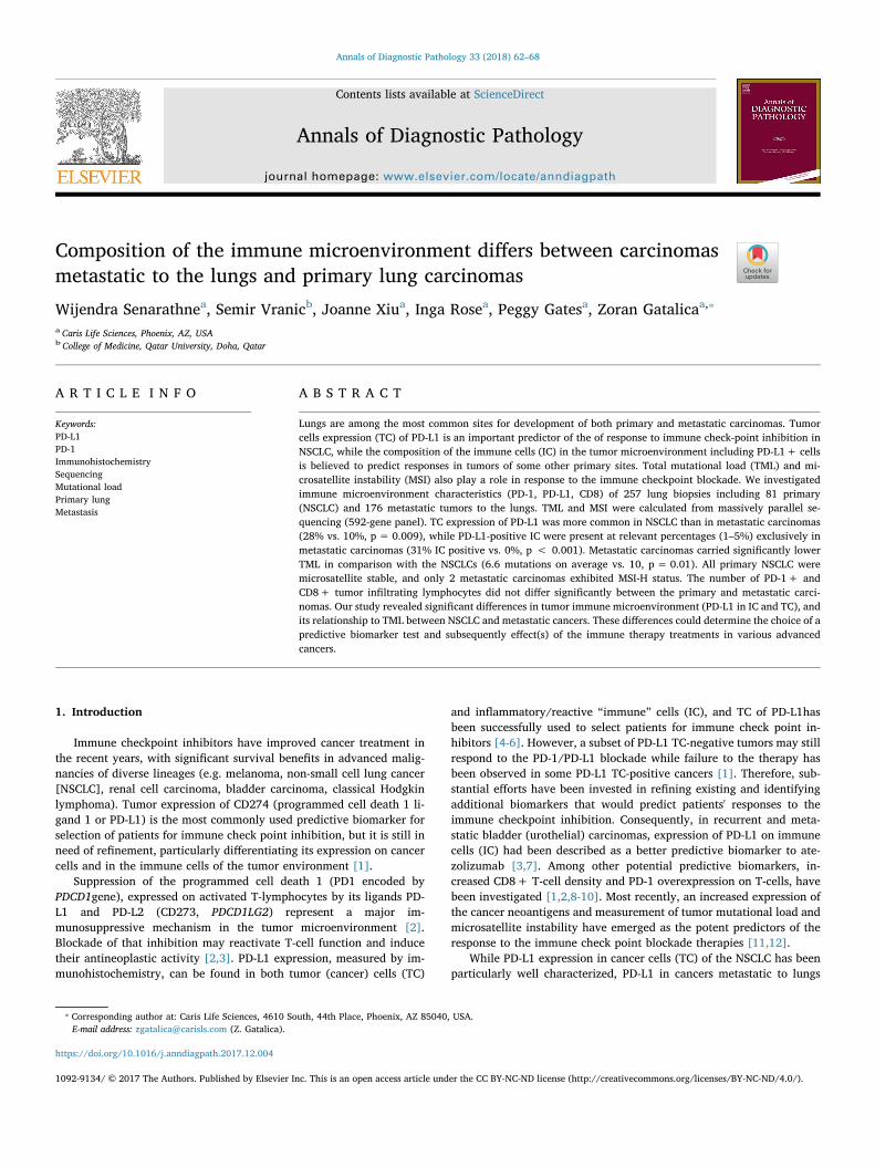
Fig. 1. Various PD-L1 expression in tumor cells inprimary (NSCLC) and metastatic tumor to the lung(colorectal carcinoma).
W. Senarathne et al. Annals of Diagnostic Pathology 33 (2018) 62–68
65
melanomas exhibited particularly high TML, which, in part, elucidatetheir increased sensitivity to the PD-1/PD-L1 blockade. Interestingly, aheterogeneous group of metastatic carcinomas to the lung showed nosignificantly different TML.
Recently, tumor microsatellite instability has raised to the level oflineage-agnostic biomarker of the response to the immune check pointinhibitors [12,32]. However, in our cohort, only a small proportion
(1.3%) of metastatic cancers and none of the primary NSCLC exhibitedMSI-H.
Beside its confirmed prognostic value, increased number of CD8+T-lymphocytes within the tumor (“T-cell density”) has been proposed asanother biomarker associated with more favorable response to theimmune check point inhibitors [33,34]. Our study revealed no differ-ences in the CD8+ T-cell density between the primary NSCLC and
Fig. 2. Various PD-L1 expression in inflammatory(immune) cells in primary (NSCLC) and metastatictumor to the lung (colorectal carcinoma).
Fig. 3. PD-1 and CD8 distributions (T-lymphocytes) in a case of metastatic colorectal carcinoma (upper figures) and primary (NSCLC) (lower figures).
W. Senarathne et al. Annals of Diagnostic Pathology 33 (2018) 62–68
66
metastatic tumors including metastatic carcinomas. Although a study ofBrown et al. indicated that immunogenic mutations in several solidmalignancies (lung, ovary, breast, colorectal, brain, and kidney cancerin combined analysis) correlated with T-cell density, our study couldnot confirm these observations [35]. Of note, our study included a re-latively small number of metastatic breast, gynecologic and kidneycarcinomas, which may be a limiting factor for the statistical analysisand comparisons.
Our study has several limitations. Firstly, we used a single mono-clonal antibody (SP142 clone) to assess the status of PD-L1 in a widerange of tumors and cell types. Although it was recently reported thatthe sensitivity of the SP142 antibody was somewhat lower in detectionof IC in NSCLC [36], in our own laboratory utilizing a modified, vali-dated laboratory developed test, SP142 performs comparably to 3 otherantibodies (SP263, 28-8, 22c3 antibodies) when all tumor types areevaluated together [37] (Gatalica Z, manuscript in preparation). Fur-thermore, SP142 is a widely used antibody clone with numerous studiesshowing its utility not only to detect PD-L1 expression in TC but also topredict a response to atezolizumab when measured in the immune cells(IC) of the recurrent and metastatic urothelial carcinomas [3]. InNSCLC, SP142 antibody has been shown to measure TC and IC. Whileresponse was seen regardless of PD-L1 expression, extension of overallsurvival was significantly longer in patients with higher PD-L1 ex-pression level [6]. Secondly, we used a uniform threshold for the as-sessment of PD-L1 positivity, as recently 2 different thresholds (using22c3 antibody) were introduced for the NSCLC treatment decisionbased on the previous treatments status [38]. Since we used a widevariety of cancer lineages (both primary and metastatic cancers), webelieve that a single antibody (SP142) and single threshold (5%) is bestsuited for such comparative study, when no outcome data were mea-sured. However, comparisons between the same histotypes (e.g. pri-mary squamous vs. metastatic or primary adenocarcinoma vs. meta-static) was limited by a small number of each histotype. Thirdly, asignificant proportion of the assays was performed on small biopsies.Although biopsy type (small vs. surgical) in our study had no a sig-nificant impact on PD-1, PD-L1 and CD8 expressions, this issue shouldbe taken into account and results cautiously interpreted, as shown inone previous study [39].
In conclusion, our study revealed significant differences in thepresence of PD-L1 expressing cells in the microenvironments of primaryNSCLC and carcinomas metastatic to lungs. Additionally, it also con-firmed potential value in measuring the tumor mutational load, whichmay provide for immune checkpoint blockade in selected cases.
Conflict of interest
Wijendra Senarthne, Joanne Xiu, Inga Rose, Peggy Gates and Zoran
Gatalica are all employees of Caris Life Sciences. Semir Vranic had re-ceived honoraria from Caris Life Sciences.
Acknowledgement
Preliminary results from this study were presented at the EuropeanLung Cancer Conference 2017, May 5-8, 2017, Geneva, Switzerland.
References
[1] Gibney GT, Weiner LM, Atkins MB. Predictive biomarkers for checkpoint inhibitor-based immunotherapy. Lancet Oncol 2016;17. (e542-e51).
[2] Shien K, Papadimitrakopoulou VA, Wistuba II. Predictive biomarkers of response toPD-1/PD-L1 immune checkpoint inhibitors in non-small cell lung cancer. LungCancer 2016;99:79–87.
[3] Rosenberg JE, Hoffman-Censits J, Powles T, et al. Atezolizumab in patients withlocally advanced and metastatic urothelial carcinoma who have progressed fol-lowing treatment with platinum-based chemotherapy: a single-arm, multicentre,phase 2 trial. Lancet 2016;387:1909–20.
[4] Borghaei H, Paz-Ares L, Horn L, et al. Nivolumab versus docetaxel in advancednonsquamous non-small-cell lung cancer. N Engl J Med 2015;373:1627–39.
[5] Herbst RS, Baas P, Kim DW, et al. Pembrolizumab versus docetaxel for previouslytreated, PD-L1-positive, advanced non-small-cell lung cancer (KEYNOTE-010): arandomised controlled trial. Lancet 2016;387:1540–50.
[6] Rittmeyer A, Barlesi F, Waterkamp D, et al. Atezolizumab versus docetaxel in pa-tients with previously treated non-small-cell lung cancer (OAK): a phase 3, open-label, multicentre randomised controlled trial. Lancet 2017;389:255–65.
[7] Fehrenbacher L, Spira A, Ballinger M, et al. Atezolizumab versus docetaxel for pa-tients with previously treated non-small-cell lung cancer (POPLAR): a multicentre,open-label, phase 2 randomised controlled trial. Lancet 2016;387:1837–46.
[8] Gelsomino F, Barbolini M, Spallanzani A, Pugliese G, Cascinu S. The evolving role ofmicrosatellite instability in colorectal cancer: a review. Cancer Treat Rev2016;51:19–26.
[9] Danilova L, Wang H, Sunshine J, et al. Association of PD-1/PD-L axis expressionwith cytolytic activity, mutational load, and prognosis in melanoma and other solidtumors. Proc Natl Acad Sci U S A 2016;113. (E7769-E77).
[10] Liu X, Cho WC. Precision medicine in immune checkpoint blockade therapy for non-small cell lung cancer. Clin Transl Med 2017;6:7.
[11] Le DT, Uram JN, Wang H, et al. PD-1 blockade in tumors with mismatch-repairdeficiency. N Engl J Med 2015;372:2509–20.
[12] Le DT, Durham JN, Smith KN, et al. Mismatch repair deficiency predicts response ofsolid tumors to PD-1 blockade. Science 2017;357:409–13.
[13] Powles T, Eder JP, Fine GD, et al. MPDL3280A (anti-PD-L1) treatment leads toclinical activity in metastatic bladder cancer. Nature 2014;515:558–62.
[14] Taube JM, Klein A, Brahmer JR, et al. Association of PD-1, PD-1 ligands, and otherfeatures of the tumor immune microenvironment with response to anti-PD-1therapy. Clin Cancer Res 2014;20:5064–74.
[15] Carbognin L, Pilotto S, Milella M, et al. Differential activity of Nivolumab,Pembrolizumab and MPDL3280A according to the tumor expression of pro-grammed death-ligand-1 (PD-L1): sensitivity analysis of trials in melanoma, lungand genitourinary cancers. PloS one 2015;10:e0130142.
[16] Peters S, Gettinger S, Johnson ML, et al. Phase II trial of Atezolizumab as first-line orsubsequent therapy for patients with programmed death-ligand 1-selected ad-vanced non-small-cell lung cancer (BIRCH). J Clin Oncol 2017;35:2781–9.
[17] Joneja U, Vranic S, Swensen J, et al. Comprehensive profiling of metaplastic breastcarcinomas reveals frequent overexpression of programmed death-ligand 1. J ClinPathol 2017;70:255–9.
[18] Chabanon RM, Pedrero M, Lefebvre C, Marabelle A, Soria JC, Postel-Vinay S.
Table 4Tumor mutational load differences between the primary (NSCLC) and metastatic tumors to the lung (p < 0.001); Metastaticmelanomas exhibited particularly high TML (mean 32.70, SD: 43.721).
Histotype TML category Total
Low (≤6) Intermediate (7-16) High (≥17)
NSCLC (mean: 10.07, SD 5.608) 22 (29%) 49 (64%) 5 (7%) 76Metastatic carcinomas (mean: 6.60, SD 2.785) 64 (60%) 41 (39%) 1 (1%) 106
-Colorectal carcinoma 22 (50%) 22 (50%) 0 (0%) 44-Gynecologic carcinomas 11 (65%) 5 (29%) 1 (6%) 17-Breast carcinoma 11 (56%) 8 (42%) 0 (0%) 19-Head and neck carcinomas 10 (72%) 4 (28%) 0 (0%) 14-Pancreatic carcinoma 4 (86%) 1 (14%) 0 (0%) 7-Renal cell carcinoma 6 (84%) 1 (16%) 0 (0%) 5
Other metastatic tumors (mean 11.76, SD 21.405) 27 (57%) 12 (26%) 8 (17%) 47Total 113 102 14 229
NSCLC = Non-small cell lung carcinoma; TML = tumor mutational load; SD = standard deviation.
W. Senarathne et al. Annals of Diagnostic Pathology 33 (2018) 62–68
67
Mutational landscape and sensitivity to immune checkpoint blockers. Clin CancerRes 2016;22:4309–21.
[19] Davoli T, Uno H, Wooten EC, Elledge SJ. Tumor aneuploidy correlates with markersof immune evasion and with reduced response to immunotherapy. Science2017;355.
[20] Velcheti V, Schalper KA, Carvajal DE, et al. Programmed death ligand-1 expressionin non-small cell lung cancer. Lab Investig 2014;94:107–16.
[21] Scheel AH, Ansen S, Schultheis AM, et al. PD-L1 expression in non-small cell lungcancer: correlations with genetic alterations. Oncoimmunology 2016;5:e1131379.
[22] Gatalica Z, Feldman R, Russell K, Voss A, Reddy S. 1PD differences in expression ofpredictive biomarkers between primary and metastatic non-small cell lung cancertumors. J Thorac Oncol 2016;11:S57.
[23] Ilie M, Hofman V, Dietel M, Soria JC, Hofman P. Assessment of the PD-L1 status byimmunohistochemistry: challenges and perspectives for therapeutic strategies inlung cancer patients. Virchows Arch 2016;468:511–25.
[24] Reck M, Rodriguez-Abreu D, Robinson AG, et al. Pembrolizumab versus che-motherapy for PD-L1-positive non-small-cell lung cancer. N Engl J Med2016;375:1823–33.
[25] Massard C, Gordon MS, Sharma S, et al. Safety and efficacy of Durvalumab(MEDI4736), an anti-programmed cell death ligand-1 immune checkpoint inhibitor,in patients with advanced urothelial bladder cancer. J Clin Oncol 2016;34:3119–25.
[26] Fuchs CS, Doi T, Jang RW-J, et al. KEYNOTE-059 cohort 1: efficacy and safety ofpembrolizumab (pembro) monotherapy in patients with previously treated ad-vanced gastric cancer. J Clin Oncol 2017;35:4003.
[27] Muro K, Chung HC, Shankaran V, et al. Pembrolizumab for patients with PD-L1-positive advanced gastric cancer (KEYNOTE-012): a multicentre, open-label, phase1b trial. Lancet Oncol 2016;17:717–26.
[28] Herbst RS, Soria JC, Kowanetz M, et al. Predictive correlates of response to the anti-PD-L1 antibody MPDL3280A in cancer patients. Nature 2014;515:563–7.
[29] Rizvi NA, Mazieres J, Planchard D, et al. Activity and safety of nivolumab, an anti-
PD-1 immune checkpoint inhibitor, for patients with advanced, refractory squa-mous non-small-cell lung cancer (CheckMate 063): a phase 2, single-arm trial.Lancet Oncol 2015;16:257–65.
[30] Grigg C, Rizvi NA. PD-L1 biomarker testing for non-small cell lung cancer: truth orfiction? J Immunother Cancer 2016;4:48.
[31] Rizvi NA, Hellmann MD, Snyder A, et al. Cancer immunology. Mutational landscapedetermines sensitivity to PD-1 blockade in non-small cell lung cancer. Science2015;348:124–8.
[32] Overman MJ, McDermott R, Leach JL, et al. Nivolumab in patients with metastaticDNA mismatch repair-deficient or microsatellite instability-high colorectal cancer(CheckMate 142): an open-label, multicentre, phase 2 study. Lancet Oncol2017;18(9):1182–91.
[33] Fridman WH, Pages F, Sautes-Fridman C, Galon J. The immune contexture inhuman tumours: impact on clinical outcome. Nat Rev Cancer 2012;12:298–306.
[34] Tumeh PC, Harview CL, Yearley JH, et al. PD-1 blockade induces responses byinhibiting adaptive immune resistance. Nature 2014;515:568–71.
[35] Brown SD, Warren RL, Gibb EA, et al. Neo-antigens predicted by tumor genomemeta-analysis correlate with increased patient survival. Genome Res2014;24:743–50.
[36] Rimm DL, Han G, Taube JM, et al. A prospective, multi-institutional, pathologist-based assessment of 4 immunohistochemistry assays for PD-L1 expression in non-small cell lung cancer. JAMA Oncol 2017.
[37] Vranic S, Ghosh N, Kimbrough J, et al. PD-L1 status in refractory lymphomas. PloSone 2016;11:e0166266.
[38] Garon EB, Rizvi NA, Hui R, et al. Pembrolizumab for the treatment of non-small-celllung cancer. N Engl J Med 2015;372:2018–28.
[39] Ilie M, Long-Mira E, Bence C, et al. Comparative study of the PD-L1 status betweensurgically resected specimens and matched biopsies of NSCLC patients reveal majordiscordances: a potential issue for anti-PD-L1 therapeutic strategies. Ann Oncol2016;27:147–53.
W. Senarathne et al. Annals of Diagnostic Pathology 33 (2018) 62–68
68