Embed Size (px)
Citation preview
IntroductionUnderstanding the pathology resulting from Staphy-lococcus aureus infection is of great interest in medicinebecause of the organism’s common and increasingprevalence in humans, its growing bacterial resist-ance, and its ability to cause serious and life-threat-ening disease (1). Toxins contribute in a major way tothe pathogenicity of S. aureus, as they do with manyother bacteria (2).
S. aureus frequently infects the skin. In fact, the mostcommon bacterial infection of children is impetigo,which accounts for approximately 10% of all skin prob-lems in children (3). Of these impetigo patients, about30% have bullous impetigo, which is caused by S. aureusstrains that produce exfoliative toxins (ETs). In patientsdischarged from the newborn nursery, over 30% may becolonized with S. aureus, and organisms that produceETs are the major cause of neonatal nursery outbreaksof staphylococcal disease (1, 4). Newborns, young chil-dren, and adults with renal failure and/or who are
immunocompromised may develop a generalized formof bullous impetigo called staphylococcal scalded skinsyndrome (SSSS). In this disease, bullae and erosionsdevelop over a large area of the skin surface, resulting inthe death of probably less than 3% of affected childrenbut up to 60% of affected adults (5, 6).
The now-classic experiments of Melish et al. in theearly 1970’s established that ET produced by S. aureuscauses the blistering in bullous impetigo and SSSS (7–9).Passive transfer of the toxin to neonatal mice caused thesame skin lesions seen in humans, namely a blister in thesuperficial living epidermis caused by separation of thekeratinocytes in the granular cell layer due to splitting ofthe desmosomes as either a primary or secondary event(10, 11). Presumably, this type of blister allows the bac-teria to spread under and circumvent the stratumcorneum, a major barrier that prevents infection of theskin. Two major ETs, ETA and ETB, which share 40%identity in amino acid sequence, have been identifiedand cloned (12, 13). Recently, a third ET (termed ETD)that causes identical epidermal blisters has been identi-fied (T. Yamaguchi et al., unpublished observations). Inbullous impetigo, blisters occur at the site of infectionwith S. aureus, and are caused by local production of ET,whereas in SSSS the toxin is produced at the site of infec-tion (not necessarily in the skin) and causes generalizedblistering due to its systemic circulation. The latter dis-ease underscores the exquisite specificity of ETs that,even when they circulate through the entire body, causepathology only in the superficial epidermis.
The pathophysiologic basis of this specificity hasbeen a mystery for over 30 years, even though recent
The Journal of Clinical Investigation | July 2002 | Volume 110 | Number 1 53
Molecular mechanisms of blister formation in bullousimpetigo and staphylococcal scalded skin syndrome
Yasushi Hanakawa,1 Norman M. Schechter,1 Chenyan Lin,1 Luis Garza,1 Hong Li,1
Takayuki Yamaguchi,2 Yasuyuki Fudaba,2 Koji Nishifuji,3 Motoyuki Sugai,2
Masayuki Amagai,3 and John R. Stanley1
1Department of Dermatology, University of Pennsylvania School of Medicine, Philadelphia, Pennsylvania, USA2Department of Bacteriology, Hiroshima University Graduate School of Biomedical Sciences, Hiroshima, Japan3Department of Dermatology, Keio University School of Medicine, Tokyo, Japan
Bullous impetigo due to Staphylococcus aureus is one of the most common bacterial infections of man,and its generalized form, staphylococcal scalded skin syndrome (SSSS), is a frequent manifestationof staphylococcal epidemics in neonatal nurseries. Both diseases are mediated by exfoliative toxins(ETs), which show exquisite pathologic specificity in blistering only the superficial epidermis. Weshow that these toxins act as serine proteases with extremely focused molecular specificity to cleavemouse and human desmoglein 1 (Dsg1) once after glutamic acid residue 381 between extracellulardomains 3 and 4. Mutation of the predicted catalytically active serine to alanine completely inhibitscleavage. The mutated ETs bind specifically to Dsg1 by immunofluorescence colocalization and bycoimmunoprecipitation. Thus, ETs, through specific recognition and proteolytic cleavage of onestructurally critical peptide bond in an adhesion molecule, cause its dysfunction and allow S. aureusto spread under the stratum corneum, the main barrier of the skin, explaining how, although theycirculate through the entire body in SSSS, they cause pathology only in the superficial epidermis.
J. Clin. Invest. 110:53–60 (2002). doi:10.1172/JCI200215766.
Received for publication April 22, 2002, and accepted in revised form May 29, 2002.
Address correspondence to: John R. Stanley, Department ofDermatology, University of Pennsylvania, School of Medicine,211 Clinical Research Building, 415 Curie Boulevard,Philadelphia, Pennsylvania 19104, USA. Phone: (215) 898-3240;Fax: (215) 573-2033; E-mail: [email protected] of interest: No conflict of interest has been declared.Nonstandard abbreviations used: exfoliative toxin (ET);staphylococcal scalded skin syndrome (SSSS); desmoglein 1(Dsg1); Tris-buffered saline (TBS); horseradish peroxidase (HRP);pemphigus foliaceus (PF); extracellular cadherin domain (EC).
crystal structure and amino acid sequences of ETA andETB have suggested that they are “atypical” glutamate-specific serine proteases. The term “atypical” was usedbecause some crystal structures of ETA and ETB haveshown that the presumed catalytic site is not config-ured properly to be active, perhaps requiring activationby binding to a specific substrate or receptor (14–17).On the other hand, it has been argued that the toxicityof ETs may not be as serine proteases at all (e.g., datahas suggested that these ETs are poorly inhibited byserine protease inhibitors), but as superantigens orthrough less-direct mechanisms such as release of pro-teases by the epidermis (reviewed in refs. 1, 18). Fur-thermore, until very recently, proteolysis of a relevantbiological target had not been demonstrated.
Very recently, we identified a relevant substrate ofETA, ETB, and ETD to be desmoglein 1 (Dsg1), atransmembrane glycoprotein of desmosomes in thecadherin gene superfamily (19, 20) (T. Yamaguchi etal., unpublished observations). Dsg1, but not theclosely related Dsg3 or E-cadherin, is cleaved by ETs.Furthermore, Dsg1 is a relevant substrate becauseantibodies in the autoimmune disease pemphigusfoliaceus (PF) target Dsg1 and cause clinical and his-tologic blisters in mouse and man identical to thoseseen in bullous impetigo and SSSS. These data implythat targeting of Dsg1 can result in the pathologycharacteristic of bullous impetigo and SSSS.
Here we demonstrate that ETA, ETB, and ETD act asglutamic acid–specific serine proteases with unusuallyfocused specificity that results in a single cleavage ofDsg1, leading to its functional inactivation. The patho-physiology of bullous impetigo and SSSS can thus beattributed to hydrolysis of a single peptide bond, pre-sumably in a functionally critical area in Dsg1.
MethodsRecombinant ETs. The expressed proteins, vectors, andtheir hosts are summarized in Table 1. Shuttle vectorpCE104, containing ETA and ETA Cmu, was fromPatrick Schlievert (Department of Microbiology, Uni-versity of Minnesota Medical School, Minneapolis, Min-nesota, USA) (14). ETA Amu was constructed from thesevectors with the QuikChange Site-Directed Mutagene-sis Kit (Stratagene, La Jolla, California, USA). ETB Amu
was cloned by PCR using pCE104-ETB (20) as a tem-plate. PCR was also used to add nucleotides encodingpeptide tags, V5His (Invitrogen Corp., Carlsbad, Cali-fornia, USA) or 3X FLAG (Sigma-Aldrich, St. Louis, Mis-souri, USA), to the 3′ end of these ET constructs. ETA-V5His, ETA Cmu–V5His, ETA Amu–V5His, ETB-V5His,ETB–3X FLAG, and ETB Amu–V5His were subclonedinto the HindIII and EcoRI sites of pCE104. DNA wastransformed into the S. aureus strain RN4220 (providedby Patrick Schlievert) via electropolation. The super-natant from RN4220 transformed with these plasmidswas precipitated with 85% ammonium sulfate. Precipi-tated ETs were dialyzed against PBS with 1 mM CaCl2
(PBS-Ca). His-tagged ETs were purified on Ni-NTA
columns (QIAGEN Inc., Valencia, California, USA)using the manufacturer’s procedure and then dialyzedagainst PBS-Ca. ET concentrations were estimated witha protein assay kit from Bio-Rad Laboratories Inc. (Her-cules, California, USA).
ETD was cloned by PCR from a patient sample (T.Yamaguchi et al., unpublished data), and ETD Amu wascloned by PCR mutagenesis. The amplified DNA frag-ments were cloned into pQE70 in order to express thefusion protein with a 6xHis tag sequence on the car-boxy terminus in E. coli. The recombinant proteins werepurified using Ni-NTA resin according to the manu-facturer’s protocol (QIAGEN Inc.).
For comparison of mutant to wild-type ETs (see Fig-ure 2), the supernatants from transformed RN4220staphylococci were used, with the amount of ETs nor-malized by Coomassie blue staining of SDS-PAGE gelsin which the ETs were the major band and accountedfor over 90% of the total protein.
Cell culture and transduction with adenovirus vectors.HaCaT cell keratinocytes cultured in DMEM with 10%FBS were transduced with recombinant adenovirus vec-tors Ax-mDsg1F and Ax-mDsg3F (20), encodingmouse Dsg1 and Dsg3, respectively, with carboxy-ter-minal FLAG tags (Table 1). After 24 hours, the cellswere incubated with recombinant ETA, ETB, or ETD inculture media for 10 minutes. Then the cells werewashed with PBS-Ca, extracted, and used for immuno-precipitation or immunoblotting.
In vitro digestion of recombinant Dsg1 with ETs. The entireextracellular domain of human or mouse recombinantDsg1 with an E tag (hDsg1E and mDsg1E) on the car-boxyl terminus was produced as a secreted protein bybaculovirus, as previously described (21). (These con-structs also contained a His tag on the carboxy termi-nus.) Approximately 1 µg of mouse or human Dsg1purified with a TALON (BD Biosciences Clontech, SanJose, California, USA) or E-tag column (Amersham Bio-sciences Inc., Piscataway, New Jersey, USA) was incu-bated with recombinant ETA, ETB, or ETD in Tris-buffered saline with 1 mM CaCl (TBS-Ca) for 1 hour at
54 The Journal of Clinical Investigation | July 2002 | Volume 110 | Number 1
Table 1Vectors and hosts used for expression of ETs and desmogleins
Protein Vector Host
ETA pCE104-ETA-V5His S. aureusETA Cmu pCE104–ETA Cmu–V5His S. aureusETA Amu pCE104–ETA Amu–V5His S. aureusETB pCE104-ETB-V5His S. aureusETB-FLAG pCE104-ETB–3X FLAG S. aureusETB Amu pCE104–ETB Amu–V5His S. aureusETD pQE70-ETD-His E. coliETD Amu pQE70–ETD Amu–His E. colihDsg1E Baculo-hDsg1E Insect cell (High Five)mDsg1E Baculo-mDsg1E Insect cell (High Five)Dsg1FLAG Ax-mDsg1F Keratinocytes (HaCaT)Dsg3FLAG Ax-mDsg3F Keratinocytes (HaCaT)
Baculo, baculovirus vector; Ax, adenovirus vector.
37°C. The digested samples were subjected to SDS-PAGE, transferred to PVDF membranes, and stainedusing Coomassie brilliant blue. Stained bands were cutout and sequenced by Edman degradation at the Pro-tein Microchemistry Facility of The Wistar Institute(Philadelphia, Pennsylvania, USA).
Antibodies. Anti–E-tag mouse monoclonal antibodyconjugated with horseradish peroxidase (HRP; Amer-sham Biosciences Inc.), anti–V5-tag mouse monoclonalantibody (Invitrogen Corp.), anti–His-tag mouse mon-oclonal antibody (QIAGEN Inc.), anti–FLAG-tag mousemonoclonal antibody (M2; Sigma-Aldrich), anti-ETAsheep polyclonal antibody (Toxin Technology Inc., Sara-sota, Florida, USA), anti-ETD rabbit polyclonal anti-body (T. Yamaguchi, unpublished observations), and PFserum (anti-Dsg1) were used for immunoblotting andimmunohistochemistry. HRP-conjugated anti-IgG anti-bodies of the appropriate specificity (Bio-Rad Labora-tories Inc.) were used for immunoblotting. Anti-humanantibody conjugated with Alexa Fluor 495 and anti-mouse antibody conjugated with Alexa Fluor 590 (Mol-ecular Probes Inc., Eugene, Oregon, USA) were used forimmunohistochemistry.
Ex vivo binding of ETA Amu to mouse epidermis, andimmunohistochemistry. Mouse tail or paw skin was incu-bated in DMEM at a ratio of 1:1 with 10% FBS and ETAAmu–V5His (∼83 µM) in PBS-Ca at 4°C for 12 hoursand then cryosectioned. After being fixed with 4%paraformaldehyde in PBS-Ca, specimens were blocked
with 1% BSA in PBS-Ca. Blocked specimen was incu-bated with anti-V5 antibody followed by anti-mouseantibody conjugated with Alexa Fluor 590, and thenstained with PF serum followed by anti-human anti-body conjugated with Alexa Fluor 495. Stained sectionswere photographed using confocal microscopy (MRC1024; Bio-Rad Laboratories Inc.). For controls, skin wasfirst incubated with wild-type ETA or an irrelevanttoxin (staphylococcal enterotoxin B with a V5 tag), thenstained with PF serum or anti-V5 antibody, respective-ly. These controls showed no staining (not shown).
Immunoprecipitation. After incubation with ETs, aden-ovirus-infected cells were extracted with 1% Triton X-100in TBS-Ca with protease inhibitors (2 µg/ml aprotinin,2 µg/ml leupeptin, and 1 mM PMSF) for immunopre-cipitation. Supernatants of High Five insect cells (Invit-rogen Corp.) transduced with baculovirus encodinghDsg1E were incubated with ETs, then used directly forimmunoprecipitation. FLAG-tagged proteins were pre-cipitated with anti-FLAG agarose (Sigma-Aldrich), andE-tagged proteins were precipitated with anti–E-tagSepharose (Amersham Biosciences Inc.) at 4°C for 1hour. Immunoprecipitates were washed ten times with1% Triton X-100 in TBS-Ca, then eluted with Laemmlisample buffer at 100°C.
Immunoblotting. Cell extracts or immunoprecipitates inLaemmli sample buffer were separated by 4–20% or 10%Tris-glycine SDA-PAGE (Bio-Rad Laboratories Inc. orInvitrogen Corp.), then transferred to nitrocellulose
The Journal of Clinical Investigation | July 2002 | Volume 110 | Number 1 55
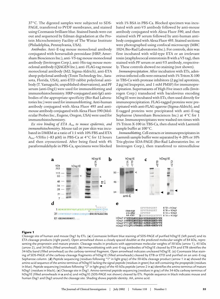
Figure 1Cleavage site of human and mouse Dsg1 by ETs. (a) Coomassie brilliant blue staining of SDS-PAGE of purified hDsg1E (left panel) and itsETA cleavage products (right panel). Open arrowhead shows a closely spaced doublet at the predicted molecular weight of 84 kDa, repre-senting the proprotein and mature protein. Cleavage results in products with approximate molecular weights of 50 kDa (arrow 1), 45 kDa(arrow 2), and 34 kDa (filled arrowhead). (b) Immunoblotting with anti–E-tag antibodies of hDsg1E cleaved by ETA and ETB identifies the34-kDa band (filled arrowhead) as the carboxy-terminal fragment. Open arrowhead indicates uncleaved hDsg1E. (c) Coomassie blue stain-ing of SDS-PAGE of the carboxy-cleavage fragments of hDsg1E (filled arrowheads) cleaved by ETB or ETD and purified on an anti–E-tagSepharose column. (d) Peptide sequencing (residues following "1" in light gray) of the 50-kDa cleavage product (arrow 1 in a) showed theamino acid sequence of the amino terminus of hDsg1E lacking the signal peptide (residues in green) but still containing the propeptide (residuesin blue). Peptide sequencing (residues following "2" in light gray) of the 45-kDa peptide (arrow 2 in a) identifies the amino terminus of maturehDsg1 (residues in black). (e) Cleavage site in Dsg1. Amino-terminal peptide sequencing (residues in gray) of the 34-kDa carboxy terminus ofhDsg1E (filled arrowheads in a and c) and mDsg1E (SDS-PAGE not shown) cleaved by ETs. Peptide sequence in black indicates mouse andhuman Dsg1 and Dsg3 around the cleavage site. Shading shows peptide identity. AA, amino acid.

sheets (Trans-Blot; Bio-Rad Laboratories Inc.). Thesheets were incubated for 1 hour in blocking buffer (5%fat-free milk powder in PBS). The first antibody, dilutedin blocking buffer, was applied for 1 hour at room tem-perature. After two washes with 0.1% Tween 20 in PBS,the sheets were incubated with HRP-conjugated sec-ondary antibody diluted in blocking buffer. In someexperiments, only one antibody, HRP-conjugated anti-ETA, was used. After the blots were washed, the signalswere detected with chemiluminescence (ECL or ECLPlus, Amersham Biosciences Inc.).
ResultsETs cleave human and mouse Dsg1 at a unique site. Previousstudies of proteolysis of Dsg1 by ETs identified only asmall carboxy-terminal fragment, leaving the extent ofdegradation uncertain. To determine whether there isone specific cleavage site as opposed to more generalproteolytic degradation, the extracellular domain ofhuman Dsg1, containing an E tag (hDsg1E) on the car-boxy terminus, was purified and cleavage was charac-terized by SDS-PAGE and amino-terminal sequenceanalysis. hDsg1E was produced by baculovirus in HighFive insect cells (21). The recombinant protein was iso-lated from insect cell supernatant with anti–E-tagSepharose and elution with excess E peptide. Coomassieblue staining of the peptide-eluted material resolved bySDS-PAGE (Figure 1a, left) indicates a closely spaceddoublet at the predicted molecular weight of 84 kDa.The isolated hDsg1E was incubated for 1 hour withETA at 37°C, and the products were separated by SDS-
PAGE (Figure 1a, right). Two major bands of approxi-mately 50 kDa (arrow marked 1) and 34 kDa (filledarrowhead) were detected by Coomassie blue staining,suggesting that hDsg1E was cleaved at a single site.Immunoblotting with anti–E-tag antibodies of hDsg1Edigests identified the 34-kDa band as the carboxy-ter-minal fragment (Figure 1b). Amino terminal peptidesequencing of this fragment shown in Figure 1a (rightside, arrowhead) was then used to determine the exactcleavage site, which was after glutamic acid residue 381(as counted from the initiating methionine of hDsg1)(Figure 1e). A similar study showed that ETA cleavedmouse Dsg1 at the same site (Figure 1e).
To determine the site of cleavage by ETB and ETD inhDsg1, we incubated unpurified hDsg1E (obtaineddirectly from baculovirus supernatant) with these ETs,then purified the carboxy terminus with anti–E-tagSepharose. Coomassie blue staining of the resultingpurified carboxy-cleavage fragments separated on SDS-PAGE is shown in Figure 1c. Amino-terminal peptidesequencing of these fragments indicated that the ETBand ETD cleavage site in Dsg1 was identical to thatproduced by ETA (Figure 1e). These data are consistentwith structural models that suggest that ETA and ETBmight cleave a substrate after a glutamic acid residue.
Amino terminal sequence analysis of the slowermigrating 50-kDa band formed by cleavage of hDsg1E(Figure 1a, arrow marked 1) revealed the amino termi-nus of hDsg1E lacking the signal peptide (which waspresumably processed by the insect cells) but still con-taining the propeptide (Figure 1d). Sequencing of theband shown immediately below the 50-kDa band (Fig-ure 1a, arrow marked 2) indicated that it was the aminoterminus of the hDsg1E mature protein (Figure 1d).These data show that the signal peptide of hDsg1E pro-duced by baculovirus in insect cells is properlyprocessed, and confirm that the sequence previouslypredicted by computer modeling to be the signal pep-tide in hDsg1 (Figure 1d, shown in green) is correct.The data also show that the proprotein produced bybaculovirus in insect cells is not efficiently processed.However, both the precursor and mature proteins areefficiently cleaved by ETA at the same site.
56 The Journal of Clinical Investigation | July 2002 | Volume 110 | Number 1
Figure 2Point mutation of serine 195 (chymotrypsin numbering), the pre-sumed catalytically active serine, of ETs inhibits cleavage of Dsg1.(a) Anti–E-tag antibody immunoblot of SDS-PAGE of hDsg1E incu-bated with RN4220 staphylococcal vector supernatant (RN), wild-type (WT) ETA, ETA Cmu (serine 195 mutated to cysteine), and ETAAmu (serine 195 mutated to alanine) shows markedly decreasedcleavage with ETA Cmu compared with wild-type ETA. ETA Amu
shows no catalytic activity. Horizontal lines indicate migration ofmolecular weight markers of 83 kDa (top) and 32 kDa. (b)Anti–FLAG-tag immunoblots of anti–FLAG-tag immunoprecipitatesof extracts of mDsg1-FLAG adenovirus–transduced cells that wereincubated with ETB or ETB Amu. ETB Amu shows no cleavage. Hori-zontal lines, from top, indicate migration of molecular weight mark-ers of 203 kDa, 115 kDa, and 93 kDa. (c) Anti–E-tag antibodyimmunoblot of SDS-PAGE of hDsg1E incubated with ETD and ETDAmu. ETD Amu does not cleave hDsg1. Horizontal lines, from top,indicate migration of molecular weight markers of 83 kDa and 34kDa. Uncleaved Dsg1 (open arrowhead) and its carboxy-terminalcleavage product (filled arrowhead) are indicated.
Figure 3ETA Amu inhibits cleavage of Dsg1 by ETA. Anti–E-tag immunoblot ofhDsg1E (open arrowhead) and its carboxy-terminal cleavage product(filled arrowhead). ETA Amu at a concentration of 13 µM incubatedwith hDsg1E inhibits cleavage by subsequent addition of wild-typeETA, but at 4 µM does not. At 8 µM, about half of the Dsg1 is free tobe cleaved. Kd can then be roughly estimated to be 8 µM. ETA Amu inthe concentrations shown (all in large excess of that of hDsg1E) wasincubated with hDsg1 at 25°C for 60 minutes, then wild-type ETAwas added for a 20-minute incubation before SDS-PAGE.

Taken together, the data in Figure 1 demonstrate thatETA, ETB, and ETD cleave hDsg1 at the same site, andthat ETA cleaves mDsg1 at the homologous site. TheseETs are known not to cleave the closely related Dsg3(19, 20). Sequence alignment of human and mouseDsg1 and Dsg3 (Figure 1e) indicated marked homolo-gy (17 of 18 identical residues) of mouse and humanDsg1, but divergence of Dsg3, around the cleavage site.
Serine 195 of ETs is critical for proteolysis of Dsg1. The infer-ence from structural studies of ETA and ETB is that theserine residue at position 195 in ETA and 186 in ETBcorresponds to the catalytically active serine 195 of chy-motrypsin. Furthermore, like chymotrypsin, ETA andETB have a functional catalytic triad consisting of ser-ine 195/186/195, histidine 72/65/57, and aspartic acid120/114/102 (ETA/ETB/chymotrypsin numbering;subsequent numbering will be for chymotrypsin only).To demonstrate that this serine is critical in the cleavageof hDsg1, it was mutated to a cysteine (resultantmutant ETA Cmu). Incubation of ETA Cmu with Dsg1 at37°C for 1 hour demonstrated a markedly decreasedrate of cleavage compared with wild-type ETA (Figure2). Assuming this activity was likely mediated by the cys-teine sulfhydryl group, similar to what has been foundin a similar mutant of trypsin (22), we mutated serine195 to alanine (ETA Amu). The ETA Amu did not cleaveDsg1 (Figure 2). We similarly showed that mutation ofserine 195 to alanine in ETB and ETD inhibited cleav-age of Dsg1 (Figure 2).
The observations that substitution of serine 195 withboth cysteine and alanine inhibit cleavage, and that themutant ETs still specifically bind Dsg1 (see below),demonstrate that serine 195 is necessary for efficientcatalytic cleavage of Dsg1.
Binding of ETs to Dsg1. Crystal structural studies of ETAhave suggested that it may be an inactive enzyme due toan inappropriate alignment of certain residues formingthe active site. It has been postulated that as a conse-quence, ETA may have to bind to its specific substrate orto a receptor in order to become catalytic (14–16, 23).(However, for an opposing view, see ref. 17.) In SSSS andin mouse models of SSSS, ETs diffuse through the entirebody, yet have exquisite specificity in causing pathology(e.g., blisters) only in the superficial epidermis. We havehypothesized that this specificity is due to specific recog-nition and cleavage of a single target, Dsg1.
If ETA binds to Dsg1, and that binding does notrequire the catalytic serine, then ETA Amu should be acompetitive inhibitor of wild-type ETA. Establishing theability of ETA Amu to protect Dsg1 from cleavage byETA would also provide an estimate of the dissociationconstant (Kd) for the reaction. To determine the Kd forthe interaction between ETA Amu and Dsg1, Dsg1 wasincubated with increasing concentrations of ETA Amu,with ETA Amu always in large excess of Dsg1. Free Dsg1was evaluated by relatively rapid treatment with wild-type ETA and by using SDS-PAGE to estimate the frac-tion cleaved and the fraction protected from cleavage(Figure 3). At concentrations of 13 µM and above, ETA
Amu mostly inhibited cleavage, but at concentrations of4 µM and below, it did not. Approximately half of theDsg1 was observed cleaved at an ETA Amu concentrationof 8 µM, allowing Kd to be roughly estimated as follows:
Equation 1
The magnitude of this Kd is within the range of ahydrolytic reaction whose specificity is due to relative-ly strong binding in the catalytic site with a relativelylow Km, but is probably not consistent with strongbinding to an site outside the substrate binding site.
We further showed that mutant ETA binds the epi-dermis where it causes a blister. This was demonstrat-ed by incubating mouse skin with ETA Amu with a V5epitope tag on the carboxy terminus at concentrationsin excess of Kd. Immunofluorescence with antibodiesagainst the V5 tag showed binding of ETA Amu to epi-dermis in a pattern identical to that of Dsg1 (Figure 4).
Finally, we confirmed by coimmunoprecipitation thatETs bind Dsg1. Incubation of active ETs with Dsg1showed coprecipitation that quickly decreased withtime as cleavage progressed (Figure 5a). The rapidchange in the ability to coprecipitate ETA with Dsg1indicates that ETA dissociates from Dsg1 after hydrol-ysis of the targeted peptide bond, consistent with
The Journal of Clinical Investigation | July 2002 | Volume 110 | Number 1 57
Figure 4Immunofluorescence colocalization of ETA Amu and Dsg1 in epider-mis. Mouse skin was incubated with ETA Amu with a V5 tag. Doubleimmunofluorescence staining with antibodies against V5 tag (red flu-orescence) and Dsg1 (green fluorescence) showed colocalization.Note that immunofluorescence localization of ETA Amu shows thetypical pattern of Dsg1 localization, with less intense staining in thebasal layer of the epidermis — a pattern opposite that of Dsg3, whichis seen most intensely in the basal layer (36).

enzyme recycling. Supporting this conclusion are stud-ies with ETA Cmu, which hydrolyzes Dsg1 at a muchslower rate. Consistent with the slower cleavage rate,coprecipitation of ETA Cmu with Dsg1 was observedover a longer time, but still decreased with increasinghydrolysis (Figure 5b). Finally, ETA Amu, which does notcleave Dsg1 at all, showed increased binding with time.
To show specificity of binding in these studies, wealso incubated Dsg3 with ETs and showed that ETs didnot cleave or bind to Dsg3, which is highly homolo-gous in amino acid sequence to Dsg1 (Figure 5c).
DiscussionETs are serine proteases that specifically bind to and cleaveDsg1 at a unique site after a glutamic acid residue. Previousstudies have suggested various possible sites and mech-anisms of action of ETs, such as binding to ganglio-sides, causing release of proteases by keratinocytes; act-ing as superantigens in stimulating the skin’s immunesystem; and acting as lipases (reviewed in ref. 1). How-ever, recent inferences from crystal structures of ETAand ETB suggest that they might be atypical serine pro-teases (14–17). These ETs have a structural homologyto the chymotrypsin family of serine proteases. Thepresumptive active site contains the classic catalytic
triad of serine 195, histidine 57, and aspartic acid 102.In addition, homology to the Streptomyces griseus pro-tease Glu-SGP suggests that ETA and ETB cleave aftera glutamic acid (or possibly aspartic acid) residue thatwould be stabilized in the active site by histidine 213,threonine 190, and lysine 216. However, the ETs areatypical because in ETA, and possibly ETB, the oxyan-ion hole, which helps to stabilize the transitional-statecomplex of catalysis, is not properly formed. This isbecause, as indicated by crystal structures, the peptidebond between proline 192 and glycine 193 is flipped180° relative to typical serine proteases. This observa-tion has led to speculation proposing the requirementfor interaction with a specific substrate or receptor thatwould impart the relatively small energy necessary toflip this bond.
Despite these predictions, only one protein substrate,melanocyte-stimulating hormone, was reported to becleaved by ETs until recently, although an ester sub-strate was reported (24, 25). The biological significanceof this protein substrate, however, was not apparent.
Recently we showed that Dsg1 is cleaved by ETA andETB (19, 20). Here we determine the molecular mecha-nism of that cleavage and of its exquisite specificity. TheETs (ETA, ETB, and ETD) specifically bind, and consis-tent with the predictions from amino acid sequence andcrystal structure, act as glutamic acid–specific serineproteases to cleave Dsg1 at a unique site. These data arealso consistent with previous findings of differentialaccumulation of ETA in newborn mouse skin comparedwith blood and other tissues (26).
Proteolysis of this one peptide bond leading to dys-function of Dsg1 and the desmosome would explainthe pathophysiology of blister formation in bullousimpetigo and SSSS (see below). It is therefore likely thatthis peptide bond is critically important to the properfunction of Dsg1. Its site in relation to the domainstructure of Dsg1 is shown in Figure 6. Interestingly,the ETs cleave at the border between extracellular cad-herin domains (ECs) 3 and 4, just toward the carboxyterminal from one of the predicted calcium-bindingdomains in EC3. Although three-dimensional struc-tures of desmogleins have not been solved, there ismarked homology in both calcium-binding sites andECs between E- and N-cadherins and Dsg1. Structuralstudies of E- and N-cadherin have suggested that theborder between ECs, through calcium binding, stabi-lizes the rigidity of the molecule and fixes its orienta-tion, and is critical for function (27–31). The results wepresent here suggest that this border between ECs inDsg1 is also critical for their proper function, since the
58 The Journal of Clinical Investigation | July 2002 | Volume 110 | Number 1
Figure 5Coprecipitation of ETs with Dsg1. (a) Immunoprecipitation followedby immunoblotting. Incubation of ETB (marked with a FLAG epitopetag) and ETD with Dsg1 showed coprecipitation at 1 minute that wasmarkedly diminished at 60 minutes, after increased cleavage of Dsg1.Similar results were found for ETA (data not shown). (b) Binding ofDsg1 with ETA Cmu and ETA Amu. ETA Cmu, which slowly cleaves Dsg1,showed decreased binding with time. (c) ETA and ETB (marked withHis epitope tags) bind to Dsg1 but not Dsg3. These ETs did not cleaveor bind to Dsg3, which is highly homologous in amino acid sequenceto Dsg1. Similar results were found with ETD (data not shown). IP,immunoprecipitation; IB, immunoblot.
Figure 6Schematic diagram of domains and ET cleavage site in Dsg1. Verticallines indicate presumptive calcium-binding sites. Arrowhead showsET cleavage site. S, signal peptide; P, propeptide sequence; TM, trans-membrane region.
cleavage of one peptide bond in this region causes dra-matic dysfunction of this desmosomal cadherin.
Pathophysiology of blister localization in PF, bullous impeti-go, and SSSS: inactivation of Dsg1 with Dsg3 compensation.In bullous impetigo and SSSS, blisters occur in thesuperficial epidermis from loss of keratinocyte adhe-sion in the granular layer. Strikingly, an autoantibody-mediated disease, PF, has very similar histopathologyto that seen in bullous impetigo and SSSS. PF is causedby anti-Dsg1 autoantibodies (32–34).
Although these antibodies are thought to inactivateDsg1, which is found throughout the epidermis and inmucous membranes, the blister occurs only in the super-ficial epidermis. This blister localization has beenexplained by the “desmoglein compensation hypothesis”(35). This hypothesis states that in areas of epitheliumwhere both Dsg3 and Dsg1 are expressed, a spontaneousblister will not occur when anti-Dsg1 antibodies inacti-vate Dsg1, because Dsg3 can compensate. However, ifonly Dsg1 is present, a blister will occur. This hypothe-sis has been validated both by clinical observation andexperimentally. In mucous membranes, both Dsg1 andDsg3 are found throughout the epithelia; therefore, noblisters are seen in PF, even though anti-Dsg1 antibod-ies bind to mucous membranes. In epidermis, Dsg1 isfound throughout, but Dsg3 is only in the deep epider-mis. Therefore, PF antibodies cause only superficial blis-ters where Dsg1 is not compensated for by Dsg3. Simi-larly, mothers with PF passively transfer anti-Dsg1antibodies to their neonates; however, these neonates donot develop PF because neonatal skin, unlike adult skin,expresses Dsg3 throughout all layers (36).
Experimental evidence for the desmoglein compen-sation hypothesis has been obtained in the neonatalmouse model of PF in which passively transferred anti-bodies from PF patients cause clinically and histologi-cally typical disease (35). Normal neonatal mice thathave Dsg3 in the deep epidermis (similar to adulthuman epidermis) and throughout the oral mucousmembranes develop only superficial epidermal blisterswhen injected with PF IgG, but Dsg3 knockout mice,which have no Dsg3 to compensate, develop blisters inthe deep epidermis and in oral mucous membraneswhen similarly injected. Furthermore, mice that expressDsg3 in the superficial epidermis (from a transgenecontaining Dsg3 cDNA driven off an involucrin pro-moter) are not susceptible to blistering from injectedPF IgG (36). These data provide an explanation forantibody inactivation of Dsg1 resulting in blisters onlyin the superficial epidermis.
ETs cause blisters with pathology identical to thoseoccurring in PF. Although ETs circulate throughoutthe body in SSSS, as do anti-Dsg1 antibodies in PF,blisters occur only in the superficial epidermis, not inthe deep epidermis or in mucous membranes, identicalto the findings in PF. Such identical pathology can beexplained by the loss of function of Dsg1 caused byantibodies in the case of PF, and by ET cleavage in thecase of SSSS or bullous impetigo. Loss of function of
Dsg1 by cleavage with ETs is also consistent with pre-vious immunofluorescence experiments in which ETscause cellular internalization and loss of cell surfacestaining of Dsg1 (19).
ETs are skin-smart. A major physiologic function of skinis to provide a barrier against infection. Much of that bar-rier resides in the stratum corneum. S. aureus, through theuse of ETs, has evolved an efficient and extremely focusedmechanism of proliferating and spreading under thatbarrier. Once introduced, the bacteria can spread effi-ciently by using ET to produce a cleavage plane rightunder the stratum corneum. To do so, the bacterium hasdeveloped a toxin that specifically binds and cleaves amolecule that is critical to adhesion in this area andcleaves it at a specific site that destroys its function.
AcknowledgmentsWe thank Michael Plotnik for helpful discussions. Thiswork was supported by grants from NIH and a Grant-in-Aid for Scientific Research from the Ministry of Educa-tion, Science, and Culture of Japan.
1. Ladhani, S., Joannou, C.L., Lochrie, D.P., Evans, R.W., and Poston, S.M.1999. Clinical, microbial, and biochemical aspects of the exfoliative tox-ins causing staphylococcal scalded-skin syndrome. Clin. Microbiol. Rev.12:224–242.
2. Schiavo, G., and van der Goot, F.G. 2001. The bacterial toxin toolkit. Nat.Rev. Mol. Cell Biol. 2:530–537.
3. Darnstadt, G.L. 2000. Cutaneous bacterial infections. In Nelson textbookof pediatrics. R.E. Behrman, R.M. Kliegman, and H.B. Jenson, editors. W.B.Saunders Co. Philadelphia, Pennsylvania, USA. 2028–2030.
4. Shinefield, H.R. 1995. Staphyolococcal infections. In Infectious disease ofthe fetus and newborn infant. J.S. Remington and J.O. Klein, editors. W.B.Saunders Co. Philadelphia, Pennsylvania, USA. 1105–1141.
5. Cribier, B., Piemont, Y., and Grosshans, E. 1994. Staphylococcal scaldedskin syndrome in adults. A clinical review illustrated with a new case. J. Am. Acad. Dermatol. 30:319–324.
6. Gemmell, C.G. 1995. Staphylococcal scalded skin syndrome. J. Med.Microbiol. 43:318–327.
7. Melish, M.E., and Glasgow, L.A. 1970. The staphylococcal scalded-skinsyndrome. Development of an experimental model. N. Engl. J. Med.282:1114–1119.
8. Melish, M.E., and Glasgow, L.A. 1971. Staphylococcal scalded skin syn-drome: the expanded clinical syndrome. J. Pediatrics. 78:958–967.
9. Melish, M.E., Glasgow, L.A., and Turner, M.D. 1972. The staphylococcalscalded-skin syndrome: isolation and partial characterization of theexfoliative toxin. J. Infect. Dis. 125:129–140.
10. Lillibridge, C.B., Melish, M.E., and Glasgow, L.A. 1972. Site of action ofexfoliative toxin in the staphylococcal scalded-skin syndrome. Pediatrics.50:728–738.
11. Elias, P.M., Fritsch, P., Dahl, M.V., and Wolff, K. 1975. Staphylococcaltoxic epidermal necrolysis: pathogenesis and studies on the subcellularsite of action of exfoliatin. J. Invest. Dermatol. 65:501–512.
12. Lee, C.Y., Schmidt, J.J., Johnson-Winegar, A.D., Spero, L., and Iandolo,J.J. 1987. Sequence determination and comparison of the exfoliativetoxin A and toxin B genes from Staphylococcus aureus. J. Bacteriol.169:3904–3909.
13. O’Toole, P.W., and Foster, T.J. 1987. Nucleotide sequence of the epider-molytic toxin A gene of Staphylococcus aureus. J. Bacteriol.169:3910–3915.
14. Vath, G.M., et al. 1997. The structure of the superantigen exfoliativetoxin A suggests a novel regulation as a serine protease. Biochemistry.36:1559–1566.
15. Cavarelli, J., et al. 1997. The structure of Staphylococcus aureus epider-molytic toxin A, an atypic serine protease, at 1.7 A resolution. Structure.5:813–824.
16. Vath, G.M., et al. 1999. The crystal structure of exfoliative toxin B: asuperantigen with enzymatic activity. Biochemistry. 38:10239–10246.
17. Papageorgiou, A.C., Plano, L.R., Collins, C.M., and Acharya, K.R. 2000.Structural similarities and differences in Staphylococcus aureus exfo-liative toxins A and B as revealed by their crystal structures. Protein Sci.9:610–618.
18. Bailey, C.J., Lockhart, B.P., Redpath, M.B., and Smith, T.P. 1995. The epi-
The Journal of Clinical Investigation | July 2002 | Volume 110 | Number 1 59
dermolytic (exfoliative) toxins of Staphylococcus aureus. Med. Microbiol.Immunol. 184:53–61.
19. Amagai, M., Matsuyoshi, N., Wang, Z.H., Andl, C., and Stanley, J.R. 2000.Toxin in bullous impetigo and staphylococcal scalded-skin syndrometargets desmoglein 1. Nat. Med. 6:1275–1277.
20. Amagai, M., et al. 2002. Staphylococcal exfoliative toxin B specificallycleaves desmoglein 1. J. Invest. Dermatol. 118:845–850.
21. Ishii, K., et al. 1997. Characterization of autoantibodies in pemphigususing antigen-specific enzyme-linked immunosorbent assays with bac-ulovirus-expressed recombinant desmogleins. J. Immunol.159:2010–2017.
22. Yokosawa, H., Ojima, S., and Ishii, S. 1977. Chemical transformation ofthe active-site serine residue of Streptomyces griseus trypsin to a cysteineresidue. J. Biochem. (Tokyo). 82:869–876.
23. Ladhani, S. 2001. Recent developments in staphylococcal scalded skinsyndrome. Clin. Microbiol. Infect. 7:301–307.
24. Rago, J.V., Vath, G.M., Bohach, G.A., Ohlendorf, D.H., and Schlievert,P.M. 2000. Mutational analysis of the superantigen staphylococcal exfo-liative toxin A (ETA). J. Immunol. 164:2207–2213.
25. Rago, J.V., et al. 2000. Staphylococcal exfoliative toxins cleave α- and β-melanocyte-stimulating hormones. Infect. Immun. 68:2366–2368.
26. Fritsch, P., Elias, P., and Varga, J. 1976. The fate of Staphylococcal exfo-liatin in newborn and adult mice. Br. J. Dermatol. 95:275–284.
27. Pokutta, S., Herrenknecht, K., Kemler, R., and Engel, J. 1994. Confor-mational changes of the recombinant extracellular domain of E-cad-herin upon calcium binding. Eur. J. Biochem. 223:1019–1026.
28. Nagar, B., Overduin, M., Ikura, M., and Rini, J.M. 1996. Structural basisof calcium-induced E-cadherin rigidification and dimerization. Nature.380:360–364.
29. Overduin, M., et al. 1995. Solution structure of the epithelial cadherindomain responsible for selective cell adhesion. Science. 267:386–389.
30. Pertz, O., et al. 1999. A new crystal structure, Ca2+ dependence andmutational analysis reveal molecular details of E-cadherin homoassoci-ation. EMBO J. 18:1738–1747.
31. Tamura, K., Shan, W.S., Hendrickson, W.A., Colman, D.R., and Shapiro,L. 1998. Structure-function analysis of cell adhesion by neural (N-) cad-herin. Neuron. 20:1153–1163.
32. Stanley, J.R., Koulu, L., Klaus Kovtun, V., and Steinberg, M.S. 1986. Amonoclonal antibody to the desmosomal glycoprotein desmoglein Ibinds the same polypeptide as human autoantibodies in pemphigus foli-aceus. J. Immunol. 136:1227–1230.
33. Koulu, L., Kusumi, A., Steinberg, M.S., Klaus Kovtun, V., and Stanley, J.R.1984. Human autoantibodies against a desmosomal core protein inpemphigus foliaceus. J. Exp. Med. 160:1509–1518.
34. Amagai, M., Hashimoto, T., Green, K.J., Shimizu, N., and Nishikawa, T.1995. Antigen-specific immunoabsorption of pathogenic autoantibod-ies in pemphigus foliaceus. J. Invest. Dermatol. 104:895–901.
35. Mahoney, M.G., et al. 1999. Explanations for the clinical and micro-scopic localization of lesions in pemphigus foliaceus and vulgaris. J. Clin.Invest. 103:461–468.
36. Wu, H., et al. 2000. Protection of neonates against pemphigus foliaceusby desmoglein 3. N. Engl. J. Med. 343:31–35.
60 The Journal of Clinical Investigation | July 2002 | Volume 110 | Number 1