Embed Size (px)
Citation preview

Abstract— Intact myocardial perfusion is a function of normal
sodium potassium adenosine triphosphatase (ATPase) pump
while normal myocardial viability is the function of intact
mitochondrium. Both molecular processes can be clinically
manipulated using non-invasive molecular imaging techniques.
Despite being a routine procedure, myocardial perfusion
imaging has inherent limitation in verifying viable myocardial
segments. Fluorodeoxyglucose (FDG), an analogue of glucose
molecule is a gold standard biomarker in demonstrating
viability. Integrating the two molecular processes in a clinical
study will define and classify viable from non viable
myocardial segments. The function of viable myocardial
segments may benefit revascularization procedure while
function has not been successfully revearsible after
revascularization in infarcted scared myocardium.
The objective of this study is to assess the concordant and
discordant results obtained when hypoperfused segments at
resting MPI are being compared with myocardial viability
study using new integrated diagnostic imaging modality
Positron Emission Tomography Computed Tomography
(PET/CT) with flourodeoxyglucose (FDG) as the biomarker.
The usefulness of flourodeoxyglucose as a potential viability
PET agent in the assessment of myocardium is elucidated
from this study.
I. METHODOLOGY
he study was conducted in collaboration between
Diagnostic Nuclear Imaging Centre, Universiti Putra
Malaysia (UPM), Cardiology Unit, Universiti Malaya
Medical Centre and Department of Cardiology and
Abdul Jalil Nordin1, Wan Himratul Azliza Wan Harun2 , Ahmad Zaid Fatah
Azman1, Ahmad Fazli Abdul Aziz1, Fathinul Fikri Ahmad Saad1, Zul Hilmi
Yaakob2 ,Annuar Rapaie3 , Wan Azman Wan Ahmad 2
1 Diagnostic Nuclear Imaging Centre, Universiti Putra Malaysia 2 Cardiology Unit, University Malaya, Kuala Lumpur, Malaysia 3 Serdang Hospital, Selangor, Malaysia
Department of Diagnostic Imaging , Serdang Hospital,
Malaysia upon approval by UPM Medical Ethic Committee.
Thirty one patients diagnosed coronary artery disease were
prospectively recruited. Only 19 patients were finally enrolled
since insufficient data in the remaining 12 patients. All
patients underwent pharmaceutical stress and rest myocardial
perfusion imaging (MPI) study using 99m
Tc-MIBI at Universiti
Malaya Medical Centre, Kuala Lumpur upon diagnosis
confirmation of ischaemic heart disease . The inclusion
criteria are clinical signs and symptoms of ischaemic heart
disease, raised cardiac enzymes, electrophysiological changes
and evident from imaging modalities like cardiac scintigraphy
using 99m
Tc-MIBI, echocardiography and Magnetic Resonance
Imaging.. Exclusion criteria include childrens and pregnant
mothers. Only patients who underwent pharmaceutical stress
and rest cardiac scintigraphy study are recruited.
Patients demonstrating perfusion defect during rest 99m
Tc-
MIBI were referred to Centre for Diagnostic Nuclear Imaging
of Universiti Putra Malaysia for further assessment using 18F-
FDG PET/CT . All patients were prepared using modified
glucose loading protocol as recommended by the updated
version of American Society of Nuclear Cardiology.
Patients were instructed to be fasting for at least 8 hours on the
day of examination. Patients known to have diabetes mellitus
follow routine medication. Fasting blood sugar was checked
early in the morning to ensure that patients are fasted. 1 hour
prior to serial blood glucose tests alternating with
subcutaneous short acting insulin, patients were given 200mg
of oral Niacin ingestion to facilitate myocardial glucose
uptake. Once the blood glucose reached the desired level, 8-
10mCi 18F-FDG given intravenously. Patient was rested for
not less than an hour prior to PET/CT image acquisition . 18F-FDG PET/CT study was conducted using 64MDCT/PET
Biograph Siemens Medical Systems, Germany. A CT scout
view was first obtained over the heart to plan the study. A low
dose CT was performed for attenuation correction and
anatomical correlation. The offset value is -30 to 0 on X and Y
respectively. List mode replay for gated study sampled 10
gates with 100% phase up to 600 second duration. The average
trigger rate is 64 per minute where trigger rejection threshold
Molecular Imaging in the clinical evaluation of
mitochondrial function in myocardium : The
potential role of integrated Positron Emission
Tomography Computed Tomography (PET/CT)
in cardiac imaging
Abdul Jalil Nordin, Wan Himratul Azliza Wan Harun, Ahmad Zaid Fatah Azman, Ahmad Fazli Abdul
Aziz, Fathinul Fikri Ahmad Saad, Zul Hilmi Yaakob, Annuar Rapaie , Wan Azman Wan Ahmad
T
Proceedings of the World Medical Conference
ISBN: 978-1-61804-036-7 118
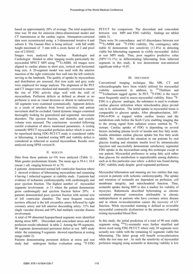
based on approximately 20% of average. The total acquisition
time was 30 min for emission (three-dimensional mode) and
CT transmission at the cardiac region. Attenuation-corrected
data were reconstructed using a 3-dimensional iterative 4 and
subsets 8. The Gausan filter is being utilized with full width
height maximum of 5 mm with a zoom factor of 2 and pixel
size of 2.03642.
Images were analyzed by Nuclear Radiologist and
Cardiologist blinded to other imaging results particularly the
myocardial SPECT MPI using 99m
Tc-MIBI. All images were
aligned to cardiac planes in short axis, vertical and horizontal
long axis. A 20-segment model was used with the anterior
insertion of the right ventricular free wall into the left ventricle
serving as the landmark. The quality of uptake by myocardium
and distribution are assessed. Hot iron scale and grey scale
were employed for image analysis. The alignment of the PET
and CT images were checked and manually corrected to ensure
the rim of FDG activity align well with the wall of
myocardium. Perfusion defects as defined as absent or no
perfusion is seen as visual decrease or absent in radioactivity.
All segments were examined systematically. Apparent defects
as a result of artefacts from bowel activities and patient
movement shall be excluded. Gated dynamic images examined
thoroughly looking for generalized and segmental movement
disorders. The ejection fraction, end diastolic and systolic
volume were assessed. The conclusions recorded following
careful evaluation of above mentioned parameters. A resting
sestamibi SPECT myocardial perfusion defect which is seen to
be reperfused during FDG PET/CT study is considered viable
or hibernating. A matched resting MPI and viability defect is
considered as infarcted or scarred myocardium. Results were
analyzed using SPSS version18.
II. RESULTS
Data from these patients (n=19) were analyzed (Table 1) .
Male gender predominate female. The mean age is 58.4 ± 10.8
(mean ± sd) ranging between 41 to 79.
6 patients demonstrated normal left ventricular function where
2 showed evidence of hibernating myocardium and remaining
4 having 1 infarcted segment at viability study . 9 patients had
evidence of ischaemic cardiomyopathy with cardiomegaly and
poor ejection fraction. The highest number of myocardial
segments involvement is 11 where the patient demonstrate
gross cardiomegaly and ejection fraction below 20%. 4
patients demonstrated poor ejection fraction with normal size
of left ventricular chamber. The most frequent vascular
territory affected is the left circumflex artery followed by right
coronary artery and left anterior descending artery. 31% had
single vascular disease and 69% had multiple vascular disease
involvement.
A total of 99 abnormal hypoperfused segments were identified
during stress MPI . Discordant and concordant stress and rest
perfusion results tabulated in table 2. Among the 99 segments,
90 segments demonstrated persistent defect at rest MPI study
while the remaining 9 segments showed reperfusion at resting
image (table 2).
Patients demonstrating persistent defects at stress and rest
study had undergone further evaluation using 18F-FDG
PET/CT for comparison. The discordant and concordant
between rest MPI and FDG viability findings are tabled
(table 3).
There were 36 concordance and 63 discordance between rest 99m
Tc-MIBI and 18F-FDG viability. The contingency table
(table 4) demonstrate low sensitivity (11.4%) in detecting
viable but hibernating segments in visibly myocardial defect
at rest MPI study. Thus, poor negative predictive value
(NPV=31.1%) in differentiating hibernating from infarcted
segments in this study. K test demonstrate non-statistical
significant with p >0.05.
III. DISCUSSION
Conventional imaging technique like MR, CT and
echocardiography have been widely utilized for myocardial
viability assessment in addition to 201
Thallium and 99m
Technetium tagged agents. In PET, 18F-FDG is the tracer
most frequently used to assess myocardial viability. Since 18F-
FDG is a glucose analogue, the substance is used to evaluate
cardiac glucose utilization where mitochondria plays pivotal
role in its utilization . The initial uptake in myocardial tissue is
comparable to glucose uptake. After phosphorylation, 18F-
FDG-6-PO4 is trapped within cardiac tissues and the
metabolism ends before the Kreb Cycle enabling imaging due
to the strong signal from radiation source emitted by 18F-
isotope. Cardiac uptake of FDG is depending on several
factors including plasma levels of insulin and free fatty acids.
Insulin stimulates cardiac glucose uptake but free fatty acids
inhibit. We mimicked the situation in our patients by oral
glucose loading and stimulate insulin level by intramuscular
injection and successfully demonstrate satisfactory segmental
FDG uptake in the myocardium using this technique except in
one patient. Myocardial preference in using fatty acid rather
than glucose for metabolism is unpredictable among diabetics
such as in this particular case where a defect was found during
FDG viability study despite good segmental perfusion.
Myocardial hibernation and stunning are two entities that may
coexist in patients with ischemic cardiomyopathy. The uptake
and retention of sestamibi are dependent on perfusion, cell
membrane integrity, and mitochondrial function. Thus,
sestamibi uptake during MPI is also a marker for viability of
myocytes. Rahimtoola described hybernating as chronic
sustained abnormal contraction attributable to chronic
underperfusion in patients who have coronary artery disease
and in whom revascularization causes the recovery of LV
function. While myocardial stunning is defined as reversible
myocardial contractile dysfunction in the presence of normal
resting myocardial blood flow .
In this study, the initial prediction of a total of 90 non viable
segments using 99m
Tc-sestamibi were further stratified and
down sized using FDG PET/CT where only 28 segments were
actually non viable with the remaining 62 segments viable but
hibernating. The latter group will benefit revascularization
while the rest may not . As such the sensitivity of myocardial
perfusion imaging using sestamibi in detecting viability is low
Proceedings of the World Medical Conference
ISBN: 978-1-61804-036-7 119

(11.4%). Therefore, careful interpretation should be exercised
involving segments with perfusion defect at rest during MPI
study.The importance of accurate ientification of viable
segments has been shown in previous study.
In studies conducted by D Carli and Marwick TH, they
revealed clinical evidence of improved heart failure
symptoms after revascularization which occurred
predominantly in patients with viable myocardium. The study
conducted using data from 333 patients also found improved
left ventricular ejection fraction (LVEF) in patients with viable
myocardium where in patients with-out viable myocardium,
the LVEF remained unchanged
In addition, informations obtained from FDG PET/CT study
can help predicting the outcome of our patients. We identified
9 patients with poor left ventricular ejection fraction (<50%)
and remodeling ( LV volume > 150 mls). These patients are
predicted having poor outcome. While 4 patients will
potentially benefit from revascularization procedure as they
demonstrated no clinical evidence of regional functional
abnormality. Rohatgi et al demonstrated that revascularization
in patients with a substantial amount of viable myocardium
reduces the number of hospital readmissions for congestive
heart failure. In our study, base on available evidence, almost
all our patients should get improvement in global function as
more than 20% of the left ventricular wall are found to be
viable.
We also derived a clinical prediction rule from our study
(table 5). Negative perfusion on MPI study and FDG viability
study or matched defect is defined as infarction (figure 1) .
While a negative perfusion on MPI study in the presence of
positive FDG uptake or mis-matching defect is termed as
viable but hibernating segments (figure 2) similar to the term
suggested by Maddahi and colleagues. This study warrants
long term follow-up in our patients to observe the final clinical
outcome and determining the actual prognosis, thus creating
better understanding in the clinical role of FDG PET/CT
viability studies .
Our study was conducted with challenging limitation where
there is lacking in real time integration of both images from
MPI and viability study. Analysis was restricted by software
incompatibility since the studies were conducted at two
different institutions.
IV. CONCLUSION
The results of our study support the importance of FDG as a
gold standard biomarker in determining myocardial viability.
Myocardial perfusion imaging using 99m
Tc MIBI are limited
by its low sensitivity and poor negative predictive value thus,
inability to recognized infarcted from viable but hibernating
myocardial segments. Without viability, those patients whom
will not benefit from high risk revascularization surgical
procedure shall be avoided.
Acknowledgement
The study was supported by Centre for Diagnostic Nuclear
Imaging of Universiti Putra Malaysia
Proceedings of the World Medical Conference
ISBN: 978-1-61804-036-7 120

No Age Sex
Race
MPI
FDG
Viability
LVF
Polar Map
Stress
Rest
Vol EF
LAD
LCX
RCA
Neg
Pos
Neg
Pos
Neg
Pos
mls
%
1 70 F Chi 1 19 1 0 0 1 233 40% Yes No Yes
2 53 M Mly 6 14 6 0 3 3 157 48% No Yes Yes
3 52 M Chi 13 7 12 1 11 2 294 18% Yes Yes Yes
4 49 F Mly 7 13 7 0 2 5 64 11% Yes Yes Yes
5 74 M Chi 4 16 4 0 0 4 105 51% Yes Yes Yes
6 41 M Mly 5 15 5 0 0 5 204 14% Yes No No
7 51 M Mly 3 17 2 1 1 2 105 51% No No Yes
8 75 M Chi 5 15 4 1 0 5 104 43% No Yes Yes
9 70 F Chi 3 17 3 0 1 2 153 38% No Yes No
10 56 M Ind 5 15 4 1 4 1 158 32% Yes Yes Yes
11 54 M Mly 5 15 5 0 4 1 160 43% Yes Yes Yes
12 68 F Mly 5 15 5 0 1 4 75 61% No Yes No
13 58 M Ind 7 13 2 5 0 7 111 21% No No No
14 50 M Chi 3 17 3 0 0 3 105 60% No No No
15 79 M Ind 3 17 3 0 0 3 84 96% Yes Yes Yes
16 48 M Ind 8 12 8 0 2 6 107 36% Yes No Yes
17 57 M Chi 5 15 5 0 0 5 113 50% No No No
18 51 M Ind 5 15 5 0 0 5 213 13% No Yes No
19 53 M Mly 6 14 6 0 0 6 203 24% No Yes No
MPI=myocardial perfusion imaging ; neg=negative; pos=positive ; FDG= Fluorodeoxyglucose; LV=left
ventricular; vol = volume; EF=Ejection Fraction ; LAD=left anterior descending; LCX=left circumflex;
RCA=Right coronary artery
Table 1. Results demonstrating patient demography, relationship between myocardial perfusion and viability, left
ventricular function and vascular territorial involvement derived from polar map. The numbers in MPI and FDG
viability columns are representing number of segments affected.
Proceedings of the World Medical Conference
ISBN: 978-1-61804-036-7 121

Rest Perfusion MIBI
Total Neg Pos
Stress
MPI
Neg
ativ
e Count 90 9 99
% Stress Perfusion MIBI 90.9% 9.1% 100.0%
% Rest Perfusion MIBI 100.0% 100.0% 100.0%
Total
Count 90 9 99
% Stress Perfusion MIBI 90.9% 9.1% 100.0%
% Rest Perfusion MIBI 100.0% 100.0% 100.0%
Neg= negative ; pos=positive
Table 2 . Stress Perfusion MIBI * Rest Perfusion MIBI * Crosstabulation
FDG Viability
Total Neg Pos
Rest MPI
Neg
Count 28 62 90
% Rest Perfusion MIBI 31.1% 68.9% 100.0%
% FDG Viability 96.6% 88.6% 90.9%
Pos
Count 1 8 9
% Rest Perfusion MIBI 11.1% 88.9% 100.0%
% FDG Viability 3.4% 11.4% 9.1%
Total
Count 29 70 99
% Rest Perfusion MIBI 29.3% 70.7% 100.0%
% FDG Viability 100.0% 100.0% 100.0%
Neg= negative ; pos=positive
Table 3 . Rest Perfusion MIBI * FDG Viability Crosstabulation
The Positive Predictive Value (PPV) and Negative Predictive Value (NPV) of MPI Rest is 88.9% and
31.1%. (Table 4) This result of PPV and NPV needs to interpret with caution. A low-ish NPV of MPI Rest
may be attributed to the high prevalence of viable/hibernating segments within the study population.
Additionally, test of agreement shows non-statistically significant, moderate agreement between the two
test (Kappa = 0.49, p = 0.29).
Proceedings of the World Medical Conference
ISBN: 978-1-61804-036-7 122

FDG Viability
+ve -ve
MPI Rest
+ve 8 1 9
-ve 62 28 90
70 29 99
Table 4. Contingency table of MPI Rest vs. FDG Viability
*Kappa = 0.49 (p = 0.20
From our study, we found 62% may benefit revascularization since more than 30% of the segments were
viable while the remaining 28% may not benefit the procedure.
Segment
n (%)
Infarct 28 (28.3)
Hibernate 62 (62.6)
Viable 8 (8.0)
Table 5: Percentage of segmental viability according to Clinical Prediction Rule
Proceedings of the World Medical Conference
ISBN: 978-1-61804-036-7 123

Figure 1 , A 52 year old man known ischaemic cardiomyopathy underwent
pharmaceutical induced stress-rest MPI study and FDG viability. Top are transaxial
images during MPI and viability during FDG PET/CT . Bottom Vertical long axis and
horizontal long axis images. There are matching defect in the apical region in keeping
with infarcted myocardium
Proceedings of the World Medical Conference
ISBN: 978-1-61804-036-7 124

Figure 2. Transaxial images of the left ventricle of a 72 year old woman demonstrating
revearsible defect in the septal region on viability study (bottom row) indicating
hibernating myocardium.
Figure 3. Longitudinal long axis of MPI and FDG viability PET/CT demonstrating stress
induced ischaemia on MPI (top row) in the apical region but viable in FDG PET/CT study
(bottom row).
Proceedings of the World Medical Conference
ISBN: 978-1-61804-036-7 125
![Positron Emission Tomography Imaging: A Quantitative ... · evidence,” i.e., a subset of biomarkers [3]. Positron Emission Tomography (PET), as a non-invasive imaging technique](https://img.pdfslide.us/doc/110x75/5c0b635309d3f2461a8c2663/positron-emission-tomography-imaging-a-quantitative-evidence-ie.jpg)