Embed Size (px)
Citation preview
Molecular Imaging Heterogeneity of Clinically Defined AD
Gil Rabinovici, M.D.Edward Fein & Pearl Landrith Endowed Professor
UCSF Memory & Aging Center
2017 Spring ADC MeetingBoston, MA
April 22, 2017

Disclosures
• Research support – Avid Radiopharmaceuticals/Eli Lilly, GE Healthcare,
Piramal Imaging– NIH, American College of Radiology, Alzheimer’s
Association, Tau Consortium, Association for Frontotemporal Degeneration, Michael J Fox Foundation
• Consulting/honoraria– Eisai, Genentech, Lundbeck, Merck, Putnam, Roche
Outline
• Heterogeneity in causes of clinical AD dementia• Amyloid-negative MCI/AD
• Prevalence• Demographics, clinical features• Biomarker signatures, relationship to SNAP
• Heterogeneity in clinical presentations of AD neuropathology• Molecular correlates of non-amnestic AD• Early age-of-onset AD (sporadic)

Rates of Aβ Biomarker Negativity in Clinical AD
Bapineuzumab: Mild-Mod AD ADNI: Late MCI and AD
Liu et al. Neurology 2015 Landau et al. Neurology 2016
6.5% 36.1%
16.4%
15%34%
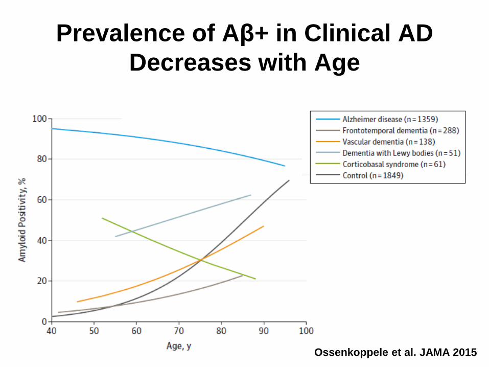
Prevalence of Aβ+ in Clinical AD Decreases with Age
Ossenkoppele et al. JAMA 2015

Characterization of Aβ- MCI/AD in ADNI• Demographics
• Older than Aβ+• M > F
• Cognition and function• Better at baseline (MCI)• Slower decline (MCI and AD)
• Lower prevalence ApoE4• MCI: Aβ- 16% vs. Aβ+ 71%• AD: Aβ- 4% vs. Aβ+ 75%
• Less abnormal neurodegeneration biomarkers• CSF t-tau, p-tau• Baseline MRI and FDG• Longitudinal MRI
ADAS
-Cog
Landau et al. Neurology 2016

Neurodegeneration in Aβ-negative Amnestic AD (N=21)
Chételat et al. Brain 2016

Suspected Non-Alzheimer Disease Pathology (SNAP)
• SNAP in MCI and AD dementia• 17%-35% of MCI• ~6%-15% of AD dementia • Older age-of-onset• Male > female• ApoE4 rates 11%-32%
• Rate of decline intermediate between A-/N- and A+/N+
• No clinical fingerprint of a single underlying disease• Increased WMH in some studies• No features of DLB• No increases (yet) in tau PET Jack et al. Nature Rev Neurol 2016

Intermediate Risk of Cognitive Decline in MCI-SNAP
Caroli et al. Neurology 2015
201 MCI from ADNI/EUFollowed up to 5 yrs (mean 2.5)Decline:
Conversion to ADMMSE decline ≥ 3 pts/yrMMSE ≤ 24

Neuropathological Diagnoses in Low Amyloid Clinical AD (N=50)
Dementia onset late 70s, death mid 80s
ApoE4 – 26%
Most common diagnoses:AD (8), VaD (8), DLB (5), HS (5), normal brain (5) FTLD (4)
PART not diagnosed but44% had Braak III/IV
Monsell et al. JAMA Neurol 2015

CTE at Autopsy in Aβ-PET Negative AD
Gardner et al. Neurol Clin Pract 2015
79 year-old retired NFL player with progressive memory loss
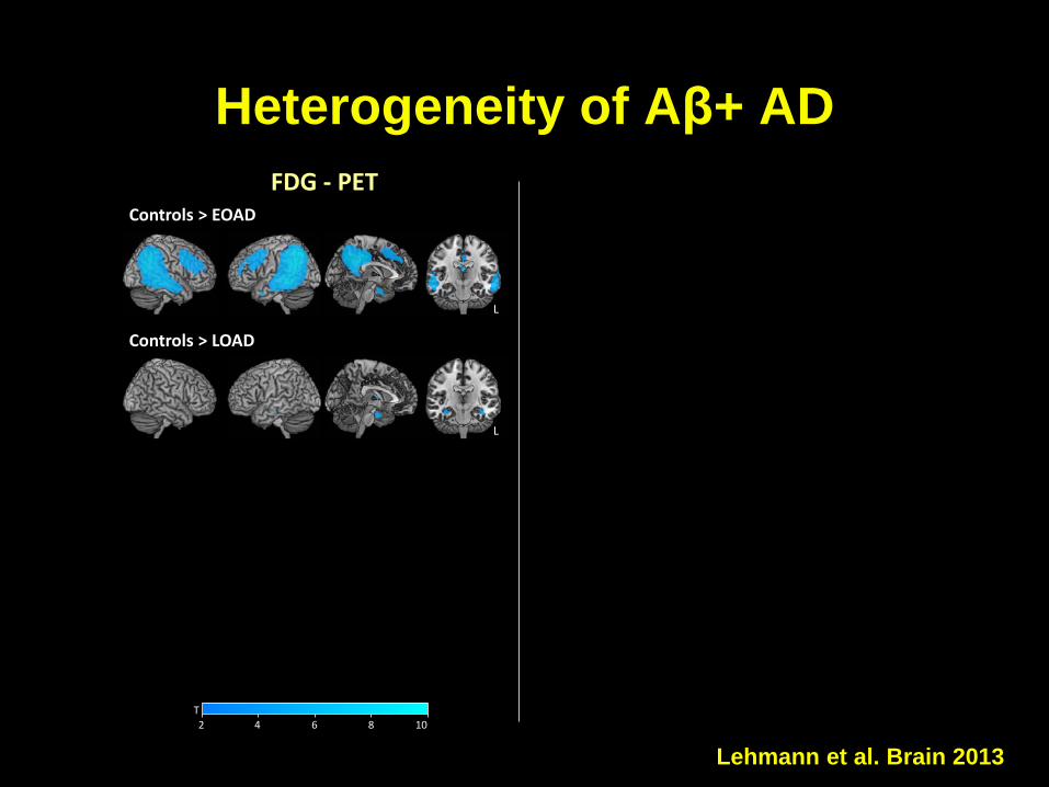
Controls > EOAD
Controls > lvPPA
Controls > PCA
L
L
L
FDG - PET
lvPPA > controls
PCA > controls
L
L
L
EOAD > controls
PIB - PET
T2 4 6 8 10
T2 4 6 8 10
Controls > LOAD LOAD > controls
L L
Lehmann et al. Brain 2013
Heterogeneity of Aβ+ AD

Tau PET Patterns Correlate with AD Phenotype
Ossenkoppele et al. Brain 2016Xia et al. JAMA Neurol 2017Day et al. Alz Dis Assoc Disord 2017
Co t o s O
Ossenkoppele et al. Brain 2016
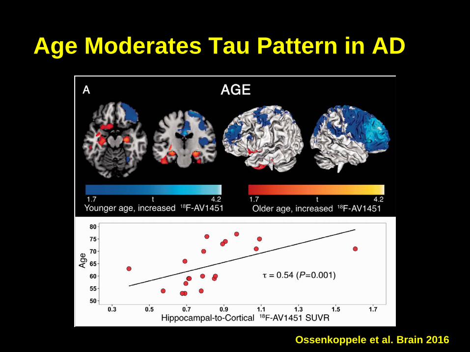
Age Moderates Tau Pattern in AD

Pontecorvo et al. Brain 2017
Tau Burden in AD is Negatively Correlated with Age

Longitudinal Tau PET in EOAD

Conclusions• Biomarkers identify patients with non-Aβ
pathologies mimicking clinical AD• Consistently ~15% of AD dementia• Associated with ApoE4 neg, older age, male• Better prognosis than Aβ+ (but not benign)• Likely represents a mix of neuropathologies
• PART, CARTS, AGD, vascular, DLB• Biomarkers can identify AD pathology as cause of
heterogeneous syndromes• Early-onset AD critical and under-studied cohort in which
to investigate mechanisms that drive heterogeneity• Dedicated study will require multi-site collaborations

UCSF-MACBruce MillerRik OssenkoppeleNagehan AyaktaViktoria BourakovaAlexandre BejaninLeonardo IaccarinoRenaud La JoieAshley MensingJulie PhamDaniel SchonhautRichard TsaiGautam TammewarAdrienne VisaniAdam BoxerLea GrinbergMarilu Gorno-TempiniAnna KarydasRobin KetelleJoel KramerZach MillerHowie RosenMiguel SantosSalvatore SpinaBill SeeleyMike Weiner
FundingNIA R01-AG045611, P01-AG1972403, P50-AG023501NINDS U54NS092089Tau ConsortiumMichael J. Fox FoundationAFTDAlzheimer’s Association Avid RadiopharmaceuticalsAmerican College of RadiologyFrench Foundation
AcknowledgmentsUC Berkeley/LBNLBill JagustSusan LandauJim O’NeillKris NortonMustafa JanabiSuzanne BakerSam Lockhart
Avid Mark MintunAndrew SiderowfMarybeth Howlett
IDEAS Study teamMaria CarrilloConstantine GatsonisBruce HillnerBarry SiegelRachel WhitmerCharlie ApgarLucy HannaJim HendrixCynthia Olson

Cognitive Trajectories By Aβ Status
Landau et al., Neurology 2016

Characterization of Aβ- MCI/AD in ADNI• Slightly older than Aβ+ (AD only)
• Mean age 78 vs. 74• Lower ApoE4
• MCI: Aβ- 16% vs. Aβ+ 71%• AD: Aβ- 4% vs. Aβ+ 75%
• Better baseline cognition and function (MCI only)
• Slower cognitive decline (both groups)
• Higher prevalence of depression and hypertension
• Lower neurodegeneration biomarkers• CSF t-tau; p-tau, baseline MRI and
FDG, longitudinal MRI

Atrophy in Aβ-Neg AD Dementia
Chételat et al, in revision
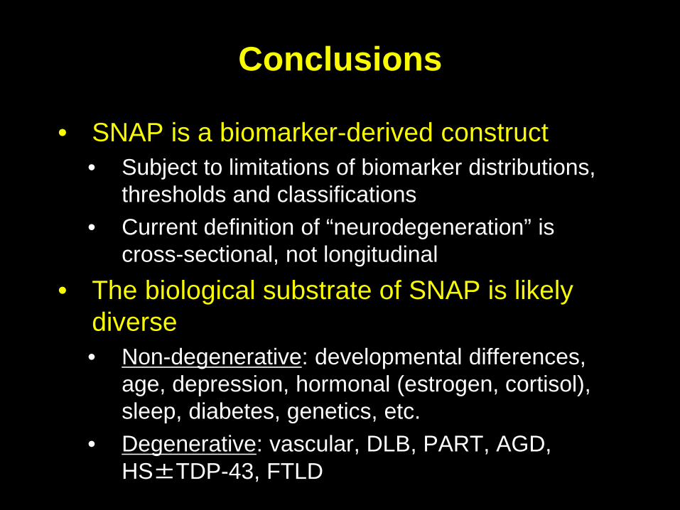
Conclusions
• SNAP is a biomarker-derived construct• Subject to limitations of biomarker distributions,
thresholds and classifications• Current definition of “neurodegeneration” is
cross-sectional, not longitudinal• The biological substrate of SNAP is likely
diverse• Non-degenerative: developmental differences,
age, depression, hormonal (estrogen, cortisol), sleep, diabetes, genetics, etc.
• Degenerative: vascular, DLB, PART, AGD, HS±TDP-43, FTLD

Conclusions• The prognosis of SNAP differs by baseline
cognitive status• Healthy elderly: relatively benign (simailar to A-N-)• MCI: intermediate between A-N- and A+N+• Dementia: majority show continued decline
• The substrate of SNAP likely differs by baseline cognitive status• Healthy elderly: greater contribution of non-
degenerative factors (or very slow pathologies)• Dementia: primarily non-AD cortical/subcortical (non-
amnestic) or limbic (amnestic) pathologies• MCI: mix of degenerative vs. non-degenerative
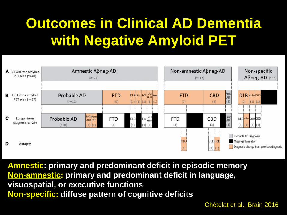
Outcomes in Clinical AD Dementia with Negative Amyloid PET
Chételat et al., Brain 2016
Amnestic: primary and predominant deficit in episodic memoryNon-amnestic: primary and predominant deficit in language, visuospatial, or executive functionsNon-specific: diffuse pattern of cognitive deficits
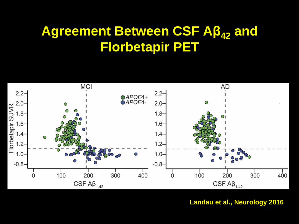
Agreement Between CSF Aβ42 and Florbetapir PET
Landau et al., Neurology 2016

Early Tau PET Data Suggest SNAP ≠ PART
Mormino et al, JAMA Neurol 2016

Intermediate Risk of Cognitive Decline in MCI-SNAP
Caroli et al, Neurology 2015 Vos et al, Brain 2015

Intermediate Risk of Cognitive Decline in MCI-SNAP
Caroli et al, Neurology 2015 Vos et al, Brain 2015

Early-Onset AD (Age ≤ 65)
• 5% of all AD patients = ~250,000 in U.S.– Only ~5%-10% harbor APP/PSEN mutations
• Study mechanisms of heterogeneity and selective vulnerability in AD– Non-amnestic clinical presentations; focal cortical
syndromes (lvPPA, PCA, fvAD)• Identify novel genetic risk factors
– Only ~50% carry ApoE4– Not represented in GWAS; will require targeted effort
• Employ biomarkers– Improve clinical diagnosis– Study mechanisms of “pure” AD: fewer co-pathologies– Under-represented in ADNI, not included in DIAN