Embed Size (px)
Citation preview

Annu. Rev. Genet. 1992.26:403-24Copyright © by Annual Reviews Inc. All rights reserved
MOLECULAR GENETICS OFHUMAN VISUAL PIGMENTS
Jeremy Nathans, Shannath L. Merbs, Ching-Hwa Sung,Charles J. Weitz, and Yanshu WangHoward Hughes Medical Institute, Department of Molecular Biology and Genetics,and Department of Neuroscience, Johns Hopkins University School of Medicine,
Baltimore, Maryland 21205
KEY WORDS: color vision, color blindness, retina, retinal disease
CONTENTS
INTRODUCTION .......................................... 403
COLOR VISION .......................................... 405RED AND GREEN PIGMENTS ................................. 406
Psychophysics of Red~Green Color Blindness ....................... 406Molecular Genetics of RedIGreen Color Blindness .................... 407Variation in Normal Color Vision .............................. 409Blue Cone Monochromacy .................................. 412
THE BLUE PIGMENT ....................................... 415Psychophysics of Tritanopia .................................. 415Molecular Genetics of Tritanopia .............................. 415
RHODOPSIN ............................................. 416Inherited Defects in Rod Vision ............................... 416Rhodopsin Mutations in Retinitis Pigmentosa ....................... 417Biochemical Characteristics of Mutant Rhodopsins .................... 418
PERSPECTIVE ........................................... 420
INTRODUCTION
Human vision is based upon four light-sensitive pigments: in dim light, it ismediated by rhodopsin, the visual pigment in rod photoreceptors, and at higherlight levels, it is mediated by three pigments that reside in three classes ofcone photoreceptors.1 The absorption spectra of rhodopsin and the cone
lThe three cone pigments and the photoreceptors within which they reside have historicallybeen referred to as "blue", "green", and "red", to indicate those colors to which their wavelengthsof maximal absorption approximately correspond.
403
0066-4197/92/1215-0403502.00
www.annualreviews.org/aronlineAnnual Reviews
Ann
u. R
ev. G
enet
. 199
2.26
:403
-424
. Dow
nloa
ded
from
arj
ourn
als.
annu
alre
view
s.or
gby
Roc
kefe
ller
Uni
vers
ity o
n 05
/14/
08. F
or p
erso
nal u
se o
nly.
404 NATHANS ET AL
pigments consist of broad bell-shaped curves that differ from one anotherprincipally by translation along the wavelength axis (Figure 1). Each visualpigment absorption spectrum~quivalent to a probability curve of photoncapture as a function of wavelength---determines the action spectrum of thephotoreceptor within which that pigment resides.
All visual pigments contain a chromophore, 11-cis retinal (or in someinstances l l-cis dehydroretinal), bound via a protonated Schiff’s base to lysine residue of an integral membrane protein. Light activates a visualpigment by photoisomerizing the retinal chromophore from cis to trans aboutthe 11-12 double bond, an event analogous to the replacement of an antagonistby an agonist within the binding pocket of a hormone receptor. Thephotoactivated visual pigment catalyzes the activation of a G-protein (trans-ducin) that in turn activates a cGMP phosphodiesterase. The resulting declinein cytosolic free cGMP concentration closes plasma membrane cation channelsthat are gated directly by cGMP, thereby decreasing the inward current andhyperpolarizing the cell (reviewed in 55,84).
Each of the four human visual pigment apoproteins is encoded by a separategene (60, 62). The nucleotide sequences of genomic and cDNA clonesencoding the four human visual pigments reveal 40-45% amino acid identitybetween rhodopsin and each of the cone pigments, and between the bluepigment and the red or green pigments. By contrast, the red and green pigmentsare 96% identical at the amino acid level. Each visual pigment sequence shows
Rh G R
~ooWAVELENGTH (nm)
Figure 1 Absorption difference spectra of the four human visual pigments following in vitroreconstitution of the recombinant apoproteins with 11-cis retinal. Left to right: blue, rhodopsin,green, and red (Ala~8°) pigment absorption spectra. Each photohleaching difference spectrum wasobtained by subtracting an absorption spectrum measured after light exposure from one measuredprior to light exposure. In the difference spectra, the positive peak is derived from the photolabilevisual pigment, whereas the negative peak at 380 nm arises from released all-trans retinal, aproduct of photobleaching. (Adapted from ref. 57)
www.annualreviews.org/aronlineAnnual Reviews
Ann
u. R
ev. G
enet
. 199
2.26
:403
-424
. Dow
nloa
ded
from
arj
ourn
als.
annu
alre
view
s.or
gby
Roc
kefe
ller
Uni
vers
ity o
n 05
/14/
08. F
or p
erso
nal u
se o
nly.

HUMAN VISUAL PIGMENTS 405
seven putative membrane-spanning segments, the most carboxy-terminal ofwhich contains the conserved lysine to which retinal is attached.
In this review, we focus on sequence variation in the human visual pigmentgenes and the effects of this variation on visual physiology. Related aspectsof human retinal physiology and pathology can be found in refs. (10, 35, 67,73).
COLOR VISION
In 1860, James Clerk Maxwell described an instrument for producing andmixing monochromatic lights in defined proportions, thereby initiating thequantitative analysis of color vision (54). Maxwell showed that for mosthumans any stimulus light can be matched by a mixture of three spectrallypure lights of suitable wavelengths or by a mixture of two spectrally purelights with the third added to the stimulus light. Human color vision istherefore said to be trichromatic.
The output from a single class of photoreceptors indicates the number ofphotoisomerizations per unit time for one type of visual pigment. From thisOutput it is impossible to extract the independent variables of intensity andwavelength composition. Information regarding the wavelength compositionof the stimulus, which we experience as color vision, requires a comparisonbetween at least two classes of photoreceptors. In humans, color vision restsupon a comparison between the three classes of cones, which accounts forthe three degrees of freedom that Maxwell observed in his color-matchingexperiments. Overlapping visual pigment absorption spectra are essential forthis comparison so that a given stimulus produces a particular ratio ofexcitation of the three cone types (Figure 1). Rods appear to contribute littleor no information to hue discrimination, principally because they are not activeat high light intensities.
The three sensitivity curves upon which human color vision is based havebeen of long-standing interest to physiological psychologists (10). Maxwellshowed that these sensitivity curves cannot be uniquely deduced from thecolor-matching data of normal trichromats; an infinite number of sets of threespectral sensitivity curves satisfy the trichromatic color-mixing equations (54).However, the three curves can be uniquely defined if one uses the additionalconstraints imposed by the color-matching data of subjects with dichromaticcolor vision (50,99; see below under Psychophysics of Red/Green ColorBlindness). The curves obtained in this manner agree remarkably well withthose determined by retinal reflectometry (79), microspectrophotometry (14),single cell-action spectra (82), electroretinography (66) and, most recently,by measurements of recombinant cone-pigment absorption spectra (57, 68).Photobleaching difference absorption spectra of the recombinant cone pig-ments have maxima at 426 nm for the blue pigment, 530 nm for the green
www.annualreviews.org/aronlineAnnual Reviews
Ann
u. R
ev. G
enet
. 199
2.26
:403
-424
. Dow
nloa
ded
from
arj
ourn
als.
annu
alre
view
s.or
gby
Roc
kefe
ller
Uni
vers
ity o
n 05
/14/
08. F
or p
erso
nal u
se o
nly.
406 NATHANS ET AL
pigment, and 552 and 557 nm for two polymorphic variants of the red pigment(57).
RED AND GREEN PIGMENTS
Psychophysics of Red~Green Color Blindness
In analyzing his color-mixing experiments, Maxwell introduced a three-di-mensional Cartesian coordinate system in which each axis represents the extentof excitation of one of the three receptors (54). Using this representation,Maxwell observed that the color space of individuals who are commonlyreferred to as color-blind occupies two rather than three dimensions. The axesthat define the two-dimensional color space correspond, to two of the threeaxes that define the usual three-dimensional color space. Maxwell correctlyinferred that in these individuals one of the three receptor classes isnonfunctional. Their color vision is said to be dichromatic.
Maxwell studied the most common dichromacies, those in which either redor green receptors are nonfunctional. They are referred to as protanopia ordeuteranopia, and abbreviated here as G+R- or G -R+, respectively. Affectedindividuals of either type have difficulty distinguishing stimuli at wavelengthsgreater than 500 nm because hue discrimination in this region of the spectrumrelies only upon a comparison of the relative number of photons captured bythe green and red pigments (Figure 1). Taking advantage of the low sensitivityof the blue receptors at long wavelengths, John William Strutt (better knownas Lord Rayleigh) designed a simpler experimental paradigm to classifyred/green anomalous subjects (76). In the Rayleigh test, the subject views spectrally pure yellow light projected onto one half of a screen, and asuperposition of red and green lights projected onto the other half. The subjectadjusts the relative intensities of the red and green lights and the intensity ofthe yellow light until the two halves of the screen appear identical. In hisoriginal design, Rayleigh chose the sodium line at 589 nm for the yellowlight, and the thallium and lithium lines at 535 nm and 670 nm for the greenand red lights, respectively. Rayleigh observed that most trichromats repro-ducibly chose a particular red/green intensity ratio, and, as predicted,dichromats accepted any red/green ratio. Rayleigh also described a third classof subjects who require a red/green ratio that differs from the one chosen bythe majority of trichromats. These subjects are referred to as anomaloustrichromats. They can be divided into protanomalous or deuteranomaloustrichromats, abbreviated here as G+R’ or G’R+, depending upon whether thevariation is in the red or green receptors, respectively. The Rayleigh matchresults suggest, and subsequent experiments have confirmed, that in theseindividuals the action spectrum of one of the receptors is shifted along thewavelength axis (70, 71, 81).
www.annualreviews.org/aronlineAnnual Reviews
Ann
u. R
ev. G
enet
. 199
2.26
:403
-424
. Dow
nloa
ded
from
arj
ourn
als.
annu
alre
view
s.or
gby
Roc
kefe
ller
Uni
vers
ity o
n 05
/14/
08. F
or p
erso
nal u
se o
nly.
HUMAN VISUAL PIGMENTS 407
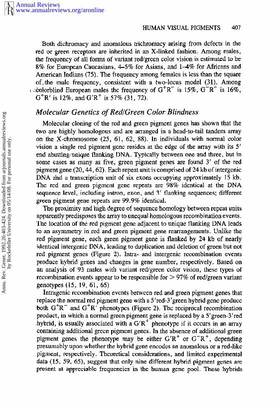
Both dichromacy and anomalous trichromacy arising from defects in thered or green receptors are inherited in an X-linked fashion. Among males,the frequency of all forms of variant red/green color vision is estimated to be8% for European Caucasians, 4-5% for Asians, and 1-4% for Africans andAmerican Indians (75). The frequency among females is less than the squareoLthe male frequency, consistent with a two-locus model (31). Among
~olorblind European males the frequency of G÷R- is 15%, G-R+ is 16%,G÷R’ is 12%, and G’R+ is 57% (31, 72).
Molecular Genetics of Red~Green Color Blindness
Molecular cloning of the red and green pigment genes has shown that thetwo are highly homologous and are arranged in a head-to-tail tandem arrayon the X-chromosome (25, 61, 62, 88). In individuals with normal colorvision a single red pigment gene resides at the edge of the array with its 5’end abutting: unique flanking DNA. Typically between one and three, but insome cases as many as five, green pigment genes are found 3’ of the redpigment gene (20, 44, 62). Each repeat unit is comprised of 24 kb of intergenicDNA and a transcription unit of six exons occupying approximately 15 kb.The red and green pigment gene repeats are 98% identical at the DNAsequence level, including intron, exon, and 3’ flanking sequences; differentgreen pigment gene repeats are 99.9% identical.
The proximity and high degree of sequence homology between repeat unitsapparently predisposes the array to unequal homologous recombination events.The location of the red pigment gene adjacent to unique flanking DNA leadsto an asymmetry in red and green pigment gene rearrangements. Unlike thered pigment gene, each green pigment gene is flanked by 24 kb of nearlyidentical intergenic DNA, leading to duplication and deletion of green but notred pigment genes (Figure 2). Intra- and intergenic recombination eventsproduce hybrid genes and changes in gene number, respectively. Based onan analysis of 93 males with variant red/green color vision, these types ofrecombination events appear to be responsible for > 97% of red/green variantgenotypes (15, 19, 61, 65)
Intragenic recombination events between red and green pigment genes thatreplace the normal red with a 5’red-3’green hybrid gene produce
G + R’pigment geneboth G+R- and phenotypes (Figure 2). The reciprocal recombinationproduct, in which a normal green pigment gene is replaced by a 5’green-3’red.hybrid, is usually associated with a G R phenotype ~f ~t occurs in an arraycontaining additional green pigment genes. In the absence of additional greenpigment genes the phenotype may be either G’R÷ or G-R÷, dependingpresumably upon whether the hybrid gene encodes an anomalous or a red-likepigment, respectively. Theoretical considerations, and limited experimentaldata (15, 59, 65), suggest that only nine different hybrid pigment genes arepresent at appreciable frequencies in the human gene pool. These hybrids
www.annualreviews.org/aronlineAnnual Reviews
Ann
u. R
ev. G
enet
. 199
2.26
:403
-424
. Dow
nloa
ded
from
arj
ourn
als.
annu
alre
view
s.or
gby
Roc
kefe
ller
Uni
vers
ity o
n 05
/14/
08. F
or p
erso
nal u
se o
nly.
408 NATHANS ET AL
R÷G+ ~ ¯ RED
[] GREEN
R’G÷.) ==-~ A
R+GP
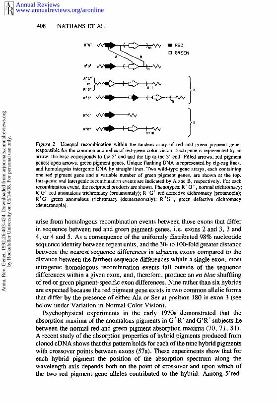
Figure 2 Unequal recombination within the tandem array of red and green pigment genesresponsible for the common anomalies of red-green color vision. Each gene is represented by anarrow: the base corresponds to the 5’ end and the tip to the 3’ end. Filled arrows, red pigmentgenes; open arrows, green pigment genes. Unique flanking DNA is represented by zig-zag lines,and homologofis intergenic DNA by straight lines. Two wild-type gene arrays, each containingone red pigment gene and a variable number of green pigment genes, are shown at the top.Intragenic and intergenic recombination events are indicated by A and B, respectively. For each
+ + .recombination event, the reciprocal products are shown. Phenotypes: R G , normal mchromacy;R’G+ red anomalous trichromacy (protanomaly); R-G~ red defective dichromacy (protanopia);R+G’ green anomalous trichromacy (deuteranomaly); R÷G-, green defective dichromacy(deuteranopia).
arise from homologous recombination events between those exons that differin sequence between red and green pigment.genes, i.e. exons 2 and 3, 3 and4, or 4 and 5. As a consequence of the uniformly distributed 98% nucleotidesequence identity between repeat units, and the 30- to 100-fold greater distancebetween the nearest sequence differences in adjacent exons compared to thedistance between the farthest sequence differences within a single exon, mostintragenic homologous recombination events fall outside of the sequencedifferences within a given exon, and, therefore, produce an en bloc shufflingof red or green pigment-specific exon differences. Nine rather than six hybridsare expected because the red pigment gene exists in two common allelic formsthat differ by the presence of either Ala or Ser at position 180 in exon 3 (seebelow under Variation in Normal Color Vision).
Psychophysical experiments in the early 1970s demonstrated that theabsorption maxima of the anomalous pigments in G+R’ and G’R+subjects liebetween the normal red and green pigment absorption maxima (70, 71, 81).A recent study of the absorption properties of hybrid pigments produced fromcloned cDNA shows that this pattern holds for each of the nine hybrid pigmentswith crossover points between exons (57a). These experiments show that foreach hybrid pigment the position of the absorption spectrum along thewavelength axis depends both on the point of crossover and upon which ofthe two red pigment gene alleles contributed to the hybrid. Among 5’red-
www.annualreviews.org/aronlineAnnual Reviews
Ann
u. R
ev. G
enet
. 199
2.26
:403
-424
. Dow
nloa
ded
from
arj
ourn
als.
annu
alre
view
s.or
gby
Roc
kefe
ller
Uni
vers
ity o
n 05
/14/
08. F
or p
erso
nal u
se o
nly.
HUMAN VISUAL PIGMENTS 409
3’green hybrid pigments in which position 180 in exon 3 is occupied by Alathe absorption spectrum approximates that of the green pigment (lambda max= 530 nm). If position 180 is occupied by Set, a 3-4 nm red shift is observedrelative to the parental green pigment. Additional red shifts of several nm areproduced by red-specific amino acid sequences within exons 2-4. Theabsorption spectra of 5’green-3’red hybrid pigments in which exons 1 and 2derive from the green pigment gene and exons 3-6 derive from the red pigmentgene are affected by the Ala/Ser polymorphism at position 180 in a mannersimilar to that described above for 5’red-3’green hybrid pigments, absorbingmaximally at 550 nm and 553 nm for alanine and serine versions, respectively.5’green-3’red hybrid pigments in which the recombination event occurredbetween exons 3 and 4 or exons 4 and 5 absorb maximally at 549 nm or 545nm, respectively, as compared to 552 nm or 557 nm for the two types of redpigment. Taken together, these data indicate that residues in exons 2, 4, and5, as well as position 180 in exon 3 are important in determining absorptiondifferences within this family of pigments.
The red and green pigments differ at 15 amino acids. Six of these differencesinvolve conservative substitutions of hydrophobic residues. Of the remainingnine differences, seven involve the substitution of similarly sized residues thatdiffer by the presence or absence of a hydroxyl group. Most likely, spectraltuning in this region of the spectrum involves differences in the intrinsic dipolemoment of amino acid side chains carrying or lacking hydroxyl groups. Giventhat an absorption shift of 10 nm at a wavelength of 550 nm corresponds toa photoexcitation energy difference of only 1 kcal/mol, it is reasonable thatdipoles of this strength could provide the requisite perturbation of the retinalchromophore (34). Predictions regarding the role of individual amino acidsubstitutions have recently come from correlations between primate visualpigment sequences and the absorption spectra of the corresponding pigmentsas determined by electroretinography (66) or microspectrophotometry (39,95). Based upon a comparison of eight primate visual pigments, Neitz et al(66) proposed that red-shifts of 6, 9, and 15 nm are produced by substitutionof Ser for Ala at position 180 in exon 3, Tyr for Phe at position 277 in exon5, and Thr for Ala at position 285 in exon 5, respectively. A second set ofcomparisons have led to the proposal that substitutions at position 233 in exon4 may also influence the absorption spectrum (39, 95). The data obtainedfrom recombinant human hybrid pigments are in good agreement with thesemodels of spectral tuning.
Variation in Normal Color Vision
Individuals who are considered to have normal color vision often show subtledifferences in color-matching. An early description of this variability can befound in Maxwell’s 1860 paper (54), in which he attributed the discrepancyin matches made by his subject "K" and himself to differences in the quantityand distribution of macular pigment, an inert yellow compound in the central
www.annualreviews.org/aronlineAnnual Reviews
Ann
u. R
ev. G
enet
. 199
2.26
:403
-424
. Dow
nloa
ded
from
arj
ourn
als.
annu
alre
view
s.or
gby
Roc
kefe
ller
Uni
vers
ity o
n 05
/14/
08. F
or p
erso
nal u
se o
nly.
4!0 NATHANS ET AL
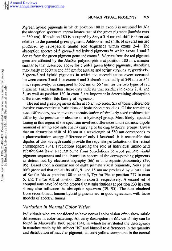
retina. Some individual variability can also be ascribed to a progressiveyellowing of the lens with age. However, recent work has shown that in thelong wavelength region of the spectrum, red pigment gene sequence poly-morphism plays the most significant role in generating person-to-personvariability in color-matching (57, 63, 96). Psychophysical experiments in the1960s and 1970s demonstrated variability in red pigment absorption spectraamong G-R÷ dichromats, and in the absorption spectra of red and/or greenpigments among normal trichromats (2, 4-6). Among male trichromats,Rayleigh matches are observed to cluster in a nearly symmetric bimodaldistribution in which the separation between the two modes is consistent witha several nm variation in red pigment absorption spectra (63, 64, 96). Femaletrichromats show a trimodal distribution in which two modes, each represent-ing approximately one quarter of the population, coincide with the malemodes, while the third mode, representing one half of the population, lies inthe interval between them (63). The segregation of Rayleigh matches withina family indicates that this variability is an X-linked trait and that the centralmode is comprised of heterozygous females (89).
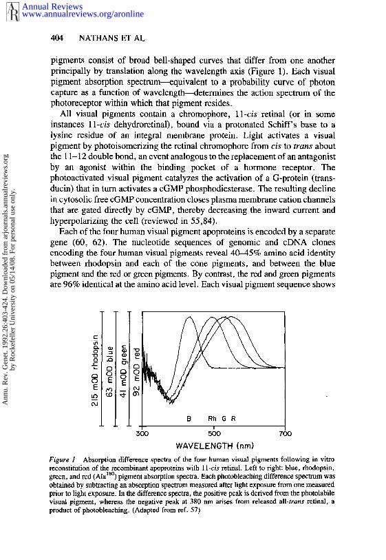
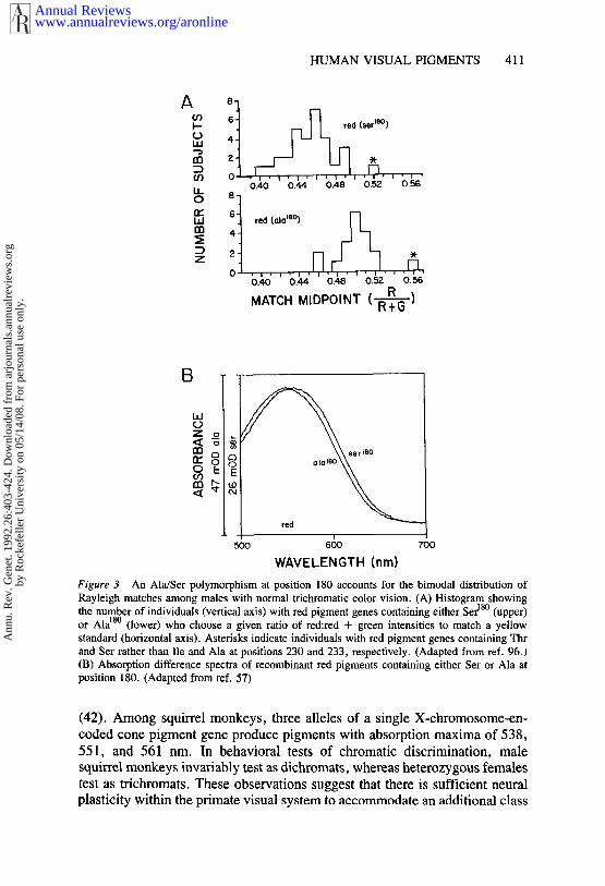
As noted earlier, there are two common alleles of the red pigment genethat differ by the presence of Ala or Ser at position 180. Two lines of evidenceindicate that this allelic variation is responsible for the bimodal distributionof Rayleigh matches. First., in a sample of 50 males with normal color vision,62% were found to have Ser and 38% were found to have Ala at position 180in the red pigment gene (96). The distribution of Ala18° alleles correlates wellwith that mode in the distribution of Rayleigh matches that requires a moreintense red light, and the distribution of Ser~8° alleles correlates well with themode that requires a less intense red light (Figure 3A). Second, recombinanthuman red pigments with Ala or Ser at position 180 absorb maximally at 552or 557 nm, respectively (Figure 3B), in good agreement with both thegenotype correlation and psychophysical predictions (57). A less commonsequence polymorphism, in which Thr and Ser replace Ile and Ala at positions230 and 233, respectively, in exon 4 of the red pigment gene, produces anindependent requirement for more red light in the Rayleigh match (96), in.agreement with the analyses of primate sequences described above (39, 95).
As a consequence of random X-inactivation, females who are heterozygousfor an X-linked trait are somatic mosaics. Psychophysical experiments showthat in females who are heterozygous for a red/green color vision defect, eachretina contains a mosaic of normal and abnormal photoreceptors (12, 33). Forexample, heterozygotes make a large number of errors when asked to identifythe colors of small stimuli that are presented too briefly to allow any significanteye movements. It seems possible, therefore, that females who are heterozy-gous for variant red or green pigments are functional tetrachromats. Thisconjecture is based upon an analogous situation in New World primates, inwhich female heterozygosity dramatically increases chromatic discrimination
www.annualreviews.org/aronlineAnnual Reviews
Ann
u. R
ev. G
enet
. 199
2.26
:403
-424
. Dow
nloa
ded
from
arj
ourn
als.
annu
alre
view
s.or
gby
Roc
kefe
ller
Uni
vers
ity o
n 05
/14/
08. F
or p
erso
nal u
se o
nly.
HUMAN VISUAL PIGMENTS 411
I’-- red (serm°)
0.40 0.44 0.48 0.52 0.56
LI.I red (ala~e°)
0,40 0.4,4 0.48 0.52 O, 56
RMATCH MIDPOINT (N--~--~)
o EE
red
500 600 700
WAVELENGTH (nm)
Figure 3 An Ala/Ser polymorphism at position 180 accounts for the bimodal distribution ofRayleigh matches among males with normal trichromatic color vision. (A) Histogram showingthe number of individuals (vertical axis) with red pigment genes containing either Ser18° (upper)or Ala~8° (lower) who choose a given ratio of red:red + green intensities to match a yellowstandard (horizontal axis). Asterisks indicate individuals with red pigment genes containing Thrand Set rather than lie and Ala at positions 230 and 233, respectively. (Adapted from ref. 96.)(B) Absorption difference spectra of recombinant red pigments containing either Ser or Ala position 180. (Adapted from ref. 57)
(42). Among squirrel monkeys, three alleles of a single X-chromosome-en-coded cone pigment gene produce pigments with absorption maxima of 538,
551, and 561 nm. In behavioral tests of chromatic discrimination, malesquirrel monkeys invariably test as dichromats, whereas heterozygous femalestest as trichromats. These observations suggest that there is sufficient neuralplasticity within the primate visual system to accommodate an additional class
www.annualreviews.org/aronlineAnnual Reviews
Ann
u. R
ev. G
enet
. 199
2.26
:403
-424
. Dow
nloa
ded
from
arj
ourn
als.
annu
alre
view
s.or
gby
Roc
kefe
ller
Uni
vers
ity o
n 05
/14/
08. F
or p
erso
nal u
se o
nly.
412 NATHANS ET AL
of cones. Among the 50% of human females who are heterozygous for thered pigment Ala/Ser polymorphism at position 180, any increase in discrim-ination is likely to be of low chromatic resolution because of the similarityin the two red-pigment absorption spectra.
To date no phenotypic effect has been ascribed to differences in the numberof "extra" green pigment genes in the array. The relative efficiency of photoncapture by the red and green cones, which shows a several-fold variationamong individuals with normal color vision (80), does not correlate with greenpigment gene number (T. P. Piantanida & J. Nathans, unpublished observa-tions). Among African-Americans with multiple green pigment genes, 21%carry 5’green-3’red hybrid genes, far higher than would be expected based onthe 4% frequency of all types of red-green variant color vision in thispopulation (44). Moreover, in a survey of 129 color vision normal Caucasianmales, four were found to carry a 5’green-3’red hybrid gene (15). Theseobservations suggest that some genes in the array are either not expressed orare expressed at reduced levels. This hypothesis has recently been tested byPCR amplification of green pigment mRNA sequences from postmortem maleretinas (J. Winderickx, L. Battisti, A. Motulsky & S. Deeb, personalcommunication). In each of 10 cases in which two green pigment genes couldbe distinguished, expression of only one was detected.
Blue Cone Monochromacy
True colorblindness, i.e. the absence of hue discrimination, is extremely rare,probably affecting not more than one person in 100,000. Two well charac-terized inherited varieties exist, rod monochromacy and blue cone mono-chromacy (1,73). Rod monochromacy is an autosomal recessive trait in whichapparently normal rods subserve all visual function. Blue cone monochrom-acy, also referred to as -rrl, atypical, or incomplete achromatopsia, is anX-linked trait in which green and red cones do not function (G-R-). In dimlight, the vision of blue cone monochromats is mediated by rods, and in brightlight, it is mediated by blue cones (37). Interestingly, at intermediate lightlevels, blue cone monochromats show a weak interaction between rod andblue cone signals which permits crude hue discrimination (78).
In the normal retina, cones are most concentrated in the fovea, a smalldepression in the retina centered on the optical axis. The fovea subserves highacuity vision, with the highest acuity deriving from the central region of 100microns in diameter. The central region contains only red and green cones,in contrast to the surrounding fovea, which contains all three cone types, apattern that may have evolved to minimize the effects of chromatic aberration(90). Because blue cone monochromats lack functional red and green cones,they experience a profound decrease in visual acuity: the average acuity inadult blue cone monochromats is 20/200 (i.e. letters that would be legible tothe normal observer at a distance of 200 feet are only legible when viewed
www.annualreviews.org/aronlineAnnual Reviews
Ann
u. R
ev. G
enet
. 199
2.26
:403
-424
. Dow
nloa
ded
from
arj
ourn
als.
annu
alre
view
s.or
gby
Roc
kefe
ller
Uni
vers
ity o
n 05
/14/
08. F
or p
erso
nal u
se o
nly.
HUMAN VISUAL PIGMENTS 413
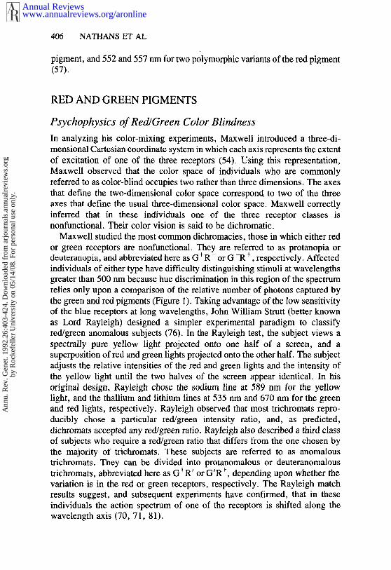
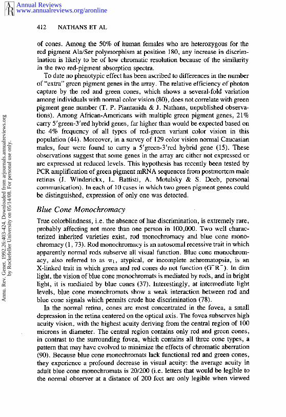
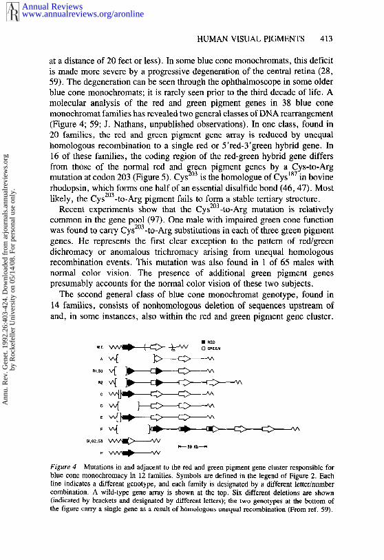
at a distance of 20 feet or less). In some blue cone monochromats, this deficitis made more severe by a progressive degeneration of the central retina (28,59). The degeneration can be seen through the ophthalmoscope in some olderblue cone monochromats; it is rarely seen prior to the third decade of life. Amolecular analysis of the red and green pigment genes in 38 blue conemonochromat families has revealed two general classes of DNA rearrangement(Figure 4; 59; J. Nathans, unpublished observations). In one class, found 20 families, the red and green pigment gene array is reduced by unequalhomologous recombination to a single red or 5’red-3’green hybrid gene. In16 of these families, the coding region of the red-green hybrid gene differsfrom those of the normal red and green pigment genes by a Cys-to-Argmutation at codon 203 (Figure 5). Cys2°3 is the homologue of Cys187 in bovinerhodopsin, which forms one half of an essential disulfide bond (46, 47). Mostlikely, the CysZ°a-to-Arg pigment fails to form a stable tertiary structure.
Recent experiments show that the Cys2°3-to-Arg mutation is relativelycommon in the gene pool (97). One male with impaired green cone functionwas found to carry Cys2°3-to-Arg substitutions in each of three green pigmentgenes. He represents the first clear exception to the pattern of red/greendichromacy or anomalous trichromacy arising from unequal homologousrecombination events. This mutation was also found in 1 of 65 males withnormal color vision. The presence of additional green pigment genespresumably accounts for the normal color vision of these two subjects.
The second general class of blue cone monochromat genotype, found in14 families, consists of nonhomologous deletion of sequences upstream ofand, in some instances, also within the red and green pigment gene cluster.
¯ RED
Figure 4 Mutations in and adjacent to the red and green pigment gene cluster responsible forblue cone monochromacy in 12 families. Symbols are defined in the legend of Figure 2. Eachline indicates a different genotype, and each family is designated by a different letter/numbercombination. A wild-type gene array is shown at the top. Six different deletions are shown(indicated by brackets and designated by different letters); the two genotypes at the bottom the figure carry a single gene as a result of homologous unequal recombination (From ref. 59).
www.annualreviews.org/aronlineAnnual Reviews
Ann
u. R
ev. G
enet
. 199
2.26
:403
-424
. Dow
nloa
ded
from
arj
ourn
als.
annu
alre
view
s.or
gby
Roc
kefe
ller
Uni
vers
ity o
n 05
/14/
08. F
or p
erso
nal u
se o
nly.
414 NATHANS ET AL
Figure 5 Schematic representation of a 5’green-3’red hybrid pigment showing the locations ofCys126 and Cys2°3 (filled circles). The seven alpha-helical segments are shown embedded withinthe membrane. The Cys2°3-to-Arg mutation is designated by the single letter code for the wild-typeamino acid, the codon number, and the introduced amino acid. N and C denote amino- andcarboxy-termini, respectively. The amino-terminus faces the extracellular space.
The deletions range in size from 0.6 kb to 55 kb. Seven different deletionshave been defined, and each is missing a 0.6 kb region that is absent fromthe chromosome with the smallest deletion (Figure 4c). A DNA rearrange-ment that includes part of the red pigment gene has also been reported ina family in which red cone function is missing, green cone function isgreatly reduced, and a progressive degeneration of the central retina isobserved (77).
Based upon the blue cone monochromat phenotype, it is reasonable tosuppose that DNA within the 0.6-kb region acts as a long-range transcriptionalcontrol element (locus control region, LCR) that is required for the activityof all of the visual pigment genes in the red-green array. In support of thismodel, recent experiments indicate a requirement for these sequences indirecting cone-specific expression of a reporter gene in transgenic mice (90a).Moreover, this small region contains sequences with a high degree ofhomology to sequences upstream of the mouse and bovine long-wavelengthpigment genes (90a). The postulated affect of this flanking sequence transcription units located over 40 kb away is reminiscent of the activity ofthe LCR located upstream of the .beta-globin gene cluster, deletion of whichresults in beta-thalassemia (91). By analogy to models in which embryonicand adult chicken globin genes compete for an enhancer (1 l), the presenceof a single LCR adjacent to the red and green pigment gene array suggests amechanism by which each red or green cone expresses only a single type ofvisual pigment gene. If transcription of a given red or green pigment generequires that the LCR physically interact with the promotor region, and if theLCR accommodates only one such interaction, then the formation of stableLCR-promotor complexes might be the critical event in determining red vsgreen cone identity.
www.annualreviews.org/aronlineAnnual Reviews
Ann
u. R
ev. G
enet
. 199
2.26
:403
-424
. Dow
nloa
ded
from
arj
ourn
als.
annu
alre
view
s.or
gby
Roc
kefe
ller
Uni
vers
ity o
n 05
/14/
08. F
or p
erso
nal u
se o
nly.
HUMAN VISUAL PIGMENTS 415
THE BLUE PIGMENT
Psychophysics of Tritanopia
Tritanopia is a disorder of color vision characterized by poor chromaticdiscrimination in the short wavelength region of the spectrum and poordiscrimination between colors that differ in the amount of blue admixed withinthem, e.g. white and yellow (73). In psychophysical tests, tritanopes performas dichromats who lack the blue-sensitive mechanism. Although individualcases of tritanopia were reported a century ago (18, 53), congenital tritanopiawas not recognized as a distinct inherited condition until Wright identifiedseveral dozen tritanopic subjects by publishing a color vision test in PicturePost (45, 98). Like the common forms of inherited variation in red-greendiscrimination, tritanopia occurs without signs or symptoms of generalizedretinal disease. Unlike the red and green pathways, the blue pathway appearsto be particularly susceptible to damage, which can lead to its acquired losssecondary to other ophthalmic disorders (73). For example, a disorderresembling tritanopia is typically seen as a secondary manifestation ofdominant juvenile optic atrophy (52, 58).
Electroretinographic records obtained from tritanopes in three unrelatedfamilies show a diminished or absent response to stimuli selective for bluecones, indicating that the defect occurs within the retina, most likely at thephotoreceptor level (58; 69). The possibility that some tritanopes have partially functional blue-sensitive pathway is suggested by the observationthat many affected subjects who perform as dichromats with stimuli subtending1 degree of visual field, make quantitatively normal trichromatic color matcheswith stimuli subtending 8 degrees (74). Simultaneous activation of a largernumber of blue cones may allow an impaired blue-sensitive pathway tocontribute more effectively to color perception.
Tritanopia is unique among inherited disorders of color vision in itsautosomal dominant transmission (45, 94). Earlier estimates of its prevalencein populations of European descent were 1 in 10,000 or less (45), but a recentstudy in the Netherlands, using testing methods with improved sensitivity andreliability, suggests that it may be as high as 1 in 500 (87).
Molecular Genetics of Tritanopia
Two groups have recently tested the hypothesis that mutations in the geneencoding the blue-sensitive pigment, located on chromosome 7 (61), can causetritanopia. In one study, 7 of 9 unrelated tritanopic subjects were found tocarry one of three different amino acid substitutions in the blue pigment gene:
,, 79 214Gly -to-Arg in two Japanese subjects, Ser -to-Pro in two Caucasiansubjects, and Pro264-to-Ser in three Caucasian subjects (92, 93). In independent study, the Pro26~-to-Ser mutation was found in the tritanopicmembers of two Caucasian families (T. Li, Zierath, P., Went, L. N., Smith,
www.annualreviews.org/aronlineAnnual Reviews
Ann
u. R
ev. G
enet
. 199
2.26
:403
-424
. Dow
nloa
ded
from
arj
ourn
als.
annu
alre
view
s.or
gby
Roc
kefe
ller
Uni
vers
ity o
n 05
/14/
08. F
or p
erso
nal u
se o
nly.
416 NATHANS ET AL
V., Pokorny, J., Cho, N. J., Applebury, M., personal communication). Thethree mutations are absent from control subjects of matched ancestry, andcoinherit with tritanopia in an autosomal dominant fashion, showing eitherincomplete penetrance (Gly79-to-Arg), almost complete penetrance (Serla-to-
Pro), or complete penetrance (Pro264-to-Ser). Some of the apparent differencesin penetrance may reflect differences in testing methodology.
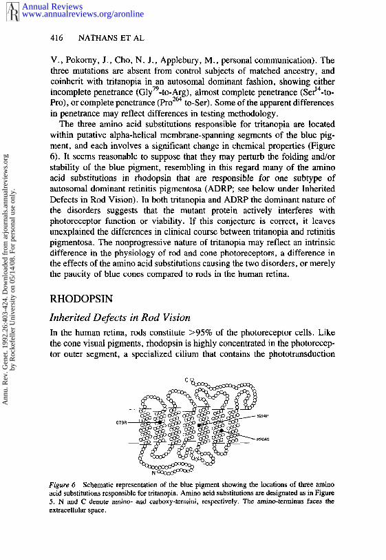
The three amino acid substitutions responsible for tritanopia are locatedwithin putative alpha-helical membrane-spanning segments of the blue pig-ment, and each involves a significant change in chemical properties (Figure6). It seems reasonable to suppose that they may perturb the folding and/orstability of the blue pigment, resembling in this regard many of the aminoacid substitutions in rhodopsin that are responsible for one subtype ofautosomal dominant retinitis pigmentosa (ADRP; see below under InheritedDefects in Rod Vision). In both tritanopia and ADRP the dominant nature ofthe disorders suggests that the mutant protein actively interferes withphotoreceptor function or viability. If this conjecture is correct, it leavesunexplained the differences in clinical course between tritanopia and retinitispigmentosa. The nonprogressive nature of tritanopia may reflect an intrinsicdifference in the physiology of rod and cone photoreceptors, a difference inthe effects of the amino acid substitutions causing the two disorders, or merelythe paucity of blue cones compared to rods in the human retina.
RHODOPSIN
Inherited Defects in Rod Vision
In the human retina, rods constitute >95% of the photoreceptor cells. Likethe cone visual pigments, rhodopsin is highly concentrated in the photorecep-tor outer segment, a specialized cilium that contains the phototransduction
Figure 6 Schematic representation of the blue pigment showing the locations of three aminoacid substitutions responsible for tfitanopia. Amino acid substitutions are designated as in Figure5. N and C denote amino- and carboxy-tennini, respectively. The amino-terminus faces theextracellular space.
www.annualreviews.org/aronlineAnnual Reviews
Ann
u. R
ev. G
enet
. 199
2.26
:403
-424
. Dow
nloa
ded
from
arj
ourn
als.
annu
alre
view
s.or
gby
Roc
kefe
ller
Uni
vers
ity o
n 05
/14/
08. F
or p
erso
nal u
se o
nly.
HUMAN VISUAL PIGMENTS 417
machinery. The 108 molecules of rhodopsin present in each rod outer segmentare replaced every ten days throughout life.
Inherited defects in rod vision are extremely heterogeneous, both geneticallyand clinically (35, 67). The most benign disorder, congenital stationarynightblindness (CSNB), is characterized by an increased rod threshold, withlittle or no progression of signs or symptoms throughout life. Retinitispigmentosa (RP), a more severe disorder, typically begins with an early lossof rod function or, in some individuals, both rod and cone function, followedby a slow progressive degeneration of the peripheral retina. The macula istypically spared until relatively late in the course of the disease. RP is one ofthe most commonly encountered retinal dystrophies, affecting approximately1 in 4000 people in all populations examined. Pedigree analysis of RP patientsin the USA reveals that in 22% the disease is due to an autosomal dominant,in 16% to an autosomal recessive, and in 9% to an X-linked gene (9). Theremaining approximately 50% of patients have no family history of RP andare presumed to comprise additional instances of autosomal recessive andX-linked transmission, as well as new mutations of all types. The most severeretinal dystrophy, Leber’s congenital amaurosis, is a diagnosis given to infantswith blinding retinal disease for which there is no infectious or metaboliccause, and for which there are no accompanying disorders elsewhere in thebody. The greater incidence of Leber’s congenital amaurosis followingconsanguinous mating suggests that some forms are inherited in an autosomalrecessive manner. Overall, the incidence is approximately one per 35,000.
Rhodopsin Mutations in Autosomal Dominant RetinitisPigmentosa
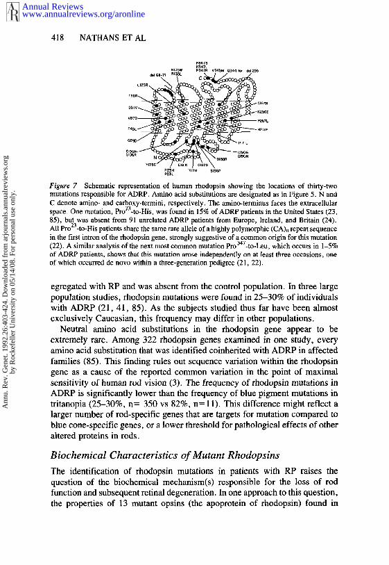
The first indication that mutations in the rhodopsin gene might be responsiblefor some cases of RP came from linkage analysis in a large Irish family withautosomal dominant RP (ADRP; 56). In this family, the disease was shownto coinherit with that region of the long ann of chromosome 3 to which therhodopsin gene had previously been mapped (61). Based upon this linkagestudy, a number of laboratories began to look for mutations in the rhodopsingene in patients with ADRP. PCR amplification of coding region exonsfollowed either by direct sequencing (23, 40), single strand conformationpolymorphism gel electrophoresis (21, 22), denaturing gradient gel electro-phoresis (83, 85), or hydrolink polyacrylamide gel electrophoresis (32, 48) has, to date, revealed 32 different rhodopsin mutations (Figure 7).Twenty-nine of the mutations are single nucleotide substitutions (28 missensemutations and one nonsense mutation), one mutation involves the substitutionsof 2 adjacent nucleotides within one codon, and two mutations are small,in-frame deletions. Most of the mutations have been tested for co-inheritancewith RP in affected families, and for their presence or absence in a controlpopulation with normal vision. In each case examined, the mutation cos-
www.annualreviews.org/aronlineAnnual Reviews
Ann
u. R
ev. G
enet
. 199
2.26
:403
-424
. Dow
nloa
ded
from
arj
ourn
als.
annu
alre
view
s.or
gby
Roc
kefe
ller
Uni
vers
ity o
n 05
/14/
08. F
or p
erso
nal u
se o
nly.
418 NATHANS ET AL
P23H TITM Sl~tPP25L
Figure 7 Schematic representation of human rhodopsin showing the locations of thirty-twomutations responsible for ADRP. Amino acid substitutions are designated as in Figure 5. N andC denote amino- and carboxy-termini, respectively. The amino-terminus faces the extracellularspace. One mutation, Pro23-to-His, was found in 15% of ADRP patients in the United States (23,85), but was absent from 91 unrelated ADRP patients from Europe, Ireland, and Britain (24).All Pro23-to-His patients share the same rare allele of a highly polymorphic (CA)n repeat sequencein the first intron of the rhodopsin gene, strongly suggestive of a common origin for this mutation(22). A similar analysis of the next most common mutation Pro 347-to-Leu, which occurs in 1-5%of ADRP patients, shows that this mutation arose independently on at least three occasions, oneof which occurred de novo within a three-generation pedigree (21, 22),
egregated with RP and was absem from the control population. In three largepopulation studies, rhodopsin mutations were found in 25-30% of individualswith ADRP (21, 41, 85). As the subjects studied thus far have been almostexclusively Caucasian, this frequency may differ in other populations.
Neutral amino acid substitutions in the rhodopsin gene appear to beextremely rare. Among 322 rhodopsin genes examined in one study, everyamino acid substitution that was identified coinherited with ADRP in affectedfamilies (85). This finding rules out sequence variation within the rhodopsingene as a cause of the reported common variation in the point of maximalsensitivity of human rod vision (3). The frequency of rhodopsin mutations ADRP is significantly lower than the frequency of blue pigment mutations intritanopia (25-30%, n= 350 vs 82%, = 11). T his d ifference m ight r eflect alarger number of rod-specific genes that are targets for mutation compared toblue cone-specific genes, or a lower threshold for pathological effects of otheraltered proteins in rods.
Biochemical Characteristics of Mutant Rhodopsins
The identification of rhodopsin mutations in patients with RP raises thequestion of the biochemical mechanism(s) responsible for the loss of rodfunction and subsequent retinal degeneration. In one approach to this question,the properties of 13 mutant opsins (the apoprotein of rhodopsin) found
www.annualreviews.org/aronlineAnnual Reviews
Ann
u. R
ev. G
enet
. 199
2.26
:403
-424
. Dow
nloa
ded
from
arj
ourn
als.
annu
alre
view
s.or
gby
Roc
kefe
ller
Uni
vers
ity o
n 05
/14/
08. F
or p
erso
nal u
se o
nly.
HUMAN VISUAL PIGMENTS 419
ADRP patients were analyzed following their production in a human embry-onic kidney cell line (86). In this tissue culture expression system, wild-typehuman opsin is targeted to the plasma membrane where it accumulates to aconcentration of approximately 3 x 10⁶ molecules per cell. Addition of 11-cisretinal to a crude membrane fraction containing expressed wild type opsingenerates a photolabile rhodopsin with the predicted absorbance properties(Figure 1). The 13 opsin mutants fall into two distinct biochemical classes.Three mutants (class I: Phe45-to-Leu, Glu344-to-Ter, i.e. deletion of residues344 to 348, and Pro347-to-Leu) resemble the wild type in yield, regenerabilitywith 11-cis retinal, and plasma membrane localization. Ten mutants (class I1:ThrlT-to-Met, Pro23-to-His, ThrSS-to-Arg, ValS7-to-Asp, Glysg-to-Asp, Gly1°6_
to-Trp, Arg135-to-Leu, Arg135-to-Trp, Tyr~TS-to-Cys, and Asplg°-to-Gly) ac-cumulate to significantly lower concentrations, regenerate variably or not at allwith 11-cis retinal, and are transported inefficiently to the plasma membrane,remaining partially or predominantly in the endoplasmic reticulum (ER).
The finding of a biochemical defect in 10 of 13 mutant rhodopsins (classII mutants) associated with ADRP strongly supports the genetic inference thatthey are responsible for the disease. The simplest interpretation of the classII biochemical phenotype is that these mutant opsins fail to fold correctly, or,once folded, are unstable. Under this interpretation, their partial or completeretention in the ER reflects an intrinsic ability of the ER to recognize andretain unfolded or improperly folded proteins (38, 51). The behavior of classII mutants closely resembles that of a large number of bovine rhodopsinmutants in which alterations were constructed in the extracellular domains(16), suggesting that many additional sites in rhodopsin may, if mutated, leadto ADRP. Many class II mutations reside near Cys11° or Cys187 (Figure 7),two conserved residues that form an essential disulfide bond in bovinerhodopsin (46, 47). These mutations may destabilize the protein by interferingwith disulfide bond formation. In one class II mutant, ThrlT-to-Met, one oftwo sites of Asn-linked glycosylation (Asn-X-Ser/Thr) is eliminated. Thismutation may produce rod dysfunction by a mechanism related to thatassociated with tunicamycin treatment, in which inhibition of Asn-linkedglycosylation is accompanied by a breakdown in the orderly assembly of theouter segment (29, 30). The pathogenic mechanisms associated with class mutants are not yet apparent. Two of three class I amino acid changes residenear opsin’s carboxy terminus, a region that shows a significant clustering ofmutations (Figure 7).
Of interest is the subcellular localization of the mutant rhodopsins in thephotoreceptor cell. One type of cellular pathology might arise if, in thephotoreceptor cell, the class H mutant opsins accumulate in the ER as theydo in tissue culture. A slow death of rod photoreceptors might occur secondaryto the metabolic costs associated with inefficiencies in ER function or
www.annualreviews.org/aronlineAnnual Reviews
Ann
u. R
ev. G
enet
. 199
2.26
:403
-424
. Dow
nloa
ded
from
arj
ourn
als.
annu
alre
view
s.or
gby
Roc
kefe
ller
Uni
vers
ity o
n 05
/14/
08. F
or p
erso
nal u
se o
nly.
420 NATHANS ET AL
degradation of the mutant protein. (In the normal case, the photoreceptors arespared the costs of degrading visual pigment because outer segment degrada-tion is carried out exclusively by the retinal pigment epithelium.) A secondtype of pathology might arise if some fraction of the mutant opsin istransported to the outer segment, where it could interfere with phototransduc-tion. This last conjecture may relate to the observation of a marked decreasein the rate of dark adaptation as well as an elevation in the dark adaptedthreshold in many patients with ADRP (67).
Studies aimed at correlating clinical findings with the type of mutation areunderway in several laboratories, and the first results of these studies suggestsome degree of allele specificity to the pattern and severity of retinaldysfunction (7, 8, 26, 27, 35, 43, 49). In particular, recent measurements dark adaptation in patients with rhodopsin gene mutations ThrlT-to-Met,Pro23-to-His, ThrSS-to-Arg, Arg135-to-Leu, Arg13~-to-Trp, and Glu344-to-Ter,show greater similarity in the time course of dark adaptation between patientscarrying the same mutation, as compared to those carrying different mutations(43, 49).
PERSPECTIVE
Inherited variation in human vision has long been a source of fascination forthose interested in sensory mechanisms. Anomalies of color vision wereaccurately described two centuries ago (13), and those of rod vision, includingretinitis pigmentosa, over one century ago (17). The present wealth knowledge about these variations has been made possible by several experi-mentally favorable attributes of the human visual system. First, humansrecognize and can accurately report abnormalities in their vision. As a result,many people with heritable visual impairments come to the attention of anophthalmologist. Second, human visual psychophysics is a highly developedscience that offers accurate, sensitive, and noninvasive tests for definingphenotypes. Third, the retina is the only part of the central nervous systemthat can be viewed directly. Monitoring its appearance through the ophthal-moscope makes it possible to follow at a tissue level the natural history ofretinal disease. And fourth, inherited variations in the visual system rarelyaffect longevity or fecundity, with the result that the responsible alleles aredestined to increase in the human gene pool.
ACKNOWLEDGMENTS
The authors gratefully acknowledge the support of the Howard HughesMedical Institute, the National Eye Institute, and the National RetinitisPigmentosa Foundation.
www.annualreviews.org/aronlineAnnual Reviews
Ann
u. R
ev. G
enet
. 199
2.26
:403
-424
. Dow
nloa
ded
from
arj
ourn
als.
annu
alre
view
s.or
gby
Roc
kefe
ller
Uni
vers
ity o
n 05
/14/
08. F
or p
erso
nal u
se o
nly.
HUMAN VISUAL PIGMENTS 421
Literature Cited
1. Alpern, M. 1974. What is it thatconfines in a world without color? Inv.Ophthalmol. 13:(:¢,8-74
2. Alpern, M. 1979. Lack of uniformityin colour matching. J. Physiol. 288:85-105
3. Alpern, M. 1987. A note on the actionspectrum of human rod vision. VisionRes. 27:1471-80
4. Alperu, M., Moeller, J. 1977. The redand green cone visual pigments ofdeuteranomalous tfichromacy. J. Phys-iol. 266:647-75
5. Alpem, M., Pugh, E. N. 1977. Vari-ation in the action spectrum oferythrolabe among deuteranopes. J.Physiol. 266:613-46
6. Alpern, M., Wake, T. 1977. Conepigments in human deutan colour visiondefects. J. Physiol. 266:595-612
7. Berson, E. L., Rosner, B., Sandberg,M. A., Dryja, T. P. 1991. Ocularfindings in patients with autosomaldominant retinitis pigmentosa and arhodopsin gene defect (pro-23-his).Arch. Ophthalmol. 109:92-101
8. Berson, E. L., Rosner, B., Sandberg,M. A., Weigel-DiFranco, C., Dryja,T. P. 1991. Ocular findings in patientswith autosomal retinitis pigmentosa andrhodopsin proline-347-1eucine. Am. J.Ophthalmol. 111:614--23
9. Boughman, J. A., Fishman, G. A.1983. A genetic analysis of retinitispigmentosa. Br. J. Ophthalmol. 67:449-54
10. Boynton, R. M. 1979. Human ColorVision. New York: Holt, Rinehart, Win-ston. 438 pp.
11. Choi, O-R. B,, Engel, J. D. 1988.Developmental regulation of beta-glo-bin gene switching. Cell 55:17-26
12. Cohn, S. A., Emmerich, D. S., Carl-son, E. A. 1989. Differences in theresponses of heterozygous carriers ofcolorblindness and normal controls tobriefly presented stimuli. Vision Res.29:255-62
13. Dalton, J. 1798. Extraordinary factsrelating to the vision of colours, withobservations. Mem. Lit. Philos. Soc.London 5:28-45
14. Dartnall, H. J. A., Bowmaker, J. K.,Mollon, J. D. 1983. Human visualpigments: microspectrophotometric re-suits from the eyes of seven persons.Proc. R. Soc. London, Ser. B 220:115-30
15. Deeb, S. S., Lindsey, D. T., Sanocki,E., Winderickx, J., Teller, D. Y.,Motulsky, A. G. 1992. Genotye-phe-notype relationships in human red/greencolor vision defects: molecular andpsychophysical studies. Am. J. Hum.Genet. In press
16. Doi, T., Molday, R. S., Khorana, H.G. 1990. Role of the intradiscal domainin rhodopsin assembly and function.Proc. Natl. Acad. Sci. USA 87:4991-95
17. Donders, F. C. 1857. Beitrage zurpathologischen Anatomie des Auges:Pigmentbildung in der Netzhaut.Graefes Arch. Clin. Exp. Ophthalmol.3:139-50
18. Donders, F. C. 1880. Remarks oncolours and colour blindness. Br. Med.J. 2:767-69
19. Drummond-Borg, M., Deeb, S. S.,Motulsky, A. G. 1988. Molecular basisof abnormal color vision: a family withthree types of color vision defects.Am. J. Hum. Genet. 43:675-83
20. Drummond-Borg, M., Deeb, S. S.,Motulsky, A. G. 1989. Molecular pat-terns of X chromosome-linked colorvision genes among 134 men of Eu-ropean ancestry. Proc. Natl. Acad. Sci.USA 86:983 87
21. Dryja, T. P., Hahn, L. B., Cowley,G. S., McGee, T, L., Berson, E. L.1991. Mutation spectrum of the rho-dopsin gene among patients with au-tosomal dominant retinitis pigmentosa.Proc. Natl. Acad. Sci. USA 88:9370-74
22. Dryja, T. P., McGee, T. L., Hahn,L. B., Cowley, G. S., Olsson, J. E.,Reichel, E., et al. 1990. Mutationswithin the rhodopsin gene in patientswith autosomal dominant retinitispigmentosa. N. Engl. J. Med. 323:1302-7
23. Dryja, T. P., McGee, T. L., Reichel,E., Hahn, L. B., Cowley, G. S.,.etal. 1990. A point mutation of therhodopsin gene in one form of retinitispigmentosa. Nature 343:364-66
24. Farrar, G. J., Kenna, P., Redmond,R., McWilliam, P., Bradley, D. G.,et al. 1990. Autosomal dominant ret-initis pigmentosa: absence of the rho-dopsin proline to histidine (codon 23)in pedigrees from Europe. Am, J. Hum.Genet. 47:941-45
25. Feil, R., Aubourg, P., Helig, R., Man-del, J. L. 1990. A 195-kb cosmid walkencompassing the human Xq28 color
www.annualreviews.org/aronlineAnnual Reviews
Ann
u. R
ev. G
enet
. 199
2.26
:403
-424
. Dow
nloa
ded
from
arj
ourn
als.
annu
alre
view
s.or
gby
Roc
kefe
ller
Uni
vers
ity o
n 05
/14/
08. F
or p
erso
nal u
se o
nly.
422 NATHANS ET AL
vision pigment genes. Genomics 6:367-73
26. Fishman, G. A., Stone, E. M., Gilbert,L. D., Kenna, P., Sheffield, V. C.1991. Ocular findings associated witha rhodopsin gene codon 58 transversionmutation in autosomal dominant reti-nitis pigmentosa. Arch. Ophthalmol.109:1387-93
27. Fishman, G. A., Stone, E. M., Shef-field, V. C., Gilbert, L. D., Kimura,A. E. 1992. Ocular findings associatedwith rhodopsin gene codon 17 andcodon 182 transition mutations in dom-inant retinitis pigmentosa. Arch. Op-hthalmol. 110:54-62
28. Fleischman, J. A., O’Donnell, F. E.1981. Congenital X-linked incompleteachromatopsia: evidence for slow pro-gression, carrier fundus findings, andpossible genetic linkage with glucose-6-phosphate dehydrogenase locus.Arch. Ophthalmol. 99:468--72
29. Fliesler, S. J., Rapp, L. M., FIollyfield,J. G. 1984. Photoreceptor-specific de-generation caused by tunicamycin. Na-ture 311:575-77
30. Fliesler, S. J., Raybom, M. E., Holly-field, J. G. 1985. Membrane morpho-genesis in retinal rod outer segments:inhibition by tunicamycin. J. Cell Biol.100:574-87
31. Francois, J. 1961. Heredity in Oph-thalmology. St Louis: Mosby. 731 pp.
32. Gal, A., Artlich, A., Ludwig, M.,Niemeyer, G., Olek, K., et al. ~991.Pro347-arg mutation of the rhodopsingene in autosomal dominant retinitispigmentosa. Genomics 11:468-70
33. Grutzner, P., Born, G., Hemminger,H. J. 1976 Coloured stimuli withinthe central visual field of carders ofdichromatism. Mod. Probl. Op-hthalmol. 17:147-50
34. Hays, T. R., Lin, S. H., Eyring, H.1980). Wavelength regulation in rho-dopsin: effects of dipoles and aminoacid side chains. Proc. Natl. Acad.Sci. USA 77:6314-18
35. Heckenlively, J. R. 1988. RetinitisPigmentosa. Philadelphia: Lippincott.269 pp.
36. Heckenlively, J. R., Rodriguez, J. A.,Daiger, S. P. 1991. Autosomal dom-inant sectoral retinitis pigmentosa: twofamilies with transversion mutation incodon 23 of rhodopsin. Arch. Op-hthalmol. 109:84-91
37. Hess, R. F., Mullen, K. T., Sharpe,L. T., Zrenner, E. 1989. The photo-receptors in atypical achromatopsia. J.Physiol. 417:123-49
38. Hobbs, H. H., Russell, D. W., Brown,
M. S., Goldstein, J. L. 1990. TheLDL receptor locus in familialhypercholesterolemia: mutational anal-ysis of a membrane protein. Annu.Rev. Genet. 24:133-70
39. Ibbotson, R. E., Hunt, D. M., Bowmaker,J. K., Mollon, J. D. 1992. Sequencedivergence and copy number of themiddle- and long-wave photopigmentgenes in old world monkeys. Proc. R.Soc. London, Ser. B 247:145-54
40. Inglehearn, C. F., Bashir, R., Lester,D. H., Jay, M., Bird, A. C., Bhattaeh-arya, S. S. 1991. A 3-bp deletion inthe rhodopsin gene in a family withautosomal dominant retinitis pigmen-rosa. Am. J. Hum. Genet. 48:26-30
41. Ingleheam, C. F., Keen, T. J., Bashir,R., Jay, M., Fitzke, F., et al. 1992.A completed screen for mutations ofthe rhodopsin gene in a panel of patientswith autosomal dominant retinitis pig-mentosa. Hum. Mol. Genet. 1:41-45
42. Jacobs, G. I-I., Neitz, J. 1987. Inher-itance of color vision in a new worldmonkey (Saimid sciureus). Proc. Natl.Acad. Sci. USA 84:254549
43. Jacobson, S. G., Kemp, C. M., Sung,C-H., Nathans, J. 1991. Retinal func-tion and rhodopsin levels in autosomaldominant retinitis pigmentosa with rho-dopsin mutations. Am. J. Ophthalmol.112:256-71
44. Jorgensen, A. L., Deeb, S. S.,Motulsky, A. G. 1990. Molecular ge-netics of X chromosome-linked colorvision among populations of Africanand Japanese ancestry: high frequencyof a shortened red pigment gene amongAfro-Americans. Proc. Natl. Acad. Sci.USA 87:6512-16
45. Kalmus, H. 1955. The familial distri-bution of congenital tritanopia withsome remarks on some similar condi-tions. Ann. Hum. Genet. 20:39--56
46. Karnik, S. S., Khorana, H. G. 1990.Assembly of functional rhodopsin re-quires a disulfide bond between cysteineresidues 110 and 187. J. Biol. Chem.265:17520-24
47. Karnik, S. S., Sakmar, T. P., Chen,H. -B., Khorana, H. G. 1988. Cysteineresidues 110 and 187 are essential forthe formation of correct structure inbovine rhodopsin. Proc. Natl. Acad.Sci. USA 85:8459-63
48. Keen, T. J., Inglehearn, C. F., Lester,D. H., Bashir, R., Jay, M., et al.1991. Autosomal dominant retinitispigmentosa: four new mutations in rho-dopsin, one of them in the retinalattachment site. Genotnics 11:199-205
49. Kemp, C. M., Jacobson, S. G., Roman,
www.annualreviews.org/aronlineAnnual Reviews
Ann
u. R
ev. G
enet
. 199
2.26
:403
-424
. Dow
nloa
ded
from
arj
ourn
als.
annu
alre
view
s.or
gby
Roc
kefe
ller
Uni
vers
ity o
n 05
/14/
08. F
or p
erso
nal u
se o
nly.
HUMAN VISUAL PIGMENTS 423
A. J., Sung, C-H., Nathans, J. 1992.Abnormal rod dark adaptation in an-tosomal dominant retinitis pigmentosawith proline-23-histidine rhodopsin mu-tation. Am. J. Ophthalmol. 113:165-74
50. Koenig, A., Dieterici, C. 1893. DieGrundempfindungen in normalen undanomalen Farbensystemen und ihreIntensit~itsverteilung im Spektrum. Z.Psychol. Physiol. Sinnesorg. 4:241-347
51. Klausner, R. D., Sitia, R. 1990. Proteindegradation in the endoplasmic reticu-lum. Cell 62:611-14
52. Krill, A. E., Smith, V. C., Pokomy,J. 1971. Further studies supporting theidentity of congenital tritanopia andhereditary dominant optic atrophy. Inv.Ophthalmol. 10:457-65
53. Levy, M. 1905. Uber einen Fall yonangeborener beiderseitiger Tritanopie(Blaublindheit). Graefes Arch. Op-hthalmol. 62:464-80
54. Maxwell, J. C. 1860. On the theoryof compound colours, and the relationsof the colours of the spectrum. Philos.Trans. R. Soc. London 150:57-84
55. McNaughton, P. 1990. Light responseof vertebrate photoreceptors. Physiol.Rev. 70:847-83
56. McWilliam, P., Farrar, G. J., Kenna,P., Bradley, D. G., Humphries, M.M., et al. 1989. Autosomal dominantretinitis pigmentosa (ADRP): localiza-tion of an ADRP gene to the longarm of chromosome 3. Genomics5:619-22
57. Merbs, S. L., Nathans, J. 1992. Ab-sorption spectra of human cone pig-ments. Nature 356:433-35
57a. Merbs, S. L., Nathans, J. 1992. Ab-sorption spectra of the hybrid pigmentsresponsible for anomalous color vision.Science. In press
58. Miyake, Y., Yagasaki, K., Ichikawa,H. 1985. Differential diagnosis of con-genital tritanopia and dominantly in-herited juvenile optic atrophy. Arch.Ophthalmol. 103:1496-501
59. Nathans, J., Davenport, C. M., Mau-menee, I. H., Lewis, R. A..Hejtmancik, J. F., et al. 1989. Mo-lecular genetics of human blue conemonochromacy. Science 245:831-38
60. Nathans, J., Hogness, D. S. 1984.Isolation and nucleotide sequence ofthe gene encoding human rhodopsin.Proc. Natl. Acad. Sci. USA 81:4851-55
61. Nathans, J., Piantanida, T. P., Eddy,R. L., Shows, T. B., Hogness, D. S.1986. Molecular genetics of inheritedvariation in human color vision. Science232:203-10
62. Nathans, J., Thomas, D., Hogness, D.
S. 1986. Molecular genetics of humancolor vision: the genes encoding blue,green, and red pigments. Science 232:193-202
63. Neitz, J., Jaeobs, G. FI. 1986. Poly-morphism of the long-wavelength conein normal human color vision. Nature323:623-25
64. Neitz, J., Jacobs, G. H. 1990. Poly-morphism in normal human color visionand its mechanism. Vision Res. 30:621-36
65. Neitz, J., Neitz, M., Jacobs, G. H.1989. Analysis of fusion gene andencoded photopigment of colour-blindhumans. Nature 342:679-82
66. Neitz, M., Neitz, J., Jacobs, G. H.1991. Spectral tuning of pigments un-derlying red-green color vision. Science252:971-74
67. Newsome, D. 1988. Retinal Dystro-phies and Degenerations. New York:Raven. 382 pp.
68. Oprian, D. D., Asenjo, A. B., Lee,N., Pelletier, S. L. 1991. Design,chemical synthesis, and expression ofgenes for the three human color visionpigments. Biochemistry 30:11367-72
69. Padmos, P., van Norren, D., Jaspers-Faijer, J. W. 1978. Blue cone functionin a family with an inherited tritandefect, tested with electroretinographyand psychophysics. Invest. Ophthalmol.Vis. Sci. 117:43(~41
70. Piantanida, T. P., Sperling, H. G.1973. Isolation of a third chromaticmechanism in the protanomalous ob-server. Vision Res. 13:2033-47
71. Piantanida, T. P., Sperling, H. G.1973. Isolation of a third chromaticmechanism in the deuteranomalous ob-server. Vision Res. 13:2049-58
72. Pickford, R. W. 1957. A practicalanomaloscope for testing colour visionand colour blindness. Br. J. Physiol.Optics 14:2-26
73. Pokomy, J., Smith, V. C., Verriest,G., Pinckers, A. J. L. G. 1979.Congenital and Acquired Color VisionDefects. New York: Grune & Stratton.409 pp.
74. Pokorny, J., Smith, V. C., Went, L.N. 1981. Color matching in autosomaldominant tritan defect. J. Opt. Soc.Am. 71:1327-34
75. Post, R. H. 1962. Population differ-ences in red and green color visiondeficiency: a review, and a query onselection relaxation. Eugen. Q. 9:131-46
76. Rayleigh, L. 1881. Experiments oncolour. Nature 25:64-66
77. Reichel, E., Bruce, A. M., Sandberg,
www.annualreviews.org/aronlineAnnual Reviews
Ann
u. R
ev. G
enet
. 199
2.26
:403
-424
. Dow
nloa
ded
from
arj
ourn
als.
annu
alre
view
s.or
gby
Roc
kefe
ller
Uni
vers
ity o
n 05
/14/
08. F
or p
erso
nal u
se o
nly.
424 NATHANS ET AL
M. A., Berson, E. 1989. An electro-retinographic and molecular geneticstudy of X-linked cone degeneration.Am. J. Ophthalmol. 108:540~47
78. Reitner, A., Sharpe, L. T., Zrenner,E. 1991. Is colour vision possible withonly rods and blue-sensitive cones?Nature 352:798-800
79. Rushton, W. A. H. 1972. Visual pig-ments in man. In Handbook of SensoryPhysiology: Photochemistry of Vision,ed. H. J. A. Dartnall, 7:364-94. Berlin:Springer-Verlag. 810 pp.
80. Rushton, W. A. H., Baker, H. D.1964. Red/green sensitivity in normalcolor vision. Vision Res. 4:75-85
81. Rushton, W. A. H., Powell, D. S.,White, K. D. 1973. Pigments inanomalous trichromats. Vision Res. 13:2017-31
82. Schnapf, J. L., Kraft, T. W., Baylor,D. A. 1987. Spectral sensitivity ofhuman cone photoreceptors. Nature325:439-41
83. Sheffield, V. C., Fishman, G. A.,Beck, J. S., Kimura, A. E., Stone,E. M. 1991. Identification of novelrhodopsin mutations associated withretinitis pigmentosa by GC-clampeddenaturing gradient gel electrophoresis.Am. J. Hum. Genet. 49:699-706
84. Stryer, L. 1986. Cyclic GMP cascadeof vision. Annu. Rev. Neurosci. 9:87-119
85. Sung, C-H., Davenport, C. M., Hen-nessey, J. C., Maumenee, I. H., Jacob-son, S. G., et al. 1991. Rhodopsinmutations in autosomal dominant ret-initis pigmentosa. Proc. Natl. Acad.Sci. USA 88:6481-85
86. Sung, C-H., Schneider, B., Agarwal,N., Papermaster, D. S., Nathans, J.1991. Functional heterogeneity of mu-tant rhodopsins responsible for autoso-mal dominant retinitis pigmentosa.Proc. Natl. Acad. Sci. USA 88:8840-44
87. van Heel, L., Went, L. N., van Norren,D. 1980. Frequency of tdtan distur-bances in a. population study. ColorVision Defic. 5:256-60
88. Vollrath, D., Nathans, J., Davis, R.W. 1988. Tandem array of humanvisual pigment genes at Xq28. Science240:1669-72
89. Waaler, G. H. M. 1967. Heredity of
two types of normal color vision. Na-ture 215:406
90. Wald, G. 1967. Blue-blindness in thenormal fovea. J. Opt. Soc. Am. 57:1289-301
90a. Wang, Y., Macke, J. P., Merbs, S.L., Zack, D. J., Klaunberg, B., et al.1992. A locus control region adjacentto the human red and green pigmentgenes. Neuron. In press
91. Weatherall, D. J., Clegg, J. B., Higgs,D. R., Wood, W. G. 1989. The hemo-globinopathies. In The Metabolic Basisof Inherited Disease, ed. C. R. Scriver,A. L. Beaudet, W. S. Sly, D. Valle,pp. 2281-39. New York: McGraw-Hill
92. Weitz, C. J., Miyake, Y., Shinzato,K., Montag, E., Zrenner, E., et al.1992. Human tritanopia associated withtwo amino acid substitutions in theblue sensitive opsin. Am. J. Hum.Genet. 50:498-507
93. Weitz, C. J., Went, L. N., Nathans,J. 1992. Human tritanopia associatedwith a third amino acid substitution inthe blue-sensitive visual pigment. Am.J. Hum. Genet. In press
94. Went, L. N., Pronk, N. 1985. Thegenetics of tfitan disturbances. Hum.Genet. 69:255-452
95. Williams, A. J., Hunt, D. M., Bowma-ker, J. K., Mollon, J. D. 1992. Thepolymorphic photopigments of the mar-moset: spectral tuning and genetic basis.EMBO J. 51:444-46
96. Winderickx, J., Lindsey, D. T.,Sanocki, E., Teller, D. Y., Motulsky,A. G., Deeb, S. S. 1992. Polymor-phism in red photopigment underliesvariation in colour matching. Nature356:431-33
97. Winderickx, J., Sanocki, E., Lindsey,D. T., Teller, D. Y., Motulsky, A.G., Deeb, S. S. 1992. Defective colorvision associated with a missense (cys-203-arg) mutation in the human greenvisual pigment gene. Nature Genet.11:2039-45
98. Wright, W. D. 1952. The character-istics of tritanopia. J. Opt. Soc. Am.42:509-21
99. Wyszecki, G., Stiles, W. S. 1982.Color Science: Concepts and Methods,Quantitative Data and Formulae. NewYork: Wiley. 950 pp.
www.annualreviews.org/aronlineAnnual Reviews
Ann
u. R
ev. G
enet
. 199
2.26
:403
-424
. Dow
nloa
ded
from
arj
ourn
als.
annu
alre
view
s.or
gby
Roc
kefe
ller
Uni
vers
ity o
n 05
/14/
08. F
or p
erso
nal u
se o
nly.
Ann
u. R
ev. G
enet
. 199
2.26
:403
-424
. Dow
nloa
ded
from
arj
ourn
als.
annu
alre
view
s.or
gby
Roc
kefe
ller
Uni
vers
ity o
n 05
/14/
08. F
or p
erso
nal u
se o
nly.
Ann
u. R
ev. G
enet
. 199
2.26
:403
-424
. Dow
nloa
ded
from
arj
ourn
als.
annu
alre
view
s.or
gby
Roc
kefe
ller
Uni
vers
ity o
n 05
/14/
08. F
or p
erso
nal u
se o
nly.