Embed Size (px)
Citation preview
Horse Genetics
Module Three
Equine Molecular Genetics
NNNiiiggghhhttt OOOwwwlll EEEddduuucccaaatttiiiooonnn aaannnddd EEEqqquuueeessstttrrriiiaaannn
Night Owl Education and Equestrian
Module three: Equine Molecular Genetics
Lesson seven - An introduction to molecular geneticsIntroductionThe structure of DNADNA and chromosome structure Genes and proteinsThe genetic codeWhat is a gene?Redundant DNAThe Horse GenomeSummary
Lesson eight - Finding and characterising genes for a particular phenotypeIntroductionPhysical mapping of the equine SCID gene Heterohybridoma panelsFluorescence in situ hybridisation (FISH)Comparative mappingReplicating DNA in a tubeDNA microarrays and chipsSummaryReferences
Lesson nine - molecular genetics testingIntroductionImpressive: a tale of triumph and tragedyGenetic TestingAllele specific PCR: genetic testing the modern wayLinkage testingPyroSequencing DNA microarrays and chipsFingerprintingExample of fingerprinting: conservation of the Przewalski horseSummaryReferences
Module three assignment
Night Owl Education and Equestrian
Seven: An introduction to molecular genetics
Introduction
Molecular genetics provides explanations for the various kinds of inheritance we have
observed, including, for example, how dominance, pleiotropy and epistasis work.
Understanding some basic molecular genetic concepts will also help you to appreciate and
better grasp some of the very many applications that have resulted from the molecular
revolution. From conservation and evolution, to colour and medical genetics, molecular
genetics is making a big impact. The scientists involved with the horse genome project
described at the end of this lesson were, and are, mainly interested in the diseases and
disorders of horses. They realised that they could better and more speedily address their
current and future research if more information were available on the horse genome. As a
result many genetic tests now exist to help breeders, and many more are likely to become
available over the next few years. In addition researchers are making, and will continue to
make, progress in various areas of horse medicine. We touch on this in this lesson but
explore it in more depth in following lessons. First though, and in order to understand the
exciting developments of late, we must have a basic knowledge of the structures and
functions of DNA, genes, proteins and the genome at the molecular level.
Note: Before reading this lesson please be somewhere where you can access the
internet, if at all possible. In many parts of the lesson I refer you to short animated video
clips from YouTube, which I’d like you to view before moving on. Although they don’t
contain any essential new information these animations may help you to a better and more
visual understanding of the concepts presented here.
1
Night Owl Education and Equestrian
The structure of DNA
We’ve already learned that genetic material is made up of deoxyribonucleic acid (DNA). In
contrast to other cell constituents DNA is not metabolised. It remains stable and intact as a
large macromolecule. The basic building blocks of DNA are called nucleotides. A
nucleotide contains one of four possible organic bases, one deoxyribose sugar unit and
one phosphate group. The four bases in DNA are adenine (A) and guanine (G) (the
purine bases) and thymine (T) and cytosine (C) (the pyrimidine bases).
Nucleotides are linked together into long chains called polynucleotides by a process
called polymerisation. The process is in a specific chemical orientation so that the chains
which are formed have a direction, and can only be added to at one end.
In 1953 James Watson and Francis Crick were the first to work out the 3D structure of
DNA, for which they were awarded a Nobel Prize for Medicine. They worked out how the
polynucleotides were organised within the DNA molecule, taking into account the findings
of Erwin Chargaff (1949). Chargaff’s had found that:
1. The number of purine bases (A + G) = the number of pyrimidine bases (T + C).
2. The number of cytosine bases = the number of guanine bases (i.e. ratio of G:C = 1:1).
3. The number of adenine bases = the number of thymine bases (i.e. ratio of A:T = 1:1).
From this Watson and Crick’s realised that adenine was always paired with thymine, and
guanine was always paired with cytosine. This can only be achieved if DNA consists of
two strands held together by specific base pairing. Here is a summary of the key
features of the structure of DNA worked out by Watson and Crick:
1. The DNA molecule is a double helix, made up of two interlocked polynucleotide chains
coiled around the same axis. They can only be separated by untwisting - not simply
pulling apart sideways.
2. The sugar-phosphate groups are on the outside “backbone” of DNA, while the bases
are on the inside. The molecule can be thought of as a ladder in which the base pairs
are the rungs and the sugar-phosphate backbones represent the two sides. The ladder 2
Night Owl Education and Equestrian
is then twisted into a double helix.
3. The chains are held together by hydrogen bonding between specific pairs of bases,
according to Chargaff’s rules. Thymine pairs with adenine (hence the 1:1 ratio of A:T).
Guanine pairs with cytosine (hence the 1:1 ratio of G:C).
4.The specific base pairing means that the sequence of nucleotides in one strand
determines the sequence in the other strand. The two strands are said to be
complementary.
5. The bases in the two strands will only fit together if the sugar molecules to which they
are attached point in opposite directions. The strands are said to be anti-parallel.
As homework I’d like you to watch a couple of short clips (about a minute each) on DNA
structure on YouTube, if it’s at all possible for you. Please do so before going any further in
the lesson.
http://uk.youtube.com/watch?v=l-hrLs03KjY&feature=related
http://uk.youtube.com/watch?
v=qy8dk5iS1f0&feature=PlayList&p=6667B09F73CF7FE4&playnext=1&index=23
DNA molecules are extremely long and can be composed of hundreds of millions of
nucleotides. Each chromatid of a horse chromosome contains one continuous
molecule of DNA double helix running throughout its length. The arrangement of
bases in the Watson and Crick model of DNA structure has two important features:
1. Nucleotides can occur along one of the polynucleotide strands in any order. The
sequence of bases is important for storing and encoding the genetic information.
2. Complementary base pairing means that for any given sequence of bases in one of
the strands, the sequence in the other one is determined. This gives a mechanism by
which DNA can self-replicate to make more identical copies of itself. The two strands
unwind from one another and each one then serves as a template for the synthesis of a
new complementary strand. An enzyme called DNA-polymerase “zips up” the bases
during the synthesis of new polynucleotide chains. This enzyme can be used to direct
the synthesis of new molecules of DNA in a test-tube.
3
Night Owl Education and Equestrian
As homework I’d like you to watch a couple of short clips (about a minute each) on DNA
replication on YouTube, if it’s at all possible for you. Please do so before going any further.
http://uk.youtube.com/watch?v=AGUuX4PGlCc&feature=related
http://uk.youtube.com/watch?v=4jtmOZaIvS0&feature=related
DNA and chromosome structure
As we’ve seen a horses cell nucleus contains 32 pairs of chromosomes. Each
chromosome is made up of a complex of protein and DNA, known as nucleoprotein or
chromatin. The DNA carries the genetic information while the proteins are mainly
concerned with chromosome “packaging“. The DNA is “packaged” and not present in a
fully extended form in the chromosome. Various “levels” of packaging ensure the
chromosomes stay intact and don’t become tangled. This organisation allows the DNA to
be replicated when necessary, to be partially unravelled for gene activity and to be tightly
packed and shortened when the chromosomes are moved during cell division.
As homework I’d like you to watch a short clip on chromosome structure on YouTube, if it’s
at all possible for you. Please do so before going any further. The clip shows how DNA is
wrapped around proteins (called histones) and then further coiled and wrapped to make up
chromosomes.
http://uk.youtube.com/watch?v=AF2wwMReTf8&NR=1
It is also interesting to have a quick look at these beautiful horse chromosome photos,
which show chromosomes with various levels of packing. The photo top right shows all the
chromosomes in a cell, in tissue that has been especially prepared for the purpose.
http://www.nzetc.org/etexts/Bio12Tuat02/Bio12Tuat02_095a(h280).jpg
4
Night Owl Education and Equestrian
Remember• DNA is a long molecule made up of two polynucleotide chains twisted together into
a double helix.
• Polynucleotides contain one of four kinds of organic bases - adenine, thymine,
cytosine and guanine - linked together in a long chain by their attachment to sugar-
phosphate backbones.
• Hydrogen bonding between specific pairs of bases holds the polynucleotides
together and also provides the mechanism for their self-replication.
• The sequence of bases along a polynucleotide encodes the genetic information.
• Horse chromosomes are composed of nucleoprotein fibres that are a complex of
DNA and protein.
• The organisation of horse chromosomes is complicated but in essence each
chromatid consists of one molecule of double helix running through its length.
Genes and proteins
Proteins are organic compounds composed of amino acids linked together in long chains
called polypeptides. Enzymes are proteins that do jobs. Twenty different amino acid subunits are commonly found in proteins. Each polypeptide has a specific sequence,
which is essential to its structure and function. This sequence gives the protein molecule
its primary structure. There is virtually an infinite number of ways in which 20 amino
acids can be assembled to make up a polypeptide, giving a potentially limitless number of
possible proteins.
Amino acids in the chain interact in various ways to give a secondary structure, while
disulphide bonds between sulphur-containing amino acids stabilise the molecule and give
a three-dimensional or tertiary structure. Many proteins contain more than one
polypeptide which interact to give a quaternary structure. The correct organisation of a
protein depends on the sequence in the primary chain. In enzymes it is therefore also
essential to its function. A change in amino acid sequence can lead to a loss, reduction or
change of function of the protein.
5
Night Owl Education and Equestrian
As early as 1909 the English physician Archibald Garrod suggested that genes work
through their control over the production of enzymes. Then in 1941 Beadle and Tatum
showed that a mutation in a single gene resulted in a change in the activity of a single
enzyme. They realised that all biochemical processes are under genetic control. These
processes progress through a series of steps, with each step being controlled by a single
enzyme, in turn coded for by a single gene. They supposed that genes might act by
determining the structure of enzymes. With some minor exceptions Beadle and Tatum’s
ideas have been shown to be essentially correct.
Enzymes are only one sort of protein. There are others that also play important roles in
structure and metabolism, including structural proteins, antibody proteins of the
immune system, some hormones (e.g. insulin) and tubulin, which is concerned with
moving chromosomes move during cell division.
The structure of all of these proteins, and not just the enzymes, is encoded in the genes of
the DNA: each gene encodes a polypeptide that either makes up a protein, or
combines with other polypeptides to make a protein.
The genetic code
Once it was realised that genes specify the amino acid sequence of polypeptide chains it
became obvious that their information must be carried in the sequence of bases in the
DNA. The way in which the genetic information is encoded in DNA is referred to as the genetic code. It’s now known that triplets of bases code each amino acid. The triplets are
known as codons, and the code of DNA has been completely known for some time.
In horses the DNA is in the nucleus, while the assembly of amino acids into proteins
occurs outside the nucleus, in the cytoplasm. Information is transferred from the site from
the nucleus by another kind of nucleic acid known as ribonucleic acid (RNA). RNA
molecules are actively engaged in the manufacture of proteins. Like DNA, RNA is
6
Night Owl Education and Equestrian
composed of nucleotides polymerised into polynucleotide chains, although there are some
slight differences in the compositions of RNA and DNA. RNA is a single-stranded
molecule, folded into various forms containing some double-stranded regions.
Three different types of RNA molecules play key roles in the biosynthesis of proteins.
Messenger RNA (mRNA) carries the genetic message from the DNA to the site of protein
synthesis in the cytoplasm. The DNA double helix unwinds in the region of the gene being
expressed. A strand of mRNA is made that is complementary to one of the DNA strands,
known as the template, in a process known as transcription (copying). An enzyme called
RNA polymerase catalyses transcription. The mRNA polynucleotide is unzipped from the
DNA template as it’s made. The completed mRNA molecules are then transported to the
site of protein synthesis, which occurs on structures called ribosomes. These are made
up of protein and ribosomal RNA and are vital to proper polypeptide synthesis.
Ribosomes can be thought of as polypeptide “factories”.
Transfer RNA (tRNA) pick up amino acids and carry them to the ribosomes so that they
can be joined together into polypeptides. There are at least 20 different tRNA molecules,
one for each amino acid. One end of each tRNA contains a triplet of exposed nucleotides,
known as the anticodon, which is complementary to one (or more) of the codons carried
in mRNA. The other end has a site for attachment to a specific amino acid. Each tRNA
picks up a particular amino acid and matches its anticodon with the complementary codon
in mRNA. This ensures the amino acids can be assembled in the correct sequence. The
amino acids are then linked to form polypeptides. There are special stop signals that end
polypeptide synthesis. The tRNAs that pair with their codons do not carry an amino acid.
The decoding of mRNA into a polypeptide chain is called translation. Several ribosomes
may attach to a mRNA molecule, one behind another, so that several polypeptides are
made from each mRNA molecule. Once synthesised polypeptides dissociate from the
ribosome and are released into the cytoplasm where they may undergo post-translational modification to form a functional protein.
As homework I’d like you to watch a short clip (about 4 minutes) on DNA transcription and
translation on YouTube, if it’s at all possible for you. Please do so before going any further.
7
Night Owl Education and Equestrian
http://uk.youtube.com/watch?v=41_Ne5mS2ls&NR=1
What is a gene?
So far we have thought of a gene as being a unit of heredity. Now that we know about the
structure of DNA we can add that genes are sequences of nucleotide pairs along a DNA
molecule which code for polypeptide products. However transfer and ribosomal RNA
molecules are also coded for by genes, made directly by transcription from the DNA, in the
same way as mRNA. So, in molecular terms, a gene is a sequence of nucleotide pairs along a DNA molecule which codes for polypeptide or RNA products.
Redundant DNA
Most horse genes have far more DNA in them than is actually needed to code for the
amino acids in their polypeptide products. Within the coding DNA, known as exons, are
stretches of non-coding DNA, called introns. Many genes are composed mostly of
introns. When transcription occurs all of the DNA bases are copied into the mRNA
transcript. An enzyme then snips out the introns to form the mature mRNA.
In addition there are large regions of the chromosomes that don’t contain any genes at all.
Some of these are composed of repetitive DNA where short base sequences are repeated
millions of times. This repetitive DNA has no known function. Some of it is transcribed, but
we have no idea why.
The function of redundant DNA is not known, but quite possibly it doesn‘t have one. It may
be a product of genome evolution that is of no benefit to the horse, but exists for its own
sake.
The Horse Genome
The total DNA content of a cell is called its genome. The nuclear genome is the total DNA
8
Night Owl Education and Equestrian
content of the haploid nucleus. Some cell organelles, such as mitochondria, have their
own genomes. Genomes are species specific, compared to genotypes that are specific to
individuals within a species.
The Horse Genome Project was started in 1995. It is an international cooperative project
involving over a hundred scientists in twenty countries. Initially the goal of the Horse
Genome Project was to make a genetic map for the horse. The 32 pairs of chromosomes
were characterised and genetic “landmarks” identified on each chromosome. In this way
points of reference were established to relate the horse genome to the human genome
sequence. Information from the human genome could then be used without the great
expense of sequencing the horse genome. The map now includes the positions of both
genes and non-coding “marker” sequences. It will also help in the identification of areas of
the genome contributing to multigenic (or quantitative) traits, such as behavioural and
performance traits and disease susceptibility. Information on genetic variability in horses,
will help to address issues about the relatedness of different breeds and the evolution of
horses.
During the first ten years the scientists used “genomics” to address important health issues
of horses, including the finding, isolation, characterisation and sequence determination of
some important genes like that for equine severe combined immune disorder, which is
discussed elsewhere in the course. Scientists believe that genome information will help
them gain a better understanding of inherited medical problems, and also of disease
organisms. They expect it will help them make progress in the prevention and diagnosis of
diseases and disorders, and the development of vaccines, therapies and treatments.
Sequencing of the horse genome began in February 2006, using techniques developed
for, and information gained from, the human genome project. By July the entire genome
had been chopped into 30,000,000 pieces and the DNA sequence of every piece was
determined! Re-assembling the sequences into the correct order was not a trivial task and
took until January 2007. The sequence was published on public databases for use by
biomedical and veterinary researchers. It is now available online at
http://genome.ucsc.edu/.
9
Night Owl Education and Equestrian
The DNA sequence of the horse genome consists of about 2.7 billion DNA base pairs. The
DNA used for sequencing was from a Thoroughbred mare at Cornell University College of
Veterinary Medicine, US. Researchers are working to improve the accuracy and resolution
of the horse genome sequence, and also to look at variation. Many scientists are now
working on diverse aspects of horse inheritance, disease and medical biochemistry, based
on the data provided from the horse genome project. A list of these is given at
http://www.uky.edu/Ag/Horsemap/hgpprojects.html.
A useful spin-off of the horse genome project was the discovery of the genetic basis for
many simple genetic traits in horses, including coat colour and several hereditary
diseases. Molecular tests have been developed and are now commercially available to
horse breeders, a list and some details of these is given in another lesson.
Genomics has shown that there are only around 20,000 genes in mammals, not the
100,000 or more that was once imagined, representing only about 2% of chromosomal
DNA. The rest of the genome does not encode genes, as we‘ve discussed already.
Remember
• genes act by determining the structure of proteins, including enzymes
• one gene is responsible for specifying the amino acids sequence of one polypeptide
chain
• DNA is encoded so that one triplet of bases carries the information specifying one
amino acid
• DNA is transcribed into a molecule of single-stranded messenger RNA
• the introns of mRNA are removed
• mRNA is translated into protein through the involvement of ribosomal and transfer
RNA
• ribosomal and transfer RNA is transcribed directly from DNA
• a gene is a sequence of nucleotides of DNA that codes for an RNA or protein
product
• there is a large amount of non-coding DNA, much of which may have no function
10
Night Owl Education and Equestrian
Summary
DNA is a long molecule made up of two polynucleotide chains twisted together into a
double helix. Polynucleotides contain one of four kinds of organic bases - adenine,
thymine, cytosine and guanine - linked together in a long chain by their attachment to
sugar-phosphate backbones. Hydrogen bonding between specific pairs of bases holds the
polynucleotides together and also provides the mechanism for their self-replication. The
sequence of bases along a polynucleotide encodes the genetic information.
Horse chromosomes are composed of nucleoprotein fibres that are a complex of DNA and
protein. The organisation of eukaryote chromosomes is complicated but in essence each
chromatid consists of one molecule of double helix running through its length.
Genes act by determining the structure of proteins, including enzymes. One gene is
responsible for specifying the amino acids sequence of one polypeptide chain, a
polypeptide being a protein or part of it. DNA is encoded so that one triplet of bases carries
the information specifying one amino acid. DNA is transcribed into a molecule of single-
stranded messenger RNA, with the introns being removed after transcription. The
processed mRNA is translated into protein through the involvement of ribosomal and
transfer RNA, both of which are themselves transcribed directly from DNA.
From a molecular point of view a gene can be defined as a sequence of nucleotides of
DNA that codes for an RNA or protein product. Besides genes chromosomes contain a
large amount of non-coding redundant DNA, much of which may have no function.
11
Night Owl Education and Equestrian
Lesson eight: Finding and characterising genes for a particular phenotype
Introduction
This chapter gives an insight into how molecular genetics research proceeds, including
some techniques used for finding and characterising genes. The linkage of equine severe
combined immunodeficiency (equine SCID) to molecular markers was discussed in the
lesson on linkage (lesson five), and this example is expanded on here. Having found two
genetic markers linked to the gene for equine SCID researchers were in a position to
locate the approximate position of the gene in the genome. Establishing the genetic
linkage of a gene of interest to markers whose approximate location is known (or
can easily be found) is called linkage mapping. Mapping a gene is the first essential
step before it can be isolated and characterized. Once the sequence of a gene and it’s
mutant alleles are known it’s possible to develop diagnostic kits, in order to genotype
potential carriers, for example. It also becomes possible to start to determine the structure,
action and role of the gene product, and how it influences the phenotype. This is
particularly important for genes of medical significance: understanding a disease or
disorder is essential for developing preventative and management strategies, and
ultimately working towards cures. Often cross species comparisons can be made, so that
advances in one species can benefit research in another. The completion of the
sequencing of the equine genome, and the sequencing of the genomes of various other
species, has lead to an explosion of new research, and a speeding up of research which
would have previously progressed more slowly.
Physical mapping of the equine SCID gene
Physically mapping a gene (or marker) is the method by which it is located at a particular chromosomal location. We’ll look at two common methods which have both
12
Night Owl Education and Equestrian
been used to locate horse genes and genetic markers, including those used for equine
SCID. These are the use of heterohybridoma panels and fluorescence in situ
hybridisation (FISH).
Mapping in one organism often makes use of knowledge gained from others, as we’ll see
in the case of equine SCID. Similar conditions exist in humans and mice and these have
been studied at the molecular level, although they involve different mutant genes in the
different animals.
In humans the condition called ADA-SCID - adenosine deaminase deficient severe
combined immunodeficiency disease – is a very serious disorder. Until recently children
suffering from this condition were kept in germ-free bubbles, and couldn’t lead a normal
life. Such children often died from illnesses considered minor in other children. Sufferers of
ADA-SCID lack the enzyme adenosine deaminase, causing toxic levels of
deoxyadenosine (the ADA substrate) to build up. The toxin kills the T lymphocytes (white
blood cells) of the immune system. The gene for ADA-SCID was one of first human
disease genes to be cloned and characterised. Now that the molecular basis of ADA-SCID
is understood, children with the condition are given weekly injections of PEG-ADA, to
prevent the build up of toxin (PEG is polyethylene glycol, which stabilises the ADA and
protects the enzyme from being broken down in the body). Initially it was thought that the
ADA gene might also be affected in equine SCID foals. However biochemical tests showed
that ADA levels in equine SCID foals are normal and it was concluded that a different
gene must be affected in humans and horses.
A similar condition in mice is caused by a deficiency of DNA-protein kinase (DNA-PK),
due to a mutant DNA-PK gene. This knowledge prompted further biochemical tests which
revealed that SCID affected foals are also DNA-PK deficient. This suggests that the gene
for either DNA-PK, or for a cofactor of DNA-PK, is defective in horses. Researchers
reasoned that if DNA-PK is linked to HTG4 & HTG8 (the two markers the SCID disease
gene was shown to be linked to) then it’s likely that SCID in horses is also caused by a
mutation in the DNA-PK gene. This was tested using somatic cell genetic techniques involving the use of heterohybridoma panels.
13
Night Owl Education and Equestrian
Heterohybridoma panels
Hybridomas are cells made by the fusion between tumour cells (myeloma cells, which
are essentially immortal) and other somatic cells (usually white blood cells).
Heterohybridomas are hybridomas where the fused cells are from different species. Such
cell lines are perpetuated in culture and used for genetic mapping and other genetics and
immunological research – they are sometimes called somatic cell hybrids, although this
broader term includes other kinds of cell hybrids, e.g. between plant cells.
Heterohybridoma panels are made up of a series of clones of somatic hybrid cells from which some of the chromosomes of one of the species have been lost.
For equine studies somatic hybrid cells are made by fusing horse white blood cells (B or
T lymphocytes) to rodent tumour cells, in the case of equine SCID mouse myeloma cells
were used.
First horse and mouse cell cultures are mixed together. The cells are fused either by
incubating them with a particular virus (a sendai virus) which forms bridges between cells,
or with the chemical polyethylene glycol (PEG), which induces the fusion of cell
membranes.
The cell mixture that results includes some unfused cells (parental cells). There are also
some fusions between cells of the same type (i.e. between 2 horse cells or 2 mouse cells).
These are called homokaryotic cells. There are a few heterokaryotic cells – fusions
between horse and mouse cells (the word heterokaryotic refers to there being “different nuclei”). Heterokaryotic cells must be selected from the mixture. The selection process
makes use of biochemical complementation. Each cell line is mutant for one enzyme
required for growth on a particular growth medium. The horse and mouse cell lines are
deficient for different enzymes essential for cell survival and growth. Once the cell mixture
is transferred to the growth medium only the hybrid cells grow – the deficiency in the
mouse cell line is complemented by the normal enzyme in the horse cell line, and vice-
versa.
14
Night Owl Education and Equestrian
One commonly used system involves the use of a medium containing hypoxanthine
aminopterin thymidine (HAT). The aminopterin in the medium blocks the normal synthesis
of DNA, and therefore the cells can‘t divide and form colonies. An alternative “salvage”
pathway for DNA synthesis can occur in cells able to use the hypoxanthine and thymidine
in the HAT medium. For this working copies of two particular genes are required to provide
the enzymes needed for the salvage pathway. Each type of cell has one enzyme that the
other doesn’t have. Only heterokaryotic cells have both enzymes.
As the hybrid cells divide by mitosis their nuclei are unstable (the cells have been forcibly
combined and haven’t evolved to contain all those chromosomes of such diverse origin!).
The horse chromosomes are lost at random resulting in cells with different numbers of
different horse chromosomes (it‘s not known why the horse chromosomes are lost in this
case, but not the mouse ones). Eventually each small group of heterokaryotic cells have
one or a few different horse chromosomes (the chromosome containing the gene allowing
the cell to grow on the selective medium is, of course, always maintained). The hybrid cells
are transferred to a growth medium that helps to slow or stop further chromosome loss. A
panel of cell lines are established, each with a different genotype with regard to which
horse chromosomes are present and absent. A heterohybridoma panel is established
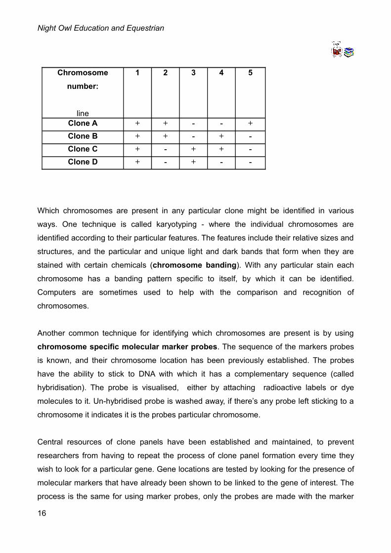
such that each chromosome can be uniquely identified. A simplified example for an organism with 5 chromosomes illustrates the how heterohybridoma panel work.
The clone panel consists of 4 cell lines A-D. There are 5 chromosomes (numbered), each
of which can be uniquely identified by its presence or absence in clones A-D.
Chromosome 1 appears in all cell lines, and is the one carrying the essential
complementation gene. Chromosome 2 appears in only cell lines A and B, which isn’t true for any of the other chromosomes. Look at the table carefully and you will see that
no two chromosomes are present in the same way in the clones.
15
Night Owl Education and Equestrian
Chromosome number:
line
1 2 3 4 5
Clone A + + - - +Clone B + + - + -Clone C + - + + -Clone D + - + - -
Which chromosomes are present in any particular clone might be identified in various
ways. One technique is called karyotyping - where the individual chromosomes are
identified according to their particular features. The features include their relative sizes and
structures, and the particular and unique light and dark bands that form when they are
stained with certain chemicals (chromosome banding). With any particular stain each
chromosome has a banding pattern specific to itself, by which it can be identified.
Computers are sometimes used to help with the comparison and recognition of
chromosomes.
Another common technique for identifying which chromosomes are present is by using
chromosome specific molecular marker probes. The sequence of the markers probes
is known, and their chromosome location has been previously established. The probes
have the ability to stick to DNA with which it has a complementary sequence (called
hybridisation). The probe is visualised, either by attaching radioactive labels or dye
molecules to it. Un-hybridised probe is washed away, if there’s any probe left sticking to a
chromosome it indicates it is the probes particular chromosome.
Central resources of clone panels have been established and maintained, to prevent
researchers from having to repeat the process of clone panel formation every time they
wish to look for a particular gene. Gene locations are tested by looking for the presence of
molecular markers that have already been shown to be linked to the gene of interest. The
process is the same for using marker probes, only the probes are made with the marker
16
Night Owl Education and Equestrian
sequence. In the example in the table, if the marker probes always hybridised to DNA of
clones B and C but never to that of A and D one would conclude that the markers, and the
gene of interest, are located on chromosome 4.
It isn’t always necessary to know exactly which chromosomes are which in order to get
some useful information about the location of a gene. In the case of equine SCID the
researchers didn’t know the identity of all the chromosomes in their clone panel.
Nevertheless in a large panel of clones HTG4 and HTG8 were always with the DNA-PK gene. This they tested using marker probes for HTG4 and HTG8, and a biochemical
test for DNA-PK.
This showed that DNA-PK was linked to the markers HTG4 and HTG8. Given the
connection of DNA-PK and SCID in mice, and the lack of DNA-PK in equine SCID foals,
this evidence of linkage makes DNA-PK a likely equine SCID candidate gene. In other
words it looked as though the disease gene was DNA-PK. The linkage was confirmed, and
chromosome location finally demonstrated, using a technique called fluorescence in situ
hybridisation (FISH). (Actually two different research groups worked on the problem at
once, one using heterohybridoma panels, the other using FISH.)
Fluorescence in situ hybridisation (FISH)
FISH is the direct visualization of the chromosome location of a gene or molecular marker using the hybridisation of probes with attached fluorescent molecules.
Special treatments are used that make chromosomes easy to visualise. Appropriately
treated cells are put on glass slides, stained and viewed using a microscope. The
chromosomal DNA is denatured in situ (in place) by dipping the slide in alkali (the
chromosomes essentially stay where they are but the strands of the DNA helices
disassociate from one another). In this state the DNA will hybridise with complementary
DNA probes that are added to the preparation.
The site of the hybridisation is visualised by attaching fluorescent dye “labels” to the
17
Night Owl Education and Equestrian
probes. Using different dye molecules the order and relative positions of various genes
and markers can be established. The chromosomes themselves are identified using a
chromosome banding technique, as discussed earlier for the clone panel method. In the
equine SCID study the HTG4 and HTG8 markers were used as probes, leading to the
discovery that the markers - and therefore the equine SCID gene - were on the short arm
of chromosome 9.
Comparative mapping
Many genes are the same in different animals. Accurate predictions about horse
genomes can often be made by comparison to the genomes of other species. This
speeds up mapping and gene isolation. As an example the gene for hyperkalemic periodic
paralysis disease in horses is the same as one in humans.
In the case of equine SCID the human DNA-PK was used to make a FISH probe. This
technique of using cross-species probes is called zoo-FISH. In this way DNA-PK was
localised to the short arm of chromosome 9, band 12 (symbolised 9p12; p stands for
petite, from the French for small). We’ve already seen that the equine SCID gene must
also be in this location, more evidence that the equine SCID gene is the DNA-PK gene
itself.
This completes our story of how the equine SCID gene was mapped and a likely candidate
gene identified. The mapping information was used in order to isolate and sequence the
normal and disease alleles of the gene.
Remember
• Mapping is the first essential step before a gene can be isolated and
characterized.
• Linkage analysis can determine which molecular markers a gene is close to,
the chromosome location of these markers can easily found using physical 18
Night Owl Education and Equestrian
mapping.
• There are various methods of physical mapping, including the use of
heterohybridoma panels and fluorescence in situ hybridisation (FISH).
• Heterohybridoma panels are made up of a series of clones of somatic hybrid
cells from which some of the chromosomes of one of the species have been lost.
• In heterohybridoma panels each chromosome can be uniquely associated
with a particular clone of cells.
• FISH is the direct visualization of the chromosome location of a gene or
molecular marker using the hybridisation of probes with attached fluorescent
molecules.
• Accurate predictions about horse genomes can often be made by
comparison to the genomes of other species, e.g. using zoo-FISH.
Replicating DNA in a tube
Whole genomic DNA can be extracted from blood, hair follicle cells, meat, bones, teeth,
semen and urine. Researchers are usually interested in studying a particular bit of DNA. It
might be that they want to sequence the DNA in a particular region mapped to be
associated with a particular phenotype, such as a genetic disease like equine SCID, or a
colour or pattern. Similarly genetic tests, discussed in the next lesson, assay only one or a
few genes, sometimes even only part of a gene, which is a tiny fraction of the genome. In
either case whole DNA is extracted first, and then a technique is used to
preferentially replicate the bit of DNA which is of interest, so there are lots of copies
of it. Most genetic tests available to horse breeders involve extracting whole DNA from
hair follicle cells, which are at the base of hairs pulled from the mane or tail. Usually pulling
20-40 hairs is all that’s necessary to conduct one or several of these tests.
The technique of preferential replication was invented by Kary Mullis, and is known as the
polymerase chain reaction (PCR). PCR can be used to amplify even tiny amounts of
DNA, so it can be used on fossil samples as well as fresh ones. The development of this
19
Night Owl Education and Equestrian
technique was hugely important to molecular geneticists, and is now routinely used
for most molecular genetics research and applications.
PCR requires that short sequences of DNA are known on either side of the piece to
be copied. Copies of these short sequences are made and are known as primers. The
DNA is heated up to separate the two strands of the DNA helices (a process called
denaturation). The primers bind to this denatured DNA and provide “starting places” for
DNA replication. Often primers can be used across species: those developed to work with
one species will also work with others. Other ingredients are required for successful DNA
replication, including bases (raw materials) and an enzyme called Taq polymerase (the
enzyme is from a heat tolerant microbe called Thermophilus aquaticus).
PCR results in the exponential increase in the number of copies of DNA between the
primers, as illustrated:
Exponential increase in DNA:start 11 cycle 22 cycles 43 cycles 84 cycles 16...10 cycles 1024
x starting amountof DNA
DNA Sequencing
DNA Sequencing is done to determine the order of nucleotides within it. It is the
nucleotide sequence that makes up the genetic code. As we saw in the introductory
molecular genetics lesson the nucleotide sequence of a gene determines the amino acid
sequence of the protein produced from it. By knowing the sequence of a gene we can
20
Night Owl Education and Equestrian
learn about the protein it produces and the biochemical processes with which this is
involved. This can be made easier if the gene turns out to be similar to one that has
already been characterised in other organisms.
In the case of equine SCID sequencing revealed a five base pair deletion in part of the
gene for a DNA-PK subunit. A DNA test is now available through Vetgen to identify carriers
of equine SCID, and is discussed further in the next lesson on genetic testing.
There are various ways of sequencing DNA, and the process is usually automated.
Just one, relatively new technique, will be briefly described here. I chose to describe this
technique as one genetic testing company told me they were looking at the technique as a
possible basis for doing some of their molecular equine tests in the future. The technique
is called pyrosequencing.
Pyrosequencing is based on "sequencing by synthesis”, and involves synthesizing
the complementary strand of DNA to be sequenced (Ronaghi et al, 1998). The template
DNA is immobilized and its complementary strand is synthesised one base pair at a time.
Solutions of the four possible nucleotides are added and removed sequentially (i.e. one at
a time), along with the necessary enzymes for DNA synthesis and chemi-luminescent
signal molecules. If the nucleotide complements the first unpaired base of the DNA the
DNA is extended and light is given off. A camera records the flash of light. If it does not
then nothing happens and apyrase degrades the free nucleotides in the mixture so that the
next nucleotide can be tried. The light signals show which nucleotide bases have been
added, and in what order, so determining the DNA sequence as it is synthesised.
DNA microarrays and chips
Microarrays are a way of performing lots of DNA or RNA assays all at once.
Depending on the application testing can be against tens, hundreds, thousands, or even
hundreds of thousands of sequences. An array contains these sequences fixed to a
surface. Common surfaces include glass and silicon chips. When silicon chips are used
21
Night Owl Education and Equestrian
the microarray might be referred to as a DNA chip. Each sequence is fixed in a particular
known position on the array (either by spotting or printing technique): a tiny spot of DNA
among many, all arranged in a grid pattern.
There are lots of uses of such arrays. Studies of gene expression can determine which
gene sequences are turned on and off, for example in normal individuals and in those with
a particular disease or genetic disorder. In this case it might be the mRNA from the test
samples that are tested against the sequences on the microarray. Another application is to
look at the genotype of an individual at many genes at once. This can be extended to
compare individuals within a population, for example to assess genetic diversity and
inbreeding, or to compare populations, for example to determine evolutionary
relationships.
Whatever the application if sequences from the samples of interest hybridize with the
immobilized sequences on the array fluorescent molecules bind too, producing fluorescent
signals. All un-hybridized DNA and unbound fluorescent molecules are then washed away.
The fluorescent signals are then analyzed by a computer to see which sequences
hybridize.
Remember
• The PCR technique is used to preferentially replicate the bit of DNA of
interest in extracted whole genomic DNA, so there are lots of copies of it.
• DNA Sequencing is done to determine the order of nucleotides within it. It is
the nucleotide sequence that makes up the genetic code.
• There are various ways of sequencing DNA, and the process is usually
automated.
• Pyrosequencing is based on "sequencing by synthesis", and may be used for
equine molecular tests in the near future.
• Microarrays are a way of performing lots of DNA or RNA assays all at once.
• There are lots of uses of micro arrays, including studying gene expression,
genotyping an individual at many genes at once, and assessing genetic diversity,
22
Night Owl Education and Equestrian
inbreeding and evolutionary relationships.
Summary
There are now many and varied, ingenious and eloquent molecular techniques for all
manner of research and practical applications, the study of which could easily fill a course
or so of their own. Nevertheless you now have some idea of some of the key techniques
involved in finding and characterising genes for a particular phenotype, and can appreciate
the importance of their application to veterinary medicine. In the next lesson we consider
genetic testing in more depth, and give you some idea of the range of applications for such
tests.
References
Bailey, E., Graves, KT, Cothran, E.G., Reid, R., Lear, T.L. and Ennis, R.B. 1995. Synteny
mapping horse microsatellite markers using a heterohybridoma panel. Animal
Genetics 26 (3), 177-180.
Bailey, E., Reid, R., Skow, L.C., Mathiason, K., Lear, T.L. and McGuire, T.C. 1997. Linkage
of the gene for equine combined immunodeficiency disease to microsatellite markers
HTG8 and HTG4; Synteny and FISH mapping to ECA9. Animal Genetics 28 (4), 268-
273.
Metalinos, D.L., Bowling, A.T. Rine, J. 1998. A missense mutation in the endotheline-B
receptor gene is associated with Lethal White Foal Syndrome: an equine version of
Hirschsprung Disease. Mammalian Genome 9, 426-431.
Pitra, C., Curson, A., Nurnberg, P., Krawczak, M. and Brown, S. 1996. An assessment of
inbreeding in Asian wild horse (Equus przewalskii Poliakov 1881) populations using
DNA fingerprinting. Arch. Tierzucht, Dummertorf 39 (6), 589-596.
Ronaghi, M., Uhlén, M. and Nyrén, P. 1998. A sequencing method based on real-time
pyrophosphate". Science 281: 363. doi:10.1126/science.281.5375.363. PMID
9705713.23
Night Owl Education and Equestrian
Shin EK, Perryman L, Meek K. A kinase negative mutation of DNA-PKcs in equine SCID
results in defective coding and signal joint formation. The Journal of Immunology 158 (8), 3565-3569.
Weiler R, Leber R, Moore BB, VanDyk LF, Perryman LE, Meek K. Equine severe combined
immunodeficiency: A defect in V(D)J recombination and DNA-dependent protein
kinase activity. Proc Natl Acad Sci USA 92, 1148-2249.
Vetgen. SCID. http://www.vetgen.com/equine-scid-service.html
24
Night Owl Education and Equestrian
Lesson nine: Molecular genetics testing
Introduction
This lesson looks at various types of genetic testing and their application. Such tests are
usually based on knowing the sequence of a gene and it’s mutant alleles, as discussed in
the last lesson. Sometimes though the sequence is not yet known, when linkage analysis
can be useful. To give you an idea of how genetic disorders can be quickly spread through
horse populations we start off by considering the story of the spread of hyperkalemic
periodic paralysis. When you have read this you will appreciate that genetic testing can be
a very important tool in reducing (and hopefully eliminating) such disorders. New ways of
genetic testing are constantly being developed and a few techniques are briefly discussed
which are likely to be used for horse genetic tests in the future.
Another aspect of genetic testing does not look at genes at all, but instead uses several
non-coding molecular markers to make “fingerprints”. Such fingerprints are highly variable
between individuals and have various uses. They are especially useful tools in the
conservation of wild and rare horses.
Impressive: a tale of triumph and tragedy
Before going on to discuss genetic testing in more detail it is interesting to see how genetic
disorders can be spread through a horse population. From there we can realise the
importance a simple genetic test can play in reducing (and hopefully one day eliminating)
the disorder from that population. The tale of Impressive is a good one for breeders to
remember, especially if it helps them to take care when planning their own breeding
programmes.
On the 15th April 1968 (and on the first birthday of my brother!) Impressive was born. He
was a chestnut Appendix American Quarter Horse colt foal, with royal thoroughbred 25
Night Owl Education and Equestrian
breeding on both sides of his pedigree. He became one of the most famous and
successful Quarter Horses in history.
Impressive changed hands a number of times, his price rising at each new sale. He was a
successful halter horse, and also raced for a while. In 1974, at age six, he became the first
World Champion Open Aged halter stallion, with 48 halter points. Apparently his owner
was offered $300,000 for him but refused the offer, saying that there "ain't nobody in this
world got enough money to buy this horse."!
Impressive was in demand as a sire, popular for his muscular and refined form, he turned
out one champion after another, siring almost 30 World Champions! Even though at one
time his stud fee was a staggering $25,000 he eventually fathered about 2,250 foals,
including Noble Tradition, a four-times World Champion halter stallion and a highly
successful sire in his own right. In 1992 13 of the top 15 halter horses were descendants
of this amazing horse. In 1993 he was estimated to have in excess of 55,000 living
descendants, including Quarter Horses, Paints and Appaloosas. He died on the 20th
March 1995, at the grand age of thirty-seven. Although he died, he is not forgotten, and is
now estimated to have over 100,000 living descendants!
It seemed that Impressive would go down in history as one hell of a horse: Impressive by
name, impressive by nature! And then tragedy struck when Impressive was linked to
something far less happy: his genetic legacy included a mutation recently implicated in the
rare muscular disorder known as known as hyperkalemic periodic paralysis (HYPP). The
disorder is inherited as a dominant condition. As such it requires only one parent to have
and pass on the gene and the disease. Breeding of an affected mare or stallion to a
normal horse will result in a 50% chance of an affected foal. If two affected horses are
bred together then there’s a 75% chance of the foal being affected.
The big problem with HYPP is that sometimes heterozygous animals (with one copy of the
mutant HYPP gene) appear to be asymptomatic, as was Impressive himself: in genetics
language HYPP is not fully penetrant. It also has variable expressivity (more severe in
some animals than others). In other horses the disorder only shows itself later in life
26
Night Owl Education and Equestrian
(again, in genetics language, HYPP is a late onset disorder). This can make it difficult to
know whether a horse has, and will therefore pass, on the mutant gene. These
characters are expected for a widespread dominant harmful mutation: if the health
of every carrier was severely affected before breeding then few would be used for
breeding and the mutation would become very rare.
It gradually became evident that many descendants of Impressive were inflicted with the
painful, alarming and often fatal disease. To my knowledge the disorder has never been
observed in horses of other lineages.
As Impressive ascended to the top of the sire's list owners of his foals began to notice a
strange muscular twitching that often left their horses temporarily unable to move. These
episodes, which varied widely in degree and duration, were usually mis-diagnosed as
tying-up syndrome or colic, but they are now known to be caused by hyperkalemic periodic
paralysis. At the time Impressive's descendants continued to make history in the show ring
and as breeding stock, some making their owners large amounts of money. It hadn’t been
proved that the disorder was exclusive to his line, and no one wanted to be the first to
publicly implicate Impressive as the source of HYPP. Many owners of affected horses
considered HYPP an inconvenience, not a reason to refrain from showing and breeding
their valuable horses. Most quarter horse owners were still unaware of HYPP.
AQHA and the University of California-Davis Equine Research Laboratory collaborated in
research to learn more about the disorder. They found that a mutation disrupts sodium ion
channels in the muscles, causing uncontrolled sodium influxes that alter the voltage
current of muscle cells. This in turn causes uncontrolled muscle twitching, stiffness and
profound muscle weakness. Horses with HYPP can experience unpredictable attacks of
paralysis which, in severe cases, can lead to collapse and sudden death.
The disorder is inherited as a dominant condition, but must involve interactions with other
genes and/or the environment since not all horses with the mutation show symptoms, or if
they do then the symptoms may occur intermittently. Foals homozygous for the mutant
gene have respiratory problems and may not survive. In horses where HYPP has been
27
Night Owl Education and Equestrian
diagnosed a combination of regular exercise and a diet low in potassium can help to keep
the disease under control.
The mutant HYPP gene occurs due to a single nucleotide change in the wild type HYPP
gene. The DNA test for HYPP detects the presence or absence of this specific mutation in
the HYPP gene.
During the past few years the pressure built to reveal the extent and nature of HYPP, and
to eliminate HYPP by selective breeding. A genetic test was made available to horse
breeders in 1992, and can identify affected horses with virtual certainty. The simple blood
test is available at the University of California at Davis School of Veterinary Medicine,
Department of Medicine, or through the AQHA. Breeders were pressurised to show that
their Impressive bred horses were negative for HYPP, or to remove them from breeding.
The lucky ones protected their investments by advertising their negative test results along
with their stud services and sales. If you are considering buying a horse with Impressive
bloodlines, whether for breeding or not, you would be wise to ensure that the horse is
HYPP negative before committing to buy.
At the AQHA 2004 convention a motion was passed to set January 1st 2007 as the date
after which foals testing homozygous for HYPP would no longer be registerable, with
mandatory testing for HYPP for the descendants of Impressive. The Appaloosa
Association followed suit and have disallowed the registry of homozygous foals from
January 1st 2008. The American Palomino registries have gone all the way and passed
rulings against both homozygous and heterozygous HYPP horses, beginning January 1st
2007. Part and half-blood registries need to follow suit and pass rulings on both testing
and registration. If enough pressure is brought to bear it might eventually be possible to
eliminate this awful disease from horse populations, so that Impressive may be
remembered for his impressive legacy rather than as the founder of a genetic disorder.
Genetic Testing
The list of molecular genetic tests now available to horse-breeders is increasing all the
28
Night Owl Education and Equestrian
time. The methods that underlie the tests are generally similar, even when the
testing is for very different traits. As it happens the method used for testing for
hyperkalemic periodic paralysis disease (HYPP) is the same as that used to test for the
“red factor” gene.
The red factor test distinguishes the alleles of the extension gene, which is useful
information for people wanting to breed black horses. Black horses may be of genotype
EE or Ee. Breeding together heterozygous blacks may therefore produce chestnut foals.
The test is for black horses whose genotype at the extension locus is ambiguous.
Researchers at the Swedish University of Agricultural Sciences found that the alleles
producing black and red pigment differed by a single nucleotide that resulted in a single
amino acid substitution.
For both tests the part of the gene coding for the horse muscle sodium channel or the
extension locus is amplified from whole blood samples using the polymerase chain
reaction, discussed in the last lesson. The next part of the test relies on the fact that in
both cases mutation has affected a site in gene called a host restriction endonuclease enzyme cutting site.
Host restriction endonuclease enzymes are sequence specific DNA cutters: they
recognise a specific base sequence and chop the DNA at that point. They occur naturally,
with a purpose of destroying the DNA of pathogenic organisms. Different enzymes
recognise different specific sequences. These enzymes have been put to use by molecular
geneticists for many tasks where DNA has to be precisely cut, including so that it can be
joined to other DNA, to make DNA molecules "to order".
Sometimes alleles of a particular gene differ in whether they possess sequences for
particular host restriction endonuclease enzymes. This is the case for HYPP and the
extension locus, and provides a useful test. Some alleles will be cut into pieces by a
particular enzyme or enzymes. Some won’t. The cut up alleles are therefore in smaller
pieces of DNA. These pieces can by separated and visualised by a process called
electrophoresis. The DNA pieces travel through a rectangular gel, under the influence of
29
Night Owl Education and Equestrian
an electric current. The small bits travel farther than the big bits, so that the DNA pieces
are separated according to their size. In this way, and by the use of dyes or radio-active
labels, it is possible to "see" which alleles are present in DNA samples.
The PCR fragments are then cut using a host restriction enzyme which recognizes and
cuts the specific base sequences in the DNA. In both cases the mutation affects the host restriction enzyme cutting site. The mutant and normal wild-type alleles therefore yield a different number of fragments on cutting. Each of the three possible
genotypes can be distinguished from one another according to the number and size of
fragments observed after electrophoresis.
Allele specific PCR: genetic testing the modern way
For many genes the differences between alleles do not happen to be at the site of a
restriction cutting enzyme. I contacted several genetic testing companies and they
confirmed that the majority of horse genetic tests are now based on some variation of an
allele specific PCR test.
One variant of allele specific PCR is used for testing alleles that differ in length, due to
deleted or added DNA. It is used for testing for equine SCID, discussed in the last lesson.
A company called VetGen do the test for potential SCID carriers among Arabian horses
and part-breds. A single pair of PCR primers are used to amplify both the normal and
mutant alleles, but as the two differ in size by five base pairs they can be readily
distinguished by electrophoresis.
More direct allele specific PCR is used for other genes. The diagnostic test now available
to identify horses at risk of producing lethal white overo (LWO) foals, is an example of this.
LWO has been shown to be due to a mutation in the endotheline-B receptor gene, with
there being a single nucleotide difference between the wild type and mutant genes. PCR
primers are used that will recognise and attach to the mutant sequence, but which will not
initiate replication of the wild-type allele. DNA amplification therefore only occurs in
30
Night Owl Education and Equestrian
samples containing the mutant gene, i.e. in those from carriers of LWO. Fluorescent
molecules are used for DNA replication and are incorporated into the PCR products, if
there are any. This allows immediate genotyping, with fluorescence indicating that the
mutation is present.
Discrimination of alleles in a single tube can also be achieved using two pairs of allele specific primers, one for each allele. This method of genotyping is used for
distinguishing single nucleotide polymorphisms of medical importance in humans, and may
in the future be used for horse tests too. Depending on the genotype of the sample, a
mixed signal of fluorescence is observed for a heterozygote while a single signal is
observed for a homozygote. The newer methods for allele specific genotyping are
relatively simple and cheap, with the advantage that (usually) no post-PCR manipulation is
required.
Linkage testing
Sometimes the specific mutation causing a genetic disorder is not known, as is the case
for cerebellar abiotrophy (CA), a recessively inherited neurological condition found almost
exclusively in Arabian horses. If the gene has nevertheless been mapped to a particular
region then tests can be carried out for a group of nearby genetic molecular markers
instead. In the case of CA markers that are usually inherited with the disorder are used as
a diagnostic tool, to identify affected foals and potential carriers of the disease. Arab
breeders can test their horses before breeding in order to avoid breeding two suspected
carriers together.
The Veterinary Genetics Laboratory at the University of California Davis, US, also
performs a dun test using linkage analysis as the gene for dun hasn’t yet been identified.
31
Night Owl Education and Equestrian
PyroSequencing, DNA microarrays and chips
Pyrosequencing, based on "sequencing by synthesis”, is discussed in the last lesson.
Some companies are now considering the use of pyrosequencing for horse genetic
testing. If such tests do become available it is possible that new allele variants might be
identified that were previously unknown, if they exist. This includes the occurrence, or
otherwise, of further agouti alleles. Students should note though that it is most likely that
only small parts of the gene in the region of the known allele variations will actually be
sequenced.
Microarrays and DNA chips could be used to look at the genotype of an individual at many
genes at once, as discussed in the last lesson. At least one horse genetic testing facility is
looking into this possibility. It is noted that microarrays might be used to test for polygenic
traits, allowing for selection for quantitative traits, as discussed in the lesson on complex
traits and polygenic inheritance.
Remember
• Genetic tests can be used to determine the genotype of horses for a particular
phenotype of interest. This can include colours and patterns, as well as genetic
disorders and potentially other traits too.
• Sometimes we accidentally breed genetic disorders into our horses without
realising, sometimes they occur anyway. Genetic tests can help in the
elimination of genetic disorders and diseases from horses.
• The methods that underlie the tests are generally similar, even when the testing
is for very different traits.
• Sometimes alleles of a particular gene differ in whether they possess sequences for
particular restriction cutting enzymes. This provides a useful test since some alleles
are cut into pieces, while others aren‘t. Electrophoresis can be used to “see” which
alleles are present in a sample.
• The majority of horse genetic tests are now based on some variation of an allele
32
Night Owl Education and Equestrian
specific PCR test.
• Variations of allele specific PCR can distinguish alleles either according to their
sizes or some aspect of sequence difference.
• When the specific gene causing a phenotype is not known linkage analysis might still
be used to infer genotype.
• PyroSequencing, DNA microarrays and DNA chips may well be used for future
equine genetics testing.
• Microarrays might be used to test for polygenic traits, allowing for selection for
quantitative polygenic traits.
Fingerprinting
Fingerprinting is a technique invented by UK scientist Alec Jeffrey’s in 1984, and famous
for its application to forensic science, e.g. in the OJ Simpson case. The technique has
been applied for many other purposes, including for studying horses and their relatives.
Equine applications of fingerprinting include:
• Paternity and maternity analysis for stud books, conservation research and
maintaining breed purity.
• Forensic testing, including for proving identity when trying to detect race doping.
• The conservation and management of wild, feral and ancient horse breeds, and
other equine species such as zebras. In particular assessing genetic diversity and
maintaining it through inbreeding avoidance.
• Establishing evolutionary relationships between equine species, and
between equine and other species.
Fingerprinting involves the use of non-coding molecular markers, which are usually
assumed to have a neutral effect on the phenotype. These are used to describe
patterns of variation in marker DNA that are unique, or nearly unique, to individuals.
Markers commonly used are called variable number tandem repeats (VNTRs). Their
useful feature is that they consist of a short DNA sequence, with different alleles varying 33
Night Owl Education and Equestrian
according to the number of repeat units. For example, if AT is the sequence then 3
possible various alleles might "look" as follows (only one strand of DNA is shown, AT is
said to be a dinucleotide repeat):
AT AT AT AT AT AT AT AT AT AT AT
AT AT AT AT AT AT AT AT
AT AT AT AT AT AT
Different alleles are therefore distinguished by their different sizes. The difference is
determined according to how far they travel when subjected to electrophoresis. There may
be many different possible alleles at any particular locus. Several loci will be looked at,
depending on what the study is for. The characteristic "bar-code" arrangement of
different alleles on an electrophoresis gel is what’s called the fingerprint. The
probability of two randomly chosen individuals sharing the same fingerprint is low even if
only a few polymorphic loci are used.
The fingerprint may be transferred to a nylon or nitro-cellulose membrane (called Southern
hybridisation or Southern blotting, after its inventor Edward Southern), and photographed
for more permanent records. Software is sometimes used to analyse complex fingerprints,
but other fingerprints may be easy enough to analyse by eye.
The relatedness of horses is determined according to the degree of band sharing
observed in the fingerprints. The markers used for fingerprinting are carefully chosen.
Population data is used to determine the likelihood of band sharing in unrelated
individuals, and the number of bands used for any particular fingerprint depends on the
marker diversity within the population.
Example of fingerprinting: conservation of the Przewalski horse
Initial attempts at saving the unique Przewalski’s were hampered with problems. Captive
breeding programs had the aim of re-introduction into their former habitat. In an early
attempt to allow captive-bred horses to return to a wild state five horses were released in
34
Night Owl Education and Equestrian
to a French reserve. Although they survived the first winter four later died of congenital
defects associated with inbreeding, (inbreeding leads to an increase in the occurrence
of deleterious recessive disorders, as will be discussed in detail in another lesson). The
fifth horse died of a heart-attack after being struck by lightning. To say this was unfortunate
seems somewhat of an understatement.
While nothing can be done about lightning, conservationists can make a reasonable
attempt to avoid inbreeding. By incorporating genetic fingerprint tests into their stud-books,
they can help to determine the relatedness of individuals. By carefully choosing
unrelated mates they can minimize inbreeding and help to maintain the genetic diversity of
the population, which is important for future evolution. Also since unrelated individuals are
more likely to produce genetically healthy offspring there is more chance of them surviving
re-introduction, and producing offspring of their own.
Thirteen micro-satellites are used. DNA is replicated using PCR, with primers isolated from
the domestic horse E. caballus. The genotype at the markers is then assessed using
electrophoresis.
Mostly there are different alleles present at each locus in the different species, E. caballus
and E. przewalskii: in only 4 loci is the predominant allele the same in both species. These
micro-satellites can therefore also be used as species specific markers, for example check
that no crossing occurs between the two species (which could spoil the conservation
effort).
Remember
• Fingerprinting involves the use of non-coding molecular markers, which are
usually assumed to have a neutral effect on the phenotype. These are used to
describe patterns of variation in marker DNA that are unique, or nearly unique, to
individuals.
• Applications of fingerprinting include paternity and maternity analysis, forensic
testing, conservation and management, assessing genetic diversity and establishing
evolutionary relationships.
35
Night Owl Education and Equestrian
• Determining the relatedness of individuals is important to avoid inbreeding
and maintain genetic diversity in rare species. Congenital defects are often
associated with inbreeding, as they are in the Przewalski’s horse.
Summary
Genetic tests can be used to determine the genotype of horses for a particular phenotype
of interest. This includes colours and patterns, genetic disorders and potentially other traits
in the future. Sometimes genetic disorders are accidentally spread through horses by our
breeding practices. Genetic tests can help to eliminate genetic disorders that have spread
in this way.
The methods that underlie the tests are generally similar, even when the testing is for very
different traits. The majority of horse genetic tests are now based on some variation of an
allele specific PCR test, which can distinguish alleles either according to their sizes or
some aspect of sequence difference. When the specific gene causing a phenotype is not
known linkage analysis might still be used to infer genotype. PyroSequencing, DNA
microarrays and DNA chips may well be used for future equine genetics testing.
Microarrays might be used to test for polygenic traits, allowing for selection for quantitative
polygenic traits.
Fingerprinting involves the use of non-coding molecular markers, which are usually
assumed to have a neutral effect on the phenotype. These are used to describe patterns
of variation in marker DNA that are unique, or nearly unique, to individuals. Applications of
fingerprinting include paternity and maternity analysis, forensic testing, conservation and
management, assessing genetic diversity and establishing evolutionary relationships.
Determining the relatedness of individuals is important to avoid inbreeding and maintain
genetic diversity in rare species. Serious congenital defects are often associated with
inbreeding, as they are in the Przewalski’s horse.
36
Night Owl Education and Equestrian
References
Bailey, E., Graves, KT, Cothran, E.G., Reid, R., Lear, T.L. and Ennis, R.B. 1995. Synteny
mapping horse microsatellite markers using a heterohybridoma panel. Animal
Genetics 26 (3), 177-180.
Bailey, E., Reid, R., Skow, L.C., Mathiason, K., Lear, T.L. and McGuire, T.C. 1997. Linkage
of the gene for equine combined immunodeficiency disease to microsatellite markers
HTG8 and HTG4; Synteny and FISH mapping to ECA9. Animal Genetics 28 (4), 268-
273.
Metalinos, D.L., Bowling, A.T. Rine, J. 1998. A missense mutation in the endotheline-B
receptor gene is associated with Lethal White Foal Syndrome: an equine version of
Hirschsprung Disease. Mammalian Genome 9, 426-431.
Pitra, C., Curson, A., Nurnberg, P., Krawczak, M. and Brown, S. 1996. An assessment of
inbreeding in Asian wild horse (Equus przewalskii Poliakov 1881) populations using
DNA fingerprinting. Arch. Tierzucht, Dummertorf 39 (6), 589-596.
Ronaghi, M., Uhlén, M. and Nyrén, P. 1998. A sequencing method based on real-time
pyrophosphate". Science 281: 363. doi:10.1126/science.281.5375.363. PMID
9705713.
Shin EK, Perryman L, Meek K. A kinase negative mutation of DNA-PKcs in equine SCID
results in defective coding and signal joint formation. The Journal of Immunology 158 (8), 3565-3569.
Weiler R, Leber R, Moore BB, VanDyk LF, Perryman LE, Meek K. Equine severe combined
immunodeficiency: A defect in V(D)J recombination and DNA-dependent protein
kinase activity. Proc Natl Acad Sci USA 92, 1148-2249.
Vetgen. SCID. http://www.vetgen.com/equine-scid-service.html
37
Night Owl Education and Equestrian
Module three assignment
Q1. Which of the following statements about DNA structure is false?
A. DNA consists of organic bases, deoxyribose sugar units and phosphate
groups.
B. The basic building blocks of DNA are called nucleotides.
C. The four bases in DNA are adenine, guanine, thymine and cytosine.
D. The bases make up the backbone of DNA, being on the outside of the
molecule.
E. Specific base pairing means that the sequence of nucleotides in one
strand of DNA determines the sequence in the other strand.
[1,1]
Q2. Which describes the important features about the arrangement of bases in
the Watson and Crick model of DNA structure ?
A. It allows the storage of genetic information and it gives a mechanism by
which DNA can self-replicate to make more identical copies of itself.
B. The number of cytosine bases equals the number of thymine bases.
C. The bases in the two strands will only fit together if the sugar molecules
to which they are attached point in the same direction.
D. The base sequence in one strand is identical to that in the
complementary strand, indicating how self-replication occurs.
E. The arrangement of the bases could not be used to store genetic
information, which is in the sugar-phosphate backbone of the DNA. [1,2]
page 1 of 13
Night Owl Education and Equestrian
Q3. Which is the best molecular description of genes?
A. All genes code for a single enzyme.
B. All genes code for a single polypeptide.
C. Genes are sequences of nucleotide pairs along a DNA molecule which
code for polypeptide products.
D. Genes are sequences of nucleotide pairs along a DNA molecule which
code for either polypeptide or RNA products.
E. Genes are units of heredity.
[1,3]
Q4. Which of the following statements about transcription is false?
A. During transcription mRNA is made that is complementary to the coding
strand of the gene.
B. Transcription occurs outside of the cell nucleus.
C. Transcription is catalysed by an enzyme called RNA polymerase.
D. The mRNA polynucleotide is unzipped from the DNA template as it’s
made.
E. mRNA molecules are transported from the site of transcription to the site
of protein synthesis. [1,4]
Q5. What is translation?
A. The formation of mRNA by copying of DNA.
B. The copying of new strands of DNA using the complementary strand as a
template.
C. The decoding of transfer RNA into a polypeptide chain.
D. The decoding of ribosomal RNA into a polypeptide chain
E. The decoding of messenger RNA into a polypeptide chain.
[1,5]
page 2 of 13
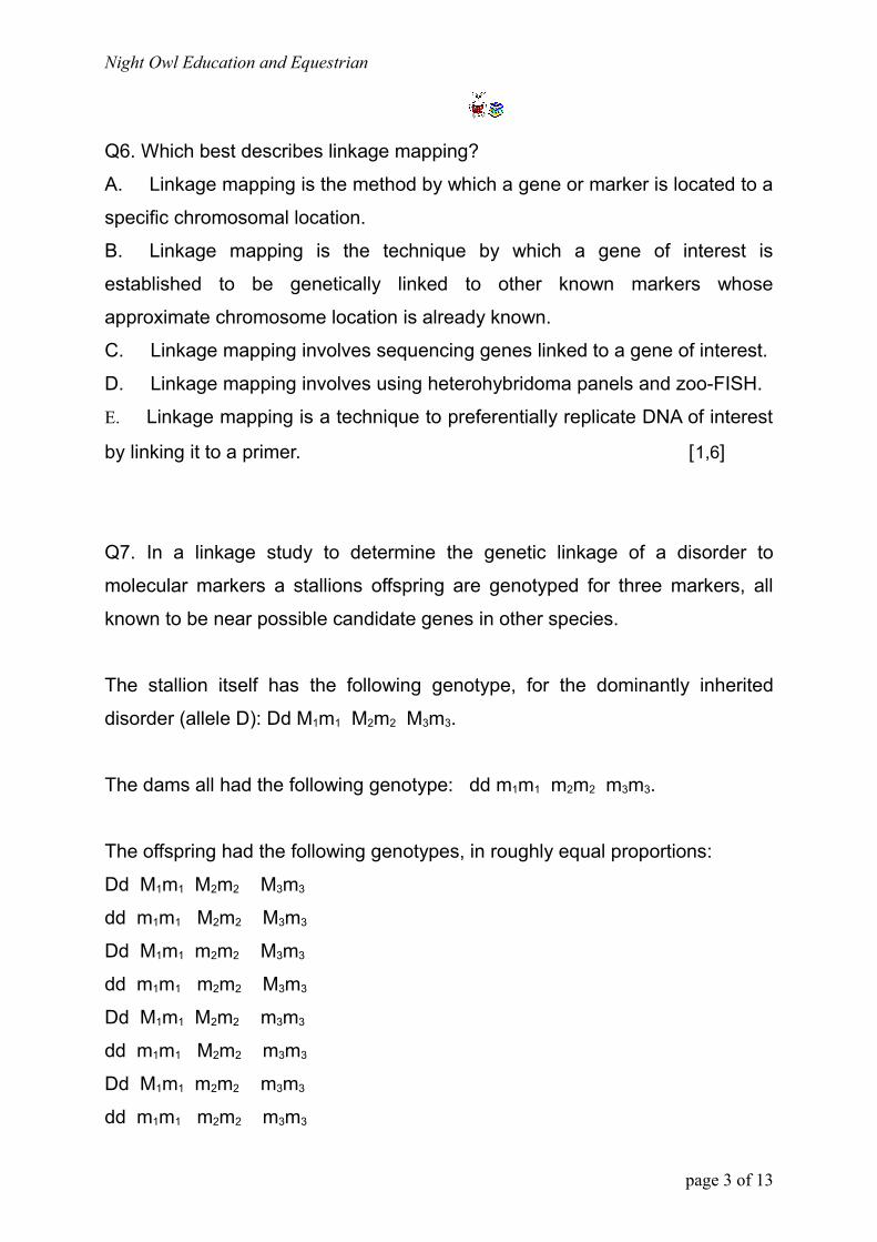
Night Owl Education and Equestrian
Q6. Which best describes linkage mapping?
A. Linkage mapping is the method by which a gene or marker is located to a
specific chromosomal location.
B. Linkage mapping is the technique by which a gene of interest is
established to be genetically linked to other known markers whose
approximate chromosome location is already known.
C. Linkage mapping involves sequencing genes linked to a gene of interest.
D. Linkage mapping involves using heterohybridoma panels and zoo-FISH.
E. Linkage mapping is a technique to preferentially replicate DNA of interest
by linking it to a primer. [1,6]
Q7. In a linkage study to determine the genetic linkage of a disorder to
molecular markers a stallions offspring are genotyped for three markers, all
known to be near possible candidate genes in other species.
The stallion itself has the following genotype, for the dominantly inherited
disorder (allele D): Dd M1m1 M2m2 M3m3.
The dams all had the following genotype: dd m1m1 m2m2 m3m3.
The offspring had the following genotypes, in roughly equal proportions:
Dd M1m1 M2m2 M3m3
dd m1m1 M2m2 M3m3
Dd M1m1 m2m2 M3m3
dd m1m1 m2m2 M3m3
Dd M1m1 M2m2 m3m3
dd m1m1 M2m2 m3m3
Dd M1m1 m2m2 m3m3
dd m1m1 m2m2 m3m3
page 3 of 13
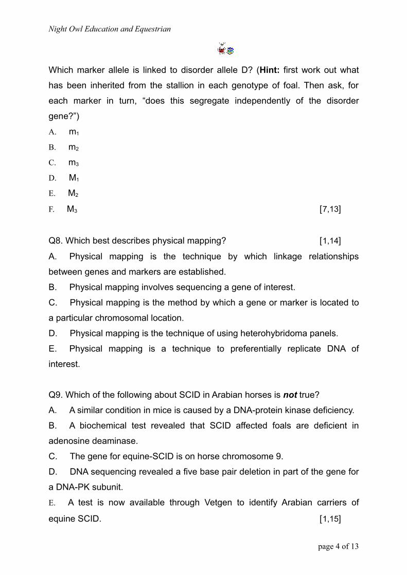
Night Owl Education and Equestrian
Which marker allele is linked to disorder allele D? (Hint: first work out what
has been inherited from the stallion in each genotype of foal. Then ask, for
each marker in turn, “does this segregate independently of the disorder
gene?”)
A. m1
B. m2
C. m3
D. M1
E. M2
F. M3 [7,13]
Q8. Which best describes physical mapping? [1,14]
A. Physical mapping is the technique by which linkage relationships
between genes and markers are established.
B. Physical mapping involves sequencing a gene of interest.
C. Physical mapping is the method by which a gene or marker is located to
a particular chromosomal location.
D. Physical mapping is the technique of using heterohybridoma panels.
E. Physical mapping is a technique to preferentially replicate DNA of
interest.
Q9. Which of the following about SCID in Arabian horses is not true?
A. A similar condition in mice is caused by a DNA-protein kinase deficiency.
B. A biochemical test revealed that SCID affected foals are deficient in
adenosine deaminase.
C. The gene for equine-SCID is on horse chromosome 9.
D. DNA sequencing revealed a five base pair deletion in part of the gene for
a DNA-PK subunit.
E. A test is now available through Vetgen to identify Arabian carriers of
equine SCID. [1,15]
page 4 of 13
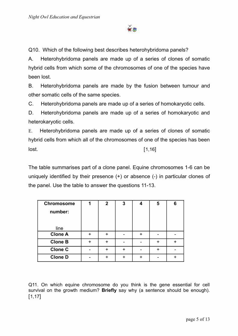
Night Owl Education and Equestrian
Q10. Which of the following best describes heterohybridoma panels?
A. Heterohybridoma panels are made up of a series of clones of somatic
hybrid cells from which some of the chromosomes of one of the species have
been lost.
B. Heterohybridoma panels are made by the fusion between tumour and
other somatic cells of the same species.
C. Heterohybridoma panels are made up of a series of homokaryotic cells.
D. Heterohybridoma panels are made up of a series of homokaryotic and
heterokaryotic cells.
E. Heterohybridoma panels are made up of a series of clones of somatic
hybrid cells from which all of the chromosomes of one of the species has been
lost. [1,16]
The table summarises part of a clone panel. Equine chromosomes 1-6 can be
uniquely identified by their presence (+) or absence (-) in particular clones of
the panel. Use the table to answer the questions 11-13.
Chromosome number:
line
1 2 3 4 5 6
Clone A + + - + - -Clone B + + - - + +Clone C - + + - + -Clone D - + + + - +
Q11. On which equine chromosome do you think is the gene essential for cell survival on the growth medium? Briefly say why (a sentence should be enough). [1,17]
page 5 of 13
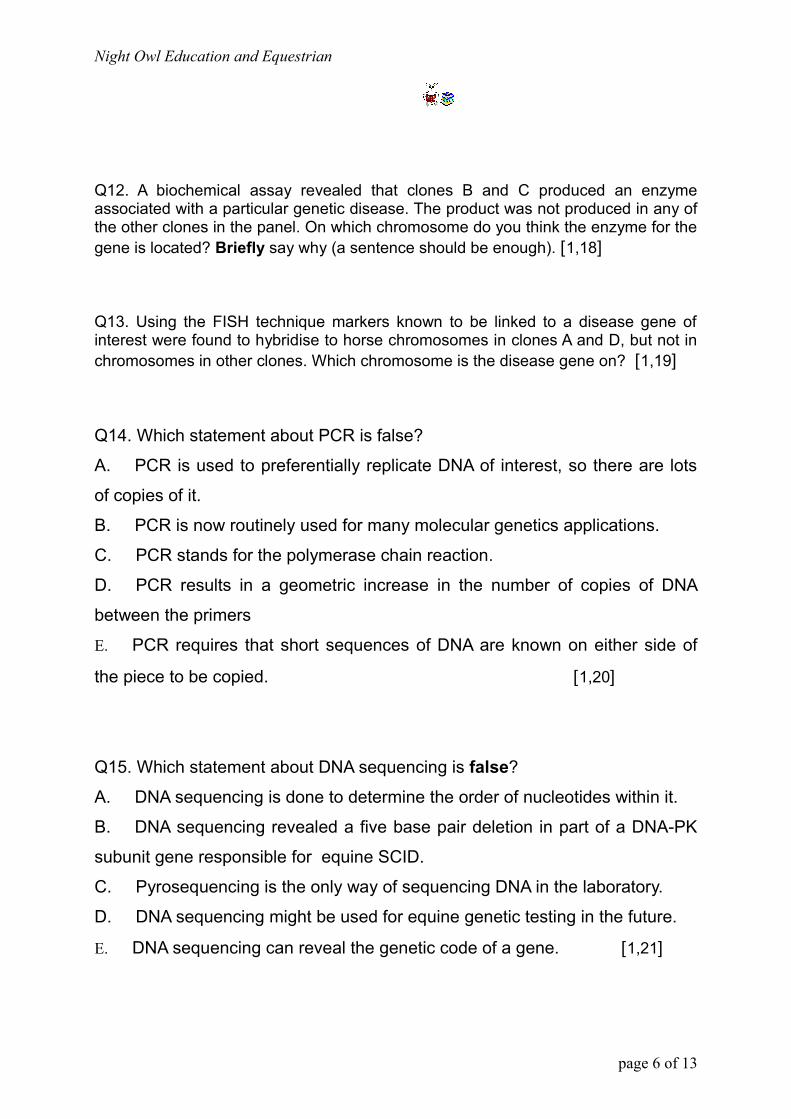
Night Owl Education and Equestrian
Q12. A biochemical assay revealed that clones B and C produced an enzyme associated with a particular genetic disease. The product was not produced in any of the other clones in the panel. On which chromosome do you think the enzyme for the gene is located? Briefly say why (a sentence should be enough). [1,18]
Q13. Using the FISH technique markers known to be linked to a disease gene of interest were found to hybridise to horse chromosomes in clones A and D, but not in chromosomes in other clones. Which chromosome is the disease gene on? [1,19]
Q14. Which statement about PCR is false?
A. PCR is used to preferentially replicate DNA of interest, so there are lots
of copies of it.
B. PCR is now routinely used for many molecular genetics applications.
C. PCR stands for the polymerase chain reaction.
D. PCR results in a geometric increase in the number of copies of DNA
between the primers
E. PCR requires that short sequences of DNA are known on either side of
the piece to be copied. [1,20]
Q15. Which statement about DNA sequencing is false?
A. DNA sequencing is done to determine the order of nucleotides within it.
B. DNA sequencing revealed a five base pair deletion in part of a DNA-PK
subunit gene responsible for equine SCID.
C. Pyrosequencing is the only way of sequencing DNA in the laboratory.
D. DNA sequencing might be used for equine genetic testing in the future.
E. DNA sequencing can reveal the genetic code of a gene. [1,21]
page 6 of 13
Night Owl Education and Equestrian
Q16. Which statement about DNA microarrays is false? [1,22]
A. Microarrays are a way of performing lots of DNA or RNA assays all at
once.
A. A DNA chip is a DNA microarray on a silicon chip.
A. Microarrays can be used to compare gene expression in healthy and
diseased patients.
A. Microarrays can only be used to assay DNA.
A. Microarrays can allow genotyping of individuals at many genes at once.
Q17. What are the reasons that the dominant disorder HYPP could be spread
so extensively through a horse population?
A. It is not fully penetrant so that some heterozygotes appear to be
asymptomatic. It shows variable expressivity between horses and may be late
onset in some horses. Therefore heterozygous horses may be bred from
without anyone realising that their foals may be affected, or carriers.
A. All heterozygotes are asymptomatic, allowing the gene to be passed on
to foals without anyone knowing.
A. The disorder is always late onset, so that horses are bred before they are
diagnosed with HYPP.
A. All horses homozygous for HYPP are asymptomatic, allowing the gene to
be passed on to foals without anyone knowing.
A. Although HYPP is fully penetrant and early-onset it shows variable
expressivity, so that heterozygous horses may be bred from without anyone
realising they have HYPP. [1,23]
page 7 of 13
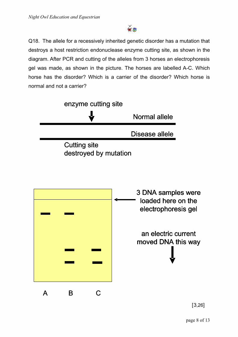
Night Owl Education and Equestrian
Q18. The allele for a recessively inherited genetic disorder has a mutation that
destroys a host restriction endonuclease enzyme cutting site, as shown in the
diagram. After PCR and cutting of the alleles from 3 horses an electrophoresis
gel was made, as shown in the picture. The horses are labelled A-C. Which
horse has the disorder? Which is a carrier of the disorder? Which horse is
normal and not a carrier?
Disease allele
Normal allele
enzyme cutting site
Cutting site destroyed by mutation
3 DNA samples were loaded here on the electrophoresis gel
an electric current moved DNA this way
A B C
Disease allele
Normal allele
enzyme cutting site
Cutting site destroyed by mutation
3 DNA samples were loaded here on the electrophoresis gel
an electric current moved DNA this way
3 DNA samples were loaded here on the electrophoresis gel
an electric current moved DNA this way
A B C[3,26]
page 8 of 13
Night Owl Education and Equestrian
Q19. Sarah has a patterned horse that is probably tobiano, but might also
carry the overo gene (so called tovero). It’s difficult to be sure from the
phenotype, and she isn’t sure about the exact breeding of her mare either. She
wants to produce a nicely marked foal and has her eye on a particular overo
stallion as a possible sire. She wants to know if she is at risk of producing a
lethal white overo foal. Briefly say what you can tell her about the chances of
a LWO foal, and advice you would give her, before using the stallion.
[5,31]
Q20. Andy has a field full of mares with which he’s run a favourite stallion.
Unfortunately a young stallion got in with the mares afterwards. Andy isn’t
certain if the youngster might’ve mated with one or two of the mares, who may
not have gotten pregnant the first time. The following diagram shows a
fingerprint from the two foals of uncertain sire, their dams and the two potential
sires. For each foal say which stallion is the sire.
[4,35]
page 9 of 13
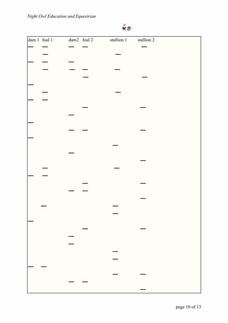
Night Owl Education and Equestrian
dam 1 foal 1 dam2 foal 2 stallion 1 stallion 2— — — — — — —— — — — — — — — ——
— —— —
— ——
—— — —
— —
— —
— —— —
— —— —
—— —
——
— ———
— —
— — — —
— — —
page 10 of 13

Night Owl Education and Equestrian
Q21. Conservationists are trying to decide the best potential mate for a particular mare. Their fingerprints at various markers are shown. Say which stallion you would choose, and briefly explain your decision. Do you think the mare is directly related to any of the stallions, or any of the stallions directly related to each other? If so how?
mare stallion 1 stallion 2 stallion 3
— — — — — — — — —
—— —
—— — —
— —— ——
— —
—— — —
— —— —
— —
— —
———
[8,43]
page 11 of 13
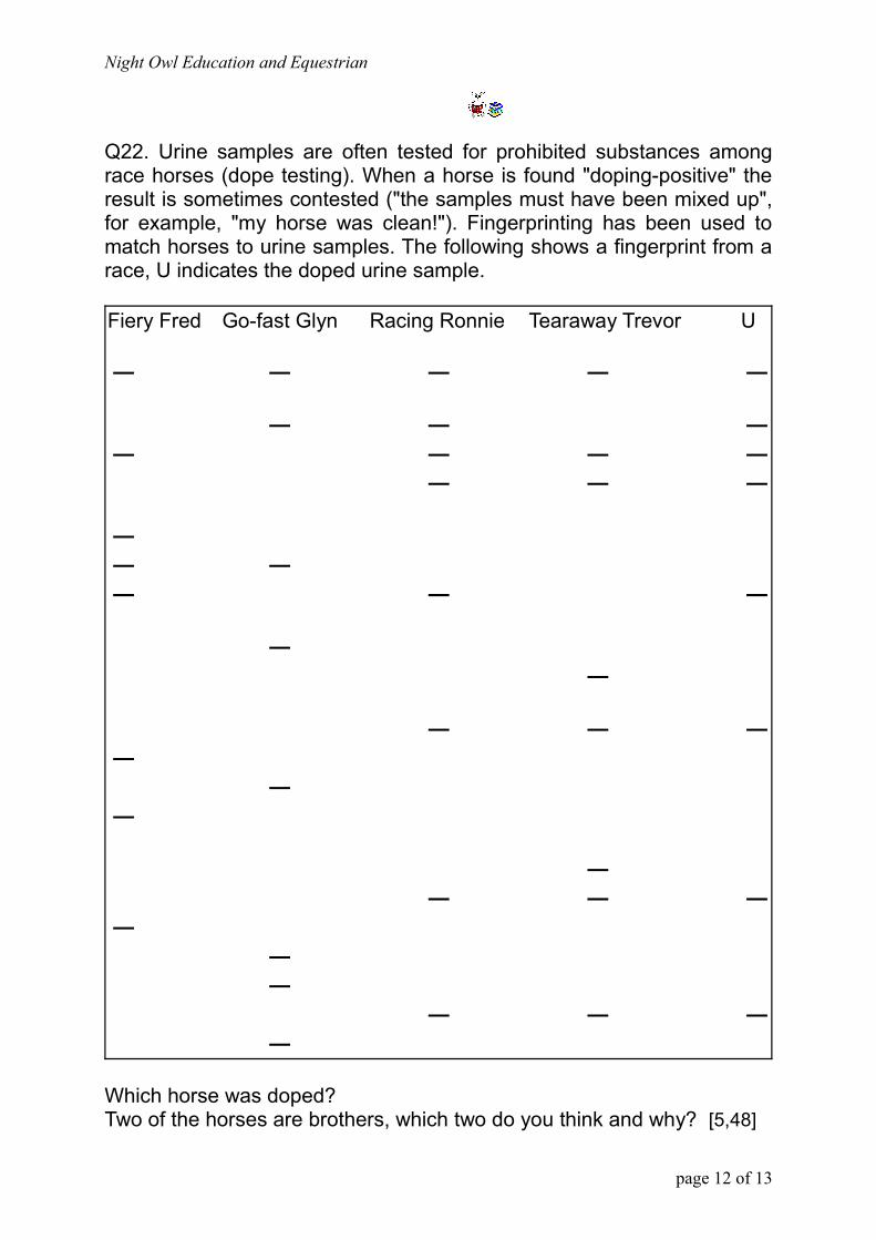
Night Owl Education and Equestrian
Q22. Urine samples are often tested for prohibited substances among race horses (dope testing). When a horse is found "doping-positive" the result is sometimes contested ("the samples must have been mixed up", for example, "my horse was clean!"). Fingerprinting has been used to match horses to urine samples. The following shows a fingerprint from a race, U indicates the doped urine sample.
Fiery Fred Go-fast Glyn Racing Ronnie Tearaway Trevor U
— — — — —
— — — — — — —
— — —
— — — — — —
— —
— — — —
— —
— — — —
— — —
— — — —
Which horse was doped? Two of the horses are brothers, which two do you think and why? [5,48]
page 12 of 13
Night Owl Education and Equestrian
Q23. Which of the following statements about chromosomes is true? A. Chromosomal DNA molecules are really short and composed of only a few hundred nucleotides each. A. Each chromatid of a horse chromosome contains one continuous molecule of DNA double helix running throughout its length. A. Horse chromosomes are made up of nucleoprotein, which contains various types of protein but no DNA. A. Proteins carry genetic information, which is packaged into chromosomes by being wrapped up with DNA. A. If chromosomes were packaged then the DNA could never be replicated or unravelled for gene activity. [1,49]
Q24. Which of the following statements about DNA is false? A. Most horse genes have far more DNA in them than is actually needed to code for the amino acids in their polypeptide products. A. Within genes in general there is coding DNA, known as exons, and stretches of non-coding DNA, called introns. Most horse genes don’t have any introns.A. Large regions of the chromosomes don’t contain any genes at all.A. Repetitive DNA, which may consist of a short base sequence repeated millions of times, has no known function. Some of it is transcribed, but we have no idea why. A. The function of redundant DNA is not known, but quite possibly it doesn‘t have one. It may be a product of genome evolution that is of no benefit to the horse, but exists for its own sake. [1,50]
page 13 of 13