Embed Size (px)
Citation preview
HAL Id: hal-03121524https://hal.archives-ouvertes.fr/hal-03121524
Submitted on 3 May 2021
HAL is a multi-disciplinary open accessarchive for the deposit and dissemination of sci-entific research documents, whether they are pub-lished or not. The documents may come fromteaching and research institutions in France orabroad, or from public or private research centers.
L’archive ouverte pluridisciplinaire HAL, estdestinée au dépôt et à la diffusion de documentsscientifiques de niveau recherche, publiés ou non,émanant des établissements d’enseignement et derecherche français ou étrangers, des laboratoirespublics ou privés.
Molecular docking studies and in vitro degradation offour aflatoxins (AFB 1 , AFB 2 , AFG 1 , and AFG 2 )by a recombinant laccase from Saccharomyces cerevisiaeYingli Liu, Huijia Mao, Chuanqin Hu, Thierry Tron, Junfang Lin, Jing Wang,
Baoguo Sun
To cite this version:Yingli Liu, Huijia Mao, Chuanqin Hu, Thierry Tron, Junfang Lin, et al.. Molecular docking studiesand in vitro degradation of four aflatoxins (AFB 1 , AFB 2 , AFG 1 , and AFG 2 ) by a recombinantlaccase from Saccharomyces cerevisiae. Journal of Food Science, Wiley, 2020, 85 (4), pp.1353-1360.�10.1111/1750-3841.15106�. �hal-03121524�
1
Molecular docking studies and in vitro degradation of four 1
aflatoxins(AFB1, AFB2, AFG1 and AFG2) by recombinant 2
laccase from Saccharomyces cerevisiae 3
Yingli Liu1, Huijia Mao1, Chuanquin Hu,1 Thierry Tron2, Junfang Lin3, Jing Wang1∗, 4
Baoguo Sun1 5
1 Beijing Advanced Innovation Center for Food Nutrition and Human Health, China-Canada Joint 6
Lab of Food Nutrition and Health (Beijing), Beijing Engineering and Technology Research Center 7
of Food Additives, Beijing Technology and Business University (BTBU), 11 Fucheng Road, Beijing, 8
100048, China 9
2Aix Marseille Université, Centrale Marseille, CNRS, iSm2 UMR 7313, 13397, Marseille, France. 10
3College of Food Science & Institute of Food Biotechnology, South China Agriculture University, 11
Guangzhou 510640, China 12
13
14
15
16
17
18
19
* corresponding authors: Jing Wang 20
address: Beijing Technology & Business University (BTBU), 11 Fucheng Road, Beijing 100048, 21
China. 22
Tel.: +86-010-68985334 Fax: +86-010-68985378 23
E-mail address: [email protected] 24
2
ABSTRACT: Aflatoxins are widely distributed mycotoxins with high concentration in 24 food and feed. They may have negative impacts on human health, animal productivity 25 and the economy, these being accompanied by removal difficulties. Enzymatic 26 degradation of aflatoxins is recently becoming an efficient strategy to ensure food and 27 feed safety . Here, molecular docking was used to predict and compare interactions 28 between laccase and four aflatoxins (AFB1, AFB2, AFG1 and AFG2) as well as their 29 degradation at a molecular level. Docking simulation studies indicated that aflatoxins 30 may interact near the T1 copper center through H-bonds and hydrophobic interactions 31 with amino acid residues His481 and Asn288, His481, Asn288 and Asp230, His481 and 32 Asn288. Removal tests were performed in vitro in the presence of a recombinant fungal 33 laccase. Degradation increased as incubation time increased from12h to 60h and the 34 maximum degradation obtained for AFB1, AFB2, AFG1 and AFG2 was 90.33%, 74.23%, 35 85.24% and 87.58%, respectively. Maximum degradation of aflatoxins was obtained 36 with a laccase total activity 4U at 30 °C in 0.1M phosphate buffer, pH 5.7 after 48h 37 incubation. The experimental results are consistent with that of docking calculation on 38 the removal of four aflatoxins by laccase. 39 40 Key Words: Laccases; degradability; aflatoxins. 41 42
3
INTRODUCTION 43
Mycotoxins are low-molecular weight molecules produced as secondary metabolites 44 by several species of fungi that contaminate various agricultural products, such as 45 cereals, maize, oilseeds and nuts, either before or under post-harvest conditions[1, 2]. 46 Among various types of mycotoxins, aflatoxins produced by Aspergillus flavus, A. 47 parasiticus and A. nomius, pose serious threat to public health and livestock 48 productivity, even at relatively low concentration. They constitute a group of closely 49 related compounds leading to highly toxic, mutagenic and carcinogenic[3, 4]. Therefore, 50 it is necessary to remove them timely from foodstuffs. 51
Among the 20 well-known aflatoxins, aflatoxinB1 (AFB1), aflatoxinB2 (AFB2), 52 aflatoxinG1 (AFG1), aflatoxinG2 (AFG2) are the most toxic ones (toxicity of AFB1>53 AFG1>AFB2>AFG2). They are both frequently found in many human dairy 54 foodstuffs and animal feeds and difficult to remove. AFB1 is the most potent naturally 55 occurring hepatotoxic, immunotoxin and teratogenic properties reported to date 56 (classified as Group 1 carcinogen by International Agency for Research on Cancer). 57 Although it affects primarily the liver, ABF1 is pluripotent acting on multiple organs 58 and multiple systems [5], and is causing both acute and chronic toxicity [6]. The main 59 toxic structures are the coumarin lactone ring, the double bond in the furan ring and the 60 cyclopentenone ring. 61
Currently, many researchs are focused on aflatoxins degradation or removal, and 62 various strategies have been reported to reduce aflatoxins levels. The most common 63 way to remove aflatoxin is by physical treatment, such as heating at high temperatures, 64 plasma degradation [7], extrusion or electromagnetic radiation treatment[8]. In addition, 65 chemical treatments such as ammoniation, nixtamalization, ozonation [9], acids and 66 bases treatments are also effective. However, these methods are so far not leading to 67 practical applications because of consecutive nutritional quality losses and because they 68 hardly comply with security, cost and productivity requirements for commercialization, 69 as well as they may cause environmental damages. Biological processes, including 70 degradation by plant extracts[10], microorganisms and enzymes, or the direct 71 adsorption to the cell surface of microorganisms like Saccharomyces cerevisiae [11] 72 and Lactobacillus casei[12], which are gentle, low cost and effective with less or no 73 intermediates harmful to humans and animals, have been identified as promising routes. 74
Laccases (EC1.10.3.2) belong to the family of the blue multicopper oxidases. They 75 contain four copper ions, a type 1 copper acting as a primary electron acceptor from 76 reductant species and a trinuclear cluster (TNC) involved in dioxygen reduction. 77 Widely distributed in plants, fungi, bacteria, and insects, these enzymes have the 78 catalytic ability to oxidize a wide range of aromatic substrates with a concomitant 79 reduction of dioxygen to water[13]. Globally, fungal laccase have better pH/or 80 temperature stability,higher metal tolerance and are able to oxidize a wider range of 81 substrates as enzymes from other origin[14]. At present, they are broad application 82 prospects of these enzymes in environment, food, medicine, cosmetics and other fields. 83 Considering the structural characteristics of aflatoxins, laccases may have a great 84 potential in AFs’ biodegradation applications. 85

4
The use of laccases may result in rapid and significant recalcitrant substrate 86 degradation[15]. With the maturity of computer simulation technology and the deep 87 understanding of the three-dimensional structure of laccase, it becomes easier and faster 88 to study the relationship between laccase a and substrate through molecular docking. 89 Laccase is taken as redox enzyme while molecular docking is used to conduct 90 simulation studies focusing on substrates binding abilities and binding modes. Catalytic 91 reactions then occur only if substrates get close enough to the T1 copper center of the 92 enzyme. Molecular docking has been used in a good deal of researches to study the 93 interaction of laccase with substrates. They agree that molecular docking is an effective 94 analytical tool to assess the enzyme-substrate inter atom. E.D. Morales-Alvarez[16] has 95 used molecular docking to evaluate interactions between the recombinant laccases 96 GlLCC1 and POXA 1B enzymes with Crystal Violet (CV) or Malachite Green (MG) 97 dyes. L. Dellafiora[17] have investigated the enzyme-substrate interaction for various 98 enzyme isoforms through 3D molecular docking techniques. D. Mo[18] has employed 99 molecular docking to study the interaction mechanism between nonylphenol and 100 octylphenol isomers and a laccase from Trametes versicolor. Y. Zhang[13] has used 101 molecular docking method to analyze the interactions between laccase produced by 102 Trametes versicolor and Triton X-100. Comparing three-dimensional models of 103 laccases A.K.S. Kameshwar[19] has performed molecular docking studies using lignin 104 model compounds to understand the structural and functional propertied of laccase. K.K. 105 Sharma[20] has used molecular docking to study the interaction of different 106 biomolecules with laccases from C. neoformans and different enteropathogenic bacteria 107 to understand their probable role in the oxidation of cellular metabolites and the 108 formation of the reactive intermediates. 109
In this work, we investigated the enzymatic degradation of aflatoxins by a laccase. 110 An homology model of the enzyme was first obtained then molecular docking analysis 111 was used to evaluate interactions between the laccase and four aflatoxins(AFB1, AFB2, 112 AFG1 and AFG2). This analysis was used to better understand the basis of the enzyme–113 substrates interaction at a molecular level, highlighting interaction areas around the 114 catalytic site and specific differences among the aflatoxins in terms of enzyme surface-115 ligand recognition. Results were compared to degradation tests performed alongside. 116 The comparison of theoretical and experimental data help to broaden our knowledge on 117 the degradation of aflatoxins and provide a basis for later molecular modifications of 118 laccase. 119
MATERIALS AND METHODS 120
Chemicial and Reagents 121 LAC3 from Trametes versicolor produced heterologously in Saccharomyces 122
cerevisiae was gift from Dr. Thierry Tron’s lab. Aflatoxins (purity>99%) was obtained 123 from J&K. ABTS was purchased from sigma. Methanol and formic acid were of high 124 performance liquid chromatography grade. Britton-Robinson buffer consisting of 125 0.04M phosphoric, 0.04M acetic acid, 0.04M boric acid was used during the 126 experiments. Ultrapure water was used throughout all of the experiments. 127
5
Preparation of Aflatoxins Working Solution 128 Solid aflatoxins were dissolved into 10mL of methanol to obtain 0.1 mg/mL standard 129
aflatoxins solutions and subsequentlystored at -20℃. 130
Preparation of Laccase 131 Laccase secreting Saccharomyces cerevisiae cells were cultivated at 30℃ for 4 days 132
in S-Gal medium on a rotary shaker incubator at 160 rpm. Cells were pelleted by 133 centrifugation (8000rpm for 30min). The recombinant laccase was found in the 134 supernatant. Purification was carried out as previously described[21] 135
Laccase Activity 136 Laccase assay was based on 2,2’-azino-bis(3-ethylbenzothiazoline-6-sulfonate) 137
(ABTS) oxidation in 0.04M Britton-Robison buffer (pH 4.0) at 30℃. Oxidation of 138 ABTS was monitored spectroscopically by absorbance measurements at 420nm (ε= 139 3.6×10⁴M−1cm−1). One unit of laccase oxidizes 1μmol of substrate per minute[22]. 140
Extraction of residual Aflatoxins and analysis by HPLC-MS 141 Aflatoxins were extracted three times from samples with chloroform (1 :1, v/v) as 142
described by Teniola et al[23]. Then chloroform fractions were pooled and evaporated 143 under nitrogen to dry. 144
The samples were dissolved in methanol, filtered (0.22μm) and separated on an 145 Inertsil ODS-3 C18 column,(4.6×150mm,3μm) coupled to a 1260 HPLC system 146 equipped with 6240 Triple Quad LC/MS (Agilent, United States) detector. LC-MS 147 conditions for Aflatoxins analysis were the followings: for regular analysis, 5μL of 148 sample or standard solutions were injected; the mobile phase, pumped at a flow rate of 149 0.2mL/min, consisted in an isocratic mixture of aqueous solution of 0.1% formic 150 acid/methanol (3:7, v/v); column temperature was maintained at 30℃ ; the total 151 operation time was 30 min; compounds were analyzed in positive mode. MS conditions 152 were as follows: capillary temperature 300℃; sheath gas flow and auxiliary gas flow 153 (11L/min and 3L/min, respectively) were adjusted to get stable spray,; Fragmentor 154 Voltage, Collision Energy, and Cell Accelerator Voltage were 135V, 30eV, 4KV, 155 respectively. Data were collected in MRM Scan Type with the precursor ions of AFB1, 156 AFB2, AFG1, AFG2 measured at m/z 313, 314, 328, 330 and product ions measured at 157 m/z 285, 287, 243 and 245, respectively. 158
Aflatoxins Degradation by Laccase 159 Aflatoxins degradation experiments (methanol solution) were carried out in 10mL 160
tubes. Laccase was incubated with toxins (0.1mg/mL) at intervals of enzyme activity, 161 incubation periods and temperature. The control was prepared with addition of 0.1 M 162 PBS pH 5.7 instead of laccase. Samples were placed in a shaker-incubator and 163 incubated under constant agitation (200rpm/min) at 30℃. Afterward, the toxins content 164 in the reaction mixture was extracted and determined as described above. Experiments 165 were performed independently for each aflatoxin. 166
The effect of a variation of the enzyme concentration on degradation rates was 167
6
conducted with enzyme loads corresponding to 1-5U. The effect of the incubation time 168 on degradation rates was investigated sampling the reactions every 12h for 60h. The 169 effect of temperature on degradation rates was investigated in the range of 25-45℃. 170
All experiments were performed in triplicate and the percentage (%) of degradation 171 was calculated with the following equation: 172
C(%) = [(A - B) / A]*100 173 Where, A corresponds to the initial mass of the aflatoxins and B is the residual mass 174
of the aflatoxins, C is the degradation rate of laccase for aflatoxins. 175
Homology Modeling of the laccase LAC3 176 The target sequence LAC3 was retrieved from Uniprot (Uniprot ID Q6TH77). 177
Template crystal structures were screened through a BLAST search; the structure with 178 the highest homology score was downloaded from the RCSB Protein Data Bank (PDB 179 ID: 3KW7). Homology modeling was conducted in MOE v2014.0901 (Chemical 180 Computing Group Inc, 1010 Sherbooke St. West, Suite#910, Montreal, QC, Canada, 181 H3A 2R7. 2014). The protonation state of the protein and the orientation of the 182 hydrogen atoms were optimized by LigX at a pH of 7 and a temperature of 300 K. First, 183 the target sequence was aligned to the template sequence, and ten independent 184 intermediate models were built. These different homology models were the result of the 185 permutational selection of different loop candidates and side chain rotamers. Then, the 186 intermediate model with the highest score according to the GB/VI scoring function was 187 chosen as final model, and then subjected to further energy minimization using the 188 AMBER12/EHT force field. 189
Modeling of Aflatoxins 190 AFB1、AFB2、AFG1、AFG2 were used to perform docking analysis with the model 191
constructed for LAC3 from Trametes sp. C30. 2D structures of the AFB1(CAS: 1162-192 65-8), AFB2(CAS: 7220-81-7), AFG1(CAS: 1165-39-5) and (AFG2, CAS: 7241-98-7) 193 were drawn in ChemBio Draw 2014 and converted to 3D in MOE v2014.0901 through 194 energy minimization. 2D structure of ligands are shown in Table 1. 195
Molecular Docking Experiments 196 MOE Dock in MOE v2014.0901 was used for molecular docking simulations of the 197
aflatoxins and prediction of their “binding affinity” to the homology model of LAC3. 198 The 3D structure of the LAC3 was predicted through homology modeling. And then, 199 the protonation state of the enzyme and the orientation of the hydrogens were optimized 200 by LigX, at the PH of 7 and temperature of 300 K. Prior to docking, the force field of 201 AMBER12: EHT and the implicit solvation model of Reaction Field (R-field) were 202 selected. The docking workflow followed the “induced fit” protocol, in which the side 203 chains of the receptor pocket were allowed to move according to ligand conformations, 204 with a constraint on their positions. The weight used for tethering side chain atoms to 205 their original positions was 10. All docked poses of aflatoxins molecules were ranked 206 by London dG scoring first, then a force field refinement was carried out on the top 30 207 poses followed by a rescoring of GBVI/WSA dG. 208
7
Statistical Analysis 209 All the experiments were performed in triplicate. All the data are graphically presented 210
as means ± standard deviation of triplicates(n=3), and the data were analyzed using 211 single factor analysis of variance(ANOVA). ANOVA was performed using SPSS 212 software(Version 8.5). Difference among the means were considered for significance at 213
P<0.05 using Duncan’s multiple range test. 214
RESULTS 215
Interaction of Aflatoxins with the laccase 216 In the absence of an available 3D structure of LAC3, the investigation of the binding 217
mode of aflatoxins with this enzyme required first the construction of an homology 218 model. Results are displayed in Figure 1 and Table 1. Analysis of the Ramachandran 219 plot revealed that ninety-nine percent of LAC3 residues map in allowed regions which 220 underlines that our homology model of LAC3 conforms to stereo chemical rules (Figure 221 2). Therefore, we docked aflatoxins AFB1, AFB2, AFG1 and AFG2 on the LAC3 222 homology model. 223
Docking scores of interactions between aflatoxins and LAC3 are reported in Table 3. 224 The minor differences observed among the values obtained for each aflatoxins may be 225 related to their chemical similarities. The different binding models between ligands and 226 laccase may result from different binding ability. The computational result indicates 227 that each of the aflatoxins tested can interact with laccase with a binding ability of 228 AFB1>AFG2>AFG1>AFB2. 229
The lowest energy configurations of aflatoxins and their binding mode at the surface 230 of LAC3 are illustrated in Figure 3. We assume that these predictions correspond to the 231 most likely configurations. In our models, the oxygen atom of the methoxy group of 232 AFB1, regarded as hydrogen bond acceptor, forms one hydrogen bond with the side 233 chain of His481 which is coordinated to the nearby copper ion (i.e. T1 CuII) [24] 234 whereas the oxygen atom of the terminal furan ring, regarded as hydrogen bond 235 acceptor, forms one hydrogen bond with the side chain of Asn288. Similarly, a carbon 236 atom of the benzene ring in AFB2, regarded as hydrogen bond acceptor, forms one 237 hydrogen bond with the sidechain of His481. Two carbon atoms of the terminal furan 238 ring of AFG1, regarded as hydrogen bond acceptors, form two hydrogen bonds with the 239 sidechain of Asn288 whereas the oxygen atom of 5-membered heterocyclic ring of 240 AFG1, regarded as hydrogen bond acceptors, form one hydrogen bond with the 241 sidechain of Asp230. The carbon and oxygen atoms of the terminal furan ring of AFG2, 242 regarded as hydrogen bond acceptors, forms two hydrogen bonds with the side chain 243 of Asn288 in laccase. Eventually, the carbon atom of methoxy group of AFG2, regarded 244 as hydrogen bond acceptor, forms one hydrogen bond with the side chain of His481. 245
Degradation of Aflatoxins using the Recombinant Laccase LAC3 Produced in 246 Saccharomyces cerevisiae 247
The aflatoxins(B1, B2, G1 and G2) detoxifying efficacy of LAC3 was tested at 248
8
different incubation times, enzyme activities and temperatures. Generally speaking, the 249 degradation of aflatoxins by the laccase was a rapid process. 250
The kinetic of degradation of aflatoxins was investigated at an enzyme activity of 3U 251 and at 30℃. As it can been seen from Figure 4(A), degradation rate increased first from 252 12 to 48h and then no significant difference was observed from 48 to 60h. In these 253 conditions, the maximum degradation of AFB1, AFG1 and AFG2, respectively 90.33%, 254 85.24%, 87.58%, was observed after 48h of incubation time, while for AFB2 the 255 maximum of degradation was reached after 60h of incubation. This may be explained 256 that the activity sites of laccase were more and the concentration of aflatoxins were 257 higher during the beginning of the reaction, the declining activity sites limited the 258 reaction. 259
Degradation experiments with laccase were tested at different enzyme activity (i. e. 260 different enzyme loads). Experiments were performed at 30℃ for 12h and results are 261 presented in Figure 4(B). Expectedly, degradation increased with an increase in the 262 enzyme load. Starting to plateau at 4U of laccase, the maximum degradation of AFB1, 263 AFB2, AFG1, AFG2, respectively 74.87%, 62.36%, 66.94%, 70.88%, was obtained at 264 5U of laccase. It is more likely to predict that the substrate-binding sites may have 265 reached to the saturation point as the activity of laccase was above 4U. 266
The effect of temperature on degradation rate of aflatoxins was conducted at an 267 enzyme activity of 3U for 12h and the results are shown in Figure 4(C). The percentage 268 of degradation of aflatoxins by laccase increased slightly from 25 to 40℃ and then 269 decreased with an increase of temperature from 40 to 45℃. The maximum degradation 270 of AFB1, AFB2, AFG1, AFG2 respectively, i.e.,77.45%, 51.84%, 56.06%, 68.33%, was 271 observed at 30℃. LAC3, like most of fungal laccase, has been described as a fairly 272 temperature tolerant enzyme. However, temperature tolerance is function of the 273 exposure time. Therefore, the decrease in activity observed when the reaction is 274 performed at a temperature over 40°C is probably a consequence of the long exposure 275 (12h) of the enzyme at this temperature. Hence, the optimal temperature was set at 30℃ 276 in future studies. 277
All together the experimental are in agreement with the theoretical study that 278 predict aflatoxins “binding affinity” for LAC3, in particular that of AFB2 that was found 279 minimum among the aflatoxins tested. 280
DISCUSSION AND CONCLUSION 281 It is important to reduce the mycotoxin contents in food and feed for public health 282
and globe trade. A wide number of effective strategies are already used to control toxin 283 accumulation levels at pre- or post-harvest. Comparing with cultivation techniques, 284 conventional breeding, genetic engineering and other physical or chemical methods, 285 biological treatments have good application potential due to their ability to transform 286 toxins to non - or less harmful compounds during food processing [25]. Among these, 287 the degradation by means of enzymes is considered as the strategy with the most highest 288 potential for the mitigation of toxins content. So far, J.F. Alberts et al. have tried to 289 degrade AFB1 by laccase from white rot fungi Peniophora, Pleurotus ostreatus and 290 Trametes. versicolor achieving degradation of 40.45%, 35.90% and 87.34%, 291
9
respectively[26]. Zeinvand-Lorestani H et al. also applied a commercial laccase 292 preparation from Trametes versicolor to AFB1 with 67% of the toxic substrate 293 removal[27]. Loi M et al. have shown that AFB1 and AFM1 degradation by a pure 294 enzyme form Pleurotus pulmonarius is greatly enhanced from 23% to more than 90% 295 in the presence of redox mediators [28]. However, screening diversity to find out proper 296 active enzymes is challenging and time-consuming, and more importantly does not 297 allow to study an interaction mechanism between ligands and the target enzyme. 298 Investigation of potential enzyme-substrate interactions by in silico methods could be 299 used as screening tool as it provides increasingly reliable and informative insights [17]. 300 While taken alone there may be still some deviations due to computer simulation, 301 performing alongside molecular docking analysis and in vitro experiment allows a 302 benchmarking of methodologies. The consistency between in vitro experimental results 303 and that of docking calculation will contribute to find out a faster, more precise and 304 effective way in aflatoxins removal. 305
Here, we employed molecular docking to study the interaction of four aflatoxins with 306 the surface of a typical fungal laccase aiming to highlight possible differences in ligand-307 enzyme interactions. According to our models, relevant differences exist among 308 aflatoxins in terms of interaction with the enzyme surface. AFB1 is likely interacting 309 the most favorably with laccase among all the aflatoxins. Docking simulation studies 310 indicate that the residue His481 present in the coordination sphere of the T1 copper 311 may interact with aflatoxins an therefore could mediate the electron transfer during 312 oxidation[29]. Different hydrogen bonding patterns arise from calculation: in addition 313 to His481, AFB1 may interact with and Asn288, AFB2 may interact with His481, AFG1 314 may interact with both Asn288 and Asp230, AFG2 may interact with both His481 and 315 Asn288. AAmino acid residues involved in H-bonds are considered as key residues for 316 the interaction of laccase with a given ligand[18]. H-bonds details as shown in Table 4 317 suggest different interactions between the laccase and the ligands which may 318 correspond to differences in binding ability with AFB1>AFG2>AFG1>AFB2, an order 319 consistent with that drawn from experimental results on the biodegradation of 320 aflatoxins by LAC3 by , and the of aflatoxins at the same time is 321 AFB1>AFG2>AFG1>AFB2. 322
The binding affinity is related to the efficiency of enzyme-ligand interactions which 323 is affected by structural characteristics and distorting extent of ligands and the shape 324 complementation between the ligand and the enzyme surface[30, 31].With the higher 325 binding affinity, the ligands have stronger capability to reach the pocket-like structure 326 of enzyme. Differences in toxin degradation appears to be related primarily to group 327 contacts established with the laccase surface. The stronger the aflatoxin-laccase 328 interaction is the more efficient is the oxidation of the toxin. 329
In silico analysis provides insight into the interaction of aflatoxins with laccase and 330 allows to point structures interactions relevant for the degradation. An in-depth 331 mapping of enzyme-ligand potential interactions will provide a theoretical basis for 332 molecular modification strategies of laccase set to improve degradation or for screening 333 for natural laccase with high toxins degrading capabilities from a variety of 334 microorganisms. In both cases, molecular docking will be advantageous to improve the 335

10
biodegradation techniques. 336 Acknowledgement 337
This work was supported by National Natural Science Foundation of China (No. 338 31601564), Beijing Excellent Talents Funding for Youth Scientist Innovation Team 339 (2016000026833TD01) and High-level Teachers in Beijing Municipal Universities 340 (IDHT20180506). 341
REFERENCES: 342
[1] R. Liu, Q. Jin, G. Tao, L. Shan, Y. Liu, X. Wang, LC–MS and UPLC–Quadrupole 343
Time-of-Flight MS for Identification of Photodegradation Products of Aflatoxin B1, 344
Chromatographia 71(1-2) (2009) 107-112. 345
[2] S.K. Pankaj, H. Shi, K.M. Keener, A review of novel physical and chemical 346
decontamination technologies for aflatoxin in food, Trends in Food Science & 347
Technology 71 (2018) 73-83. 348
[3] M.G. Theumer, Y. Henneb, L. Khoury, S.P. Snini, S. Tadrist, C. Canlet, O. Puel, I.P. 349
Oswald, M. Audebert, Genotoxicity of aflatoxins and their precursors in human cells, 350
Toxicol Lett 287 (2018) 100-107. 351
[4] C. Verheecke, T. Liboz, F. Mathieu, Microbial degradation of aflatoxin B1: Current 352
status and future advances, Int J Food Microbiol 237 (2016) 1-9. 353
[5] S. Agriopoulou, A. Koliadima, G. Karaiskakis, J. Kapolos, Kinetic study of 354
aflatoxins' degradation in the presence of ozone, Food Control 61 (2016) 221-226. 355
[6] A. Raiola, G.C. Tenore, L. Manyes, G. Meca, A. Ritieni, Risk analysis of main 356
mycotoxins occurring in food for children: An overview, Food Chem Toxicol 84 (2015) 357
169-80. 358
[7] A. Sakudo, Y. Toyokawa, T. Misawa, Y. Imanishi, Degradation and detoxification 359
of aflatoxin B 1 using nitrogen gas plasma generated by a static induction thyristor as 360

11
a pulsed power supply, Food Control 73 (2017) 619-626. 361
[8] F. Wang, F. Xie, X. Xue, Z. Wang, B. Fan, Y. Ha, Structure elucidation and toxicity 362
analyses of the radiolytic products of aflatoxin B1 in methanol-water solution, J Hazard 363
Mater 192(3) (2011) 1192-202. 364
[9] X. Luo, R. Wang, L. Wang, Y. Wang, Z. Chen, Structure elucidation and toxicity 365
analyses of the degradation products of aflatoxin B1 by aqueous ozone, Food Control 366
31(2) (2013) 331-336. 367
[10] W. Iram, T. Anjum, M. Iqbal, A. Ghaffar, M. Abbas, Mass spectrometric 368
identification and toxicity assessment of degraded products of aflatoxin B1 and B2 by 369
Corymbia citriodora aqueous extracts, Sci Rep 5 (2015) 14672. 370
[11] P.H. Shetty, B. Hald, L. Jespersen, Surface binding of aflatoxin B1 by 371
Saccharomyces cerevisiae strains with potential decontaminating abilities in indigenous 372
fermented foods, Int J Food Microbiol 113(1) (2007) 41-6. 373
[12] A. Hernandez-Mendoza, H.S. Garcia, J.L. Steele, Screening of Lactobacillus casei 374
strains for their ability to bind aflatoxin B1, Food and Chemical Toxicology 47(6) (2009) 375
1064-1068. 376
[13] Y. Zhang, Z. Zeng, G. Zeng, X. Liu, Z. Liu, M. Chen, L. Liu, J. Li, G. Xie, Effect 377
of Triton X-100 on the removal of aqueous phenol by laccase analyzed with a combined 378
approach of experiments and molecular docking, Colloids Surf B Biointerfaces 97 379
(2012) 7-12. 380
[14] B. Legerska, D. Chmelova, M. Ondrejovic, Decolourization and detoxification of 381
monoazo dyes by laccase from the white-rot fungus Trametes versicolor, J Biotechnol 382
12
285 (2018) 84-90. 383
[15] F.J. Ruiz-Duenas, A.T. Martinez, Microbial degradation of lignin: how a bulky 384
recalcitrant polymer is efficiently recycled in nature and how we can take advantage of 385
this, Microb Biotechnol 2(2) (2009) 164-77. 386
[16] E.D. Morales-Alvarez, C.M. Rivera-Hoyos, S.A. Poveda-Cuevas, E.A. Reyes-387
Guzman, A.M. Pedroza-Rodriguez, E.A. Reyes-Montano, R.A. Poutou-Pinales, 388
Malachite Green and Crystal Violet Decolorization by Ganoderma lucidum and 389
Pleurotus ostreatus Supernatant and by rGlLCC1 and rPOXA 1B Concentrates: 390
Molecular Docking Analysis, Appl Biochem Biotechnol 184(3) (2018) 794-805. 391
[17] L. Dellafiora, G. Galaverna, M. Reverberi, C. Dall'Asta, Degradation of Aflatoxins 392
by Means of Laccases from Trametes versicolor: An In Silico Insight, Toxins (Basel) 393
9(1) (2017). 394
[18] D. Mo, G. Zeng, X. Yuan, M. Chen, L. Hu, H. Li, H. Wang, P. Xu, C. Lai, J. Wan, 395
C. Zhang, M. Cheng, Molecular docking simulation on the interactions of laccase from 396
Trametes versicolor with nonylphenol and octylphenol isomers, Bioprocess Biosyst 397
Eng 41(3) (2018) 331-343. 398
[19] A.K.S. Kameshwar, R. Barber, W. Qin, Comparative modeling and molecular 399
docking analysis of white, brown and soft rot fungal laccases using lignin model 400
compounds for understanding the structural and functional properties of laccases, J Mol 401
Graph Model 79 (2018) 15-26. 402
[20] K.K. Sharma, D. Singh, S. Rawat, Molecular dynamics simulation studies suggests 403
unconventional roles of non-secretary laccases from enteropathogenic gut bacteria and 404

13
Cryptococcus neoformans serotype D, Comput Biol Chem 73 (2018) 41-48. 405
[21] Y. Liu, A.M. Cusano, E.C. Wallace, Y. Mekmouche, S. Ullah, V. Robert, T. Tron, 406
Characterization of C-terminally engineered laccases, International Journal of 407
Biological Macromolecules 69 (2014) 435-441. 408
[22] A. Klonowska, C. Gaudin, M. Asso, A. Fournel, M. Réglier, T. Tron, LAC3, a new 409
low redox potential laccase from Trametes sp. strain C30 obtained as a recombinant 410
protein in yeast, Enzyme and Microbial Technology 36(1) (2005) 34-41. 411
[23] O.D. Teniola, P.A. Addo, I.M. Brost, P. Farber, K.D. Jany, J.F. Alberts, W.H. van 412
Zyl, P.S. Steyn, W.H. Holzapfel, Degradation of aflatoxin B(1) by cell-free extracts of 413
Rhodococcus erythropolis and Mycobacterium fluoranthenivorans sp. nov. 414
DSM44556(T), Int J Food Microbiol 105(2) (2005) 111-7. 415
[24] O.A. Glazunova, K.M. Polyakov, K.V. Moiseenko, S.A. Kurzeev, T.V. Fedorova, 416
Structure-function study of two new middle-redox potential laccases from 417
basidiomycetes Antrodiella faginea and Steccherinum murashkinskyi, Int J Biol 418
Macromol 118(Pt A) (2018) 406-418. 419
[25] G. Jard, T. Liboz, F. Mathieu, A. Guyonvarc'h, A. Lebrihi, Review of mycotoxin 420
reduction in food and feed: from prevention in the field to detoxification by adsorption 421
or transformation, Food Addit Contam Part A Chem Anal Control Expo Risk Assess 422
28(11) (2011) 1590-609. 423
[26] J.F. Alberts, W.C.A. Gelderblom, A. Botha, W.H. van Zyl, Degradation of aflatoxin 424
B-1 by fungal laccase enzymes, International Journal of Food Microbiology 135(1) 425
(2009) 47-52. 426
14
[27] H. Zeinvand-Lorestani, O. Sabzevari, N. Setayesh, M. Amini, A. Nili-Ahmadabadi, 427
M.A. Faramarzi, Comparative study of in vitro prooxidative properties and 428
genotoxicity induced by aflatoxin B1 and its laccase-mediated detoxification products, 429
Chemosphere 135 (2015) 1-6. 430
[28] M. Loi, F. Fanelli, P. Zucca, V.C. Liuzzi, L. Quintieri, M.T. Cimmarusti, L. Monaci, 431
M. Haidukowski, A.F. Logrieco, E. Sanjust, G. Mule, Aflatoxin B-1 and M-1 432
Degradation by Lac2 from Pleurotus pulmonarius and Redox Mediators, Toxins 8(9) 433
(2016). 434
[29] N.J. Christensen, K.P. Kepp, Setting the stage for electron transfer: Molecular basis 435
of ABTS-binding to four laccases from Trametes versicolor at variable pH and protein 436
oxidation state, Journal of Molecular Catalysis B: Enzymatic 100 (2014) 68-77. 437
[30] H.J. Gabius, S. Andre, J. Jimenez-Barbero, A. Romero, D. Solis, From lectin 438
structure to functional glycomics: principles of the sugar code, Trends Biochem Sci 439
36(6) (2011) 298-313. 440
[31] C.E. Cassidy, W.N. Setzer, Cancer-relevant biochemical targets of cytotoxic 441
Lonchocarpus flavonoids: a molecular docking analysis, J Mol Model 16(2) (2010) 442
311-26. 443
444
15
Table Legends 445
Table 1: Homology modeling of LAC3 (Trametes sp. C30) 446
Table 2: 2D structure of ligands 447
Table 3: Docking score of molecules binding to Laccase 448
Table 4: H-bonding distances between laccase and aflatoxins 449

16
Figure Legends 450
Fig. 1: Homology models of laccase protein sequences (Trametes sp. C30) 451
452
Fig. 2: Ramachandran Plot: dark green dots represent the residues in most favored 453
regions; yellow dots represent the residues in additional allowed regions; Small red 454
cross represents the residues in not-allowed regions. 455
456
Fig. 3: Models of AFB1, AFB2, AFG1 and AFG2 interacting with the laccase LAC3. A. 457
The binding model of AFB1 on molecular surface of the laccase. B. The interaction 458
model of AFB2 with the laccase. C. The binding model of AFB2 on molecular surface 459
of the laccase. D. The interaction model of AFB2 with the laccase. E. The binding model 460
of AFG1 on molecular surface of the laccase. F. The interaction model of AFG1 with 461
the laccase. G. The binding model of AFG2 on molecular surface of the laccase. H. The 462
interaction model of AFG2 with the laccase. The ligands are colored in cyan, the T1 463
copper ion is colored in pink and the surrounding residues in the binding pockets are 464
colored in orange. The backbone of the receptor is depicted as lightblue ribbon. 465
466
Fig. 4: Effect of incubation time (A), enzyme activity (B) and temperature (C) on the 467
degradation of aflatoxins by the laccase LAC3. Values are means of three replicates and 468
their standard errors. Means with different letters representing differences within the 469
group are significantly different according to Duncan’s test(P≤0.05). 470

Table Legends
Table 1:
Protein Template Query Cover Identity
Laccase 3KW7 , A 93% 77%
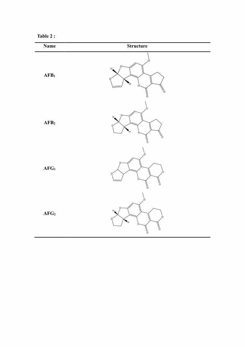
Table 2 :
Name Structure
AFB1
AFB2
AFG1
AFG2
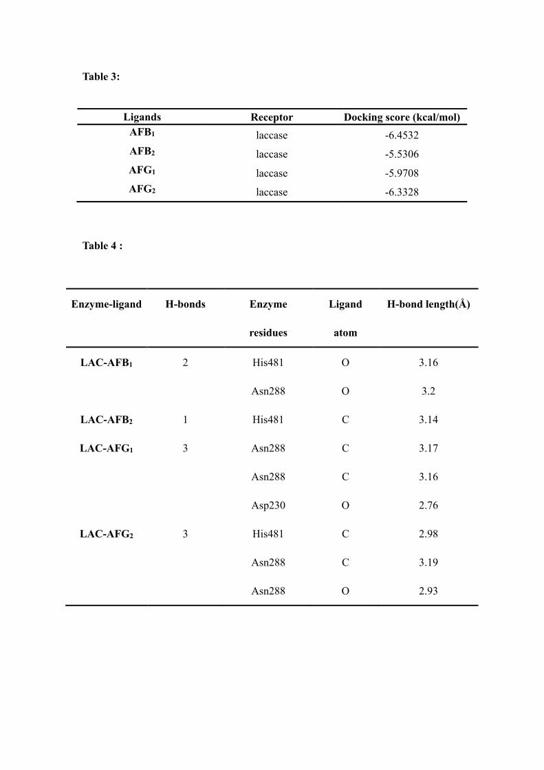
Table 3:
Ligands Receptor Docking score (kcal/mol)
AFB1 laccase -6.4532 AFB2 laccase -5.5306 AFG1 laccase -5.9708 AFG2 laccase -6.3328
Table 4 :
Enzyme-ligand H-bonds Enzyme
residues
Ligand
atom
H-bond length(Å)
LAC-AFB1 2 His481 O 3.16
Asn288 O 3.2
LAC-AFB2 1 His481 C 3.14
LAC-AFG1 3 Asn288 C 3.17
Asn288 C 3.16
Asp230 O 2.76
LAC-AFG2 3 His481 C 2.98
Asn288 C 3.19
Asn288 O 2.93

Figure Legends
Fig. 1
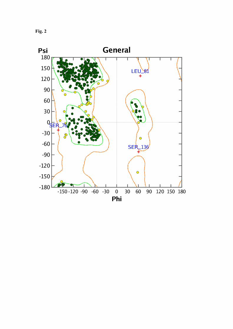
Fig. 2
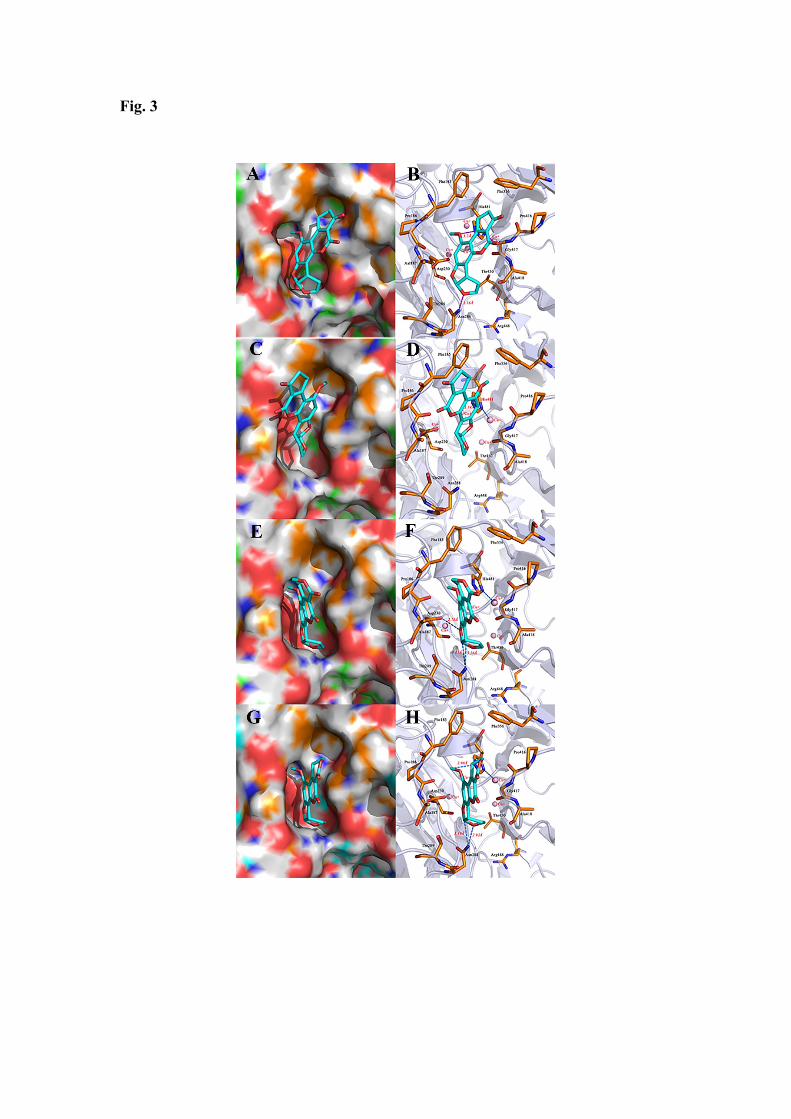
Fig. 3
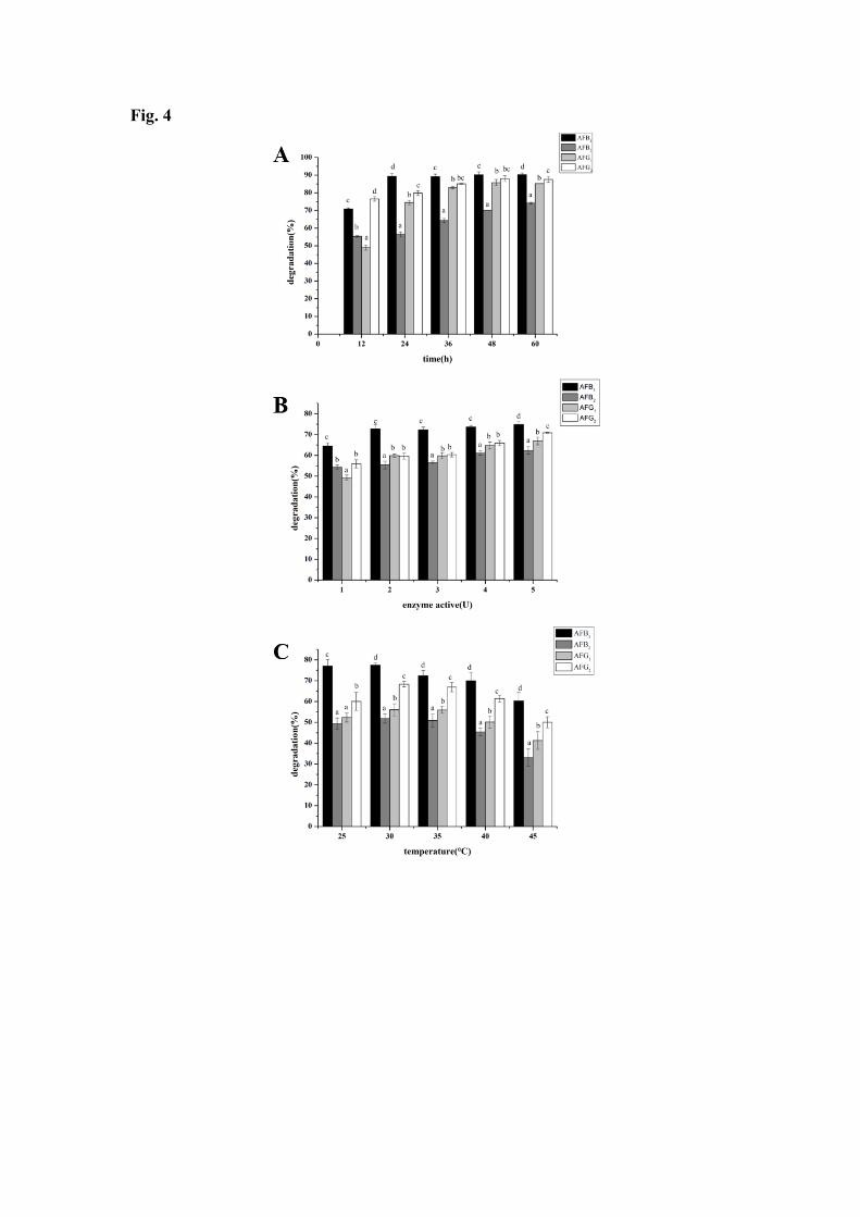
Fig. 4

![IspE Inhibitors Identified by a Combination of In Silico ... · docking and in vitro high-throughput screening [29,30,31,32,33,34,35,36,37,38]. These studies suggest that often the](https://img.pdfslide.us/doc/110x75/5f2ee20b7759a50bd9270253/ispe-inhibitors-identified-by-a-combination-of-in-silico-docking-and-in-vitro.jpg)