Embed Size (px)
Citation preview
Vol. 184, No. 2, 1992 BIOCHEMICAL AND BIOPHYSICAL RESEARCH COMMUNICATIONS
April 30, 1992 Pages 640-646
Molecular cloning and nucleotide sequence of leukocidin F-component gene (l&F) from methicillin resistant Staphylococcus aureus
Arifur Rahmanl , Hirobumi Nariyal , Kazuo Izaki’ , Iwao KaW, and Yoshiyuki Kamio’ *
1 Department of Agricultural Chemistry, Faculty of Agriculture, Tohoku University, Aoba-ku, Sendai, 981 Japan
2 Department of Oral Bacteriology, School of Dentistry, Showa University, Shinagawa-ku, Tokyo, 142 Japan
Received March 12, 1992
SUMMARY: A 1ukF gene encoding F-component of Staphylococcal leukocidin from methi- cillin resistant Staphylococcus aureus (MRSA) was cloned. The nucleotide sequence of lukF gene was determined. The sequence data have revealed an open reading frame, which encodes a polypeptide with 323 amino acid residues. Inspection of the amino acid sequence deduced from nucleotide sequence of 1ukF and that from F-component of leukocidin from S. aureus V8 clarified that pre-matured F-component contains a typical signal peptide at the NH2 terminus and ATG starting codon for pre-matured F-component was present one base downstream to the TGA which is translation termination codon for S-component of leukocidin [A. Rahman et al. (1991) Biochem. Biophys. Res. Commun. 181, 138-1441. The nucleotide sequence of 5’-flanking region of 1ukF showed the presence of the consensus sequence of ribosome binding site in the internal region of the structural gene of S-component. The 1ukF was transcribed in the same direction as that of 1ukS. No Pribnow box can be discerned in the intercistronic region between the 1ukS and 1ukF genes. The amino acid sequence homology between S- and F-components was 31%. F-component was expressed in Escherichia coli DH5a harboring plasmid pFRK92 which contained 1ukF gene. 0 199.2 Academic Press, Inc.
Staphylococcal leukocidin consists of two proteins: the S- and F-components act
synergistically to induce cytotoxic changes in human and rabbit polymorphonuclear
leukocytes (1). Little is known about the relation between the chemical structure
and function of these components on the mode of action of leukocidin. One method
of studying the role of the toxin in pathogenesis is to examine the virulence of
toxin-deficient mutant in experimental infections. To facilitate the isolation of
leukocidin-deficient mutant, and to study the chemical structure and function of
the S- and F-components of leukocidin, we have started to clone the genes for two
components and determine the nucleotide sequences of the genes. In the previous
paper, we have described the cloning and complete nucleotide sequence of S-
* To whom correspondence should be addressed.
CUO6-291X/92 $1.50 Copyight 0 1992 by Academic Press. IIK. All rights of reproduction in any form reserved. 640
Vol. 184, No. 2, 1992 BIOCHEMICAL AND BIOPHYSICAL RESEARCH COMMUNICATIONS
component gene (lukS) from methicillin resistant Staphylococcus aureus (MRSA)
strain No.4 (2). In the present study, cloning, expression and complete nucleotide
sequence of the 1ukF gene and its flanking regions are described, and a co-
transcriptional expression of the 1ukS and 1ukF genes are suggested. The amino
acid sequence homology of F- and S-components also compared.
MATERIALSANDMETHODS
Bacterial strains and plasmid. MRSA strain No.4 was used for preparation of chromosomal DNA. Escherichia coli DH5a(recAl, A-, A(lac-proAB), endAl, gyrA96, thi, hsdR17, relA1, supE44,[F', traD36, proAB, la& &Y15]} was used as a host strain for recombinant plasmids. Plasmid pUC119, pUC118 and Charomid 9-36 (Nippon gene Ltd. Tokyo) were used as cloning vectors. E. coli MV1184{ara, A(lac-pro), &z-A, thi, (@do lacZAbf15), ZAbfkj]},
d(srl-recA)306::TnlO (tet ), [f7, traD36, proAB, lacl" , and M13K07 were used as a host and a helper phage, respectively for
preparation of single stranded DNA.
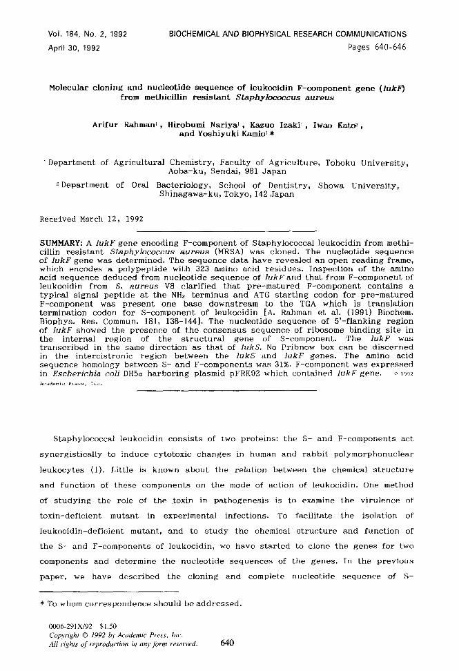
Plasmid construction. Plasmid pFRK92: Plasmid pSA9-36 was previously constructed by inserting 6-8 kbp Sau3A digested chromosomal DNA of MRSA No.4 strain into BamHI site of Charomid 9-36 vector DNA and transduced into E. coli DH5a. The pSA9-36 was further digested with EcoRI. About 4 kbp EcoRI fragment was ligated into EcoRI site of pUC119, and plasmid pFRK92 was obtained (Fig.1).
Preparation of mixed oligonucleo@de probes The N-terminal amino acids sequence of purified F-component preparation from S. aureus V8 was determined by Edman degradation method. Oligonucleotides were synthesized by a MilliGen/Biosearch, CyclonT fl Plus DNA synthesizer. Synthetic oligonucleotides were labeled with y-3? P-ATP( >llOTBq/mmol) as described by Maniatis et a1.(3).
Southern blot hybridization. Southern blot hybridization was performed according to the method as described by Maniatis et a1.(3).
DNA sequencing. A series of deletion derivatives were obtained from plasmid pFRK92 by exonuclease III and mung bean nuclease digestion (Fig.2) and subcloned into both pUC119 and pUC118. Resulting plasmids were introduced into E. coli MV1184 to isolate ss-DNA using bacteriophage M13K07. DNA sequencing was performed by cycle sequencing of DNA with Dye primer -2lM13 (Applied biosystem) using DNA sequencer (Mole1 373A, DNA sequencing System, Applied biosystem).
Preparation of cell extracts and determination of 1ukF gene product. The cells of the plasmid-containing E. coli DH5a were grown at 37” C in 2 ml of LB medium containing ampicillin (100 Mg/ml). At exponential phase, IPTG (1mM) was added, and allowed to grow for more two hours. The cells were harvested and then suspended in 0.3 ml of 10 mM Tris-HCl buffer (pH7.5). The periplasmic preparation from the cells was obtained according to the method of Heppel(4). The periplasm and shocked cell fractions were heated in solubilizing buffer for SDS-PAGE and subjected to SDS- PAGE. The IukF gene product was determined by Western immuno-blot analysis by using polyclonal antibody raised against F-component of leukocidin from S. aureus strain V8.
RESULTS ANDDISCUSSION
Analysis of N-terminal sequence of F-component. We determined N-terminal 41 amino
acids sequence of the purified F-component of leukocidin from S. aureus V8 as
follows; Ala’ -Glu-Gly-Lys-Ile” -Thr-Pro-Val-Ser-Vail 0 -Lys-Lys-Val-Asp-Asp15 -Lys-Val-
Thr-Leu-Tyr20-Lys-Thr-Thr-Ala-Thr’5 -Ala-Asp-Ser-Asp-Lys? 0 -Phe-Lys-Ile-Ser-Glu3 5
-Ile-Leu-Thr-Phe-Asn4 0 -Phe. According to the amino acids sequence, the oligo-
641
Vol. 184, No. 2, 1992 BIOCHEMICAL AND BIOPHYSICAL RESEARCH COMMUNICATIONS
*-- +-- -
E c--x-- EKB
pFRK92
0 1 2 3 4 I I 1
KB
F~J& Restriction endonuclease map of 4 kbp EcoRI-EcoRI fragment and sequencing strategy. The S- and F-components of leukocidin coding region are indicated by thick horizontal arrows. Thin arrows indicate the direction and sequence determined region of deleted DNA clones. Double and single lines are inserted 4 kbp fragment and polylinker region of vector plasmid pUC119, respectively. Abbreviations: B, BarnHI; C, ClaI; E, EcoRI; H, ffindII1; K, KpnI; X, XbaI. F, F-component gene; S, S-component gene.
nucleotide probes were synthesized. The probes were as follows: (i) from the amino
acid position 10-15: 5’-GTI-AA(A,G)-AA(A,G)-GTI-GA(T,C)-GA-3’ and (ii) from the
amino acid position 16-21: 5’-AA(A,G)-GTI-ACI-(C,T,)TI-TA(T,C)-AA-3’ (I; Inosine).
Cloning of 1ukF in E. coli Plasmid pSA9-36, which covered full length of the
structural gene of S-component (1ukS) from MRSA strain No.4 was also found to be
strongly hybridized to the two mixed probes (Data not shown). The plasmid DNA was
isolated from pSA9-36 and its physical map was determined. About 4.0 kbp (EcoRI-
EcoRI) fragment which hybridized with the two probes was assumed to contain the
full length of the structural gene of F-component. Accordingly, the fragment was
subcloned into EcoRI site of plasmid vector pUC119 and transformed into E. coli
DH5a. This plasmid was designated as pFRK92. The physical map of plasmid pFRK92 is
shown in Fig.1.
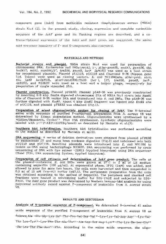
Nucleotide sequence of the 1ukF gene from pFRK92. We have determined the nucleo-
tide sequence containing 1ukF gene according to the sequencing strategy shown in
Fig.1. The nucleotide sequence of the fragment was comprised of 2,012 bp (Fig.2).
Within this sequence, we can identify an open reading frame which begins with ATG
codon at position 378 and terminates with TGA codon at position 1,347. The ATG
codon as initiating codon for F-component gene was present just one base
downstream of ‘the termination codon for S-component of leukocidin (Fig.2). This 969
nucleotides encode 323 amino acids, and the molecular weight deduced from these
amino acids sequence was 36,508 dalton, which coincides with that determined by
SDS-PAGE of the product of pFRK92-directed transcription/translation system (5)(un-
published data). The N-terminal 41 amino acid residues of the purified F-component
from S. aureus V8 were identical to the deduced 41 amino acid residues from 26th N-
terminal amino acid residue to 66th one. The total amino acid composition of
642

Vol. 184, No. 2, 1992 BIOCHEMICAL AND BIOPHYSKAL RESEARCH COMMUNICATIONS
190 200 210 220 230 240 250 260 270 AAC~GATA~AAGCGAATTTGAAATTACTTACGGAAGAAACATGGATGTCACTCATGCCATTAAAAGAT~AA~~~ATTAT~~~~A~A~TT~
SerAspThrS~rGluPheCl~IleThrTyrGlyAr~AsnMetA~pValThrHiSAl8IleLYSA~8S~~Th~~~~TY~~lYA~ns~~Tyr
280 290 300 310 320 330 340 350 360 TTTAGACGGACATAGAGTCCATAATGCATTCGTAAATAGA~CTATACTGTTAAATACGAGGTCAATTGGAAGACTCATGA~TC~AG~~
L~~AspGlyHi~A~gValHi8AsnAlaPheValAsnArgAsnTyrTh~valLy~Ty~Gl~ValA~~T~PLY~Th~~~~~l~Il~Ly~v~1
LysGlyGlnAsn.** MetLyaMetAsnLysLeuValLy~SerS~rValAlaThrSerMetAlaLeuLeuLeULeuSerGlyThrAlaAsn
460 470 490 490 500 510 520 530 640 ATGCTGAAGGTAAAATAACACCAGTCAGCGTAAAAAAA GTCGATGACAAAGTTACTTTATACAAAACAACAGCCACAGCAGATTCTGATA
AlaCluClyLysIleThrProValGerValLye4yeVslAapAap~~ValThrLeuTyrLy~ThrThrAlaThrAlaAapSerAspLy~ A
650 550 570 680 690 800 610 620 630 AGTTTAAAATTTCACAGATTTTAACATTTMTTTCAATA
PheLysIleSerClnIleLeuThrPheAanPheIleLyaAapLy~SerTyrAap~aAapThrLeuValLeu4~AlaThrGlyAanIle
640 660 860 670 660 690 700 710 720 TTAACTCAGGCTTTGTGAAACCTAATCCTMTGACTATOATGTATCTATAAGCTCAC
AenSerGlyPheValLyaProAsnProAenAspTyrAepPheSerLy~LeuTyrTrpGlyAlaLy~TyrAsnV~lS~rIleSerSerGln
730 740 760 700 770 780 790 800 810 AATCTAATGATTCAGTAAACGCTGTTGATTATGCAAAAATACTTTAGGCTATACATTTG
SerAsnAspSerV8lAsnAlaValA6pTyrAlaProLyaAsnGlnAsn~luGluPheGlnV~lGlnAanThrLeuGlyTyrThrPheGly
820 830 840 660 880 870 860 890 900 GTGGTGACATTAGTATCTCTAATGGTTTATCTGGTGGACTT~TGG~TACAGCTTTTTCTG~AC~TTAATTAT~C~GAAAGTT
GlyAspIleSerIleSerAsnClyLeuGerClyClyGlyL~uA~nGlyA~nTh~Al~Ph~S~rGluThrIleA~nTy~Ly~GlnGluSe~Tyr
910 920 830 940 850 980 870 880 980 ACAGAACATTAAGTCGCMCACAAATTATAIWVLTGTTGC
ArgThrLeuSerArgAsnThrAsnTyr4ysAenValGlyTrpGlyValGluAlaHi~LyaIleMetA~nGlyTrpGlyProTyrGlyArg
1000 1010 1020 1060 1070 ~A~ATA~~TT~~A~C~~~ATATGGTAATCT~~~~AG~T~~~~~~C~~C~~~~CATAC~CT~~CCA~CTTCATA~C~~~~
A~DS~~P~~S~~P~~Th~TYrClyAsnGluLeuPheLeuAlaGlyArgGlnS~rs~rAl~Ty~AlaGlyGl~~~PheIl~Al~cl~Hi~
IOU0 1100 1110 1160 1160 1170 ACCAAATGCCATTATTATCTAGAACTAACTTCAATC~~~~ATTTTT~~~~~TACTA~~~~ACA~ACAA~AT~CC~CTAA~AATCT~~
~l~~~~P~~L~~L~~S~~Ar~S~~AanPheAsnProArgPheLeuSerValLeuSerHisArgGlnA~pAlaAla~eLy~s~~Ly~Il~
1180 llS0 1200 1210 1220 1230 1240 1250 1260 TTA~AGTAACTTATC~CGTGAAATGGATTTATACC~TTCGTTGG~TGGCTTCTACTGGGCAGGCGC~ATTATAAA~CTTTAAAA
*h~V~lT~~TY~~l~A~~GIuMe~AspLe~TyrClnIleArgTrpAanGlyPheTyrTrpAlaGlyAlaA~nTyrLy~A~~Ph~LyaTh~
1270 1280 1290 1330 ~TAG~CATTT~ATCAACATATGAAATTGATT~~~~TCACA~~~~AAATT~~~~~ATACA~A~~ACT~~~~~TAAATA~C
1350
~~~~h~~h~LY~S~~~~~TYrGIuIleAaPTrRGI~anHiaLy~Val~aLeuL~uAepThrLy~GluTh~~I~A~~~~~LY~...
1380 1370 1380 1400 1410 1420 1430 1440 TA~T~ACACGGTC~CCAACA~T~TT~T~A~AC~~~~TTTT~ATTTATTATCTTA~TAA~ACT~CCATTCTTTTTCTCAAT~T~A~A
1460 1460 1470 1490 1510 1520 1530 TATAAAGGAATAG~TACMTTAAAGTGAATATTAC~~~~~~~TC~C~TTT~C~~~~~ACCCACACA~~T~TTTA~ATAATA~AT
1640 1550 1660 1680 1eoo 1610 1620 AATA~TA~~~TA~ATA~~ACT~~CTGCCTACACTT~~~~~CT~T~ATAGTAC~~~~ATTTTCAATAAATAAACAA~TG~AATA~~T
1830 1640 1660 lEQ0 ATAA~T~TAAATATAATAGAAATAATCACATTAATT~~~~TA~TA~~~~AAATAT~~~~AATAATACGCTTGAT~~~~~~~A~~~~~
1720 1730 1740 1760 1700 1770 1780 1790 1800 AACTACTTTTT~TTCCAAAGAAAGCTAATATCAATAATGCTGCCGGTGCAGATAATTGAAAATCTAATCCTGGTATAATGGACGGTATT
1610 1620 1830 1870 TTCAAAACTGCCAAAATGGTTAAIVLTCGCA~C~T~~~~~TAATTT~~~~AATATC~~~~GATGTCATACTA~AC~~~~A~ACC~~~~~
lQO0 lSl0 1920 1960 1870 1980 ATAAACTTCTTGCTTCGGTGTCCTTTCTAAAAATGA~~~~~TC~~~~~TC~TCCACAAATATTATTCT~CCTCCATA
1880 2000 2010 TCTCGTATTAACTGGTTTAATATCAAATAATC
Fig.B. Nucleotide sequence of IukF gene and the deduced amino acid sequence of F- component of leukocidin. The sequence is arranged from EcoRI and 2,012 is the last nucleotide sequenced in pFRK92 DNA. The strand shown is from the 5’ to 3’ direction. A putative ribosomal binding site, 12 base-pairs upstream from the initiation ATG codon is indicated by BOX. A signal sequence is indicated by single underline. Vertical arrow represents the processing site of the pre-matured F- component. An inverted repeat region that forms part of a possible terminator is indicated by horizontal arrow. From bp 1 to 376 is the C-terminal sequence of S- component gene.
643

Vol. 184, No. 2, 1992 BIOCHEMICAL AND BIOPHYSICAL RESEARCH COMMUNICATIONS
Table 1. Amino acid compositions of matured and pre-matured F-component. of leukocidin of S. aureus
No. of amino acid residues Amino acid(s) -..
Purified Matured Pre-matured F-componenta F-component” F-componentc
LYS Arg His Asp
Asn Glu
Gln Thr Ser GUY Ala Val Ile Leu CYS TOP Met TY~ Phe Pro
22 8 7
t 45
k 27
20 21 22 12 15 15 15
0 4 3
17 12 10
28 10
5 17
29 12
13 22 26 21 17 15 14 18
0 6 3
18 16
8
31 10
5 17
46 r
48 31 .-- 12
25 25
;: 30 22 20 17 14 23
0 6 6
18 16
8
Total residues 275 298 323 Calculated M.W. 31,189 33,917 36,808
aDetermined by the amino acid analysis of the purified F-component from S. aureus V8.
bcalculated from the predicted amino acid sequence (residues 26-323) of the matured F-component (Fig.2).
CCalculated from the predicted amino acid sequence (residues l-323) of the pre-matured F-component (Fig.2).
matured F-component of MRSA strain No.4 deduced from the nucleotide sequence (from
453 to 1,346 base) almost corresponded with that of the matured F-component of S.
aureus V8 (Table 1). A calculated isoelectric point value of 8.9 for the deduced
sequence correlated with a value of 9.0, which was determined experimentally for
the protein (6). Moreover, the molecular weight calculated from the deduced amino
acid sequence was also found to be same as that of purified F-protein from E. coli
DHSa(pFRK92). N-terminal amino acid of the purified F-component from E. coli
DHSa(pFRK92) was determined as alanine (data not shown). Therefore, the position of
Asnz5--Ala2@ of the pre-matured F-component might be processed by signal peptide to
secrete as a matured protein. No cystein residue was detected from deduced amino
acid sequence of F-component from MRSA and chemical analysis of matured F-component
from S. aureus V8 (6). These findings indicate that the open reading frame encodes
the pre-F-component of leukocidin, and that the N-terminus of the pre-form is
processed in its conversion into the active form.
644

Vol. 184, No. 2, 1992 BIOCHEMICAL AND BIOPHYSICAL RESEARCH COMMUNICATIONS
Analysis of the putative signal sequence. Sequence analysis of the N-terminal
region of the 1ukF revealed a putative 25 amino acid signal sequence that showed
several characteristics of signal peptide (7). A cluster of positively charged
amino acids adjacent to the methionine residue at the N-terminus (Met’ -Lys-Met-Asn-
Lyss ) was followed by a region rich in hydrophobic amino adid (Leu” -Val-Lys-Ser-
Ser-Val-Ala-Thr-Ser-Met-Ala-Leu-Leu-Leu-LeuZO ). There was Ala-Asn-Ala residue at
position with respect to cleavage site. These results indicate the presence of a
signal peptide.
Codon usage. The GC content of the entire sequence of 1ukF gene was relatively low
(32%) which is reflected in the preferential use of codons with A or T in the third
position. Overall, the codon usage of 1ukF gene was found to be similar to that of
1ukS (2). Obvious exceptions are preferential use of CAT for His in 1ukS. Out of 8
His residues, none of them used CAC codon in lukS gene, while in the case of 1ukF
gene, out of 5 His residues, 4 were found to use CAC codon.
The nucleotide. sequence of 5’- and 3’-flanking regions. The following observations
were made concerning DNA sequence of 5’-flanking region of 1ukF. (i) A typical
Shine-Dalgarno sequence (GAAAG) at 12 base pair upstream from the initiation codon
for F-component were found to present in the internal sequence for the C-terminal
region of S-component gene. (ii) No t ra scription n termination sequence can be
discerned in the intracistronic region between the 1ukS and IukF genes. (iii) The
lac promoter in pSRK91 (2) which has whole 1ukS gene and Z/3 of 1ukF gene,
functioned as the major promoter for expression of both 1ukS and 1ukF genes in E.
coli strains harboring pSRK91. (v) The 1ukS and 1ukF genes were transcribed in the
same direction. (iv) No potential promoter site between the promoter for S-
component and initiation codon for F-component. From these observations, 1ukS and
1ukF genes might be co-transcriptionally expressed in S. aureus. Other supporting
evidence for this hypothesis is the fact that in MRSA strain No.4 and S. aureus
V8, the ratio of chemical amount of S and F components produced is constant,
irrespective of the growth conditions. Sl mapping experiment should be needed to
support the hypothesis. A 12 bp region of dyad symmetry followed by consecutive
run of thymine bases, starting at position 1,355 in the sequence was found. This
inverted repeat sequence seems to be the transcription termination signal for 1ukS
and 1ukF genes (Fig.2).
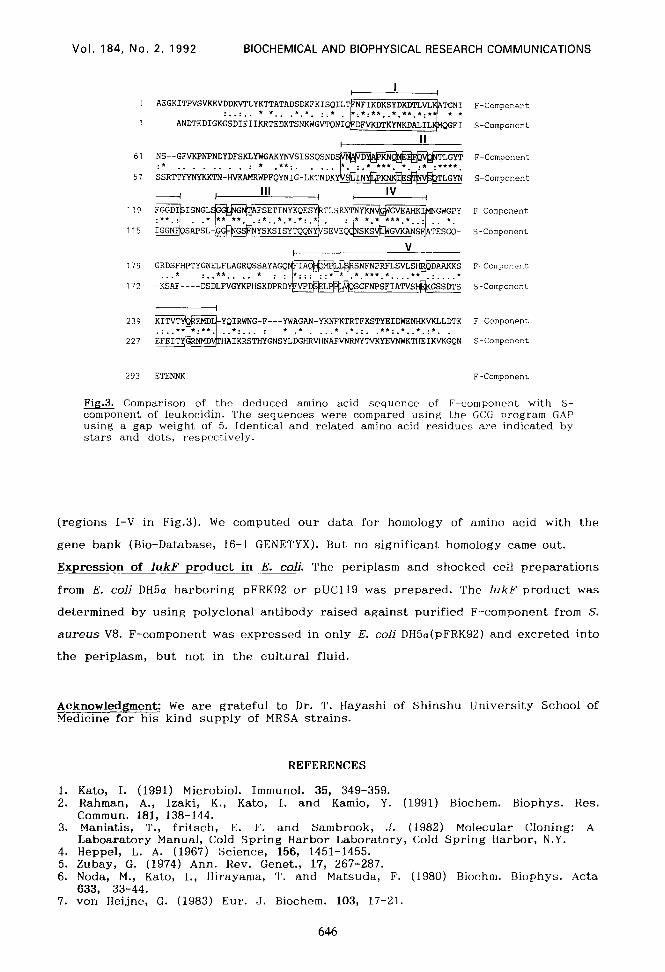
Amino acid homology study. Close investigation of the relationship between S
and F components revealed that homologies are not distributed in all alignments of
the amino acid sequences among them, but instead are localized to specific region
645
Vol. 184, No. 2, 1992 BIOCHEMICAL AND BIOPHYSICAL RESEARCH COMMUNICATIONS
:..:.. * *.. .*.*. :.* ANDTEDIGKGSDIEIIKRTEDKTSNKWGV'IQNI
F-Component
S-Component
61
57
I II
NS--GFVK?IiTNDYDFSiUYWGAKYNVSISSQSND :* . . . . . . ...* .**:. . . . .
SSR'TTYYNYKKTN-HVKAMRWPFQYNIG-LKTNDK
F-Component
S-Component
119
115
F-Component
S-Component
119
172
SNFNPRFLSVLSH@~ *.***.*....** .:....* SGFNPSFIATVS#GSSDS
F-Component
S-Component
239
227
I AI-YKNFKTRTFKSTYEIDWEM-MVKLLDTK
. . . * .*.:. .**:.*..*.:*. . HRVHNAFVNRNYTVKYEVKTHEIKVKGQN
F-Component
S-Component
293 ETENNK F-Component
Fig.J. Comparison of the deduced amino acid sequence of F-component with S- component of leukocidin. The sequences were compared using the GCG program GAP using a gap weight of 5. Identical and related amino acid residues are indicated by stars and dots, respectively.
(regions I-V in Fig.3). We computed our data for homology of amino acid with the
gene bank (Bio-Database, 16-1 GENETYX). But no significant homology came out.
Expression of 1ukF product in E. coli. The periplasm and shocked cell preparations
from E. coli DH5a harboring pFRK92 or pUC119 was prepared. The 1ukF product was
determined by using polyclonal antibody raised against purified F-component from S.
aureus V8. F-component was expressed in only E. coli DHSa(pFRK92) and excreted into
the periplasm, but not in the cultural fluid.
Acknowledgment: We are grateful to Dr. T. Hayashi of Shinshu University School of Medicine for his kind supply of MRSA strains.
REFERENCES
1. Kato, I. (1991) Microbial. Immunol. 35, 349-359. 2. Rahman, A., Izaki, K., Kato, I. and Kamio, Y. (1991) Biochem. Biophys. Res.
Commun. 181, 138-144. 3. Maniatis, T., fritsch, E. F. and Sambrook, J. (1982) Molecular Cloning: A
Laboaratory Manual, Cold Spring Harbor Laboratory, Cold Spring Harbor, N.Y. 4. Heppel, L. A. (1967) Science, 156, 1451-1455. 5. Zubay, G. (1974) Ann. Rev. Genet., 17, 267-287. 6. Noda, M., Kato, I., Hirayama, T. and Matsuda, F. (1980) Biochm. Biophys. Acta
633, 33-44. 7. von Heijne, G. (1983) Eur. J. Biochem. 103, 17-21.
646