Embed Size (px)
Citation preview
Staphylococcus aureus Coordinates Leukocidin Expression andPathogenesis by Sensing Metabolic Fluxes via RpiRc
Divya Balasubramanian,a Elizabeth A. Ohneck,a Jessica Chapman,b Andy Weiss,c Min Kyung Kim,d Tamara Reyes-Robles,a
Judy Zhong,e Lindsey N. Shaw,c Desmond S. Lun,d Beatrix Ueberheide,b,f Bo Shopsin,a,g Victor J. Torresa
Department of Microbiology, New York University School of Medicine, New York, New York, USAa; Proteomics Resource Center, Office of Collaborative Science, New YorkUniversity School of Medicine, New York, New York, USAb; Department of Cell Biology, Microbiology and Molecular Biology, University of South Florida, Tampa, Florida,USAc; Center for Computational and Integrative Biology and Department of Computer Science, Rutgers University, Camden, New Jersey, USAd; Department of PopulationHealth, Division of Biostatistics, New York University School of Medicine, New York, New York, USAe; Department of Biochemistry and Molecular Pharmacology, New YorkUniversity School of Medicine, New York, New York, USAf; Division of Infectious Diseases, Department of Medicine, New York University School of Medicine, New York,New York, USAg
D.B. and E.A.O. contributed equally to this work.
ABSTRACT Staphylococcus aureus is a formidable human pathogen that uses secreted cytolytic factors to injure immune cellsand promote infection of its host. Of these proteins, the bicomponent family of pore-forming leukocidins play critical roles inS. aureus pathogenesis. The regulatory mechanisms governing the expression of these toxins are incompletely defined. In thiswork, we performed a screen to identify transcriptional regulators involved in leukocidin expression in S. aureus strain USA300.We discovered that a metabolic sensor-regulator, RpiRc, is a potent and selective repressor of two leukocidins, LukED andLukSF-PV. Whole-genome transcriptomics, S. aureus exoprotein proteomics, and metabolomic analyses revealed that RpiRcinfluences the expression and production of disparate virulence factors. Additionally, RpiRc altered metabolic fluxes in the tri-carboxylic acid cycle, glycolysis, and amino acid metabolism. Using mutational analyses, we confirmed and extended the obser-vation that RpiRc signals through the accessory gene regulatory (Agr) quorum-sensing system in USA300. Specifically, RpiRcrepresses the rnaIII promoter, resulting in increased repressor of toxins (Rot) levels, which in turn negatively affect leukocidinexpression. Inactivation of rpiRc phenocopied rot deletion and increased S. aureus killing of primary human polymorphonu-clear leukocytes and the pathogenesis of bloodstream infection in vivo. Collectively, our results suggest that S. aureus sensesmetabolic shifts by RpiRc to differentially regulate the expression of leukocidins and to promote invasive disease.
IMPORTANCE The bicomponent pore-forming leukocidins play pivotal roles in the ability of S. aureus to kill multiple host im-mune cells, thus enabling this pathogen to have diverse tissue- and species-tropic effects. While the mechanisms of leukocidin-host receptor interactions have been studied in detail, the regulatory aspects of leukocidin expression are less well characterized.Moreover, the expression of the leukocidins is highly modular in vitro, suggesting the presence of regulators other than theknown Agr, Rot, and S. aureus exoprotein pathways. Here, we describe how RpiRc, a metabolite-sensing transcription factor,mediates the repression of two specific leukocidin genes, lukED and pvl, which in turn has complex effects on the pathogenesis ofS. aureus. Our findings highlight the intricacies of leukocidin regulation by S. aureus and demonstrate the involvement of fac-tors beyond traditional virulence factor regulators.
Received 6 May 2016 Accepted 17 May 2016 Published 21 June 2016
Citation Balasubramanian D, Ohneck EA, Chapman J, Weiss A, Kim MK, Reyes-Robles T, Zhong J, Shaw LN, Lun DS, Ueberheide B, Shopsin B, Torres VJ. 2016. Staphylococcusaureus coordinates leukocidin expression and pathogenesis by sensing metabolic fluxes via RpiRc. mBio 7(3):e00818-16. doi:10.1128/mBio.00818-16.
Editor Steven J. Projan, MedImmune
Copyright © 2016 Balasubramanian et al. This is an open-access article distributed under the terms of the Creative Commons Attribution 4.0 International license.
Address correspondence to Victor J. Torres, [email protected].
Staphylococcus aureus is a daunting human pathogen that causesa range of diseases, from mild skin and soft tissue infections to
debilitating and life-threatening bacteremia. In order to establisha successful infection, S. aureus secretes a variety of immuno-modulatory proteins and virulence factors, a substantial numberof which target leukocytes (1, 2). A complex family of these se-creted proteins is the bicomponent pore-forming leukocidins(here referred to as leukocidins) (3, 4). These toxins consist of twodifferent subunits that are secreted as water-soluble monomers.The binding subunit anchors to leukocytes through proteinaceoushost receptors, recruits the other subunit, oligomerizes, and sub-
sequently forms �-barrel pores within the host plasma membrane,leading to cell death (3, 4). Most S. aureus species carry five differ-ent leukocidins: leukocidin AB (LukAB, also known as LukHG),leukocidin ED (LukED), Panton-Valentine leukocidin (LukSF-PV, also known as PVL), and the gamma hemolysins HlgAB andHlgCB. Unique and central to the action of these leukocidins istheir ability to lyse immune cells in a species-specific and cell-type-specific manner (3, 5). For example, while all of the leukoci-dins can target human polymorphonuclear leukocytes (hPMNs orneutrophils), LukAB, PVL, HlgAB, and HlgCB display greater tro-pism to human neutrophils than to murine neutrophils, whereas
RESEARCH ARTICLE
crossmark
May/June 2016 Volume 7 Issue 3 e00818-16 ® mbio.asm.org 1
on August 16, 2019 by guest
http://mbio.asm
.org/D
ownloaded from
LukED is active against neutrophils from all of the species tested(6, 7). Similarly, the different leukocidins preferentially target spe-cific immune cells on the basis of cell surface receptors (3, 5, 8–11).
Given their potential damaging effects, it is not surprising thatthe expression of leukocidins is tightly regulated by a highly com-plex, multifaceted regulatory network involving two-componentsystems (TCSs), winged helix-turn-helix (HTH) DNA-bindingtranscription factors, and at least one effector small RNA. Vital tothe regulation of leukocidins is the master quorum-sensing regu-latory system in S. aureus, which comprises the accessory generegulator (Agr) proteins (12). At quorum (mimicked by postex-ponentially growing bacteria in vitro), agr produces an autoinduc-ing peptide that, in the process of activating its own synthesis,leads to the production of the effector RNA molecule known asRNAIII (12, 13). RNAIII acts directly and indirectly on many vir-ulence genes, up- or downregulating their translation and proteinproduction (14, 15). Increased production of RNAIII leads to anincrease in the abundance of exoproteins (including toxins andexoenzymes) and a decrease in the abundance of surface proteinsinvolved in immune evasion (12–17). One critical target ofRNAIII is the repressor of toxins (Rot) (14), which directly targetsthe leukocidin promoters (17, 18). In addition to the Agr-Rotregulatory network, the S. aureus exoprotein (Sae) expression TCS(SaeRS-TCS) has been implicated in the regulation of leukocidins(19–24). In contrast to Agr, which is a “self”-sensing system, theSaeRS-TCS responds to host-derived molecules and/or condi-tions likely encountered in vivo, such as pH, osmolarity, andneutrophil-derived proteins and peptides (21, 25, 26). Furtherlinking Agr, Rot, and Sae is the observation that Rot negativelyregulates one of the saeRS promoters, leading to robust repressionof toxins (27). In sum, early in S. aureus infection, the agr locus isinactive, allowing enhanced production of surface molecules andadhesins, while toxins are repressed by Rot. At later stages of in-fection, the Agr system is activated and the expression of secretedvirulence factors such as leukocidins is upregulated by RNAIII-mediated inhibition of Rot translation (7, 17). This circuit is thencompleted by relief of the inhibition of the sae locus, which leadsto the further upregulation of leukocidins (23).
The success of S. aureus as a versatile pathogen relies in part onits ability to infect nearly all sites of the body. This adaptabilitydepends on the ability of S. aureus to fine-tune the production of
its virulence factors by sensing and responding to a diversity ofexternal stimuli, leading to optimal pathogenesis, depending onthe environment it is inhabiting (13, 28, 29). Therefore, identify-ing and characterizing regulators of toxins that sense and respondto disparate environmental conditions may shed light on S. aureuspathogenesis, enabling better therapeutic approaches for S. aureusclearance from specific infection sites.
In this study, we sought to identify transcriptional regulators ofleukocidins in USA300, which is the leading cause of the currentcommunity-associated methicillin-resistant S. aureus epidemic inthe United States (here referred to as USA300). We demonstratedthat inactivation of a metabolic regulatory gene, rpiRc, increasesS. aureus cytotoxicity for human neutrophils. RpiRc belongs to afamily of transcriptional regulators with roles in the regulation ofenzymes involved in sugar catabolism in many bacterial species.Using mutational analyses, we demonstrated that RpiRc differen-tially regulates the expression of leukocidins. Specifically, RpiRc isa potent repressor of the lukED and lukSF-PV loci. Importantly,we found that RpiRc-mediated gene regulation is critical to thefine-tuning of S. aureus pathogenesis.
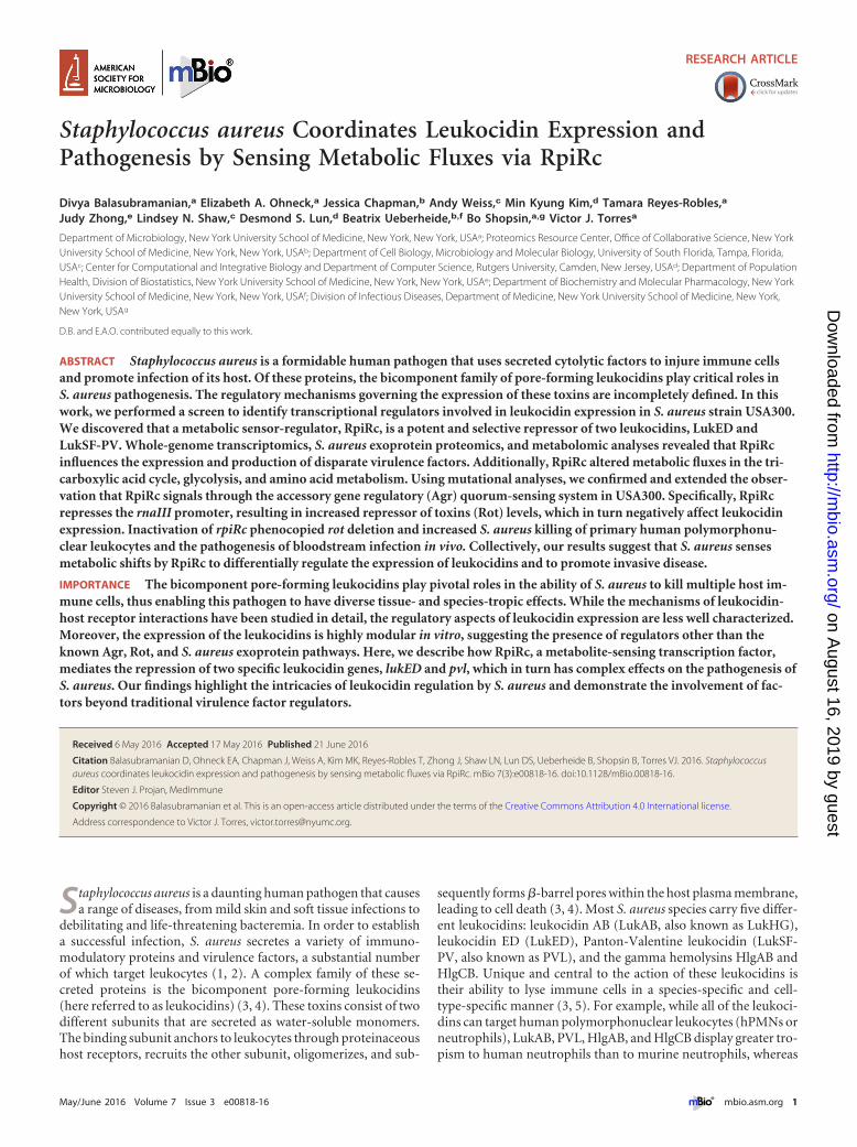
RESULTSThe protein levels and promoter activities of the leukocidinsvary during S. aureus growth in vitro. Postexponential-phaseS. aureus cultures produce and secrete a variety of virulence fac-tors, a major class of which are the leukocidins. Proteomic analy-ses of S. aureus culture filtrates revealed that the comparative levelsof the five leukocidins differed greatly during the postexponential-phase growth of USA300 strain LAC in tryptic soy broth (TSB)(Fig. 1A). Specifically, LukAB and LukSF-PV were highly abun-dant in culture filtrates, while minimal levels of LukED andHlgACB were detected.
To test if these differential protein levels were due to differen-tial regulation of the leukocidin promoters, we used toxin pro-moter sequences fused to luciferase reporter genes and measuredthe respective leukocidin promoter activity (17, 30). As shown inFig. 1B, in the postexponential growth phase, the promoters of thedifferent toxins were active to various degrees, and for the mostpart, expression profiles showed changes in protein levels(Fig. 1A). Among the leukocidins, the lukSF promoter activity andthe corresponding PVL levels were the highest, whereas the lukED
FIG 1 Leukocidin protein abundances and promoter activities vary during growth in vitro. (A) Quantitative mass spectrometry analyses (by LFQ) ofpostexponentially grown USA300 JE2 culture filtrates. (B) Leukocidin promoter activity in TSB measured by luminescence of postexponentially grown USA300harboring plasmids of leukocidin promoter sequences fused to the luciferase gene. The values shown are averages of two independent experiments eachperformed with three colonies of each strain � the standard deviation.
Balasubramanian et al.
2 ® mbio.asm.org May/June 2016 Volume 7 Issue 3 e00818-16
on August 16, 2019 by guest
http://mbio.asm
.org/D
ownloaded from
and hlgA promoters were minimally active, which was reflected intheir respective protein amounts (Fig. 1A and B). Of note, theactivation of the leukocidin promoters is highly dependent on thegrowth medium used (see Fig. S1 in the supplemental material).These results suggest that the differentially secreted leukocidins inUSA300 are due primarily to variance among toxin promoter ac-tivities.
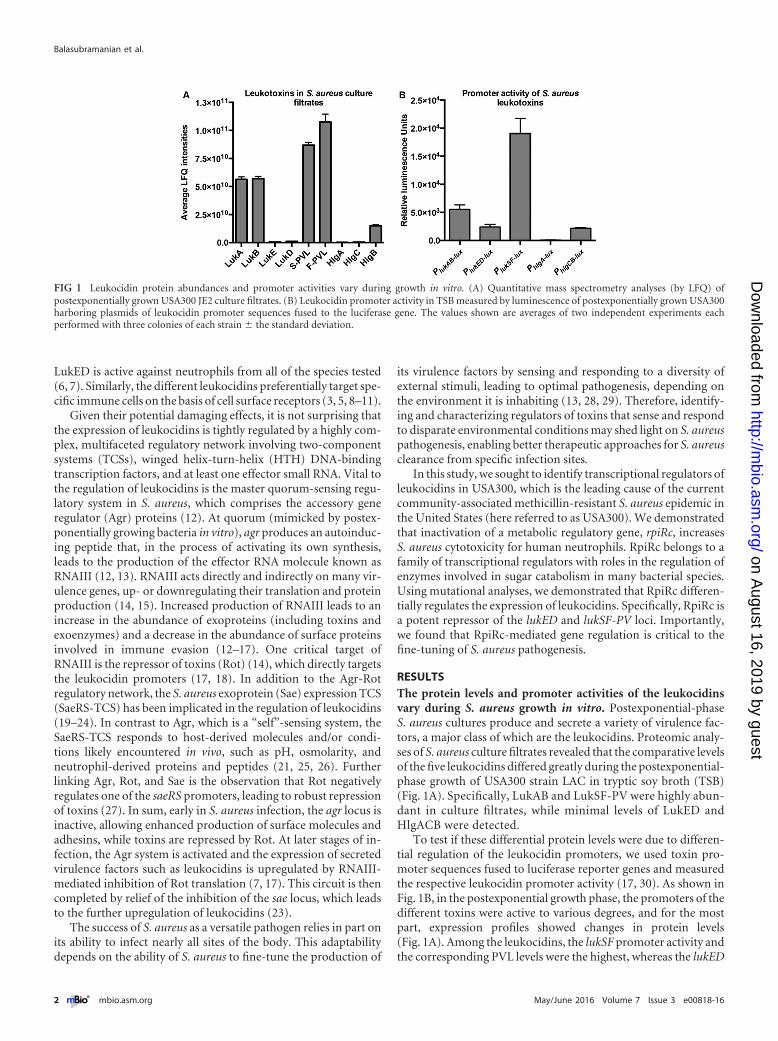
Screen to identify transcriptional regulators that alter S. au-reus cytotoxicity. The differential activity of the leukocidin pro-moters may be due to the action of transcription factors at thesepromoters. In order to screen regulatory genes that may alter leu-kocidin production, we created a “regulator” sublibrary from theUSA300 Nebraska transposon mutant library collection (31). Thissublibrary consisted of 251 mutants of the JE2 strain (a laboratoryversion of USA300 LAC) that included gene products with anypotential regulatory roles, including ones with nucleotide-bindingdomains, putative or confirmed HTH motifs, two-componentregulatory systems, terminators, and antiterminators. For thefunctional annotations of the sublibrary, see Table S1 in the sup-plemental material.
All of the leukocidins are known to target hPMNs. Therefore,
we screened supernatants collected from the 251 mutants for theability to lyse hPMNs. Supernatants collected from the sublibrarygrown for 3 h were used to intoxicate hPMNs isolated from fourhuman donors. We found several mutants that exhibited alteredcytotoxicity for hPMNs. For this study, we chose candidates thatshowed hypercytotoxicity with the goal of identifying novel re-pressors involved in the expression of lukED and hlgACB (seeTable S1 in the supplemental material).
Compared to wild-type-induced cytotoxicity, mutants withchanges in known leukocidin repressors, such as rot and sigB, wereidentified as hypercytotoxic in our screening (see Table S1 in thesupplemental material), validating our assay. Of the mutationsthat caused increased cytotoxicity, 10 regulators led to neutrophilkilling similar to that of a rot::bursa mutant (~2.2-fold increasedcytotoxicity, Fig. 2A). In order to validate these data, we collectedsupernatants at both 3 and 6 h of bacterial growth. Of the 10regulators tested, only rot::bursa and the mutation correspondingto NE1142 (rpiRc::bursa) caused increased cytotoxicity forhPMNs at both time points (Fig. 2B and C). Thus, we decided tofocus on characterizing RpiRc and its repressive effects on S. au-reus virulence.
FIG 2 Identification of transcriptional regulators that enhance S. aureus cytotoxicity. (A) Primary intoxication screening of hPMNs with USA300 JE2supernatants at a final concentration of 5% (vol/vol). Data points represent neutrophil death caused by an individual mutant relative to that caused by wild-type(WT) bacteria (lower dotted line). Supernatants from each mutant were tested on hPMNs from four donors, and cell viability was measured with CellTitermetabolic dye. A 2.2-fold cutoff was used to identify candidate mutants for further screening (upper dotted line). The data point indicated by the trianglerepresents the cytotoxicity of a rot::bursa mutant. (B and C) Validation intoxication screening of hPMNs isolated from two donors with supernatants from selectS. aureus mutants. Wild-type and mutant bacteria were grown for 3 (B) and 6 (C) h postinoculation. Error bars indicate the standard error of the mean.
S. aureus Senses Metabolic Fluxes via RpiRc
May/June 2016 Volume 7 Issue 3 e00818-16 ® mbio.asm.org 3
on August 16, 2019 by guest
http://mbio.asm
.org/D
ownloaded from
RpiRc controls the production of secreted virulence factorsin S. aureus. The RpiR class of proteins has been found in manydifferent bacterial species, including Gram-negative Escherichiacoli and Pseudomonas putida and Gram-positive Bacillus subtilis(32–34). This family of proteins is traditionally thought to includetranscriptional regulators involved in sugar metabolism, althoughtheir regulons, binding sites, and exact functions are poorly char-acterized. The RpiR prototype contains an N-terminal HTHDNA-binding motif and a C-terminal sugar isomerase-sensing(SIS) domain (33, 35). A recent study identified three RpiR ho-mologs in S. aureus, namely, rpiRa, rpiRb, and rpiRc (36). Muta-tions of these genes in methicillin-susceptible S. aureus strainUAMS-1 implicated these regulators in the control of the pentosephosphate pathway (PPP). Interestingly, inactivation of rpiRc alsoled to increased RNAIII synthesis and hemolysis and reduced bio-film formation (36).
To further evaluate the interactions among RNAIII, virulence,and RpiRc in USA300, we first examined the exoproteomes of
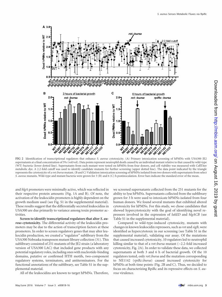
wild-type and rpiRc::bursa mutant strains. We observed that var-ious secreted proteins were differentially produced by the strains.The most dramatic difference in abundance was observed in pro-tein bands corresponding to the size of leukocidins (~35 kDa).There were notably higher levels of proteins in that size range inthe JE2 rpiRc::bursa culture filtrate than in that of wild-type JE2(Fig. 3A), whereas mutations in the other rpiR genes had no effecton these toxins.
To demonstrate that the observed phenotype of the JE2 rpiRc::bursa mutant was due to the transposon-mediated disruptionof rpiRc, the mutated allele was transduced into USA300 LACstrain AH1263 (referred to as LAC in this report), anothererythromycin-sensitive LAC clone (37). Compared to the wild-type strain, the isogenic LAC rpiRc::bursa mutant also exhibitedincreased production of proteins that run at the size of leukocidins(Fig. 3B). Importantly, this phenotype was fully complemented bythe insertion of rpiRc in single copy at the SaPI1 attachment site(referred to as the rpiRc� strain in this study) (Fig. 3B).
FIG 3 RpiRc is a potent regulator of S. aureus secreted proteins. (A) Exoprotein profiles of USA300 JE2 wild-type (WT) and rpiR mutant bacteria, as assessedby Coomassie staining. The asterisk indicates the approximate leukocidin protein size. (B) Exoprotein profile of USA300 LAC wild-type, rpiRc, and rpiRc�
isogenic strains. (C) Heat map of LAC wild-type and rpiRc mutant secretomes as assessed by mass spectrometry. (D to F) Levels of exoenzymes (D), surface andimmunomodulatory proteins (E), and cytotoxins (F) � the standard deviation in exoproteomes of wild-type versus rpiRc mutant USA300 LAC.
Balasubramanian et al.
4 ® mbio.asm.org May/June 2016 Volume 7 Issue 3 e00818-16
on August 16, 2019 by guest
http://mbio.asm
.org/D
ownloaded from
To gain better insight into the effects of RpiRc on exoproteinproduction in USA300, we analyzed the in vitro culture filtrates bymass spectrometry. Exoproteins were collected from three inde-pendent colonies of wild-type LAC and the rpiRc mutant straingrown to postexponential phase, and the protein profiles wereanalyzed by label-free quantitative mass spectrometry. We ob-served tremendous reproducibility among the biological repli-cates, as demonstrated by the clustering of the proteins in the heatmap (Fig. 3C). We identified 96 (2-fold up or down in abundance)of the total 483 secreted proteins of USA300 to be altered in themutant, of which 31 were positively impacted and 65 were nega-tively impacted in the rpiRc mutant (see Table S2 in the supple-mental material). Among these proteins, we found increased pro-duction of most exoenzymes, such as proteases (4- to 6-foldincrease), while a few exoenzymes, such as coagulase, were lowerin abundance (Fig. 3D). Proteins involved in immune evasion andadhesion (including superantigens, Sbi, protein A, and ClfB)were, for the most part, lower in abundance in the rpiRc mutant(Fig. 3E). We found a stark increase in the production of cytotox-ins (leukocidins and phenol-soluble modulins) in the rpiRc mu-tant (Fig. 3F). Interestingly, RpiRc seems to differentially regulatethe production of leukocidins, as LukSF-PV and LukED were no-tably upregulated in the rpiRc mutant strain, whereas other leuko-
cidins, such as gamma hemolysin and LukAB, were minimallyimpacted in this strain (Fig. 3F).
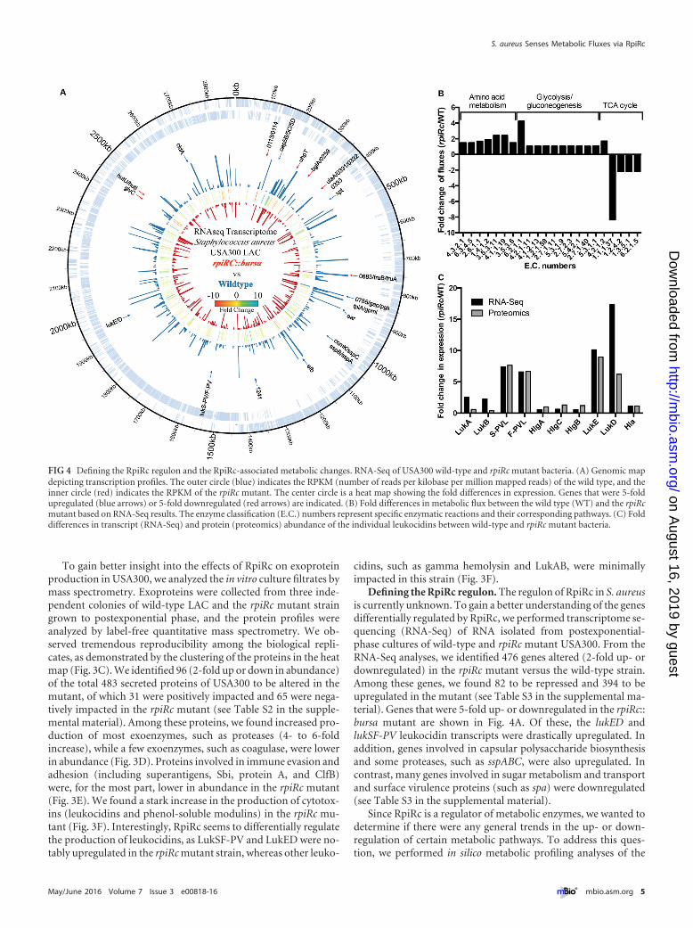
Defining the RpiRc regulon. The regulon of RpiRc in S. aureusis currently unknown. To gain a better understanding of the genesdifferentially regulated by RpiRc, we performed transcriptome se-quencing (RNA-Seq) of RNA isolated from postexponential-phase cultures of wild-type and rpiRc mutant USA300. From theRNA-Seq analyses, we identified 476 genes altered (2-fold up- ordownregulated) in the rpiRc mutant versus the wild-type strain.Among these genes, we found 82 to be repressed and 394 to beupregulated in the mutant (see Table S3 in the supplemental ma-terial). Genes that were 5-fold up- or downregulated in the rpiRc::bursa mutant are shown in Fig. 4A. Of these, the lukED andlukSF-PV leukocidin transcripts were drastically upregulated. Inaddition, genes involved in capsular polysaccharide biosynthesisand some proteases, such as sspABC, were also upregulated. Incontrast, many genes involved in sugar metabolism and transportand surface virulence proteins (such as spa) were downregulated(see Table S3 in the supplemental material).
Since RpiRc is a regulator of metabolic enzymes, we wanted todetermine if there were any general trends in the up- or down-regulation of certain metabolic pathways. To address this ques-tion, we performed in silico metabolic profiling analyses of the
FIG 4 Defining the RpiRc regulon and the RpiRc-associated metabolic changes. RNA-Seq of USA300 wild-type and rpiRc mutant bacteria. (A) Genomic mapdepicting transcription profiles. The outer circle (blue) indicates the RPKM (number of reads per kilobase per million mapped reads) of the wild type, and theinner circle (red) indicates the RPKM of the rpiRc mutant. The center circle is a heat map showing the fold differences in expression. Genes that were 5-foldupregulated (blue arrows) or 5-fold downregulated (red arrows) are indicated. (B) Fold differences in metabolic flux between the wild type (WT) and the rpiRcmutant based on RNA-Seq results. The enzyme classification (E.C.) numbers represent specific enzymatic reactions and their corresponding pathways. (C) Folddifferences in transcript (RNA-Seq) and protein (proteomics) abundance of the individual leukocidins between wild-type and rpiRc mutant bacteria.
S. aureus Senses Metabolic Fluxes via RpiRc
May/June 2016 Volume 7 Issue 3 e00818-16 ® mbio.asm.org 5
on August 16, 2019 by guest
http://mbio.asm
.org/D
ownloaded from
transcriptomic data (38–40). As shown in Fig. 4B (see Table S4 inthe supplemental material), we observed that three clusters ofmetabolic pathways were differentially activated in wild-type andrpiRc mutant bacteria. First, and notably, we observed a signatureof decreased tricarboxylic acid (TCA) cycle activity in the rpiRcmutant. Second, and in contrast, several amino acid metabolicpathways were more active in the mutant than in the wild type.Third, we observed a slight but consistent activation of the glyco-lysis and gluconeogenesis pathways. While the fold differences inmetabolic fluxes in glycolysis/gluconeogenesis between wild-typeand rpiRc mutant bacteria are only moderate, we nevertheless ob-served that many genes in these two pathways were upregulated inthe rpiRc mutant (Fig. 4B; see Table S4 in the supplemental mate-rial). Of note, we observed no growth defect in vitro when com-paring the wild-type strain and the isogenic strain lacking rpiRc(see Fig. S2 in the supplemental material), consistent with obser-vations reported previously (36). Taken together, mutation ofrpiRc seems to lead to decreased activity of the TCA cycle and anincrease in glycolysis/gluconeogenesis and certain amino acid bio-synthetic pathways. In UAMS-1, RpiRc was observed to increasethe expression and activity of some PPP genes (36). In our analy-ses, while there were no notable PPP shifts, we observed potentialpositive regulatory roles of RpiRc in the TCA cycle.
The RNA-Seq analyses also revealed differences in several vir-ulence factors between the wild type and the mutant, the mostdramatic of which was the differential expression of some leuko-cidins (Fig. 4A; see Table S3 in the supplemental material). Spe-cifically, and consistent with the proteomic analyses, we observeda striking increase in the expression of lukSF-PV and lukED in themutant (Fig. 4C), further validating that RpiRc is involved in theexpression of these specific leukocidin-coding genes.
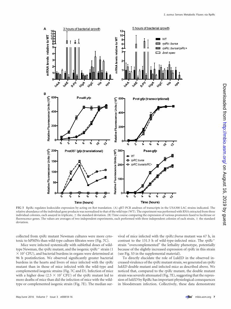
RpiRc represses leukocidin expression by acting on Rottranslation. We next validated the RNA-Seq data on the leukoci-dins by quantitative real-time PCR (qRT-PCR). Consistent withthe RNA-Seq and proteomic analysis results, levels of lukS-PV andlukE mRNAs were ~6- to 7-fold higher in the rpiRc mutant than inboth the wild-type and rpiRc� strains (Fig. 5A). In addition to theleukocidins, we also monitored the expression of alpha-toxin (en-coded by hla), another important virulence factor in S. aureus(41). The hla gene was also found to be derepressed in the USA300rpiRc mutant (Fig. 5A), data in line with the observation that de-letion of rpiRc from strain UAMS-1 produces increased hemolyticactivity (36).
To further dissect the signaling pathway(s) downstream ofRpiRc, we measured the promoter activities of the master regula-tors of toxin gene expression, rnaIII, sae, and rot, in the rpiRcmutant. Consistent with the finding that RNAIII transcript levelsare elevated in an rpiRc mutant of strain UAMS-1 (36), we ob-served 5-fold greater promoter activity of rnaIII in the USA300rpiRc mutant than in the wild-type and rpiRc� strains (Fig. 5B).Moreover, we observed that while a rot transcriptional fusion wasunaffected in the USA300 rpiRc mutant, a rot translational fusionwas severely repressed in this background (Fig. 5B), consistentwith the RNAIII-mediated translational regulation of rot (14, 42,43). Lastly, consistent with decreased levels of Rot, we observed~2-fold greater sae promoter activity in the rpiRc mutant than inthe wild-type and rpiRc� strains (Fig. 5B).
We noticed that the transcriptional profiles of the leukocidinsin the rpiRc mutant closely resembled that of the USA300 rot mu-tant (7, 17, 44), suggesting that RpiRc phenocopies the transcrip-
tional effects of Rot (Fig. 5A). To further evaluate Rot functional-ity in the rpiRc mutant strain, we monitored the expression of agene positively regulated by Rot, spa, which encodes the immunemodulator protein A (45). Both RNA-Seq and qRT-PCR showedthat spa levels were significantly lower in both the rot and rpiRcmutants (see Table S3 in the supplemental material; Fig. 5A). Col-lectively, these data suggest that in wild-type USA300, RpiRc re-presses the agr P3 promoter encoding RNAIII in a growth phase-dependent manner, which in turn leads to relief of the inhibitionof rot translation and decreased sae expression, subsequently lead-ing to the repression of specific leukocidins and other virulencefactors.
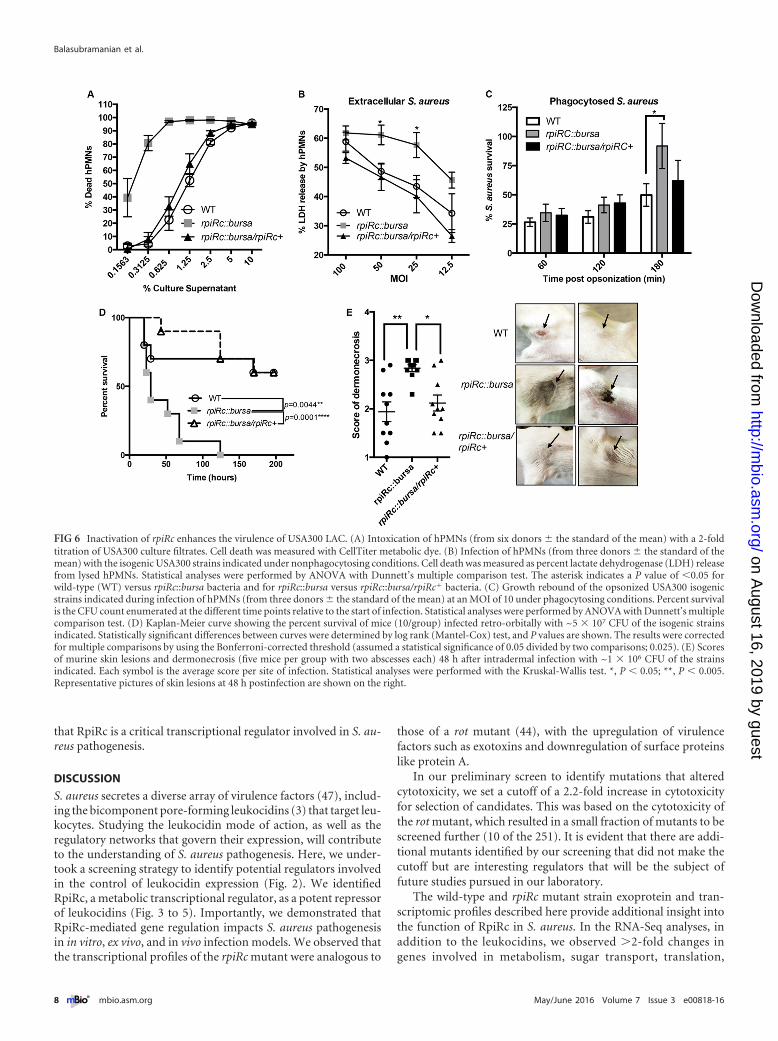
Mutation of rpiRc enhances S. aureus USA300-mediated vir-ulence. Next we elucidated the contribution of RpiRc to USA300-hPMN interactions, as neutrophils are innate immune cells criti-cal for the containment of S. aureus (46). Consistent with theincreased expression and production of secreted leukocidins de-scribed above, supernatants from the isogenic USA300 rpiRc mu-tant were more cytotoxic to hPMNs than were supernatants fromwild-type USA300, a phenotype fully complemented in the rpiRc�
strain (Fig. 6A).To further explore the role of RpiRc during USA300-hPMN
interactions, we infected hPMNs with wild-type and rpiRc mutantbacteria at different multiplicities of infection and monitoredhPMN lysis. As shown in Fig. 6B, the rpiRc mutant also causedgreater lysis of hPMNs than the wild-type and rpiRc� isogenicstrains did. Lastly, we measured the survival of bacteria after in-ducing their phagocytosis by hPMNs. These studies revealed thatthe phagocytosed rpiRc mutant was better able to recover follow-ing ingestion by neutrophils than were the wild-type and rpiRc�
strains (Fig. 6C). Thus, RpiRc in a wild-type scenario repressesS. aureus virulence factors that enhance the killing of hPMNs.
We then tested the role of rpiRc in an in vivo murine bacteremiamodel. Mice were infected intravenously with the wild-typeUSA300, rpiRc::bursa, and rpiRc::bursa/rpiRc� strains, and theirsurvival was monitored over time. Infection with the mutantstrain led to significantly more mouse deaths than the wild-typeand rpiRc� strains, strongly supporting our ex vivo data showingthat RpiRc has critical roles in S. aureus pathogenesis (Fig. 6D).
To investigate whether RpiRc is important in other murinemodels, we also infected mice intradermally with the wild-type,rpiRc::bursa, and rpiRc� strains and monitored abscess formationand dermonecrosis postinfection. We observed that infection withthe rpiRc mutant led to significantly higher dermonecrotic lesionscores than did infection with the wild-type and rpiRc� strains(Fig. 6E). The increased virulence of the rpiRc::bursa strain inthese models is likely to be due to the derepression of Hla (Fig. 3 to5), as this toxin is critical for the pathogenesis of USA300 (41).
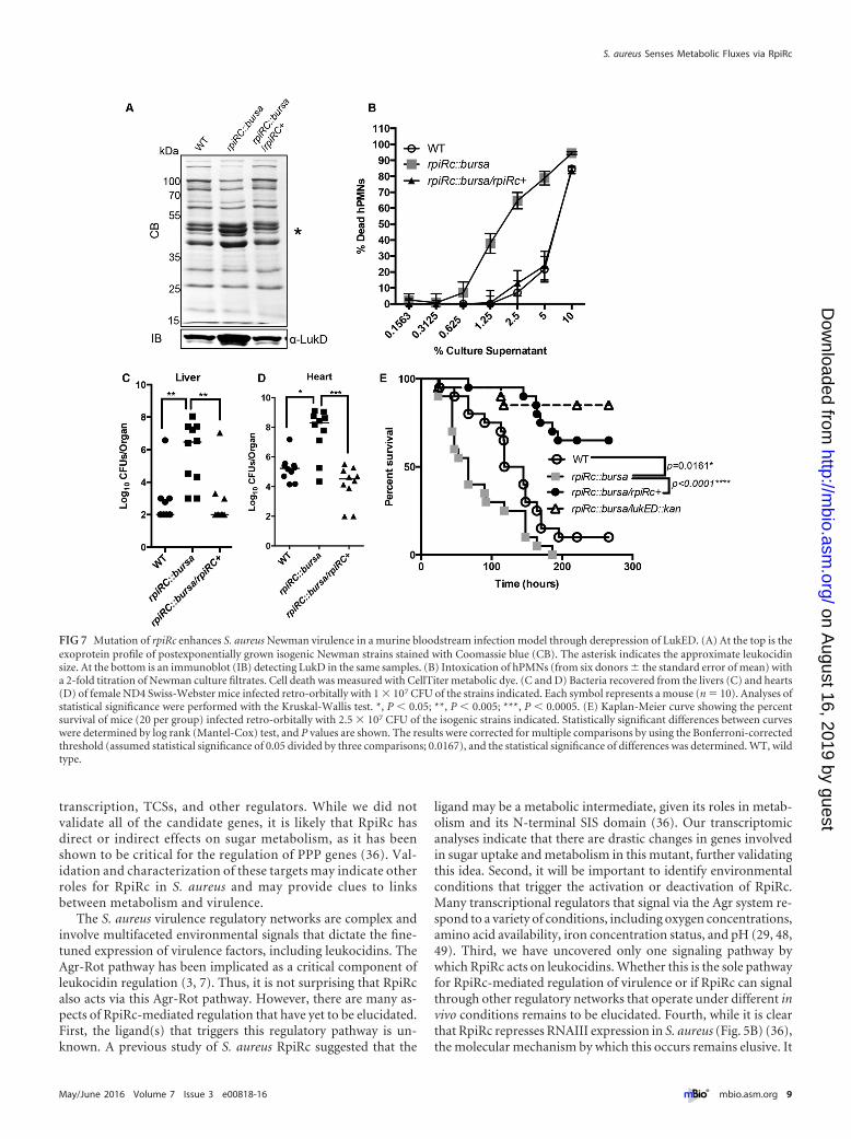
Mutation of rpiRc enhances S. aureus virulence in a murinebloodstream infection model primarily through derepressionof LukED. To understand the role of RpiRc in the virulence ofnon-USA300 strains, we created rpiRc mutant and complementedisogenic versions of S. aureus Newman, a strain that has beenwidely used to study the role of LukED in murine bacteremiamodels (6, 7, 9). Analyses of exoproteins produced by these iso-genic strains revealed that, as in USA300, mutation of rpiRc inNewman results in greater production of leukocidins (Fig. 7A,top), including increased levels of LukD (Fig. 7A, bottom) than inthe wild-type and complemented strains. Moreover, supernatants
Balasubramanian et al.
6 ® mbio.asm.org May/June 2016 Volume 7 Issue 3 e00818-16
on August 16, 2019 by guest
http://mbio.asm
.org/D
ownloaded from
collected from rpiRc mutant Newman cultures were more cyto-toxic to hPMNs than wild-type culture filtrates were (Fig. 7C).
Mice were infected systemically with sublethal doses of wild-type Newman, the rpiRc mutant, and the isogenic rpiRc� strain (1� 107 CFU), and bacterial burdens in organs were determined at96 h postinfection. We observed significantly greater bacterialburdens in the hearts and livers of mice infected with the rpiRcmutant than in those of mice infected with the wild-type andcomplemented isogenic strains (Fig. 7C and D). Infection of micewith a higher dose (2.5 � 107 CFU) of the rpiRc mutant led tomore deaths of mice than did the infection of mice with the wild-type or complemented isogenic strain (Fig. 7E). The median sur-
vival of mice infected with the rpiRc::bursa mutant was 67 h, incontrast to the 131.5 h of wild-type-infected mice. The rpiRc�
strain “overcomplemented” the lethality phenotype, potentiallybecause of the slightly increased expression of rpiRc in this strain(see Fig. S3 in the supplemental material).
To directly elucidate the role of LukED in the observed in-creased virulence of the rpiRc mutant strain, we generated an rpiRclukED double mutant and infected mice as described above. Wenoticed that, compared to the rpiRc mutant, the double mutantstrain was severely attenuated (Fig. 7E), suggesting that the repres-sion of lukED by RpiRc has important physiological consequencesin bloodstream infection. Collectively, these data demonstrate
FIG 5 RpiRc regulates leukocidin expression by acting on Rot translation. (A) qRT-PCR analyses of transcripts in the USA300 LAC strains indicated. Therelative abundance of the individual gene products was normalized to that of the wild type (WT). The experiment was performed with RNA extracted from threeindividual colonies, each assayed in triplicate, � the standard deviation. (B) Time course comparing the expression of various promoters fused to luciferase orfluorescence genes. The values are averages of two independent experiments, each performed with three independent colonies of each strain, � the standarddeviation.
S. aureus Senses Metabolic Fluxes via RpiRc
May/June 2016 Volume 7 Issue 3 e00818-16 ® mbio.asm.org 7
on August 16, 2019 by guest
http://mbio.asm
.org/D
ownloaded from
that RpiRc is a critical transcriptional regulator involved in S. au-reus pathogenesis.
DISCUSSION
S. aureus secretes a diverse array of virulence factors (47), includ-ing the bicomponent pore-forming leukocidins (3) that target leu-kocytes. Studying the leukocidin mode of action, as well as theregulatory networks that govern their expression, will contributeto the understanding of S. aureus pathogenesis. Here, we under-took a screening strategy to identify potential regulators involvedin the control of leukocidin expression (Fig. 2). We identifiedRpiRc, a metabolic transcriptional regulator, as a potent repressorof leukocidins (Fig. 3 to 5). Importantly, we demonstrated thatRpiRc-mediated gene regulation impacts S. aureus pathogenesisin in vitro, ex vivo, and in vivo infection models. We observed thatthe transcriptional profiles of the rpiRc mutant were analogous to
those of a rot mutant (44), with the upregulation of virulencefactors such as exotoxins and downregulation of surface proteinslike protein A.
In our preliminary screen to identify mutations that alteredcytotoxicity, we set a cutoff of a 2.2-fold increase in cytotoxicityfor selection of candidates. This was based on the cytotoxicity ofthe rot mutant, which resulted in a small fraction of mutants to bescreened further (10 of the 251). It is evident that there are addi-tional mutants identified by our screening that did not make thecutoff but are interesting regulators that will be the subject offuture studies pursued in our laboratory.
The wild-type and rpiRc mutant strain exoprotein and tran-scriptomic profiles described here provide additional insight intothe function of RpiRc in S. aureus. In the RNA-Seq analyses, inaddition to the leukocidins, we observed �2-fold changes ingenes involved in metabolism, sugar transport, translation,
FIG 6 Inactivation of rpiRc enhances the virulence of USA300 LAC. (A) Intoxication of hPMNs (from six donors � the standard of the mean) with a 2-foldtitration of USA300 culture filtrates. Cell death was measured with CellTiter metabolic dye. (B) Infection of hPMNs (from three donors � the standard of themean) with the isogenic USA300 strains indicated under nonphagocytosing conditions. Cell death was measured as percent lactate dehydrogenase (LDH) releasefrom lysed hPMNs. Statistical analyses were performed by ANOVA with Dunnett’s multiple comparison test. The asterisk indicates a P value of �0.05 forwild-type (WT) versus rpiRc::bursa bacteria and for rpiRc::bursa versus rpiRc::bursa/rpiRc� bacteria. (C) Growth rebound of the opsonized USA300 isogenicstrains indicated during infection of hPMNs (from three donors � the standard of the mean) at an MOI of 10 under phagocytosing conditions. Percent survivalis the CFU count enumerated at the different time points relative to the start of infection. Statistical analyses were performed by ANOVA with Dunnett’s multiplecomparison test. (D) Kaplan-Meier curve showing the percent survival of mice (10/group) infected retro-orbitally with ~5 � 107 CFU of the isogenic strainsindicated. Statistically significant differences between curves were determined by log rank (Mantel-Cox) test, and P values are shown. The results were correctedfor multiple comparisons by using the Bonferroni-corrected threshold (assumed a statistical significance of 0.05 divided by two comparisons; 0.025). (E) Scoresof murine skin lesions and dermonecrosis (five mice per group with two abscesses each) 48 h after intradermal infection with ~1 � 106 CFU of the strainsindicated. Each symbol is the average score per site of infection. Statistical analyses were performed with the Kruskal-Wallis test. *, P � 0.05; **, P � 0.005.Representative pictures of skin lesions at 48 h postinfection are shown on the right.
Balasubramanian et al.
8 ® mbio.asm.org May/June 2016 Volume 7 Issue 3 e00818-16
on August 16, 2019 by guest
http://mbio.asm
.org/D
ownloaded from
transcription, TCSs, and other regulators. While we did notvalidate all of the candidate genes, it is likely that RpiRc hasdirect or indirect effects on sugar metabolism, as it has beenshown to be critical for the regulation of PPP genes (36). Val-idation and characterization of these targets may indicate otherroles for RpiRc in S. aureus and may provide clues to linksbetween metabolism and virulence.
The S. aureus virulence regulatory networks are complex andinvolve multifaceted environmental signals that dictate the fine-tuned expression of virulence factors, including leukocidins. TheAgr-Rot pathway has been implicated as a critical component ofleukocidin regulation (3, 7). Thus, it is not surprising that RpiRcalso acts via this Agr-Rot pathway. However, there are many as-pects of RpiRc-mediated regulation that have yet to be elucidated.First, the ligand(s) that triggers this regulatory pathway is un-known. A previous study of S. aureus RpiRc suggested that the
ligand may be a metabolic intermediate, given its roles in metab-olism and its N-terminal SIS domain (36). Our transcriptomicanalyses indicate that there are drastic changes in genes involvedin sugar uptake and metabolism in this mutant, further validatingthis idea. Second, it will be important to identify environmentalconditions that trigger the activation or deactivation of RpiRc.Many transcriptional regulators that signal via the Agr system re-spond to a variety of conditions, including oxygen concentrations,amino acid availability, iron concentration status, and pH (29, 48,49). Third, we have uncovered only one signaling pathway bywhich RpiRc acts on leukocidins. Whether this is the sole pathwayfor RpiRc-mediated regulation of virulence or if RpiRc can signalthrough other regulatory networks that operate under different invivo conditions remains to be elucidated. Fourth, while it is clearthat RpiRc represses RNAIII expression in S. aureus (Fig. 5B) (36),the molecular mechanism by which this occurs remains elusive. It
FIG 7 Mutation of rpiRc enhances S. aureus Newman virulence in a murine bloodstream infection model through derepression of LukED. (A) At the top is theexoprotein profile of postexponentially grown isogenic Newman strains stained with Coomassie blue (CB). The asterisk indicates the approximate leukocidinsize. At the bottom is an immunoblot (IB) detecting LukD in the same samples. (B) Intoxication of hPMNs (from six donors � the standard error of mean) witha 2-fold titration of Newman culture filtrates. Cell death was measured with CellTiter metabolic dye. (C and D) Bacteria recovered from the livers (C) and hearts(D) of female ND4 Swiss-Webster mice infected retro-orbitally with 1 � 107 CFU of the strains indicated. Each symbol represents a mouse (n � 10). Analyses ofstatistical significance were performed with the Kruskal-Wallis test. *, P � 0.05; **, P � 0.005; ***, P � 0.0005. (E) Kaplan-Meier curve showing the percentsurvival of mice (20 per group) infected retro-orbitally with 2.5 � 107 CFU of the isogenic strains indicated. Statistically significant differences between curveswere determined by log rank (Mantel-Cox) test, and P values are shown. The results were corrected for multiple comparisons by using the Bonferroni-correctedthreshold (assumed statistical significance of 0.05 divided by three comparisons; 0.0167), and the statistical significance of differences was determined. WT, wildtype.
S. aureus Senses Metabolic Fluxes via RpiRc
May/June 2016 Volume 7 Issue 3 e00818-16 ® mbio.asm.org 9
on August 16, 2019 by guest
http://mbio.asm
.org/D
ownloaded from
is imperative to determine how RpiRc interacts with and regulatestarget promoters to determine direct versus indirect modes ofgene regulation.
In order to be a successful pathogen, S. aureus has to adapt tothe harsh environments encountered within the host. In recentyears, interest in S. aureus metabolism has reemerged, as distinctlinks between metabolism and pathogenesis are increasingly iden-tified (50, 51). In S. aureus, several transcriptional factors sensemetabolites and regulate virulence in response to these signals.Examples of metabolite-sensing regulators in S. aureus are CcpAand CcpE. These carbon catabolite repressors sense glycolytic in-termediates, and in addition to regulating uptake of nutrients suchas glucose, they also regulate the synthesis of virulence factors (52,53). Another well-studied nutrient sensor is the CodY transcrip-tional regulator, which responds to branched-chain amino acidsand GTP in S. aureus (54). In response to nutrient availability,CodY regulates the synthesis of alpha-toxin and certain adhesinsvia the agr system (55). Importantly, inactivation of many of theseregulators (including the ones cited above) alters S. aureus patho-genesis, supporting the notion that metabolism is intimatelylinked with the pathogenic lifestyle of this bacterium. The datapresented here support the idea that RpiRc is a critical transcrip-tional regulator that may respond to various environmental con-ditions to increase, decrease, or fine-tune S. aureus virulence.
MATERIALS AND METHODSEthics statement. Buffy coats were obtained from anonymous donorswith informed consent from the New York Blood Center. Because all ofthe samples were collected anonymously prior to their delivery, the NewYork University Langone Medical Center (NYULMC) Institutional Re-view Board determined that our study was exempt from further ethicsapproval requirements. All animal experiments were reviewed and ap-proved by the Institutional Animal Care and Use Committee ofNYULMC. All experiments were performed according to NIH guidelines,the Animal Welfare Act, and U.S. federal law.
Bacterial cultures and growth conditions. S. aureus strains were rou-tinely grown at 37°C on tryptic soy agar (TSA) or in TSB with antibioticsupplementation as specified. E. coli DH5� was used for cloning andpropagation of plasmids. E. coli bacteria were grown in Luria-Bertanibroth with appropriate antibiotics. Liquid cultures were grown in 5 ml ofgrowth medium in 15-ml tubes incubated at a 45° angle with shaking at180 rpm. For all experiments involving the growth of S. aureus bacteria, a1:100 dilution of overnight cultures was subcultured into fresh medium.
Construction of bacterial strains and plasmids. For all of the strains,plasmids, and oligonucleotides used in this study, see Table S5 in thesupplemental material. The LAC rpiRc::bursa mutant strain was generatedby phage transduction of the JE2 rpiRc::bursa (NE1142) strain of the Ne-braska Transposon Mutant Library with phage �80 into wild-type,erythromycin-sensitive LAC clone AH1263 (37). Complementation ofrpiRc on the chromosome was performed with suicide plasmid pJC1306(kindly provided by John Chen), which is used to stably integrate DNAinto the SaP1 site, resulting in a single-copy chromosomal insertion (56).The construction of these strains is described in Text S1 in the supplemen-tal material. A LAC rot::spec mutant strain was generated as describedpreviously (17). The mutation was then transduced into erythromycin-sensitive LAC. The Newman lukED::kan rpiRc::bursa mutant strain wasgenerated by transducing lukED::kan from strain Newman (as describedin reference 7) with rpiRc::bursa from LAC (VJT42.71).
Culture conditions for cytotoxicity screening. The regulator mutantlibrary was plated in a 96-well plate format on TSA. Overnight cultures ofthe mutants were grown in TSB on 2 independent days in 96-well round-bottom plates, subcultured into 96-well round-bottom plates, and grownfor 3 h. The plates were centrifuged at 4,000 rpm, and the supernatants
were collected into two different 96-well tissue culture-treated plates togive a 5% final concentration during intoxication and frozen. Superna-tants from this regulator mutant library were then used to intoxicatehPMNs isolated from four different donors. In the validation screening,10 mutants from the primary screening were grown as described abovebut for 3 and 6 h, and cytotoxicity to hPMNs from two donors was as-sayed.
Cytotoxicity assay. hPMNs were isolated as described in reference 57,and cytotoxicity assays were performed as described previously (17, 57).Briefly, 2 � 105 hPMNs were added to a final volume of 100 �l/well ofRPMI (Gibco) supplemented with 10 mM HEPES. Cells were intoxicatedfor 1 h at 37°C in 5% CO2. Ten microliters of CellTiter 96 Aqueous OneSolution (CellTiter; Promega) was added, and the mixture was incubatedat 37°C in 5% CO2 for 2 h. hPMN viability was assessed with a PerkinEl-mer EnVision 2103 Multilabel Reader.
Exoprotein isolation, Coomassie staining, and immunoblotting.The proteins in the culture supernatants of bacteria grown for 5 h wereprecipitated and analyzed as described in reference 17. Immunoblottingwas performed with polyclonal antibodies against LukD (1:7,500), whichwere detected with a fluorescent Alexa Fluor 680-conjugated anti-rabbitantibody (1:25,000).
Quantitative mass spectrometry analysis. Wild-type and rpiRc::bursamutant strain exoprotein isolates were analyzed in triplicate. Label-freequantification (LFQ) intensities obtained by mass spectrometry were log2
transformed, and all missing values were replaced with values from thenormal distribution. Z scores were calculated for all values, hierarchicalclustering was performed, and heat maps were generated. For detailedinformation, see Text S1 in the supplemental material.
RNA isolation, RNA-Seq, and data analyses. RNA isolation and sam-ple preparation for RNA-Seq were performed as previously outlined byCarroll et al. (58). For detailed information on the RNA-Seq methodsused, see Text S1 in the supplemental material.
qRT-PCR. A 10- to 100-ng sample of total RNA (depending on theabundance of the target gene) purified as described above was used toperform qRT-PCR in a one-step reaction with Reverse Transcriptase Mas-termix (QuantiTect) and SYBR green master mix (Qiagen) in a 7300 Real-Time PCR system (Applied Biosystems). For the primers used to detectthe specific mRNAs, see Table S5 in the supplemental material. Analysiswas performed by the 2CT method, and target genes in each strainwere normalized to the corresponding genes in wild-type cells.
Reporter assays. Strains containing reporter plasmids were grownovernight in different media containing 10 �g/ml chloramphenicol (toretain reporter plasmids) in 96-well round-bottom plates. Following sub-culture in fresh medium in 96-well black, flat-bottom plates (Corning),fluorescence and luminescence were measured with a PerkinElmer EnVi-sion 2103 Multilabel Reader immediately after subculture (T0) and every2 h for 10 to 12 h.
Computational metabolic flux prediction. A computational methodcalled E-Flux2 was used to analyze the difference in intracellular metabolicfluxes between the wild-type and rpiRc::bursa mutant strains (for details,see Text S1 in the supplemental material). For a list of the metabolicpathways with significant changes and the complete list, see Table S4 inthe supplemental material.
Ex vivo assays of S. aureus-hPMN interactions. Infection of hPMNswith extracellular S. aureus was performed as described previously (30),with RPMI supplemented with 10 mM HEPES and 5% human serumalbumin at an hPMN concentration of 2 � 105/well at 37°C in 5% CO2 for1 h. The neutrophil-mediated S. aureus killing assay was performed asdescribed previously (30). For details, see Text S1 in the supplementalmaterial.
Murine models of systemic and skin infections. Five-week-old fe-male ND4 Swiss-Webster mice (Harlan Laboratories) were anesthetizedintraperitoneally with 250 to 300 �l of Avertin (2,2,2-tribromoethanoldissolved in tert-amyl alcohol and diluted to a final concentration of 2.5%[vol/vol] in sterile saline). For systemic and skin infections, 3-h S. aureus
Balasubramanian et al.
10 ® mbio.asm.org May/June 2016 Volume 7 Issue 3 e00818-16
on August 16, 2019 by guest
http://mbio.asm
.org/D
ownloaded from
cultures were washed, resuspended in 1 � phosphate-buffered saline, andnormalized for corresponding CFU counts. For systemic infections,100 �l of inoculum was administered retro-orbitally. For experimentsevaluating bacterial burdens, mice were euthanized with CO2 at 96 hpostinfection and the organs indicated were harvested as described inreference 57. For acute/survival experiments, mice infected retro-orbitallywere monitored every 4 to 6 h for signs of morbidity (hunched posture,lack of movement, paralysis, and inability to acquire food or water), atwhich time the animals were euthanized and survival curves were plottedover time (in hours). For skin infections, bacteria grown and processed asindicated above were mixed 1:1 with Cytodex 1 microcarrier beads(Sigma) in accordance with the manufacturer’s instructions and 100 �lwas injected intradermally. Lesions were monitored every 12 h, pictureswere taken, and 10 unbiased scientific volunteers were asked to score thelesions in a blind study. The scoring key was as follows: 1, no visiblelesions; 2, milk skin lesions/dermonecrosis; 3, severe skin lesions/dermo-necrosis.
Statistical analyses. The distribution of the data was first assessed todetermine if it was normal. If the distribution was normal, then one-wayor two-way analysis of variance (ANOVA; GraphPad Prism version 5.0;GraphPad Software) was used. Dunnett’s test was used for group compar-isons as a follow-up to ANOVA. If the distribution of the data was non-parametric, we used the Kruskal-Wallis test to determine the statisticalsignificance of the difference. The statistical significance of the differencebetween survival curves was determined by the Logrank test. The resultswere corrected for multiple comparisons by using the Bonferroni-corrected threshold (assumed a statistical significance of 0.05 divided bythe number of comparisons).
SUPPLEMENTAL MATERIALSupplemental material for this article may be found at http://mbio.asm.org/lookup/suppl/doi:10.1128/mBio.00818-16/-/DCSupplemental.
Figure S1, PDF file, 0.05 MB.Figure S2, PDF file, 0.03 MB.Figure S3, PDF file, 0.04 MB.Table S1, XLSX file, 0.05 MB.Table S2, XLSX file, 0.1 MB.Table S3, XLSX file, 0.2 MB.Table S4, XLSX file, 0.1 MB.Table S5, DOCX file, 0.03 MB.Text S1, DOCX file, 0.03 MB.
ACKNOWLEDGMENTS
We are grateful to the members of the Torres laboratory for dermonecro-sis scoring surveys and fruitful comments on the manuscript. We arethankful to Evelien Berends (Torres laboratory) for help with annotationand arraying of the regulator mutant library. We also thank John Chen(National University of Singapore) for providing the S. aureus pathoge-nicity island integration plasmids.
Research reported in this publication was supported in part by theNational Institute of Allergy and Infectious Diseases (NIAID) of the Na-tional Institutes of Health (NIH) under awards R21AI101533,R01AI099394, and R01AI105129 to V.J.T. and R01AI103268 to B.S. andV.J.T. T.R.-R. was supported by individual NIAID (NIH) award F31-AI112290. The Nebraska transposon mutant library was obtained fromthe Network of Antimicrobial Resistance in S. aureus program, which wassupported under NIAID (NIH) contract HHSN272200700055C. M.K.K.and D.S.L. were supported by the Samsung Advanced Institute of Tech-nology (SAIT) through the Samsung Global Research Outreach (GRO)program and the National Science Foundation under award 1515511.L.N.S. and A.W. were supported in part by the NIAID under awardAI080626.
The content of this report is solely the responsibility of the authors anddoes not necessarily represent the official views of the National Institutesof Health.
FUNDING INFORMATIONThis work, including the efforts of Victor J Torres, was funded by HHS |NIH | National Institute of Allergy and Infectious Diseases (NIAID)(AI101533, AI099394, and AI105129). This work, including the efforts ofBo Shopsin and Victor J Torres, was funded by HHS | NIH | NationalInstitute of Allergy and Infectious Diseases (NIAID) (AI103268). Thiswork, including the efforts of Tamara Reyes-Robles, was funded by HHS| NIH | National Institute of Allergy and Infectious Diseases (NIAID)(AI112290). This work, including the efforts of Lindsey N. Shaw, wasfunded by HHS | NIH | National Institute of Allergy and Infectious Dis-eases (NIAID) (AI080626). This work, including the efforts of DesmondLun, was funded by National Science Foundation (NSF) (1515511).
REFERENCES1. Vandenesch F, Lina G, Henry T. 2012. Staphylococcus aureus hemolysins,
bi-component leukocidins, and cytolytic peptides: a redundant arsenal ofmembrane-damaging virulence factors? Front Cell Infect Microbiol 2:12.http://dx.doi.org/10.3389/fcimb.2012.00012.
2. Otto M. 2014. Staphylococcus aureus toxins. Curr Opin Microbiol 17:32–37. http://dx.doi.org/10.1016/j.mib.2013.11.004.
3. Alonzo F, III, Torres VJ. 2014. The bicomponent pore-forming leucoci-dins of Staphylococcus aureus. Microbiol Mol Biol Rev 78:199 –230. http://dx.doi.org/10.1128/MMBR.00055-13.
4. Menestrina G, Dalla Serra M, Comai M, Coraiola M, Viero G, WernerS, Colin DA, Monteil H, Prévost G. 2003. Ion channels and bacterialinfection: the case of beta-barrel pore-forming protein toxins of Staphy-lococcus aureus. FEBS Lett 552:54 – 60. http://dx.doi.org/10.1016/S0014-5793(03)00850-0.
5. DuMont AL, Torres VJ. 2014. Cell targeting by the Staphylococcus aureuspore-forming toxins: it’s not just about lipids. Trends Microbiol 22:21–27.http://dx.doi.org/10.1016/j.tim.2013.10.004.
6. Alonzo F, III, Kozhaya L, Rawlings SA, Reyes-Robles T, DuMont AL,Myszka DG, Landau NR, Unutmaz D, Torres VJ. 2013. CCR5 is areceptor for Staphylococcus aureus leukotoxin ED. Nature 493:51–55.http://dx.doi.org/10.1038/nature11724.
7. Alonzo F, III, Benson MA, Chen J, Novick RP, Shopsin B, Torres VJ.2012. Staphylococcus aureus leucocidin ED contributes to systemic infec-tion by targeting neutrophils and promoting bacterial growth in vivo. MolM i c r o b i o l 8 3 : 4 2 3 – 4 3 5 . h t t p : / / d x . d o i . o r g / 1 0 . 1 1 1 1 / j . 1 3 6 5-2958.2011.07942.x.
8. Alonzo F, III, Torres VJ. 2013. Bacterial survival amidst an immuneonslaught: the contribution of the Staphylococcus aureus leukotoxins.P L o S P a t h o g 9 : e 1 0 0 3 1 4 3 . h t t p : / / d x . d o i . o r g / 1 0 . 1 3 7 1 /journal.ppat.1003143.
9. Reyes-Robles T, Alonzo F, Kozhaya L, Lacy DB, Unutmaz D, Torres VJ.2013. Staphylococcus aureus leukotoxin ED targets the chemokine recep-tors CXCR1 and CXCR2 to kill leukocytes and promote infection. CellH o s t M i c r o b e 1 4 : 4 5 3 – 4 5 9 . h t t p : / / d x . d o i . o r g / 1 0 . 1 0 1 6 /j.chom.2013.09.005.
10. Spaan AN, Vrieling M, Wallet P, Badiou C, Reyes-Robles T, OhneckEA, Benito Y, de Haas CJ, Day CJ, Jennings MP, Lina G, VandeneschF, van Kessel KP, Torres VJ, van Strijp JA, Henry T. 2014. The staph-ylococcal toxins gamma-haemolysin AB and CB differentially targetphagocytes by employing specific chemokine receptors. Nat Commun5:5438. http://dx.doi.org/10.1038/ncomms6438.
11. Alonzo F, Torres VJ. 2013. Staphylococcus aureus and CCR5: unveilingcommonalities in host-pathogen interactions and potential treatmentstrategies. Future Microbiol 8:425– 428. http://dx.doi.org/10.2217/fmb.13.12.
12. Novick RP. 2003. Autoinduction and signal transduction in the regulationof staphylococcal virulence. Mol Microbiol 48:1429 –1449. http://dx.doi.org/10.1046/j.1365-2958.2003.03526.x.
13. Novick RP, Geisinger E. 2008. Quorum sensing in staphylococci. Annu RevG e n e t 4 2 : 5 4 1 – 5 6 4 . h t t p : / / d x . d o i . o r g / 1 0 . 1 1 4 6 /annurev.genet.42.110807.091640.
14. Boisset S, Geissmann T, Huntzinger E, Fechter P, Bendridi N, PossedkoM, Chevalier C, Helfer AC, Benito Y, Jacquier A, Gaspin C, VandeneschF, Romby P. 2007. Staphylococcus aureus RNAIII coordinately repressesthe synthesis of virulence factors and the transcription regulator Rot by anantisense mechanism. Genes Dev 21:1353–1366. http://dx.doi.org/10.1101/gad.423507.
S. aureus Senses Metabolic Fluxes via RpiRc
May/June 2016 Volume 7 Issue 3 e00818-16 ® mbio.asm.org 11
on August 16, 2019 by guest
http://mbio.asm
.org/D
ownloaded from
15. Novick RP, Ross HF, Projan SJ, Kornblum J, Kreiswirth B, MoghazehS. 1993. Synthesis of staphylococcal virulence factors is controlled by aregulatory RNA molecule. EMBO J 12:3967–3975.
16. Morfeldt E, Taylor D, von Gabain A, Arvidson S. 1995. Activation ofalpha-toxin translation in Staphylococcus aureus by the trans-encoded an-tisense RNA, RNAIII. EMBO J 14:4569 – 4577.
17. Benson MA, Ohneck EA, Ryan C, Alonzo F, Smith H, Narechania A,Kolokotronis SO, Satola SW, Uhlemann AC, Sebra R, Deikus G, Shop-sin B, Planet PJ, Torres VJ. 2014. Evolution of hypervirulence by a MRSAclone through acquisition of a transposable element. Mol Microbiol 93:664 – 681. http://dx.doi.org/10.1111/mmi.12682.
18. Killikelly A, Benson MA, Ohneck EA, Sampson JM, Jakoncic J, SpurrierB, Torres VJ, Kong XP. 2015. Structure-based functional characteriza-tion of repressor of toxin (Rot), a central regulator of Staphylococcus au-reus virulence. J Bacteriol 197:188 –200. http://dx.doi.org/10.1128/JB.02317-14.
19. Benson MA, Lilo S, Nygaard T, Voyich JM, Torres VJ. 2012. Rot andSaeRS cooperate to activate expression of the staphylococcalsuperantigen-like exoproteins. J Bacteriol 194:4355– 4365. http://dx.doi.org/10.1128/JB.00706-12.
20. Giraudo AT, Cheung AL, Nagel R. 1997. The sae locus of Staphylococcusaureus controls exoprotein synthesis at the transcriptional level. Arch Mi-crobiol 168:53–58. http://dx.doi.org/10.1007/s002030050469.
21. Zurek OW, Nygaard TK, Watkins RL, Pallister KB, Torres VJ, HorswillAR, Voyich JM. 2014. The role of innate immunity in promoting SaeR/S-mediated virulence in Staphylococcus aureus. J Innate Immun 6:21–30.http://dx.doi.org/10.1159/000351200.
22. Liang X, Yu C, Sun J, Liu H, Landwehr C, Holmes D, Ji Y. 2006.Inactivation of a two-component signal transduction system, SaeRS, elim-inates adherence and attenuates virulence of Staphylococcus aureus. InfectImmun 74:4655– 4665. http://dx.doi.org/10.1128/IAI.00322-06.
23. Nygaard TK, Pallister KB, Ruzevich P, Griffith S, Vuong C, Voyich JM.2010. SaeR binds a consensus sequence within virulence gene promotersto advance USA300 pathogenesis. J Infect Dis 201:241–254. http://dx.doi.org/10.1086/649570.
24. Rogasch K, Rühmling V, Pané-Farré J, Höper D, Weinberg C, Fuchs S,Schmudde M, Bröker BM, Wolz C, Hecker M, Engelmann S. 2006.Influence of the two-component system SaeRS on global gene expressionin two different Staphylococcus aureus strains. J Bacteriol 188:7742–7758.http://dx.doi.org/10.1128/JB.00555-06.
25. Novick RP, Jiang D. 2003. The staphylococcal saeRS system coordinatesenvironmental signals with agr quorum sensing. Microbiology 149:2709 –2717. http://dx.doi.org/10.1099/mic.0.26575-0.
26. Cho H, Jeong DW, Liu Q, Yeo WS, Vogl T, Skaar EP, Chazin WJ, Bae T.2015. Calprotectin increases the activity of the SaeRS two component systemand murine mortality during Staphylococcus aureus infections. PLoS Pat-hog 11:e1005026. http://dx.doi.org/10.1371/journal.ppat.1005026.
27. Li D, Cheung A. 2008. Repression of hla by rot is dependent on sae inStaphylococcus aureus. Infect Immun 76:1068 –1075. http://dx.doi.org/10.1128/IAI.01069-07.
28. Regassa LB, Betley MJ. 1992. Alkaline pH decreases expression of theaccessory gene regulator (agr) in Staphylococcus aureus. J Bacteriol 174:5095–5100.
29. Weinrick B, Dunman PM, McAleese F, Murphy E, Projan SJ, Fang Y,Novick RP. 2004. Effect of mild acid on gene expression in Staphylococcusaureus. J Bacteriol 186:8407– 8423. http://dx.doi.org/10.1128/JB.186.24.8407-8423.2004.
30. DuMont AL, Yoong P, Surewaard BG, Benson MA, Nijland R, vanStrijp JA, Torres VJ. 2013. Staphylococcus aureus elaborates leukocidin ABto mediate escape from within human neutrophils. Infect Immun 81:1830 –1841. http://dx.doi.org/10.1128/IAI.00095-13.
31. Fey PD, Endres JL, Yajjala VK, Widhelm TJ, Boissy RJ, Bose JL, BaylesKW. 2013. A genetic resource for rapid and comprehensive phenotypescreening of nonessential Staphylococcus aureus genes. mBio 4:e00537-12.http://dx.doi.org/10.1128/mBio.00537-12.
32. Sørensen KI, Hove-Jensen B. 1996. Ribose catabolism of Escherichia coli:characterization of the rpiB gene encoding ribose phosphate isomerase Band of the rpiR gene, which is involved in regulation of rpiB expression. JBacteriol 178:1003–1011.
33. Daddaoua A, Krell T, Ramos JL. 2009. Regulation of glucose metabolismin Pseudomonas: the phosphorylative branch and Entner-Doudoroff en-
zymes are regulated by a repressor containing a sugar isomerase domain. JB i o l C h e m 2 8 4 : 2 1 3 6 0 – 2 1 3 6 8 . h t t p : / / d x . d o i . o r g / 1 0 . 1 0 7 4 /jbc.M109.014555.
34. Yamamoto H, Serizawa M, Thompson J, Sekiguchi J. 2001. Regulationof the glv operon in Bacillus subtilis: YfiA (GlvR) is a positive regulator ofthe operon that is repressed through CcpA and cre. J Bacteriol 183:5110 –5121. http://dx.doi.org/10.1128/JB.183.17.5110-5121.2001.
35. Bateman A. 1999. The SIS domain: a phosphosugar-binding domain.Trends Biochem Sci 24:94 –95. http://dx.doi.org/10.1016/S0968-0004(99)01357-2.
36. Zhu Y, Nandakumar R, Sadykov MR, Madayiputhiya N, Luong TT,Gaupp R, Lee CY, Somerville GA. 2011. RpiR homologues may linkStaphylococcus aureus RNAIII synthesis and pentose phosphate pathwayregulation. J Bacteriol 193:6187– 6196. http://dx.doi.org/10.1128/JB.05930-11.
37. Boles BR, Thoendel M, Roth AJ, Horswill AR. 2010. Identification ofgenes involved in polysaccharide-independent Staphylococcus aureus bio-film formation. PLoS One 5:e10146. http://dx.doi.org/10.1371/journal.pone.0010146.
38. Colijn C, Brandes A, Zucker J, Lun DS, Weiner B, Farhat MR, ChengTY, Moody DB, Murray M, Galagan JE. 2009. Interpreting expressiondata with metabolic flux models: predicting Mycobacterium tuberculosismycolic acid production. PLoS Comput Biol 5:e1000489. http://dx.doi.org/10.1371/journal.pcbi.1000489.
39. Brandes A, Lun DS, Ip K, Zucker J, Colijn C, Weiner B, Galagan JE.2012. Inferring carbon sources from gene expression profiles using meta-bolic flux models. PLoS One 7:e36947. http://dx.doi.org/10.1371/journal.pone.0036947.
40. Kim MK, Lane A, Kelly J, Lun D. E-Flux2 and SPOT: validated methodsfor inferring intracellular metabolic flux distributions from transcrip-tomic data. BMC Syst Biol, in press.
41. Berube BJ, Bubeck Wardenburg J. 2013. Staphylococcus aureus alpha-toxin: nearly a century of intrigue. Toxins 5:1140 –1166. http://dx.doi.org/10.3390/toxins5061140.
42. Geisinger E, Adhikari RP, Jin R, Ross HF, Novick RP. 2006. Inhibitionof Rot translation by RNAIII, a key feature of agr function. Mol Microbiol61:1038 –1048. http://dx.doi.org/10.1111/j.1365-2958.2006.05292.x.
43. Oscarsson J, Tegmark-Wisell K, Arvidson S. 2006. Coordinated anddifferential control of aureolysin (aur) and serine protease (sspA) tran-scription in Staphylococcus aureus by sarA, rot and agr (RNAIII). Int JM e d M i c r o b i o l 2 9 6 : 3 6 5 – 3 8 0 . ht tp : / /dx .doi .org/10 .1016/j.ijmm.2006.02.019.
44. Mootz JM, Benson MA, Heim CE, Crosby HA, Kavanaugh JS, DunmanPM, Kielian T, Torres VJ, Horswill AR. 2015. Rot is a key regulator ofStaphylococcus aureus biofilm formation. Mol Microbiol 96:388 – 404.http://dx.doi.org/10.1111/mmi.12943.
45. Saïd-Salim B, Dunman PM, McAleese FM, Macapagal D, Murphy E,McNamara PJ, Arvidson S, Foster TJ, Projan SJ, Kreiswirth BN. 2003.Global regulation of Staphylococcus aureus genes by Rot. J Bacteriol 185:610 – 619. http://dx.doi.org/10.1128/JB.185.2.610-619.2003.
46. Spaan AN, Surewaard BG, Nijland R, van Strijp JA. 2013. Neutrophilsversus Staphylococcus aureus: a biological tug of war. Annu Rev Microbiol67:629 – 650. http://dx.doi.org/10.1146/annurev-micro-092412-155746.
47. Thammavongsa V, Kim HK, Missiakas D, Schneewind O. 2015. Staph-ylococcal manipulation of host immune responses. Nat Rev Microbiol13:529 –543. http://dx.doi.org/10.1038/nrmicro3521.
48. Ji CJ, Kim JH, Won YB, Lee YE, Choi TW, Ju SY, Youn H, Helmann JD,Lee JW. 2015. Staphylococcus aureus PerR is a hypersensitive hydrogenperoxide sensor using iron-mediated histidine oxidation. J Biol Chem290:20374 –20386. http://dx.doi.org/10.1074/jbc.M115.664961.
49. Kinkel TL, Roux CM, Dunman PM, Fang FC. 2013. The Staphylococcusaureus SrrAB two-component system promotes resistance to nitrosativestress and hypoxia. mBio 4:e00696-13. http://dx.doi.org/10.1128/mBio.00696-13.
50. Somerville GA, Proctor RA. 2009. At the crossroads of bacterial metab-olism and virulence factor synthesis in staphylococci. Microbiol Mol BiolRev 73:233–248. http://dx.doi.org/10.1128/MMBR.00005-09.
51. Ledala N, Zhang B, Seravalli J, Powers R, Somerville GA. 2014. Influ-ence of iron and aeration on Staphylococcus aureus growth, metabolism,and transcription. J Bacteriol 196:2178 –2189. http://dx.doi.org/10.1128/JB.01475-14.
52. Seidl K, Müller S, François P, Kriebitzsch C, Schrenzel J, Engelmann S,Bischoff M, Berger-Bächi B. 2009. Effect of a glucose impulse on the CcpA
Balasubramanian et al.
12 ® mbio.asm.org May/June 2016 Volume 7 Issue 3 e00818-16
on August 16, 2019 by guest
http://mbio.asm
.org/D
ownloaded from
regulon in Staphylococcus aureus. BMC Microbiol 9:95. http://dx.doi.org/10.1186/1471-2180-9-95.
53. Ding Y, Liu X, Chen F, Di H, Xu B, Zhou L, Deng X, Wu M, Yang CG,Lan L. 2014. Metabolic sensor governing bacterial virulence in Staphylo-coccus aureus. Proc Natl Acad Sci U S A 111:E4981–E4990. http://dx.doi.org/10.1073/pnas.1411077111.
54. Pohl K, Francois P, Stenz L, Schlink F, Geiger T, Herbert S, Goerke C,Schrenzel J, Wolz C. 2009. CodY in Staphylococcus aureus: a regulatorylink between metabolism and virulence gene expression. J Bacteriol 191:2953–2963. http://dx.doi.org/10.1128/JB.01492-08.
55. Stenz L, Francois P, Whiteson K, Wolz C, Linder P, Schrenzel J. 2011.The CodY pleiotropic repressor controls virulence in Gram-positive
pathogens. FEMS Immunol Med Microbiol 62:123–139. http://dx.doi.org/10.1111/j.1574-695X.2011.00812.x.
56. Chen J, Yoong P, Ram G, Torres VJ, Novick RP. 2014. Single-copy vectorsfor integration at the SaPI1 attachment site for Staphylococcus aureus. Plas-mid 76C:1–7. http://dx.doi.org/10.1016/j.plasmid.2014.08.001.
57. Reyes-Robles T, Lubkin A, Alonzo F, Lacy DB, Torres VJ. 2016. Ex-ploiting dominant-negative toxins to combat Staphylococcus aureuspathogenesis. EMBO Rep 17:428 – 440. http://dx.doi.org/10.15252/embr.201540994.
58. Carroll RK, Weiss A, Shaw LN. 2016. RNA-sequencing of Staphylococcusaureus Messenger RNA. Methods Mol Biol 1373:131–141. http://dx.doi.org/10.1007/7651_2014_192.
S. aureus Senses Metabolic Fluxes via RpiRc
May/June 2016 Volume 7 Issue 3 e00818-16 ® mbio.asm.org 13
on August 16, 2019 by guest
http://mbio.asm
.org/D
ownloaded from