Embed Size (px)
DESCRIPTION
uro
Citation preview
Romanian Journal of Morphology and Embryology 2007, 48(2):131–138
OORRIIGGIINNAALL PPAAPPEERR
Molecular changes in superficial bladder cancer
CAMELIA DOINA VRABIE, ANGELA PETRESCU, MARIA WALLER
“Victor Babeş” National Institute for Pathology and Biomedical Sciences, Bucharest
Abstract Urinary bladder cancers represent a spectrum of diseases that can be grouped into three general categories: superficial, invasive and metastasis. Each differs in clinical behavior, prognosis and primary management. For superficial tumors, the aim is to prevent recurrences and progression to an incurable stage, recognizing that surgical removal of the bladder (over treatment for most tumors) is curative up to a point. For more invasive disease, the issue becomes how to determine which tumors can be cured with a single therapy such as surgery, and which, by virtue of a high metastatic potential, requires an integrated systemic approach to achieve cure. For metastatic disease, combination chemotherapy is the standard yet, despite responses in more than 50% of cases, overall cure rates remain low, and progression has been minimal over the past few years. We analyzed histopathological and immunohistochemical 70 patients with bladder carcinomas searching the stage, the grade and other associated lesions. The results showed that 70% were papillary transitional carcinomas infiltrated in lamina propria (T1), and almost 22.85% represent non-invasive papillary carcinomas (Ta); we found only five cases in Tis stage (7.15%). The immunohistochemistry investigated three antibodies: p53 oncoprotein, bcl-2 oncoprotein and retinoblastoma protein (pRb). We noticed the antibodies distribution related to stage: carcinoma in situ (Cis or Tis) high percent of p53 (69) and bcl-2 (37.5%). Concerning the superficial tumors we found low values of p53 in T1 (45%) versus invasive tumors (51%); oncoprotein bcl-2 is higher in T1 (35%) versus non-invasive one (6%). Keywords: transitional carcinoma, in situ carcinoma, p53 oncoprotein, bcl-2 oncoprotein, retinoblastoma protein.
Introduction
Bladder cancer represents the fourth death cause in males and the ninths in females. Cancer of the urinary bladder accounts for about 3.2% of all cancers worldwide and is considerably more common in males than in females (ratio worldwide is about 3.5:1) [1].
In both sexes, the highest incidence rates of bladder cancer are noticed in Western Europe, North America and Australia. The median age for diagnosis is 65 years old, and the disease is seldom diagnosed before 40 years old [2].
Transitional cell carcinomas comprise 90–95% of the transitional tumors in United States. They occur anywhere along the urinary tract from the renal pelvis to the ureter, bladder and proximal two-third of the urethra, at which point a squamous epithelium predominates [3, 4].
In about 70% of newly detected, cases exophytic papillary tumors are confined to the mucosa (Ta) or the submucosa (30%) [5].
They tend to be friable and have a high propensity to bleed. These tumors may recur at the same or at other portions of the bladder, at the same or at a more advanced stage. An estimated 50% to 70% of patients with a tumor confined to the mucosa have a recurrence or a new occurrence of a transitional cell carcinoma within five years, whereas 5% to 20% of superficial tumors progress to a more advanced stage. An important area of research is determining which tumors will recur, which will progress to a higher stage, and which will metastasize [5].
The first attempts to evaluate the grade of transitional cell carcinomas (TCC) occurred in ‘70 in Broders’ works [6].
In 1973, the WHO classification defines the papilloma, papillary tumor composed of normally epithelium, from carcinoma, characterized by anaplasia criteria: high cellularity, nuclei multiplication, loss of the polarity, loss of the differentiation, cellular polymorphism, anisocaria, variation in shape and nuclear chromatin design, high number of mitosis, giant cells [6]. From point of view of ‘73 WHO Classification, the presence of smaller anaplasia imposes the carcinoma diagnosis.
At Mostofi initiative, a consensus meeting with pathologists, oncologists and researchers was held in Washington to elaborate a new classification, published in 1998 by Epstein [6]. The new classification of WHO/ ISUP (International Society of Urological Pathology) from 1998 and comprise the entities from Table 1 [7].
Some authors consider that these entity tumors with low potential of malignity (LPM) are justified a cause of their better prognosis and their peculiar immunohistochemical profile [6]. American authors consider the WHO classification (1999) irreproducible, in five grades of differentiation [8].
In Ancona, the meeting in 2001 and in Chicago in 2002, it has recommended the quit of this classification and the return of the 1973 classification, adapted and detailed.
Carcinoma in situ in bladder suffered many changes of the terminology concerning the steps of natural history of the dysplasia [9, 10] (Table 2).
Camelia Doina Vrabie et al.
132
Table 1 – The main categories in WHO/ISUP classification, 1998 (adapted from [7])
1. Normally lesions with others previous described as mild dysplasia
2. Hyperplasia lesions/papillary/flat/plane: ▪ Flat lesion with atypia; ▪ Reactive atypia; ▪ Atypia with unknown significance; ▪ Dysplasia; ▪ In situ carcinoma and other lesions formerly described as
severe dysplasia. 3. Papillary neoplasms:
a. Typical papilloma/inverted; b. Papillary neoplasm with low malignant potential; c. Papillary carcinoma of low-grade; d. Papillary carcinoma of high-grade.
4. Invasive neoplasms: a. With invasion in lamina propria; b. With invasion in muscularis propria.
It considers as predictive risk factors/ prognosis of therapeutic response the following [11, 12]: multifocality; refractory to treatment; prostatic urethra involvement; immunoreactivity of p53; ADN ploidy.
Most types of human cancer share several key traits. Hanahan and Weinberg who enumerate six essential alterations have discussed these recently: self-sufficiency in growth signals, evasion of apoptosis, sustained angiogenesis, acquisition of indefinite replicative potential and tissue invasion and metastasis. It is clear that most aggressive tumors, including muscle invasion TCC, have these acquired properties. What is less clear for TCC is the temporal sequence of these acquisitions of these properties and which are present in the low-grade superficial tumors that represent the most frequent lesion at presentation [11, 13].
Table 2 – The terms of the most recently classifications of flat lesions (adapted from [10]) Nagy (1982) Mostofi (1984) Murphy (1994) Amin (1997) OMS/ISUP (1998)
Atypia Mild dysplasia Reactive atypia
Unknown significance atypiaReactive atypia
Unknown significance atypia Moderate dysplasia CIS, grade l Severe dysplasia CIS, grade ll Dysplasia Low grade dysplasia Dysplasia
CIS CIS, grade lll CIS High grade dysplasia/CIS High grade dysplasia
Material and methods
We analyzed status of 70 inpatients from “Sf. Ioan” Hospital, Urology Department. We registered no tumor recurrence. All specimens were obtained from bladder, using transurethral resection (TURB) and then, processed for histopathology and immunohisto-chemistry:
▪ Two patients (20, 26) undergone the bladder tumor and prostate nodular hyperplasia resection, too.
▪ One case (11) undergone radical cystectomy. ▪ One case (19) was diagnosed as ”spindle cell
carcinoma” and immunohistochemistry revealed that the lesion was a leiomyosarcoma
▪ Two cases (45, 46) had multiple tumors and, for stage and grade, we considered the higher value of these.
The Urology Department applied the WHO protocol which appoints the steps in every case of urotelial carcinoma complied for every patient [14]. This protocol provides for anamnesis and clinical examination; exfoliating cytology; urinary system ultrasonography; bladder X-ray examination and intravenous urography; chest X-ray; urethrocystoscopy with/without resection.
In Department of Urology are used Storz and Wolff system for resection. In surgery files are described the tumor topography, number and appearance [15].
The specimens were brought in Pathology Department and processed.
The histopathological examination included the tumor and the other tissues associated. The 70 cases were analyzed histopatho-logical and immunohistochemical.
The tumor tissues were fixed in 10% formalin and paraffin-embedded. Histological sections were cut at 3 µm thickness and stained with Hematoxylin and Eosin.
The immunohistochemistry (IHC) was performed on 3 µm thick sections from 10% formalin fixed paraffin embedded tissues, according to the indirect tristadial Avidin–Biotin–Complex method of Hsu SM et al. [16], modified by Bussolati and Gugliotta [16, 17].
Briefly, the procedure was: deparaffinization in xylene and alcohol series, rehydration, washing in phosphate saline buffer (PBS), incubation with normal serum, for 20 minutes, incubation with primary antibody overnight, standard labeled streptavidine-antibody biotin (LSAB) kit (DAKO), washing in carbonate buffer and development in 3,3’-DAB hydrochloride/H2O2; microwave antigen retrieval in M-citrate buffer pH 6.0 was performed for it.
To ensure the reliability of the experimental study, internal quality control of immunohistochemical techniques was performed as a part of an implemented and certified quality assurance system (ISO 9001/2001). The selected cases were tested by immunohisto-chemistry by means of (using) the following antibodies (Table 3).
Table 3 – Antibodies used for 70 patients with bladder carcinoma Antibody Specificity Type Dilution Provided by
P53 Protooncogene protein p53 Monoclonal 1/100 BioGenex Bcl-2 Protooncogene protein Bcl-2 Monoclonal 1/40 DAKO pRb Retinoblastoma protein gene Monoclonal 1/100 BioGenex Ki-67 Nuclear proliferation factor Polyclonal Ready to use Dako
Molecular changes in superficial bladder cancer
133
Results
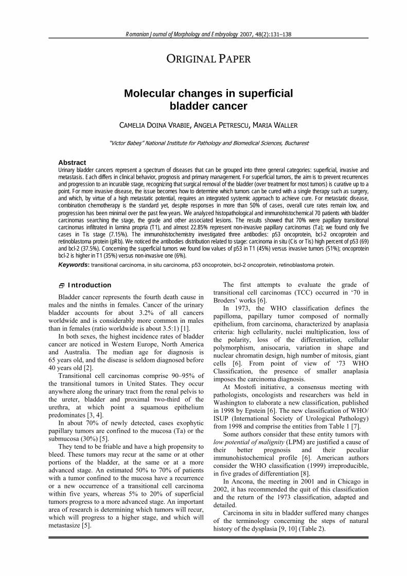
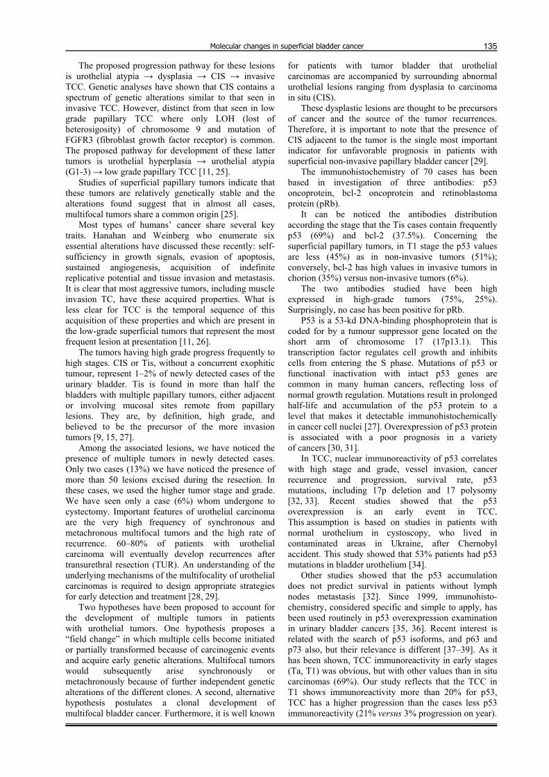
The clinical, histopathological and immunohisto-chemical analysis of 70 urothelial tumor cases revealed the next results. The distribution according to sex is showed in Figure 1. We analyzed the age range affected by bladder carcinoma and the results confirmed this (Figure 2). We analyzed bladder carcinomas in superficial stages that means in Ta (non-invasive papillary carcinoma), T1 (transitional papillary carcinoma invasive in chorion) and Tis (in situ carcinoma). The distribution of cases, regarding the stage is present in Figure 3.
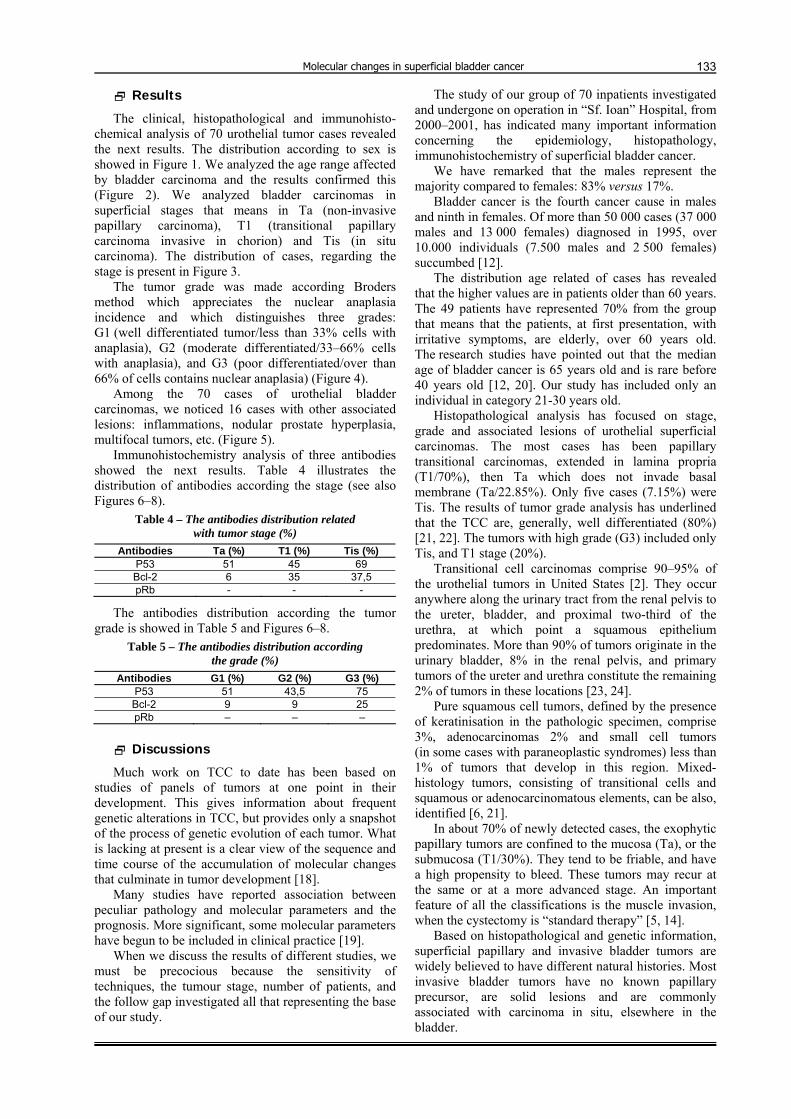
The tumor grade was made according Broders method which appreciates the nuclear anaplasia incidence and which distinguishes three grades: G1 (well differentiated tumor/less than 33% cells with anaplasia), G2 (moderate differentiated/33–66% cells with anaplasia), and G3 (poor differentiated/over than 66% of cells contains nuclear anaplasia) (Figure 4).
Among the 70 cases of urothelial bladder carcinomas, we noticed 16 cases with other associated lesions: inflammations, nodular prostate hyperplasia, multifocal tumors, etc. (Figure 5).
Immunohistochemistry analysis of three antibodies showed the next results. Table 4 illustrates the distribution of antibodies according the stage (see also Figures 6–8).
Table 4 – The antibodies distribution related with tumor stage (%)
Antibodies Ta (%) T1 (%) Tis (%) P53 51 45 69 Bcl-2 6 35 37,5 pRb - - -
The antibodies distribution according the tumor grade is showed in Table 5 and Figures 6–8.
Table 5 – The antibodies distribution according the grade (%)
Antibodies G1 (%) G2 (%) G3 (%) P53 51 43,5 75 Bcl-2 9 9 25 pRb – – –
Discussions
Much work on TCC to date has been based on studies of panels of tumors at one point in their development. This gives information about frequent genetic alterations in TCC, but provides only a snapshot of the process of genetic evolution of each tumor. What is lacking at present is a clear view of the sequence and time course of the accumulation of molecular changes that culminate in tumor development [18].
Many studies have reported association between peculiar pathology and molecular parameters and the prognosis. More significant, some molecular parameters have begun to be included in clinical practice [19].
When we discuss the results of different studies, we must be precocious because the sensitivity of techniques, the tumour stage, number of patients, and the follow gap investigated all that representing the base of our study.
The study of our group of 70 inpatients investigated and undergone on operation in “Sf. Ioan” Hospital, from 2000–2001, has indicated many important information concerning the epidemiology, histopathology, immunohistochemistry of superficial bladder cancer.
We have remarked that the males represent the majority compared to females: 83% versus 17%.
Bladder cancer is the fourth cancer cause in males and ninth in females. Of more than 50 000 cases (37 000 males and 13 000 females) diagnosed in 1995, over 10.000 individuals (7.500 males and 2 500 females) succumbed [12].
The distribution age related of cases has revealed that the higher values are in patients older than 60 years. The 49 patients have represented 70% from the group that means that the patients, at first presentation, with irritative symptoms, are elderly, over 60 years old. The research studies have pointed out that the median age of bladder cancer is 65 years old and is rare before 40 years old [12, 20]. Our study has included only an individual in category 21-30 years old.
Histopathological analysis has focused on stage, grade and associated lesions of urothelial superficial carcinomas. The most cases has been papillary transitional carcinomas, extended in lamina propria (T1/70%), then Ta which does not invade basal membrane (Ta/22.85%). Only five cases (7.15%) were Tis. The results of tumor grade analysis has underlined that the TCC are, generally, well differentiated (80%) [21, 22]. The tumors with high grade (G3) included only Tis, and T1 stage (20%).
Transitional cell carcinomas comprise 90–95% of the urothelial tumors in United States [2]. They occur anywhere along the urinary tract from the renal pelvis to the ureter, bladder, and proximal two-third of the urethra, at which point a squamous epithelium predominates. More than 90% of tumors originate in the urinary bladder, 8% in the renal pelvis, and primary tumors of the ureter and urethra constitute the remaining 2% of tumors in these locations [23, 24].
Pure squamous cell tumors, defined by the presence of keratinisation in the pathologic specimen, comprise 3%, adenocarcinomas 2% and small cell tumors (in some cases with paraneoplastic syndromes) less than 1% of tumors that develop in this region. Mixed-histology tumors, consisting of transitional cells and squamous or adenocarcinomatous elements, can be also, identified [6, 21].
In about 70% of newly detected cases, the exophytic papillary tumors are confined to the mucosa (Ta), or the submucosa (T1/30%). They tend to be friable, and have a high propensity to bleed. These tumors may recur at the same or at a more advanced stage. An important feature of all the classifications is the muscle invasion, when the cystectomy is “standard therapy” [5, 14].
Based on histopathological and genetic information, superficial papillary and invasive bladder tumors are widely believed to have different natural histories. Most invasive bladder tumors have no known papillary precursor, are solid lesions and are commonly associated with carcinoma in situ, elsewhere in the bladder.
Camelia Doina Vrabie et al.
134
PATIENT NUMBER (n=70)
83%
17%
WomenMen
AGE DISTRIBUTION
24
901
25
11
21 – 3031 – 4041 – 50 51 – 60 61 – 70 > 71
Figure 1 – The distribution according
to patients sex Figure 2 – The distribution of age
in the studied group
7%
70%
23%
Ta
T1Tis
20%
16%
64%
G1
G2
G3
Figure 3 – The distribution of cases
regarding the stage Figure 4 – The distribution of tumors
according to grade
6%
38%
13%
6%6%
31%Nodular prostatehyperplasiaLeiomyosarcoma
Inflammatory lesions
Multifocal tumours (>50)
Dysplastic lesions
Radical cystectomia
Figure 5 – The distribution of associated lesions
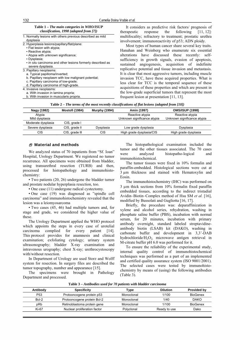
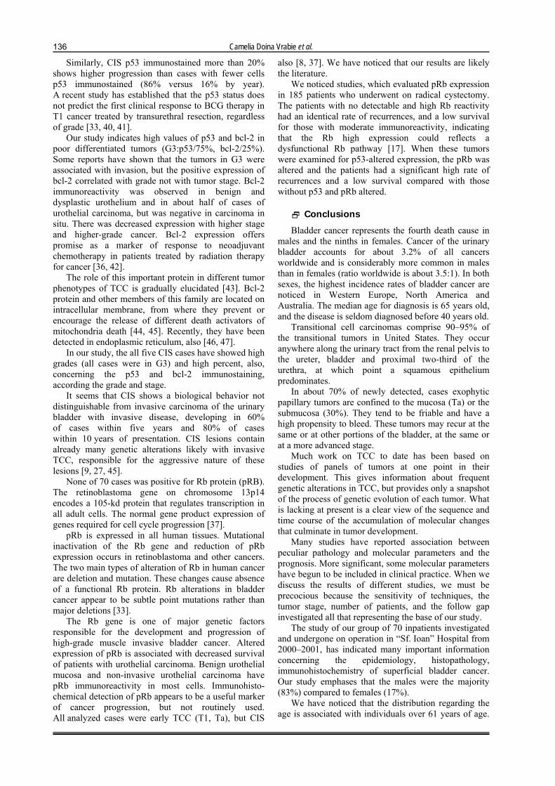
Figure 1 – Positive expression for bcl-2
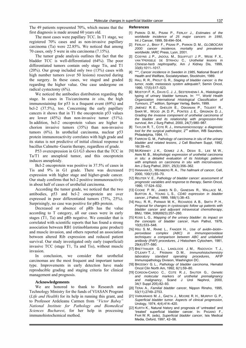
oncoprotein especially in deep layers (ABC method, 10×)
Figure 2 – Positive diffusely expression of p53 in a TCC well differentiated (G1) infiltrating the lamina propria
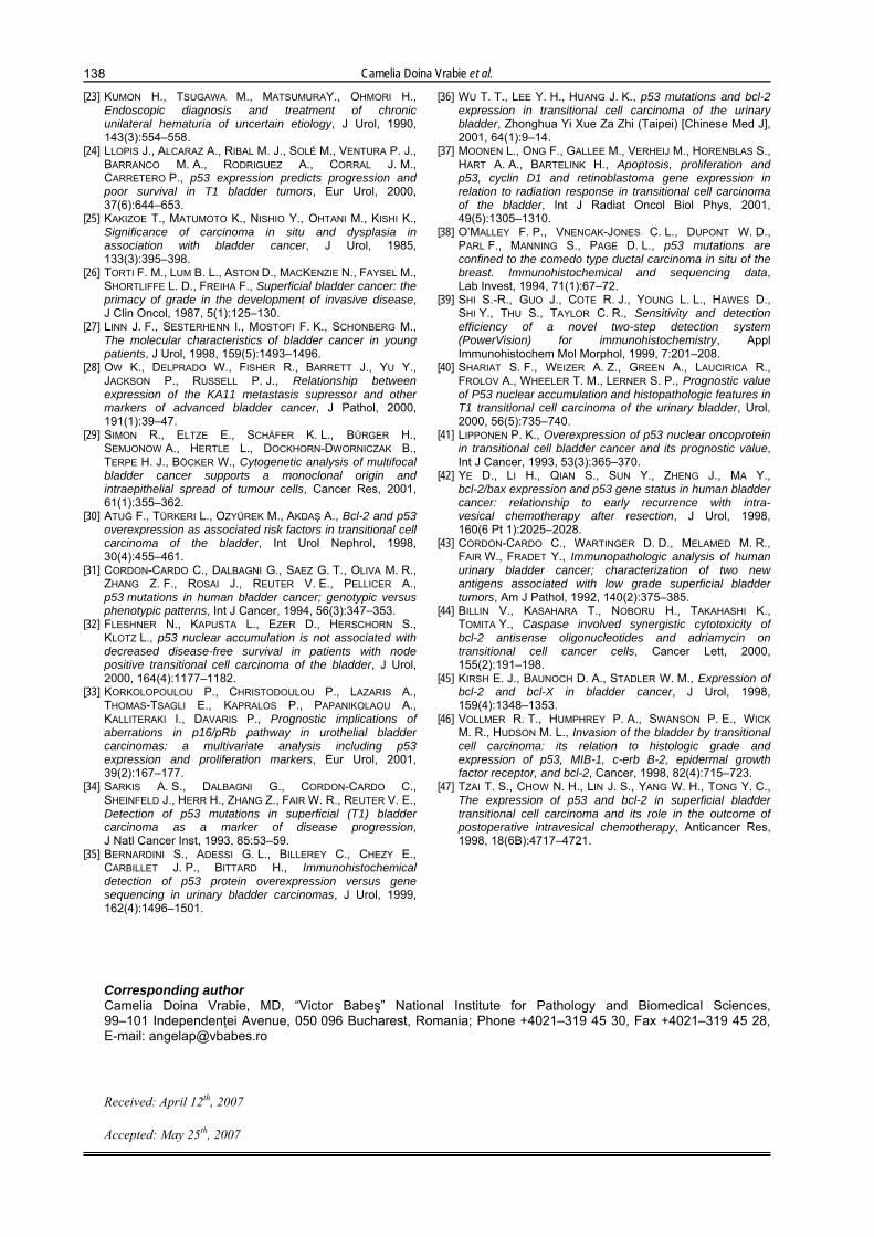
(ABC method, 10×)
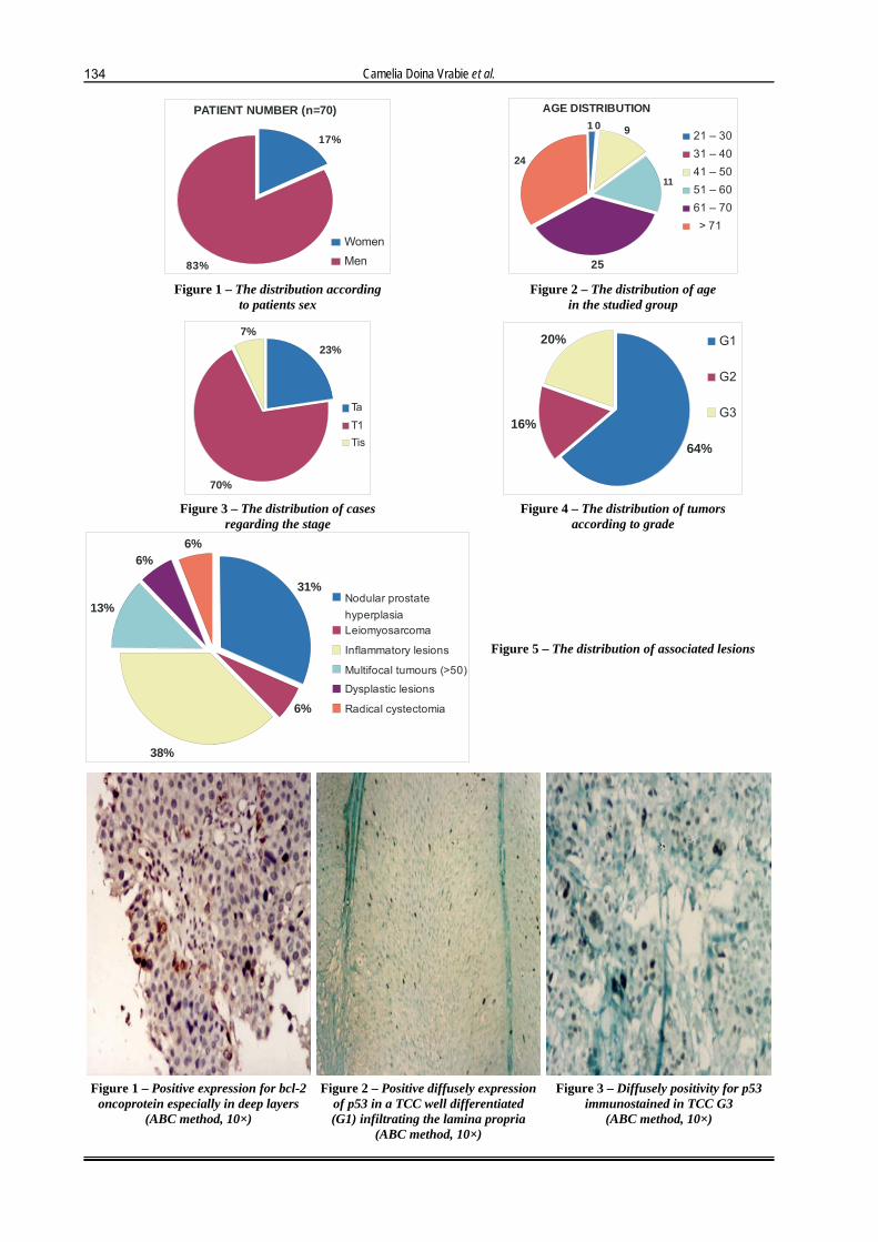
Figure 3 – Diffusely positivity for p53 immunostained in TCC G3
(ABC method, 10×)
Molecular changes in superficial bladder cancer
135
The proposed progression pathway for these lesions is urothelial atypia → dysplasia → CIS → invasive TCC. Genetic analyses have shown that CIS contains a spectrum of genetic alterations similar to that seen in invasive TCC. However, distinct from that seen in low grade papillary TCC where only LOH (lost of heterosigosity) of chromosome 9 and mutation of FGFR3 (fibroblast growth factor receptor) is common. The proposed pathway for development of these latter tumors is urothelial hyperplasia → urothelial atypia (G1-3) → low grade papillary TCC [11, 25].
Studies of superficial papillary tumors indicate that these tumors are relatively genetically stable and the alterations found suggest that in almost all cases, multifocal tumors share a common origin [25].
Most types of humans’ cancer share several key traits. Hanahan and Weinberg who enumerate six essential alterations have discussed these recently: self-sufficiency in growth signals, evasion of apoptosis, sustained angiogenesis, acquisition of indefinite replicative potential and tissue invasion and metastasis. It is clear that most aggressive tumors, including muscle invasion TC, have these acquired properties. What is less clear for TCC is the temporal sequence of this acquisition of these properties and which are present in the low-grade superficial tumors that represent the most frequent lesion at presentation [11, 26].
The tumors having high grade progress frequently to high stages. CIS or Tis, without a concurrent exophitic tumour, represent 1–2% of newly detected cases of the urinary bladder. Tis is found in more than half the bladders with multiple papillary tumors, either adjacent or involving mucosal sites remote from papillary lesions. They are, by definition, high grade, and believed to be the precursor of the more invasion tumors [9, 15, 27].
Among the associated lesions, we have noticed the presence of multiple tumors in newly detected cases. Only two cases (13%) we have noticed the presence of more than 50 lesions excised during the resection. In these cases, we used the higher tumor stage and grade. We have seen only a case (6%) whom undergone to cystectomy. Important features of urothelial carcinoma are the very high frequency of synchronous and metachronous multifocal tumors and the high rate of recurrence. 60–80% of patients with urothelial carcinoma will eventually develop recurrences after transurethral resection (TUR). An understanding of the underlying mechanisms of the multifocality of urothelial carcinomas is required to design appropriate strategies for early detection and treatment [28, 29].
Two hypotheses have been proposed to account for the development of multiple tumors in patients with urothelial tumors. One hypothesis proposes a “field change” in which multiple cells become initiated or partially transformed because of carcinogenic events and acquire early genetic alterations. Multifocal tumors would subsequently arise synchronously or metachronously because of further independent genetic alterations of the different clones. A second, alternative hypothesis postulates a clonal development of multifocal bladder cancer. Furthermore, it is well known
for patients with tumor bladder that urothelial carcinomas are accompanied by surrounding abnormal urothelial lesions ranging from dysplasia to carcinoma in situ (CIS).
These dysplastic lesions are thought to be precursors of cancer and the source of the tumor recurrences. Therefore, it is important to note that the presence of CIS adjacent to the tumor is the single most important indicator for unfavorable prognosis in patients with superficial non-invasive papillary bladder cancer [29].
The immunohistochemistry of 70 cases has been based in investigation of three antibodies: p53 oncoprotein, bcl-2 oncoprotein and retinoblastoma protein (pRb).
It can be noticed the antibodies distribution according the stage that the Tis cases contain frequently p53 (69%) and bcl-2 (37.5%). Concerning the superficial papillary tumors, in T1 stage the p53 values are less (45%) as in non-invasive tumors (51%); conversely, bcl-2 has high values in invasive tumors in chorion (35%) versus non-invasive tumors (6%).
The two antibodies studied have been high expressed in high-grade tumors (75%, 25%). Surprisingly, no case has been positive for pRb.
P53 is a 53-kd DNA-binding phosphoprotein that is coded for by a tumour suppressor gene located on the short arm of chromosome 17 (17p13.1). This transcription factor regulates cell growth and inhibits cells from entering the S phase. Mutations of p53 or functional inactivation with intact p53 genes are common in many human cancers, reflecting loss of normal growth regulation. Mutations result in prolonged half-life and accumulation of the p53 protein to a level that makes it detectable immunohistochemically in cancer cell nuclei [27]. Overexpression of p53 protein is associated with a poor prognosis in a variety of cancers [30, 31].
In TCC, nuclear immunoreactivity of p53 correlates with high stage and grade, vessel invasion, cancer recurrence and progression, survival rate, p53 mutations, including 17p deletion and 17 polysomy [32, 33]. Recent studies showed that the p53 overexpression is an early event in TCC. This assumption is based on studies in patients with normal urothelium in cystoscopy, who lived in contaminated areas in Ukraine, after Chernobyl accident. This study showed that 53% patients had p53 mutations in bladder urothelium [34].
Other studies showed that the p53 accumulation does not predict survival in patients without lymph nodes metastasis [32]. Since 1999, immunohisto-chemistry, considered specific and simple to apply, has been used routinely in p53 overexpression examination in urinary bladder cancers [35, 36]. Recent interest is related with the search of p53 isoforms, and p63 and p73 also, but their relevance is different [37–39]. As it has been shown, TCC immunoreactivity in early stages (Ta, T1) was obvious, but with other values than in situ carcinomas (69%). Our study reflects that the TCC in T1 shows immunoreactivity more than 20% for p53, TCC has a higher progression than the cases less p53 immunoreactivity (21% versus 3% progression on year).
Camelia Doina Vrabie et al.
136
Similarly, CIS p53 immunostained more than 20% shows higher progression than cases with fewer cells p53 immunostained (86% versus 16% by year). A recent study has established that the p53 status does not predict the first clinical response to BCG therapy in T1 cancer treated by transurethral resection, regardless of grade [33, 40, 41].
Our study indicates high values of p53 and bcl-2 in poor differentiated tumors (G3:p53/75%, bcl-2/25%). Some reports have shown that the tumors in G3 were associated with invasion, but the positive expression of bcl-2 correlated with grade not with tumor stage. Bcl-2 immunoreactivity was observed in benign and dysplastic urothelium and in about half of cases of urothelial carcinoma, but was negative in carcinoma in situ. There was decreased expression with higher stage and higher-grade cancer. Bcl-2 expression offers promise as a marker of response to neoadjuvant chemotherapy in patients treated by radiation therapy for cancer [36, 42].
The role of this important protein in different tumor phenotypes of TCC is gradually elucidated [43]. Bcl-2 protein and other members of this family are located on intracellular membrane, from where they prevent or encourage the release of different death activators of mitochondria death [44, 45]. Recently, they have been detected in endoplasmic reticulum, also [46, 47].
In our study, the all five CIS cases have showed high grades (all cases were in G3) and high percent, also, concerning the p53 and bcl-2 immunostaining, according the grade and stage.
It seems that CIS shows a biological behavior not distinguishable from invasive carcinoma of the urinary bladder with invasive disease, developing in 60% of cases within five years and 80% of cases within 10 years of presentation. CIS lesions contain already many genetic alterations likely with invasive TCC, responsible for the aggressive nature of these lesions [9, 27, 45].
None of 70 cases was positive for Rb protein (pRB). The retinoblastoma gene on chromosome 13p14 encodes a 105-kd protein that regulates transcription in all adult cells. The normal gene product expression of genes required for cell cycle progression [37].
pRb is expressed in all human tissues. Mutational inactivation of the Rb gene and reduction of pRb expression occurs in retinoblastoma and other cancers. The two main types of alteration of Rb in human cancer are deletion and mutation. These changes cause absence of a functional Rb protein. Rb alterations in bladder cancer appear to be subtle point mutations rather than major deletions [33].
The Rb gene is one of major genetic factors responsible for the development and progression of high-grade muscle invasive bladder cancer. Altered expression of pRb is associated with decreased survival of patients with urothelial carcinoma. Benign urothelial mucosa and non-invasive urothelial carcinoma have pRb immunoreactivity in most cells. Immunohisto-chemical detection of pRb appears to be a useful marker of cancer progression, but not routinely used. All analyzed cases were early TCC (T1, Ta), but CIS
also [8, 37]. We have noticed that our results are likely the literature.
We noticed studies, which evaluated pRb expression in 185 patients who underwent on radical cystectomy. The patients with no detectable and high Rb reactivity had an identical rate of recurrences, and a low survival for those with moderate immunoreactivity, indicating that the Rb high expression could reflects a dysfunctional Rb pathway [17]. When these tumors were examined for p53-altered expression, the pRb was altered and the patients had a significant high rate of recurrences and a low survival compared with those without p53 and pRb altered.
Conclusions
Bladder cancer represents the fourth death cause in males and the ninths in females. Cancer of the urinary bladder accounts for about 3.2% of all cancers worldwide and is considerably more common in males than in females (ratio worldwide is about 3.5:1). In both sexes, the highest incidence rates of bladder cancer are noticed in Western Europe, North America and Australia. The median age for diagnosis is 65 years old, and the disease is seldom diagnosed before 40 years old.
Transitional cell carcinomas comprise 90–95% of the transitional tumors in United States. They occur anywhere along the urinary tract from the renal pelvis to the ureter, bladder and proximal two-third of the urethra, at which point a squamous epithelium predominates.
In about 70% of newly detected, cases exophytic papillary tumors are confined to the mucosa (Ta) or the submucosa (30%). They tend to be friable and have a high propensity to bleed. These tumors may recur at the same or at other portions of the bladder, at the same or at a more advanced stage.
Much work on TCC to date has been based on studies of panels of tumors at one point in their development. This gives information about frequent genetic alterations in TCC, but provides only a snapshot of the process of genetic evolution of each tumor. What is lacking at present is a clear view of the sequence and time course of the accumulation of molecular changes that culminate in tumor development.
Many studies have reported association between peculiar pathology and molecular parameters and the prognosis. More significant, some molecular parameters have begun to be included in clinical practice. When we discuss the results of different studies, we must be precocious because the sensitivity of techniques, the tumor stage, number of patients, and the follow gap investigated all that representing the base of our study.
The study of our group of 70 inpatients investigated and undergone on operation in “Sf. Ioan” Hospital from 2000–2001, has indicated many important information concerning the epidemiology, histopathology, immunohistochemistry of superficial bladder cancer. Our study emphases that the males were the majority (83%) compared to females (17%).
We have noticed that the distribution regarding the age is associated with individuals over 61 years of age.
Molecular changes in superficial bladder cancer
137
The 49 patients represented 70%, which means that the first diagnosis is made around 60 years old.
The most cases were papillary TCC. In T1 stage, we registered 70% cases and as non-invasive papillary carcinoma (Ta) were 22.85%. We noticed that among 70 cases, only 5 were in situ carcinoma (7.15%).
The tumor grade analysis outlines the fact that the bladder TCC is well-differentiated (64%). The poor differentiated tumors contain only stage Tis, and T1 (20%). Our group included only two (13%) cases with high number tumors (over 50 lesions) resected during the surgery. In these cases, we staged and graded regarding the higher value. One case undergone on radical cystectomy (6%).
We noticed the antibodies distribution regarding the stage. In cases in Tis/carcinoma in situ, the high immunostaining for p53 is a frequent event (69%) and bcl-2 (37.5%), too. Concerning the early papillary cancers it shows that in T1 the oncoprotein p53 values are lower (45%) than non-invasive tumor (51%). In addition, bcl-2 oncoprotein values are higher in chorion invasive tumors (35%) than non-invasive tumors (6%). In urothelial carcinoma, nuclear p53 protein immunoreactivity correlates with high grade and its status is not predictive of initial clinical response to bacillus Calmette–Guerin therapy, regardless of grade.
P53 overexpression in G1/G3 shows that the TCC in Ta/T1 are aneuploid tumor, and this oncoprotein induces aneuploidy.
Bcl-2 oncoprotein was positive in 37.5% of cases in Tis and 9% in G1 grade. There was decreased expression with higher stage and higher-grade cancer. Our study confirms that bcl-2 positive immunoreactivity in about half of cases of urothelial carcinoma.
According the tumor grade, we noticed that the two antibodies, p53 and bcl-2 oncoprotein are over expressed in poor differentiated tumors (75%, 25%). Surprisingly, no case was positive for pRb protein.
Decreased or absence of pRb has the value according to T category, all our cases were in early stages (T1, Ta) and pRb negative. We consider that is correlated with scientific reports that has found a strong association between RB1 (retinoblastoma gene product) and muscle invasion, and others reported an association between altered Rib expression and reduced patient survival. Our study investigated only early (superficial) invasive TCC (stage T1, Ta and Tis), without muscle invasion.
In conclusion, we consider that urothelial carcinomas are the most frequent and important tumor type. Improvements in early detection have made reproducible grading and staging criteria for clinical management and prognosis.
Acknowledgements We are honored to thank to Research and
Technology Ministry for the funds of VIASAN Program (Life and Health) for its help in running this grant, and to Professor Ardeleanu Carmen from “Victor Babeş” National Institute for Pathology and Biomedical Sciences Bucharest, for her help in processing immunohistochemical method.
References [1] PARKIN D. M., PISANI P., FERLAY J., Estimates of the
worldwide incidence of 25 major cancers in 1990, Int J Cancer, 1999, 59:494–504.
[2] FERLAY J., BRAY F., PISANI P., PARKIN D. M., GLOBOCAN 2000: cancer incidence, mortality and prevalence worldwide, IARC Press, Lyon, 2001.
[3] COSYNS J. P., JADOUL M., SQUIFFLET J. P., WESE F. X., VAN YPERSELE DE STRIHOU C., Urothelial lesions in Chinese-herb nephropathy, Am J Kidney Dis, 1999, 33(6):1011–1017.
[4] ***, Cancer incidence in Sweden in 1985, National Board of Health and Wellfare, Socialstyrelsen, Stockholm, 1989.
[5] HALL R. R., PROUT G. R., Staging of bladder cancer: is the tumor, node, metastasis system adequate?, Semin Oncol, 1990, 17(5):517–523.
[6] MOSTOFI F. K., DAVIS C. J. J., SESTERHENN I. A., Histological typing of urinary bladder tumours. In: ***, World Health Organization International Histological Classification of Tumours, 2nd edition, Springer Verlag, Berlin, 1999.
[7] JIMENEZ R. E., GHEILER E., OSKANIAN P., TIGUERT R., SAKR W., WOOD JR. D. P., PONTES J. E., GRIGNON D. J., Grading the invasive component of urothelial carcinoma of the bladder and its relationship with progression-free survival, Am J Surg Pathol, 2000, 24(7):980–987.
[8] TAYLOR R. T., COTE R. J., Immunomicroscopy: a diagnostic tool for the surgical pathologist, 2nd edition, WB Saunders, Philadelphia, 1994, 11.
[9] FARROW G. M., Pathology of carcinoma in situ of the urinary bladder and related lesions, J Cell Biochem Suppl, 1992, 16I:39–43.
[10] MCKENNEY J. K., GOMEZ J. A., DESAI S., LEE M. W., AMIN M. B., Morphologic expression of urothelial carcinoma in situ: a detailed evaluation of its histologic patterns with emphasis on carcinoma in situ with microinvasion, Am J Surg Pathol, 2001, 25(3):356–362.
[11] HANAHAN D., WEINBERG R. A., The hallmark of cancer, Cell, 2000, 100(1):55–70.
[12] REUTER V. E., Pathology of bladder cancer: assessment of prognostic variables and response to therapy, Semin Oncol, 1990, 17:524–532.
[13] COOKE P. W., JAMES N. D., GANESAN R., WALLACE M., BURTON A., YOUNG L. S., CD40 expression in bladder cancer, J Pathol, 1999, 188(1):38–43.
[14] HALL R. R., PARMAR M. K., RICHARDS A. B., SMITH P. H., Proposal for changes in cystoscopic follow up patients with bladder cancer and adjuvant intravesical chemotherapy, BMJ, 1994, 308(6923):257–260.
[15] KOSS L. G., Mapping of the urinary bladder: its impact on the concepts of bladder cancer, Hum Pathol, 1979, 10(5):533–548.
[16] HSU S. M., RAINE L., FANGER H., Use of avidin–biotin–peroxidase complex (ABC) in immunoperoxidase techniques: a comparison between ABC and unlabeled antibody (PAP) procedures, J Histochem Cytochem, 1981, 29(4):577–580.
[17] BRETTHAUER G. L., LANGLOOS J. M., RADOVICH T. J., O’LEARY T. J., FRISMAN D. M., Immunohistochemistry laboratory standard operating procedures, AFIP Immunopathology Division, Washington DC.
[18] BRODSKY G. L., Pathology of bladder carcinoma, Hematol Oncol Clin North Am, 1992, 6(1):59–80.
[19] CORDON-CARDO C., COTE R. J., SAUTER G., Genetic and molecular markers of urothelial premalignancy and malignancy, Scand J Urol Nephrol, 2000, 34(1 Suppl 205):82–93.
[20] TERAI A., Familial bladder cancer, Nippon Rinsho, 1995, 53(11):2749–2753.
[21] VARKARAKIS M. J., GAETA J., MOORE R. H., MURPHY G. P., Superficial bladder tumor. Aspects of clinical progression, Urology, 1974, 4(4):414–420.
[22] KURTH K., Natural history and prognosis of ‘untreated’ and ‘treated’ superficial bladder cancer. In: PAGANO F., FAIR W. R. (eds), Superficial bladder cancer, Isis Medical Media, Oxford, 1997, 42–56.
Camelia Doina Vrabie et al.
138
[23] KUMON H., TSUGAWA M., MATSUMURAY., OHMORI H., Endoscopic diagnosis and treatment of chronic unilateral hematuria of uncertain etiology, J Urol, 1990, 143(3):554–558.
[24] LLOPIS J., ALCARAZ A., RIBAL M. J., SOLÉ M., VENTURA P. J., BARRANCO M. A., RODRIGUEZ A., CORRAL J. M., CARRETERO P., p53 expression predicts progression and poor survival in T1 bladder tumors, Eur Urol, 2000, 37(6):644–653.
[25] KAKIZOE T., MATUMOTO K., NISHIO Y., OHTANI M., KISHI K., Significance of carcinoma in situ and dysplasia in association with bladder cancer, J Urol, 1985, 133(3):395–398.
[26] TORTI F. M., LUM B. L., ASTON D., MACKENZIE N., FAYSEL M., SHORTLIFFE L. D., FREIHA F., Superficial bladder cancer: the primacy of grade in the development of invasive disease, J Clin Oncol, 1987, 5(1):125–130.
[27] LINN J. F., SESTERHENN I., MOSTOFI F. K., SCHONBERG M., The molecular characteristics of bladder cancer in young patients, J Urol, 1998, 159(5):1493–1496.
[28] OW K., DELPRADO W., FISHER R., BARRETT J., YU Y., JACKSON P., RUSSELL P. J., Relationship between expression of the KA11 metastasis supressor and other markers of advanced bladder cancer, J Pathol, 2000, 191(1):39–47.
[29] SIMON R., ELTZE E., SCHÄFER K. L., BÜRGER H., SEMJONOW A., HERTLE L., DOCKHORN-DWORNICZAK B., TERPE H. J., BÖCKER W., Cytogenetic analysis of multifocal bladder cancer supports a monoclonal origin and intraepithelial spread of tumour cells, Cancer Res, 2001, 61(1):355–362.
[30] ATUĞ F., TÜRKERI L., OZYÜREK M., AKDAŞ A., Bcl-2 and p53 overexpression as associated risk factors in transitional cell carcinoma of the bladder, Int Urol Nephrol, 1998, 30(4):455–461.
[31] CORDON-CARDO C., DALBAGNI G., SAEZ G. T., OLIVA M. R., ZHANG Z. F., ROSAI J., REUTER V. E., PELLICER A., p53 mutations in human bladder cancer; genotypic versus phenotypic patterns, Int J Cancer, 1994, 56(3):347–353.
[32] FLESHNER N., KAPUSTA L., EZER D., HERSCHORN S., KLOTZ L., p53 nuclear accumulation is not associated with decreased disease-free survival in patients with node positive transitional cell carcinoma of the bladder, J Urol, 2000, 164(4):1177–1182.
[33] KORKOLOPOULOU P., CHRISTODOULOU P., LAZARIS A., THOMAS-TSAGLI E., KAPRALOS P., PAPANIKOLAOU A., KALLITERAKI I., DAVARIS P., Prognostic implications of aberrations in p16/pRb pathway in urothelial bladder carcinomas: a multivariate analysis including p53 expression and proliferation markers, Eur Urol, 2001, 39(2):167–177.
[34] SARKIS A. S., DALBAGNI G., CORDON-CARDO C., SHEINFELD J., HERR H., ZHANG Z., FAIR W. R., REUTER V. E., Detection of p53 mutations in superficial (T1) bladder carcinoma as a marker of disease progression, J Natl Cancer Inst, 1993, 85:53–59.
[35] BERNARDINI S., ADESSI G. L., BILLEREY C., CHEZY E., CARBILLET J. P., BITTARD H., Immunohistochemical detection of p53 protein overexpression versus gene sequencing in urinary bladder carcinomas, J Urol, 1999, 162(4):1496–1501.
[36] WU T. T., LEE Y. H., HUANG J. K., p53 mutations and bcl-2 expression in transitional cell carcinoma of the urinary bladder, Zhonghua Yi Xue Za Zhi (Taipei) [Chinese Med J], 2001, 64(1):9–14.
[37] MOONEN L., ONG F., GALLEE M., VERHEIJ M., HORENBLAS S., HART A. A., BARTELINK H., Apoptosis, proliferation and p53, cyclin D1 and retinoblastoma gene expression in relation to radiation response in transitional cell carcinoma of the bladder, Int J Radiat Oncol Biol Phys, 2001, 49(5):1305–1310.
[38] O’MALLEY F. P., VNENCAK-JONES C. L., DUPONT W. D., PARL F., MANNING S., PAGE D. L., p53 mutations are confined to the comedo type ductal carcinoma in situ of the breast. Immunohistochemical and sequencing data, Lab Invest, 1994, 71(1):67–72.
[39] SHI S.-R., GUO J., COTE R. J., YOUNG L. L., HAWES D., SHI Y., THU S., TAYLOR C. R., Sensitivity and detection efficiency of a novel two-step detection system (PowerVision) for immunohistochemistry, Appl Immunohistochem Mol Morphol, 1999, 7:201–208.
[40] SHARIAT S. F., WEIZER A. Z., GREEN A., LAUCIRICA R., FROLOV A., WHEELER T. M., LERNER S. P., Prognostic value of P53 nuclear accumulation and histopathologic features in T1 transitional cell carcinoma of the urinary bladder, Urol, 2000, 56(5):735–740.
[41] LIPPONEN P. K., Overexpression of p53 nuclear oncoprotein in transitional cell bladder cancer and its prognostic value, Int J Cancer, 1993, 53(3):365–370.
[42] YE D., LI H., QIAN S., SUN Y., ZHENG J., MA Y., bcl-2/bax expression and p53 gene status in human bladder cancer: relationship to early recurrence with intra- vesical chemotherapy after resection, J Urol, 1998, 160(6 Pt 1):2025–2028.
[43] CORDON-CARDO C., WARTINGER D. D., MELAMED M. R., FAIR W., FRADET Y., Immunopathologic analysis of human urinary bladder cancer; characterization of two new antigens associated with low grade superficial bladder tumors, Am J Pathol, 1992, 140(2):375–385.
[44] BILLIN V., KASAHARA T., NOBORU H., TAKAHASHI K., TOMITA Y., Caspase involved synergistic cytotoxicity of bcl-2 antisense oligonucleotides and adriamycin on transitional cell cancer cells, Cancer Lett, 2000, 155(2):191–198.
[45] KIRSH E. J., BAUNOCH D. A., STADLER W. M., Expression of bcl-2 and bcl-X in bladder cancer, J Urol, 1998, 159(4):1348–1353.
[46] VOLLMER R. T., HUMPHREY P. A., SWANSON P. E., WICK M. R., HUDSON M. L., Invasion of the bladder by transitional cell carcinoma: its relation to histologic grade and expression of p53, MIB-1, c-erb B-2, epidermal growth factor receptor, and bcl-2, Cancer, 1998, 82(4):715–723.
[47] TZAI T. S., CHOW N. H., LIN J. S., YANG W. H., TONG Y. C., The expression of p53 and bcl-2 in superficial bladder transitional cell carcinoma and its role in the outcome of postoperative intravesical chemotherapy, Anticancer Res, 1998, 18(6B):4717–4721.
Corresponding author Camelia Doina Vrabie, MD, “Victor Babeş” National Institute for Pathology and Biomedical Sciences, 99–101 Independenţei Avenue, 050 096 Bucharest, Romania; Phone +4021–319 45 30, Fax +4021–319 45 28, E-mail: [email protected] Received: April 12th, 2007
Accepted: May 25th, 2007