Embed Size (px)
Citation preview

Molecular Basis of Skeletal Muscle Contraction
Dr. Abdelrahman Mustafa
Department of Basic Medical Sciences Division of Physiology Faculty of Medicine Almaarefa Colleges
بسم الله الرحمن الرحيم

ObjectivesBy the end of this lecture, you should be able to:
Understand the Molecular mechanism of skeletal muscle contraction including:
Role of calcium ions in excitation contraction coupling
Sliding Filament Theory of Contraction The role of T-tubule and sarcoplasmic
reticulum Regulation of Calcium efflux and influx from
the sarcoplasmic reticulum and into the sarcoplasm
Molecular rearrangement of Actin and Myosin Myosin-ATPase cycle and Rigor Mortis
phenomenon
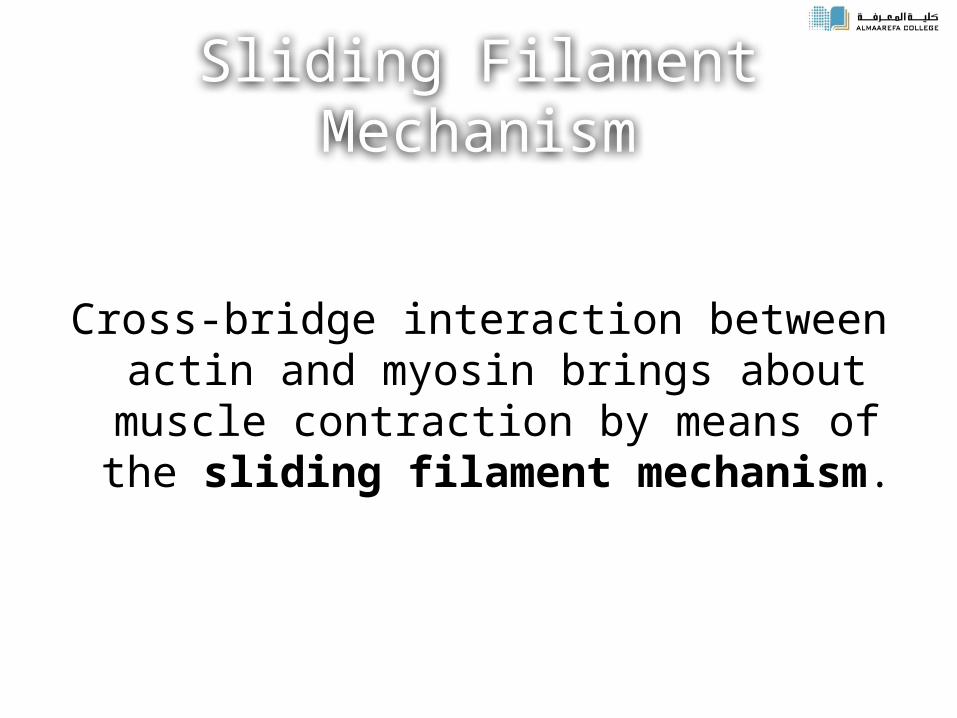
Sliding Filament Mechanism
Cross-bridge interaction between actin and myosin brings about muscle contraction by means of the sliding filament mechanism.
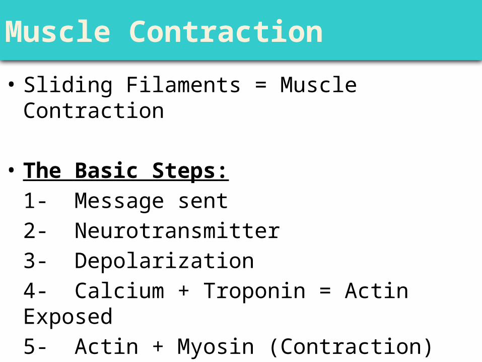
Muscle Contraction
• Sliding Filaments = Muscle Contraction
• The Basic Steps:1- Message sent2- Neurotransmitter3- Depolarization4- Calcium + Troponin = Actin Exposed5- Actin + Myosin (Contraction)
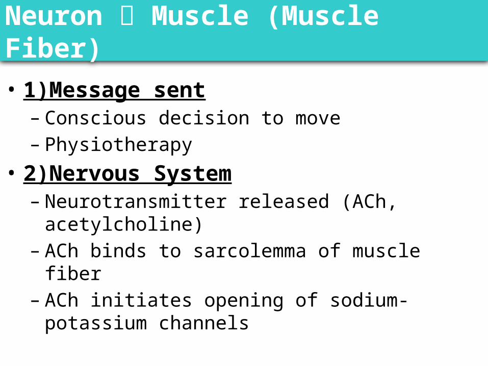
Neuron Muscle (Muscle Fiber)
• 1)Message sent– Conscious decision to move– Physiotherapy
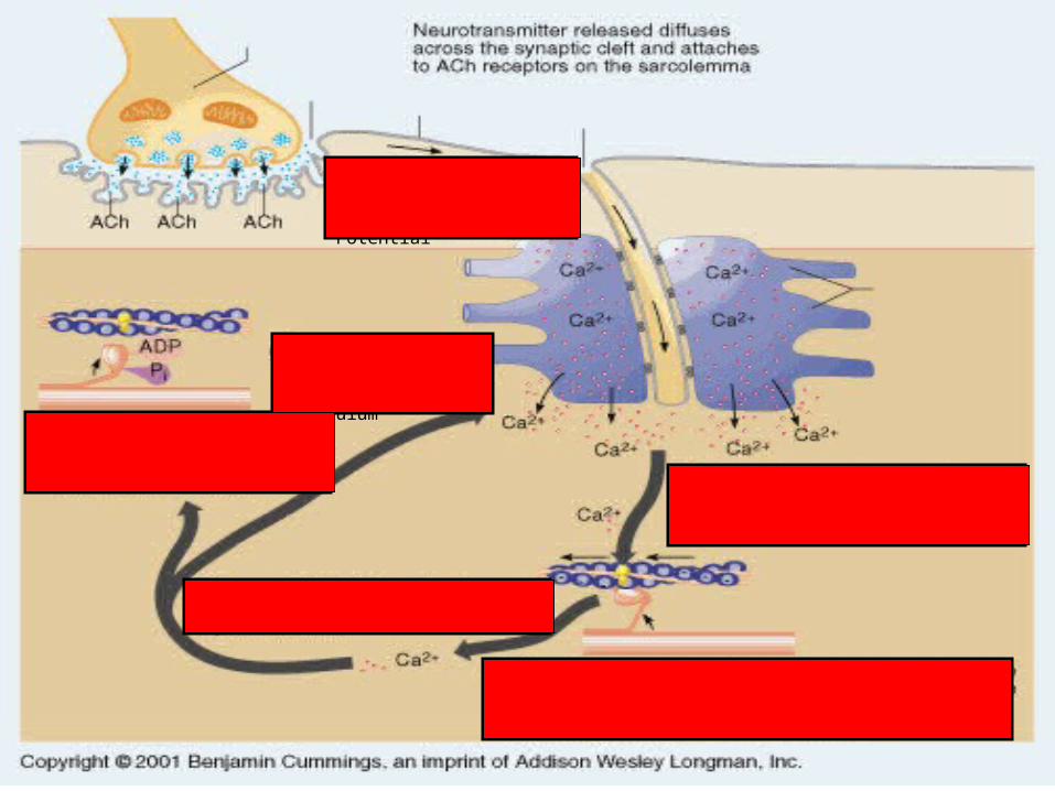
• 2)Nervous System– Neurotransmitter released (ACh, acetylcholine)– ACh binds to sarcolemma of muscle fiber– ACh initiates opening of sodium-potassium channels
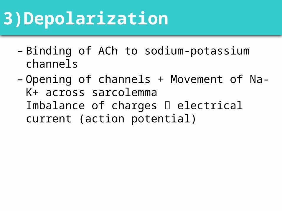
3)Depolarization
– Binding of ACh to sodium-potassium channels– Opening of channels + Movement of Na-K+ across
sarcolemmaImbalance of charges electrical current (action potential)
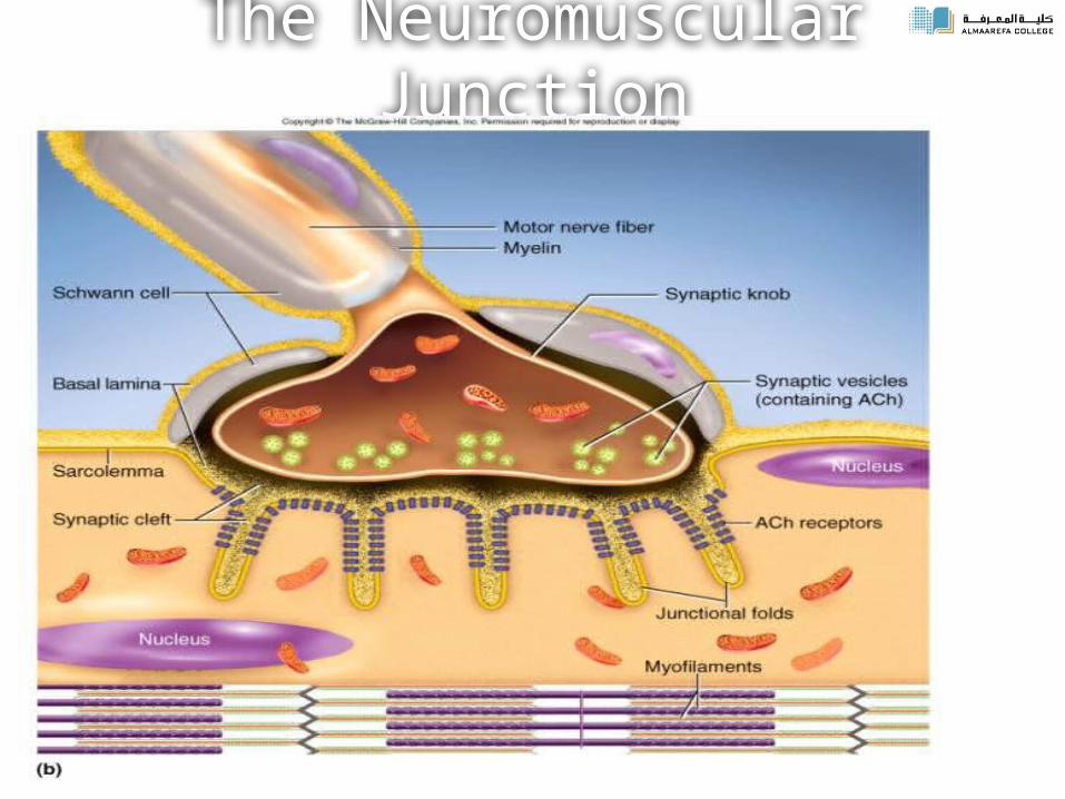
The Neuromuscular Junction
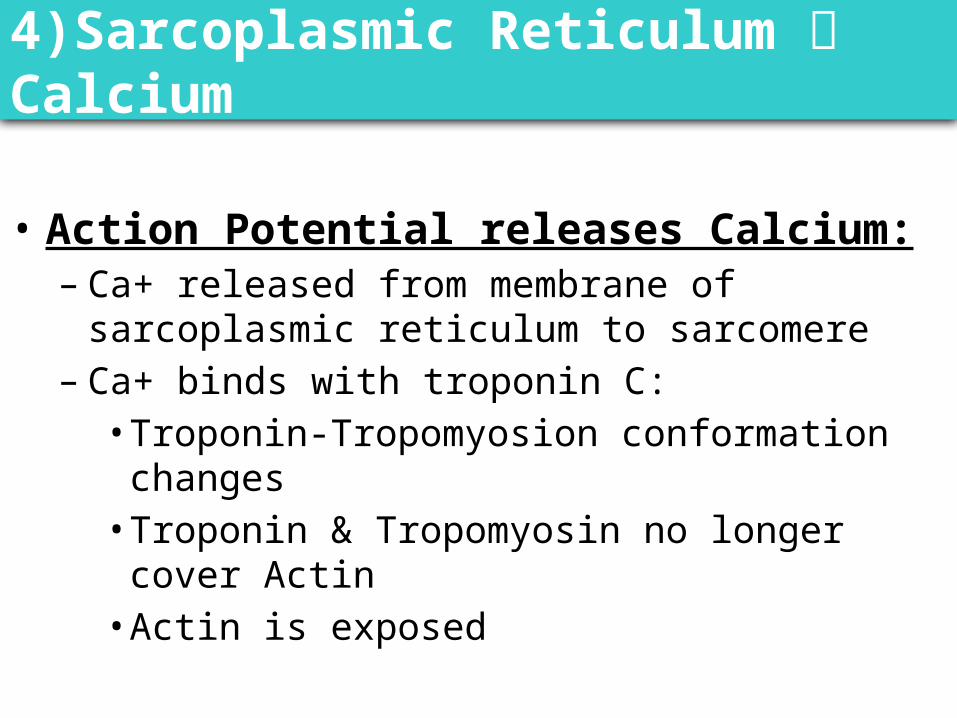
4)Sarcoplasmic Reticulum Calcium
• Action Potential releases Calcium:– Ca+ released from membrane of sarcoplasmic reticulum
to sarcomere– Ca+ binds with troponin C:• Troponin-Tropomyosion conformation changes• Troponin & Tropomyosin no longer cover Actin• Actin is exposed
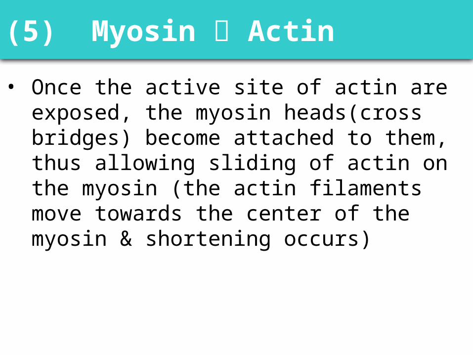
(5) Myosin Actin
• Once the active site of actin are exposed, the myosin heads(cross bridges) become attached to them, thus allowing sliding of actin on the myosin (the actin filaments move towards the center of the myosin & shortening occurs)
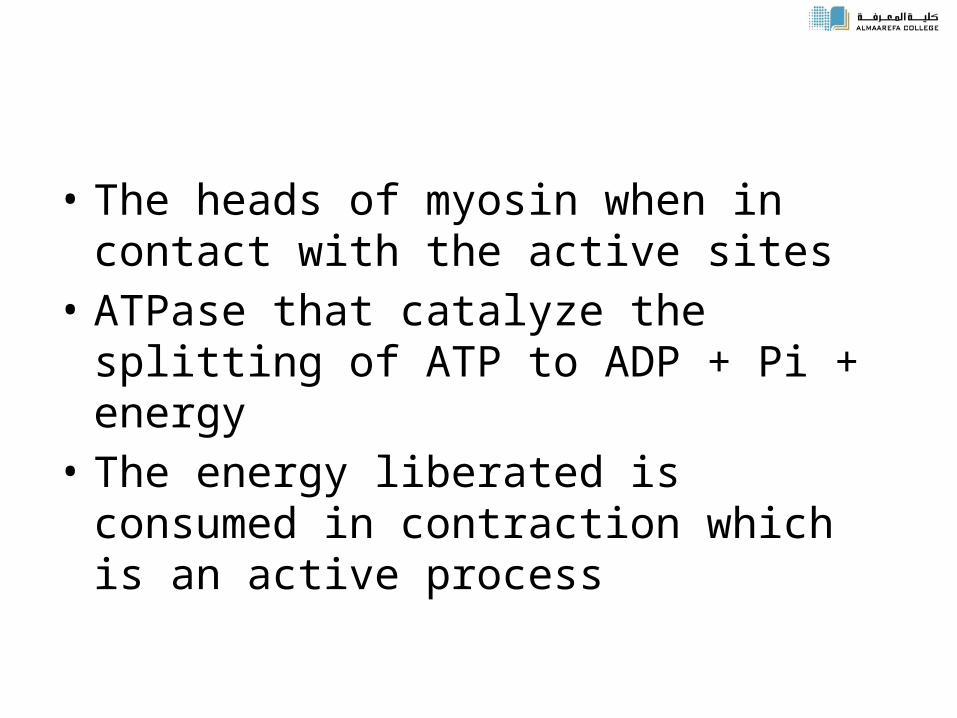
• The heads of myosin when in contact with the active sites
• ATPase that catalyze the splitting of ATP to ADP + Pi + energy
• The energy liberated is consumed in contraction which is an active process
Power Stroke• Activated cross bridge bends toward center of
thick filament, “rowing” in thin filament to which it is attached– Sarcoplasmic reticulum releases Ca2+ – Myosin heads bind to actin– Hydrolysis of ATP transfers energy to myosin head
Myosin heads (bends) toward center of sarcomere (power stroke)
– ATP binds to myosin head and detaches it from actin
©Thompson Educational Publishing, Inc. 2003. All material is copyright protected. It
is illegal to copy any of this material.This material may be used only in a course
of study in which Exercise Science: An Introduction to Health and Physical
Education (Temertzoglou/Challen) is the required textbook.
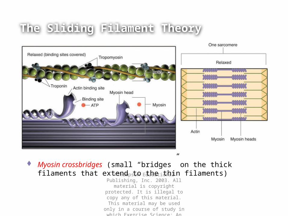
The Sliding Filament Theory
Myosin crossbridges (small “bridges” on the thick filaments that extend to the thin filaments)
©Thompson Educational Publishing, Inc. 2003. All material is copyright protected. It is illegal to copy any of this material.This material may be used only in a course of study in which Exercise Science: An Introduction to Health and Physical
Education (Temertzoglou/Challen) is the required textbook.
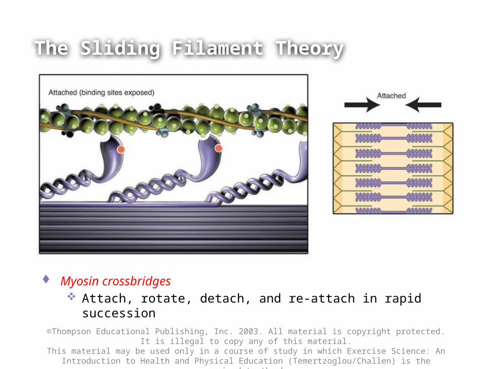
The Sliding Filament Theory
Myosin crossbridges Attach, rotate, detach, and re-attach in rapid succession
©Thompson Educational Publishing, Inc. 2003. All material is copyright protected. It is illegal to copy any of this material.This material may be used only in a course of study in which Exercise Science: An Introduction to Health and Physical
Education (Temertzoglou/Challen) is the required textbook.
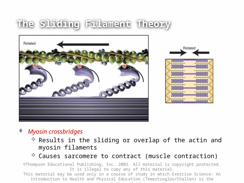
The Sliding Filament Theory
Myosin crossbridges Results in the sliding or overlap of the actin and myosin filaments Causes sarcomere to contract (muscle contraction)
(6) Relaxation
• Acetylcholinesterase breaks down ACh at neuromuscular junction
• Muscle fiber action potential stops, there no more release of Ca2+ from lateral sacs.–Myosin detaches and moves away from Actin– Troponin & Tropomyosin cover up Actin–Ca+ moves back into Sarcoplasmic Reticulum–Repolarization Sarcolemma stable again–ATP is required for myosin head to release from
actin and come back to resting state
Role of Calcium in Cross-Bridge Formation
• Increase in Ca2+ starts filament sliding• Decrease in Ca2+ turns off sliding process• Thin filaments on each side of sarcomere slide
inward over stationary thick filaments toward center of A band during contraction
• As thin filaments slide inward, they pull Z lines closer together
• Sarcomere shortens
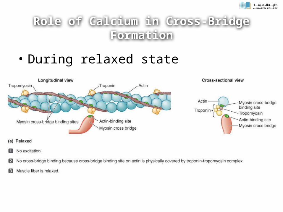
Role of Calcium in Cross-Bridge Formation
• During relaxed state
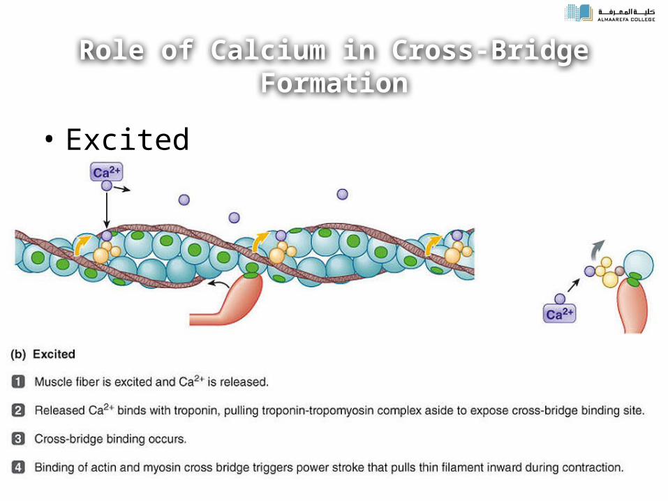
Role of Calcium in Cross-Bridge Formation
• Excited
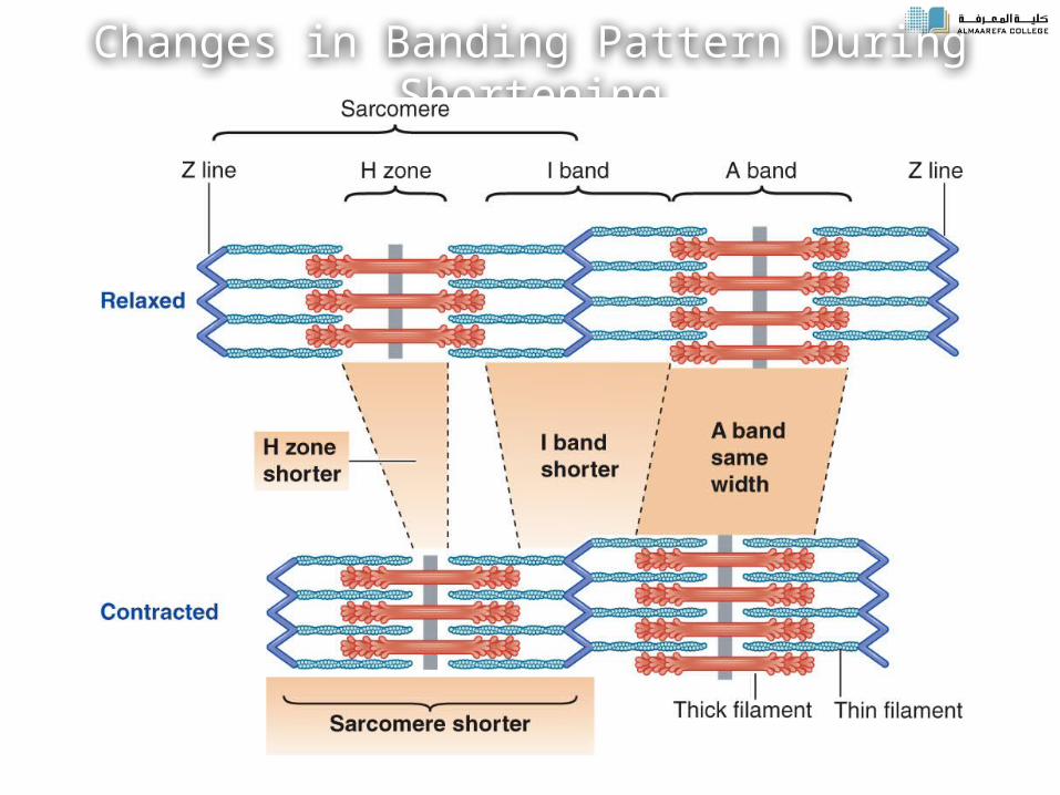
Changes in Banding Pattern During Shortening
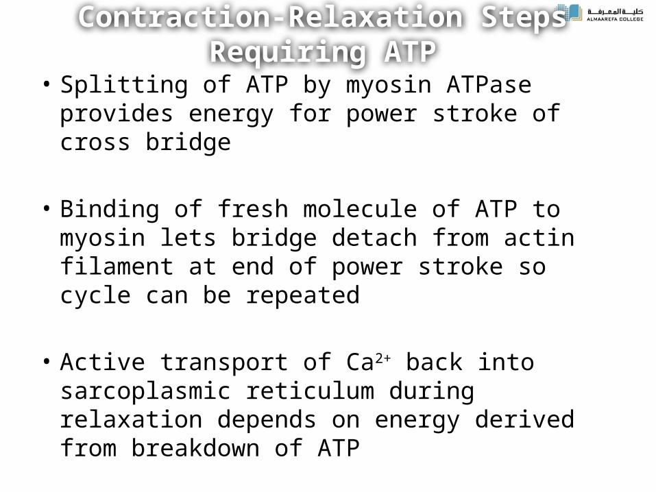
Contraction-Relaxation Steps Requiring ATP
• Splitting of ATP by myosin ATPase provides energy for power stroke of cross bridge
• Binding of fresh molecule of ATP to myosin lets bridge detach from actin filament at end of power stroke so cycle can be repeated
• Active transport of Ca2+ back into sarcoplasmic reticulum during relaxation depends on energy derived from breakdown of ATP
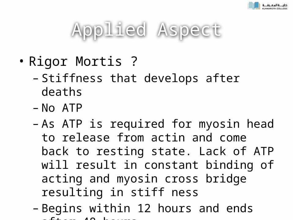
Applied Aspect
• Rigor Mortis ?– Stiffness that develops after deaths – No ATP – As ATP is required for myosin head to release from
actin and come back to resting state. Lack of ATP will result in constant binding of acting and myosin cross bridge resulting in stiff ness
– Begins within 12 hours and ends after 48 hours– After 48 hours, muscle cells begin to autolyse
Rela
tions
hip
of a
n ac
tion
pote
ntial
to
resu
ltant
mus
cle
twitc
h
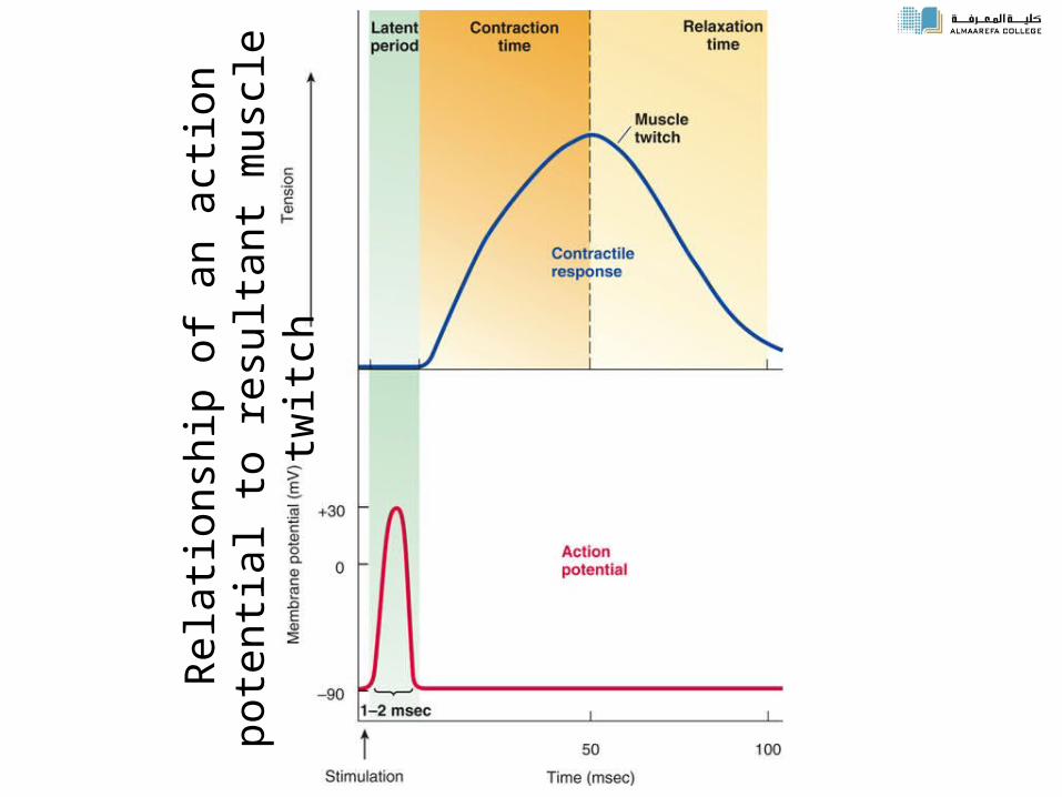
(1-2) Depolarization of Sarcolemma & Generation of Action Potential
(3) Calcium Ions released from Sarcoplasmic Reticulum
(4-6) Calcium binds to troponin, tropomyosin moves, actin exposed
(7-8) Myosin attaches to actin, Myosin moves actin, ATP released
(9) Calcium removed by active transport
(10) Everything moves back & contraction done!
Q1
Beside the Conscious decision to move skeletal muscle contract as response to A)Physiotherapy B)Tapping by clinical hummer C) Administration of drugs D)Intake large quantities of Ca2+
Q2
• The Neurotransmitter that released to conduct the massage at neuromuscular junction is :
A) Dopamin B) Acetylcholine C) Adrenaline D) Noradrenaline
Q3
• Ca+ released from membrane of sarcoplasmic reticulum to
A)Thick filment B)MyofibrilC)SarcolemmaD) Sarcomere
Q4
• When The heads of myosin contact with the active sites of Actin
A)ATPase will catalyze the splitting of ATP to ADP + Pi + energyB)Ca+2 will released from scroplasm C)Tropomyosin will cover the actin binding site D)The action potential will start