Embed Size (px)
Citation preview

14 Aug 2001 17:57 AR AR135-18.tex AR135-18.sgm ARv2(2001/05/10)P1: GPQ
Annu. Rev. Microbiol. 2001. 55:453–83
MOLECULAR ASPECTS OF PARASITE-VECTOR AND
VECTOR-HOST INTERACTIONS IN LEISHMANIASIS1
David Sacks and Shaden KamhawiLaboratory of Parasitic Diseases, National Institutes of Allergy and Infectious Diseases,National Institutes of Health, Bethesda, Maryland 20892; e-mail: [email protected]
Key Words sand flies, vector biology, lipophosphoglycan, saliva
■ Abstract Leishmania-sand fly interactions are reviewed in the context of the po-tential barriers to the complete development of the parasite that exist within the midgutenvironment of phlebotomine flies and the molecular adaptations that the parasite hasevolved that permit the development of transmissible infections to proceed. Cell sur-face and secreted phosphoglycans protect the parasite from the proteolytic activities ofthe blood-fed midgut, mediate attachment to the gut wall in order to maintain infectionduring excretion of the bloodmeal, and contribute to the formation of a biological plugin the anterior gut that may promote transmission by bite. The importance of vectorsaliva in modulating the host response to transmitted parasites is also reviewed.
CONTENTS
INTRODUCTION . . . . . . . . . . . . . . . . . . . . . . . . . . . . . . . . . . . . . . . . . . . . . . . . . . . . . 454Development of Transmissible Infections. . . . . . . . . . . . . . . . . . . . . . . . . . . . . . . . . 454Potential Barriers to Complete Development. . . . . . . . . . . . . . . . . . . . . . . . . . . . . . 457
SURVIVAL OF LEISHMANIAIN THE BLOOD-FED MIDGUT . . . . . . . . . . . . . . . . 458Susceptibility to Digestive Enzymes in the Blood-Fed Midgut. . . . . . . . . . . . . . . . 458Inhibition of Midgut Digestion in Infected Sand Flies. . . . . . . . . . . . . . . . . . . . . . . 459Parasite Molecules Controlling Early Survival. . . . . . . . . . . . . . . . . . . . . . . . . . . . . 460Analysis of Parasite Mutants. . . . . . . . . . . . . . . . . . . . . . . . . . . . . . . . . . . . . . . . . . . 461
PERSISTENCE OFLEISHMANIAFOLLOWING BLOODMEALEXCRETION . . . . . . . . . . . . . . . . . . . . . . . . . . . . . . . . . . . . . . . . . . . . . . . . . . . . . . . . 463Timing of the Loss of Infection in Refractory Sand Flies. . . . . . . . . . . . . . . . . . . . . 463Escape from the Peritrophic Membrane. . . . . . . . . . . . . . . . . . . . . . . . . . . . . . . . . . 464Attachment to Midgut Epithelial Cells—The Role of LPG. . . . . . . . . . . . . . . . . . . 465Species-Specific Midgut Attachment. . . . . . . . . . . . . . . . . . . . . . . . . . . . . . . . . . . . 466Midgut Receptors for Parasite Attachment. . . . . . . . . . . . . . . . . . . . . . . . . . . . . . . . 468
MATURATION OF TRANSMISSIBLE INFECTIONS . . . . . . . . . . . . . . . . . . . . . . . 469Stage-Specific Midgut Attachment. . . . . . . . . . . . . . . . . . . . . . . . . . . . . . . . . . . . . . 469Stage-Differentiation and Anterior Migration. . . . . . . . . . . . . . . . . . . . . . . . . . . . . . 471
1The U.S. Government has the right to retain a nonexclusive royalty-free license in and toany copyright covering this paper.
453
Ann
u. R
ev. M
icro
biol
. 200
1.55
:453
-483
. Dow
nloa
ded
from
ww
w.a
nnua
lrev
iew
s.or
g A
cces
s pr
ovid
ed b
y U
nive
rsid
ade
de S
ao P
aulo
(U
SP)
on 0
3/22
/16.
For
per
sona
l use
onl
y.

14 Aug 2001 17:57 AR AR135-18.tex AR135-18.sgm ARv2(2001/05/10)P1: GPQ
454 SACKS ¥ KAMHAWI
Transmission by Bite. . . . . . . . . . . . . . . . . . . . . . . . . . . . . . . . . . . . . . . . . . . . . . . . . 472VECTOR-HOST INTERACTIONS THAT MODULATELEISHMANIASIS: THE ROLE OF SAND FLY SALIVA . . . . . . . . . . . . . . . . . . . . 473Disease Enhancing and ImmunomodulatoryEffects of Sand Fly Saliva. . . . . . . . . . . . . . . . . . . . . . . . . . . . . . . . . . . . . . . . . . . . 473
Immunity Conferred by Presensitization to Sand Fly Saliva. . . . . . . . . . . . . . . . . . 475
INTRODUCTION
Leishmaniaare pathogenic protozoa of the order Kinetoplastida and the familyTrypanosomatidae. They have a dimorphic life cycle consisting of extracellularpromastigotes that multiply and develop within the alimentary tract of the sand flyvector and intracellular amastigotes that reside and multiply within the phagolyso-somal vacuoles of their host macrophages. Depending mainly on the species ofLeishmania, human infection can display a spectrum of clinical manifestationfrom localized cutaneous involvement to late destruction of mucous membranesto generalized systemic disease with fatal outcome. Distribution of the more than20 species and subspecies ofLeishmaniaand the diseases they produce are deter-mined by the availability of competent vectors. As far as is known, natural vectorsof all Leishmaniaspecies belong to the subfamily Phlebotominae (sand flies) of thefamily Psychodidae. The greatest concentration of phlebotomine species is in theforests of South America, where the genusLutzomyiapredominates.Phlebotomusis one of two large genera found in the Old World and is distributed largely inarid and semi-arid zones.LutzomyiaandPhlebotomusspp. account for virtuallyall known transmissions ofLeishmaniato humans, comprehensively reviewed byKillick-Kendrick (54). Moreover, even the vector-competent species often displayremarkable specificity for theLeishmaniaspecies they transmit in nature (reviewedin 53). In many instances, these restrictions cannot be accounted for by the ab-sence of sympatric (colocalized) sand flies or by host blood-meal preferences.Thus, many if not most phlebotomine species appear to be inherently refractory tothe full development ofLeishmania. A summary of the majorLeishmaniaspeciesinfecting humans, their clinical associations, geographic distribution, and provenor suspected sand fly vectors is provided in Table 1.
Understanding the basis of molecular interactions at the sand fly–Leishmaniainterface is fundamental to any study of vector competence and disease transmis-sion. This review focuses on those studies that have identified parasite- or sandfly–derived molecules that play a role in the development of transmissible in-fections and on those studies that have investigated the role of sand fly saliva inmodulating the outcome of infection in the mammalian host.
Development of Transmissible Infections
The first experimental evidence of transmission of leishmaniasis by the bite ofsand flies was by Shortt et al in 1931 (108), who achieved the transmission ofLeishmania donovanito hamsters by the bite ofPhlebotomus argentipes. Simi-lar studies involving other species ofLeishmaniaand sand flies (3, 62, 78, 126)
Ann
u. R
ev. M
icro
biol
. 200
1.55
:453
-483
. Dow
nloa
ded
from
ww
w.a
nnua
lrev
iew
s.or
g A
cces
s pr
ovid
ed b
y U
nive
rsid
ade
de S
ao P
aulo
(U
SP)
on 0
3/22
/16.
For
per
sona
l use
onl
y.

14 Aug 2001 17:57 AR AR135-18.tex AR135-18.sgm ARv2(2001/05/10)P1: GPQ
VECTOR BIOLOGY OF LEISHMANIASIS 455
TABLE 1 Proven or suspected vectors of the majorLeishmaniaspp. infecting humans,including their clinical associations and geographic distributions
Geographic Proven or suspectedParasite Clinical formsa distributions vectors
L. donovani AVL; PKDL China P. alexandriIndia, Nepal, P. argentipesBangladeshEast Africa P. martini, P. orientalis
L. infantum ZVL; ZCL Southern Europe P. ariasi, P. perniciosusEastern P. perniciosus, P. langeroniMediterraneanChina P. chinensis, P. major
L. major ZCL Middle East, P. papatasiSouthwest Asia P. papatasiAfrica P. papatasi, P. duboscqi
L. tropica ACL; LR Middle East, P. sergentiSouthwest AsiaAfrica P. sergenti, P. saevus
L. aethiopica CL; MCL; DCL East Africa P. longipes, P. pedifer
L. chagasi ZVL; ZCL Central and Lu. longipalpisSouth America
L. mexicana ZCL; DCL Central America Lu. olmeca
L. amazonensis ZCL; DCL Central and Lu. flaviscutellataSouth America
L. V. braziliensis CL; MCL Central and Lu. wellcomei, Lu. carreraiSouth America
L. V. guyanensis CL Central and Lu. umbratilis, Lu. anduzeiSouth America
L. V. panamensis CL Central and Lu. trapidoiSouth America
aAVL: anthroponotic visceral leishmaniasis; PKDL: post kala-azar dermal leishmaniasis; ZVL: zoonotic visceralleishmaniasis; ZCL: zoonotic cutaneous leishmaniasis; ACL: antroponotric cutaneous leishmaniasis; LR:leishmaniasis recidivans; MCL; mucocutaneous leishmaniasis; DCL; diffuse cutaneous leishmaniasis.
indicate that while the life cycle ofLeishmaniawithin their phlebotomine vec-tors varies depending on the parasite-vector pairs involved, some general aspectsregarding the development of suprapylarianLeishmaniaspecies appear to be con-sistent [reviewed in (52)]. SuprapylarianLeishmaniaspecies include all membersof the genus, with the exception of members of theViannasubgenus, e.g.,Leish-mania braziliensis; these species are distinguished by the fact that they do not enterthe hindgut but confine their development to the midgut of their natural vectors.
Three general stages of parasite development in the vector are depicted inFigure 1: (a) The infective bloodmeal containingLeishmaniaamastigotes is passed
Ann
u. R
ev. M
icro
biol
. 200
1.55
:453
-483
. Dow
nloa
ded
from
ww
w.a
nnua
lrev
iew
s.or
g A
cces
s pr
ovid
ed b
y U
nive
rsid
ade
de S
ao P
aulo
(U
SP)
on 0
3/22
/16.
For
per
sona
l use
onl
y.

14 Aug 2001 17:57 AR AR135-18.tex AR135-18.sgm ARv2(2001/05/10)P1: GPQ
456 SACKS ¥ KAMHAWI
Fig
ure
1St
ages
inth
ede
velo
pmen
tof
Lei
shm
ania
infe
ctio
nin
the
sand
fly,
incl
udin
gth
ese
quen
tial
appe
aran
ceof
prom
astig
ote
mor
phol
ogic
alfo
rms
and
thei
rlo
catio
nw
ithin
the
diff
eren
tasp
ects
ofth
em
idgu
tand
fore
gut.
Ann
u. R
ev. M
icro
biol
. 200
1.55
:453
-483
. Dow
nloa
ded
from
ww
w.a
nnua
lrev
iew
s.or
g A
cces
s pr
ovid
ed b
y U
nive
rsid
ade
de S
ao P
aulo
(U
SP)
on 0
3/22
/16.
For
per
sona
l use
onl
y.

14 Aug 2001 17:57 AR AR135-18.tex AR135-18.sgm ARv2(2001/05/10)P1: GPQ
VECTOR BIOLOGY OF LEISHMANIASIS 457
into the abdominal midgut, where water is removed and the blood is retained insidea well-structured peritrophic membrane (PM). The transformation of amastigotesto promastigotes occurs within 12–18 h. These initially transformed promastigotesare termed procyclics and remain short, ovoid, and only slightly motile. Intensemultiplication of these forms begins at approximately 18–24 h. Dividing pro-mastigotes are found in rosettes with flagella directed toward the center. During36–60 h, rapid multiplication within the digesting bloodmeal (arrows) continues,accompanied by the transformation of promastigotes to a long, slender, highlymotile form termed nectomonad. (b) By 60–72 h, tremendous numbers of nec-tomonads are found packed in the anterior portion of the abdominal midgut, withmany attached via their flagella to the epithelial cell microvilli. Rupture of theperitrophic membrane is first visible in the anterior parts of the envelope, andsmall amounts of digested blood may be visible in the hindgut. (c) By day 7,the passage of the digested bloodmeal is complete, and the anterior migration ofpromastigotes to the region of the cardia (thoracic midgut) and stomodeal valveproceeds until a massive accumulation of parasites behind the valve is achieved.This migration is associated with the transformation of nectomonads into short,broad forms termed haptomonads, which are occasionally seen in division, andinto metacyclic promastigotes, which are short, slender, highly active forms witha flagellum at least twice the length of the cell body and which are never seen indivision. Haptomonads attach via hemidesmosomes to the cuticular intima of thestomodeal valve or to each other by a viscous gel-like matrix that restricts theirmotility. The valve itself appears to become degenerated. Some parasites, mainlymetacyclics, migrate beyond the stomodeal valve into the esophagus, pharynx, andproboscis. It is believed that metacyclic promastigotes, derived from the foregut orfrom behind a degenerating stomodeal valve, are inoculated during blood feedingand initiate infection in the mammalian host.
Potential Barriers to Complete Development
The life cycle of suprapylarianLeishmaniaspecies within their natural or per-missive vectors suggests a number of potential barriers to complete developmentthat may have provided the evolutionary drive for expression of specific parasitemolecules required for successful development in the fly. These barriers includethe digestive enzymes in the gut, which may inhibit the early growth of parasites inthe bloodmeal; the peritrophic membrane, which may behave as a physical barrierto parasite migration out of the abdominal midgut; the excretion of the digestedbloodmeal, which may result in loss of infection from the gut; and the anatomyand physiology of the anterior gut, which may prevent the forward movement ofmetacyclic promastigotes and their egestion during blood feeding. The study of themolecular basis of these various parasite-vector interactions has been facilitated bythe fact that there are species, and in some cases, intraspecific differences amongLeishmaniain their infectivity for a particular sand fly vector. Reciprocally, thereare species differences among sand flies for a particularLeishmania. Heroic effortsat establishingLeishmania major–resistant and -susceptible lines ofPhlebotomus
Ann
u. R
ev. M
icro
biol
. 200
1.55
:453
-483
. Dow
nloa
ded
from
ww
w.a
nnua
lrev
iew
s.or
g A
cces
s pr
ovid
ed b
y U
nive
rsid
ade
de S
ao P
aulo
(U
SP)
on 0
3/22
/16.
For
per
sona
l use
onl
y.

14 Aug 2001 17:57 AR AR135-18.tex AR135-18.sgm ARv2(2001/05/10)P1: GPQ
458 SACKS ¥ KAMHAWI
papatasihave confirmed that these phenotypes are under genetic control, whichat least in the case ofP. papatasiis polygenic (132, 133). Unfortunately, theselines were not maintained, and the sand fly molecules controlling susceptibility orresistance in these crosses have not been identified. Furthermore, molecular com-parisons involving different species of sand flies that are resistant or susceptible toa particularLeishmaniahave not been pursued. Thus, it has been the identificationof species- or strain-specific parasite molecules and the generation ofLeishma-nia mutants specifically deficient in these molecules that have led to most of theadvances in our understanding of the molecular basis of vector competence.
SURVIVAL OF LEISHMANIA IN THE BLOOD-FED MIDGUT
Susceptibility to Digestive Enzymes in the Blood-Fed Midgut
The ingestion of the bloodmeal induces the synthesis and secretion of digestive en-zymes, including trypsin, chymotrypsin, aminopeptidase, carboxypeptidase, andalpha-glucosidase, into the lumen. These enzymes are released into ectoperitrophicspace and pass across the PM to digest the bloodmeal. In the midgut of unfedPhlebotomus langeroniandP. papatasithere is little baseline protease activity.Following bloodmeal ingestion, significant levels of proteases are produced in10 h and peak levels are reached between 18 and 48 h, depending on the sand flyspecies and the source of the bloodmeal (10, 22, 25). Adler (1) was the first to inves-tigate how bloodmeal digestion during the early stages of infection inP. papatasi,which normally transmitsL. major, might explain its natural resistance to otherLeishmaniaspecies. He found that by decreasing the percentage of rabbit serumin the bloodmeal, the infection rate in flies infected with an inappropriate species(presumablyLeishmania tropica) was significantly enhanced. He suggested thatproducts of serum digestion destroy the incompatibleLeishmaniaspecies and thatlowering the concentration of the serum protein components of the bloodmeallowered the level of induced proteases. It was not specified in this brief reportwhen the loss of infections involving the inappropriate strain occurred, althoughit is presumed to have occurred early, during the peak of digestive activies withinthe blood-fed midgut. Reduced parasite numbers and even dead or damaged para-sites have been observed in the midguts of sand flies infected with noncompatibleLeishmaniastrains 2–3 days following blood feeding (62, 97, 106). Pimenta et al(82) observed that the midgut environment, in the first few hours after blood feed-ing, is harmful even for a strain ofL. majorthat is capable of complete developmentin the fly, because roughly 50% of the initial parasite inoculum was killed duringthis time. Adler’s idea that these harmful conditions are due to the high concentra-tions of proteolytic enzymes has been validated in more recent studies in which theenhanced survival ofL. donovaniin P. papatasifollowing meals devoid of serum(erythrocytes, saline, and amastigotes) was correlated with delayed timing anddecreased levels of peak protease activities (97). Other treatments that reducedproteolytic activity in the gut, such as the addition of soybean trypsin inhibitor
Ann
u. R
ev. M
icro
biol
. 200
1.55
:453
-483
. Dow
nloa
ded
from
ww
w.a
nnua
lrev
iew
s.or
g A
cces
s pr
ovid
ed b
y U
nive
rsid
ade
de S
ao P
aulo
(U
SP)
on 0
3/22
/16.
For
per
sona
l use
onl
y.

14 Aug 2001 17:57 AR AR135-18.tex AR135-18.sgm ARv2(2001/05/10)P1: GPQ
VECTOR BIOLOGY OF LEISHMANIASIS 459
to the bloodmeal, also promoted the early survival ofL. donovaniin P. papatasi(10, 82).
Inhibition of Midgut Digestion in Infected Sand Flies
Implied in these results is that the relative survival of a compatibleL. majorstrainwithin P. papatasiis due either to their intrinsic resistance to digestive enyzmesin the blood-fed midgut or to their ability to reduce the levels of enzymes towhich they are exposed. Differences in the susceptibility ofLeishmaniastrains orspecies to proteolytic enzymes has not, so far as we are aware, been investigated todate. There is experimental support for the later suggestion regarding the inhibitoryeffects of parasites on digestion. Schlein et al (10, 100) reported that the proteolyticenzymes produced byP. papatasiduring digestion of a rabbit bloodmeal wereinhibited or delayed on infection withL. majorpromastigotes but notL. donovanipromastigotes. Similarly, Dillon & Lane (26) reported that inclusion of amastigotesof L. majorin a human bloodmeal delayed and depressed the peak levels of proteaseactivity in theP. papatasimidgut. The effects of inappropriateLeishmaniaspecieswere not compared. It should be noted that the survival ofL. major in P. papatasihas also been observed in the absence of any significant inhibition of or delay inpeak protease activities during infection (82). Moreover, the significance of theP. papatasidata would be reinforced were the inappropriateLeishmaniaspeciesfound to be capable of inhibiting bloodmeal digestion in their natural or permissivevectors. The only other vector species for which the effect ofLeishmaniaonprotease actvities has been studied isP. langeroni, which is considered a potentialvector forLeishmania infantumand an unnatural vector forL. major. Blood mealsconsisting of human or canine blood mixed withL. infantumresulted in reducedenzyme activity and a slower rate of blood-protein digestion compared with normalblood or blood mixed withL. major (21, 22). There was no correlation, however,between the level of the proteolytic activity and the survival of promastigotes in thegut. In each case, survival and growth of promastigotes was observed throughoutthe period of bloodmeal digestion and up to 16 days postinfection.
The idea that parasite survival depends, at least in part, on modulating theproteolytic activities in the gut would be strengthened if parasites were shownto be damaged or killed by exposure to these enzymes. The only study, so faras we are aware, to address this point directly involved a 1–2 h exposure ofL. major to lysates of a single blood-fedP. papatasimidgut in vitro (82). It is in-teresting that fresh tissue amastigotes and fully differentiated promastigotes wererelatively resistant to killing during this exposure, whereas parasites within earlystage amastigote-to-promastigote transition (2–8 h) became highly susceptible tokilling, with greater than 95% reduction in numbers of viable parasites comparedwith controls. These findings are consistent with those of Dillon & Lane (26), whoindicated that fully differentiated promastigotes thrive in the presence of trypsinin culture. These findings suggest that species- or strain-related differences inparasite survival within the early blood-fed midgut, particularly when experimentalinfections are initiated using amastigotes, might be due to differences in the kinetics
Ann
u. R
ev. M
icro
biol
. 200
1.55
:453
-483
. Dow
nloa
ded
from
ww
w.a
nnua
lrev
iew
s.or
g A
cces
s pr
ovid
ed b
y U
nive
rsid
ade
de S
ao P
aulo
(U
SP)
on 0
3/22
/16.
For
per
sona
l use
onl
y.

14 Aug 2001 17:57 AR AR135-18.tex AR135-18.sgm ARv2(2001/05/10)P1: GPQ
460 SACKS ¥ KAMHAWI
of stage transformation. A delay of only a few hours might expose the vulnerabletransitional forms to lethal concentrations of proteolytic enzymes.
Parasite Molecules Controlling Early Survival
Taken together, the data indicate that the early blood-fed midgut is a potentiallylethal environment for the developing parasite and that expression of stage- andspecies-related molecules promotes parasite survival and growth during this crit-ical time. The identification of molecules that might play a role in defending theparasite against proteolytic damage has focused on a family of glycoconjugates, thephosphoglycans, that incorpororate the common structure PO4-6Gal(β1-4)Manα1(46, 72, 77; Figure 2). These molecules are either attached to the cell surfacethrough glycosylphosphatidylinositol (GPI) lipid anchors, including the lipophos-phoglycan (LPG) and the proteophosphoglycan (PPG), or they are secreted asprotein-containing phosphoglycans, including the secreted proteophosphoglycan(sPPG) and a secreted acid phosphatase (sAP). AllLeishmaniaspecies express atleast two of these characterized phosphoglycans.
LPG is the major glycoconjugate on the surface ofLeishmaniapromastigotes.It is expressed on the entire surface, including the flagellum and is organized as
Figure 2 The major glycoconjugates ofLeishmaniapromastigotes depicting the sharedstructures of cell surface and secreted molecules. Structures depicted as eitherLPG1(dotted-lined box) orLPG2(dashed-lined box) designate those domains that are specifically affectedby mutations in these respective genes.
Ann
u. R
ev. M
icro
biol
. 200
1.55
:453
-483
. Dow
nloa
ded
from
ww
w.a
nnua
lrev
iew
s.or
g A
cces
s pr
ovid
ed b
y U
nive
rsid
ade
de S
ao P
aulo
(U
SP)
on 0
3/22
/16.
For
per
sona
l use
onl
y.

14 Aug 2001 17:57 AR AR135-18.tex AR135-18.sgm ARv2(2001/05/10)P1: GPQ
VECTOR BIOLOGY OF LEISHMANIASIS 461
a densely packed filamentous glycocalyx. LPG is a tripartite molecule consistingof a backbone of multiple repeat phosphodisaccharide units of -6Galβ1,4Manα1-PO4- that are either unsubstituted or variably substituted with a variety of sidechain oligosaccharides (see discussion of interspecies polymorphisms below).The phosphoglycan chain is linked via a hexasaccharide glycan core to a 1-O–alkyl-2-lyso-phosphatidylinositol lipid anchor, and it is capped by a nonphospho-rylated oligosaccharide. The sAP is serine- and threonine-rich, and many of theseresidues are modifed with phosphoglycans (112).Leishmaniapromastigotes syn-thesize sAP and release it from the flagellar pocket as monomeric or oligomericmolecules (29, 105). sPPG is released as a filamentous macromolecule that formsa network and appears to be associated with cell aggregation (45). The overallstructure of sPPG displays many similarities to mammalian mucins, in which thepeptide backbone comprises only a small proportion of the weight (4%), whereasthe phosphoglycan modifications of the serine residues comprise the rest (45).Recently, a gene encodingL. major PPG has been cloned, the product of whichis predominantly membrane associated and is expressed on the promastigote cellsurface anchored via a putative GPI (44). Finally,Leishmaniaproduce a relatedclass of GPI-anchored molecules that do not incorporate the nominal phosphogly-can. These molecules include the glycophosphatidylinositol lipids (GIPLs) (30)and GPI-anchored proteins such as the metalloproteinase, gp63 (15, 32). The majorglycoconjugates ofLeishmaniapromastigotes, including their unique and commonstructures, are depicted in Figure 2.
Analysis of Parasite Mutants
The use of specific mutants deficient in the biosynthesis of one or more of thesemolecules has been a powerful approach to defining their function. The biosyn-thetic genes studied to date were identified by genetic complementation of LPG-deficient lines.LPG1 was identified by transfection of theL. donovaniR2D2mutant and is involved in addition of galactofuranose to the glycan core of LPG(42, 89). As such, the defective expression of phosphoglycan containing moleculesin R2D2 or in mutants generated by targeted deletion of theLPG1gene is restrictedto LPG. These mutants continue to assemble and secrete other protein-linked phos-phoglycans (110). In contrast,LPG2, identified by complementation of anotherL.donovaniLPG-deficient mutant called C3PO, encodes a transporter for the uptakeof GDP-Man into the Golgi apparatus, and as such, C3PO or mutants generatedby targeted deletion ofLPG2are deficient in the synthesis of all phosphoglycans(24, 69). Study of the interaction of R2D2 withP. argentipesor of theL. major-LPG1-deficient mutant withP. papatasirevealed that, in each case, promastigotegrowth was only slightly reduced within the early blood-fed midgut (92). In con-trast, C3PO or aL. donovaniLPG2-targeted null mutant were completely killedduring their first 24-h exposure to the blood-fed midgut. Transfection of C3P0and Ldlpg2– with LPG2 restored their surface LPG expression, their ability toassemble other phosphoglycans, and their capacity to survive the conditions inthe digesting bloodmeal. Taken together, the results with theLPG1 and LPG2
Ann
u. R
ev. M
icro
biol
. 200
1.55
:453
-483
. Dow
nloa
ded
from
ww
w.a
nnua
lrev
iew
s.or
g A
cces
s pr
ovid
ed b
y U
nive
rsid
ade
de S
ao P
aulo
(U
SP)
on 0
3/22
/16.
For
per
sona
l use
onl
y.

14 Aug 2001 17:57 AR AR135-18.tex AR135-18.sgm ARv2(2001/05/10)P1: GPQ
462 SACKS ¥ KAMHAWI
mutants suggest that one or more of the other released phosphoglycan-containingstructures, e.g., sAP or PPG, play important roles in protecting the parasite duringits early exposure to digestive enzymes in the gut. Released molecules bearing thephosphoglycan epitope were detected in high abundance in midguts ofP. papatasiinfected withL. majoras early as day 2 (23), and the fibrous network of secretedPPG and sAP produced by someLeishmaniaspecies in vitro may correspond toa similar matrix observed in the abdominal midguts of infected sand flies (111).An abundance of these secreted phosphoglycan-containing products, by virtueof their negative charge, might protect the promastigote by acting as a transientbarrier against digestive enzymes in the vicinity of the parasite. This would beanalogous to the capacity of gastric mucins to protect intestinal epithelial cells inhumans.
These data are consistent with an earlier observation by Schlein et al (101),which suggests that released glycoconjugates from culturedL. major promastig-otes, but not similarly released material fromL. donovani, could promote earlysurvival of a glycoconjugate-deficientL. major strain in theP. papatasimidgut.The glycoconjugates were not characterized, but it is likely that sAP and/or PPGwere present in the released material. In addition to promoting the survival of theglycoconjugate-deficientL. major strain, the released material caused a delay inthe digestion of the bloodmeal, which suggests that the amount or activities of theproteolytic enyzmes in the gut had been reduced. Modulation of enzyme activityin the midgut ascribes a different role to these molecules than that suggested forthe mucin-like structures, which are thought to protect the parasite surface againstthe proteolytic enzymes to which they are exposed. Certain secreted enzymes suchas sAP may be capable of dephosphorylating and thus inhibiting the activity offly digestive enzymes. This would not explain why the released glycoconjugatesfrom L. donovanifailed to achieve the same effect. It is also not known how theinterspecies polymorphisms in phosphoglycan domains (see below) could affectthe inhibitory activities that these molecules might have for proteolytic enzymesin the gut. Clearly, further studies using well-defined glycoconjugates directly as-sayed for inhibitory effects on gut digestive enzymes would help to clarify theessential role that these molecules play during the parasite’s earliest encounterwith the invertebrate host.
The other major class of parasite molecules for which a role inLeishmania–sandfly interactions has been explored is gp63. Also referred to as leishmanolysin, it isa 63-kDa zinc metalloproteinase containing a GPI anchor and is expressed on thesurface of promastigotes of diverse species ofLeishmania(11). The presence ofgp63-like proteinase and gp63 gene-homologues in monoxenous trypanosomatids(e.g., Crithidia) (31) has led to the speculation that gp63 may play a role in parasitesurvival within the insect gut. Targeted deletion of theL. major gp63 genes didnot, however, significantly alter the growth and development of the parasite withinthe phlebotomine vector (47). The mutants grew normally in the blood-engorgedmidgut of bothP. argentipesandP. papatasi. Thus, gp63 does not appear to beneeded to confer resistance to proteolytic enzymes in the gut, nor does it appear
Ann
u. R
ev. M
icro
biol
. 200
1.55
:453
-483
. Dow
nloa
ded
from
ww
w.a
nnua
lrev
iew
s.or
g A
cces
s pr
ovid
ed b
y U
nive
rsid
ade
de S
ao P
aulo
(U
SP)
on 0
3/22
/16.
For
per
sona
l use
onl
y.

14 Aug 2001 17:57 AR AR135-18.tex AR135-18.sgm ARv2(2001/05/10)P1: GPQ
VECTOR BIOLOGY OF LEISHMANIASIS 463
to be required for nutrient utilization in the bloodmeal during the early stages ofdevelopment in the fly.
PERSISTENCE OF LEISHMANIA FOLLOWINGBLOODMEAL EXCRETION
Timing of the Loss of Infection in Refractory Sand Flies
The majority of studies that have carefully followed the development of variousLeishmaniaspecies within inappropriate vectors have not observed an early inhi-bition of parasite survival and growth. Instead, the loss of infection occurs later andis associated with the excretion of the digested bloodmeal. Most of these studiespertain to the development of unnaturalLeishmaniaspecies inP. papatasi. Heyne-man (39) examined the development of a newly isolated strain ofL. donovanifromSudan in two laboratory-reared colonies ofP. papatasiand found uniformly highintensity midgut infections at days 1 and 2, a moderate reduction in parasite num-bers at days 3 and 4, followed by rapid loss of most parasites through the hindguton day 5. In more recent studies (83), the survival and growth ofL. donovanistrainsfrom Sudan and India inP. papatasiwere found to be comparable to that observedfor the appropriateL. major species during the first 2–3 days after feeding. Ondays 4–7, shortly after the bloodmeals had been digested and passed, the infectionrate remained>90% inP. papatasiinfected withL. majorbut 0% in flies infectedwith L. donovani. When a laboratory colony ofP. papatasifrom Afghanistan wasfed through a membrane on a high dose ofL. tropicaamastigotes from the sameplace, only 6% developed heavy anterior infections (55). Since the flies were onlyexamined at late time points (days 9–11), it is not possible to know at what stagethe infections were lost. In more recent studies, the growth of a strain ofL. tropicain P. papatasi, both originating from the Jordan Valley, was heavy during the first2–3 days following the infective feed, and the loss of midgut promastigotes wasclearly associated with defecation of the bloodmeal remnants during days 4–7(50, 83). The early development ofLeishmania panamensisin P. papatasiwasgenerally similar to its development in a natural host,Lutzomyia gomezi, withintense multiplication of procyclic promastigotes and generations of elongate nec-tomoads. While 86% of the flies were infected during bloodmeal digestion, only34% remained infected following bloodmeal excretion (125). Finally, the inabilityof certain substrains ofL. major to produce mature infections inP. papatasiorPhlebotomus duboscqiwas also associated with the rapid loss of heavy midgutinfections immediately following defecation of the digested bloodmeal (19). Itshould be added that even when the early survival ofL. donovaniwithin the P.papatasimidgut appeared to be compromised (5% of flies infected on day 3) andcould be rescued by addition of trypsin inhibitor (90% of flies infected on day 3),there was still a substantial loss of parasites associated with bloodmeal excretion(26% of flies infected on day 6) that is not typically observed when flies are infectedwith a compatible strain ofL. major (97).
Ann
u. R
ev. M
icro
biol
. 200
1.55
:453
-483
. Dow
nloa
ded
from
ww
w.a
nnua
lrev
iew
s.or
g A
cces
s pr
ovid
ed b
y U
nive
rsid
ade
de S
ao P
aulo
(U
SP)
on 0
3/22
/16.
For
per
sona
l use
onl
y.

14 Aug 2001 17:57 AR AR135-18.tex AR135-18.sgm ARv2(2001/05/10)P1: GPQ
464 SACKS ¥ KAMHAWI
The inability ofPhlebotomus sergentito support the complete development ofany species other than the one it transmits in nature,L. tropica, is consistentlyassociated with bloodmeal excretion. Hindle (40) observed that compared withthe natural vector species (Phlebotomus major), P. sergentiseems to be an equallyfavorable host for the early development of a Chinese strain ofLeishmania(pre-sumablyL. donovani), but that the persistence of flagellates in the midgut wasdependent on the presence of a bloodmeal. When the alimentary canal no longercontained any food material, the flagellates disappeared. Killick-Kendrick (53) ex-amined the development ofL. infantumin wild-caughtP. sergentiand in a naturalvector,Phlebotomus perniciosus. Both species were initially infected in similarproportions; however, the parasites inP. sergentiwere lost with the feces passedon days 3–4. Identical findings were reported whenL. major, L. donovani, or L.tropicaamastigotes were used to infectP. sergenti(50). At days 3 and 4, followingthe ingestion of blood, the mean parasite load was high in each case. While maturemidgut infections developed in a high proportion ofL. tropica-infected flies ondays 7–13, the complete loss ofL. major andL. donovaniin P. sergenticloselyfollowed the loss of the digested bloodmeal (days 4–7). Persistence ofL. majorandL. donovaniin P. papatasiandP. argentipes, respectively, was confirmed inparallel infections.
The abundance of apparently healthy promastigotes at relatively late time pointsin refractory flies argues against a role for killing by digestive enzymes in theblood-fed midgut, particularly since the peak concentration of these enzymes isthought to occur at 18–36 h postfeeding. The strong correlation between the lossof bloodmeal and the rapid loss of promastigotes suggests that the inappropriatestrains are not being killed but are passed out of the midgut along with the digestedmeal. Two general mechanisms have been proposed to explain the inability ofpromastigotes to persist in the gut during this time: (a) failure to escape from theenclosed peritrophic membrane and (b) failure to atttach to midgut epithelial cells.
Escape from the Peritrophic Membrane
In blood-feeding Diptera, including sand flies, the peritrophic membrane or matrix(PM) is secreted by the midgut epithelium., Within the first 1–4 h, the PM formsa cylindrical sheet that completely surrounds the food in the abdominal midgut(9, 34, 124). It consists of a network of chitin in a matrix composed of proteins andproteoglycans. One of the major roles ascribed to the PM is the compartmental-ization of digestive events by acting as a permeable barrier for digestive enzymes(113). In P. papatasi, addition of exogenous chitinase to the bloodmeal, whichcompletely blocked PM formation, resulted in greater killing of developing para-sites within a few hours following the infective bloodmeal (82). It was suggestedthat the PM creates a barrier to the rapid diffusion of digestive enzymes and limitsthe exposure of parasites to these enzymes when they are especially vulnerableto proteolytic damage. WhileLeishmaniamay exploit PM formation for its earlysurvival, at later stages it can clearly act as a physical barrier to development, as
Ann
u. R
ev. M
icro
biol
. 200
1.55
:453
-483
. Dow
nloa
ded
from
ww
w.a
nnua
lrev
iew
s.or
g A
cces
s pr
ovid
ed b
y U
nive
rsid
ade
de S
ao P
aulo
(U
SP)
on 0
3/22
/16.
For
per
sona
l use
onl
y.

14 Aug 2001 17:57 AR AR135-18.tex AR135-18.sgm ARv2(2001/05/10)P1: GPQ
VECTOR BIOLOGY OF LEISHMANIASIS 465
has been described for other arthropod vectors. Lewis (65) showed that most mi-crofilariae ofOnchocerca volvulusbecome trapped within the peritrophic matrixof Simulium damnosumand are subsequently lost. In some instances, the devel-opment of malaria parasites was limited by the inability of ookinetes to cross themosquito PM (8). Addition of allosamidin, a specific inhibitor of chitinase, com-pletely blocked oocyst development in a natural vector by preventing ookinetesecretion of a chitinase required for their penetration of the PM (102). There areat least two examples in which the loss ofLeishmaniainfection in unnatural vec-tors appeared to be due, at least in part, to the failure of the parasite to escapefrom the PM. In each of these studies, the first involvingL. donovaniinfections inPhlebotomus mongolensis(33) and the second involvingL. panamensisinfectionsin P. papatasi(125), the membrane did not break down during blood digestion,and the trapped parasites were excreted from the gut. In comparisons of differentL. major strains inP. papatasi, the strain that developed most successfully wasthe one that escaped from the peritrophic membrane most rapidly (19). Studiesinvolving a chitinase inhibitor have provided the most direct evidence that the PMcan be a physical barrier to parasite development in the fly (82). InP. papatasifedon bloodmeals containing 1 mM allosamidin, the PM appeared thicker and wassustained as an intact structure for longer (up to 7 days). The effect of allosamidinonL. majordevelopment was to prevent their escape from the bloodmeal. So longas the bloodmeal was retained within the abdominal midgut, the promastigotes re-mained viable and the flies remained infected. In every case when the bloodmealsin the allosamidin-treated flies were finally passed, the parasites were also lost.
These data indicate that the actions of a parasite and/or sand fly-derived chitinaseare required for the parasites to escape containment by the PM. Schlein et al (99)were the first to identify chitinolytic enzymes inLeishmaniaand to suggest thatthey are responsible for the observed break up of the anterior part of the membranein infected flies. The intact membranes that have been observed in some infectedflies raise the possibility that there are species or strain-related differences inLeishmaniachitninases, or else differences in the substrate components of the PMin different flies. A secretory chitinase has since been cloned inL. donovaniandwas found to be antigenically and ezymatically conserved in diverseLeishmaniaspecies (103, 104). Deletion of this gene would provide the clearest indicationof its role in vector interactions, especially because sand flies possess their ownchitinolytic system that might be exploited by the parasite for escape from the PM.
Attachment to Midgut Epithelial Cells—The Role of LPG
Killick-Kendrick et al (58) first confirmed by electron microscopy that promastig-otes (Leishmania amazonensis) anchor themselves to the epithelial lining of themidgut (Lutzomyia longipalpis) by inserting their flagella between the microvilli.Extensive and ordered attachment of promastigotes to the epithelium of the ab-dominal and thoracic midgut has been consistently observed in light and electronmicroscopic studies performed onLeishmania-sand fly pairs in which mature
Ann
u. R
ev. M
icro
biol
. 200
1.55
:453
-483
. Dow
nloa
ded
from
ww
w.a
nnua
lrev
iew
s.or
g A
cces
s pr
ovid
ed b
y U
nive
rsid
ade
de S
ao P
aulo
(U
SP)
on 0
3/22
/16.
For
per
sona
l use
onl
y.

22 Aug 2001 10:16 AR AR135-18.tex AR135-18.sgm ARv2(2001/05/10)P1: GPQ
466 SACKS ¥ KAMHAWI
infections are seen (48, 62, 78, 126–128). There is general agreement that this at-tachment serves to anchor the parasite against the peristaltic action of the gut. Aseries of studies has investigated the role of the surface LPG in midgut adhesion.In initial studies confined toL. major, the binding of promastigotes toP. papatasimidguts in vitro could be completely inhibited in a dose-dependent manner usingLPG (84). Purified LPG was also shown by immuno-gold labeling to bind directlyto dissected midguts, with localization to the microvillar lining. Similar in vitroanalyses have been extended to demonstrate LPG-mediated binding ofL. donovanito P. argentipesmidguts (93), and ofL. tropica to P. sergentimidguts (50). Again,the most powerful evidence implicating a role for LPG in midgut adhesion is thedata obtained using LPG-deficient mutants (92). As mentioned above,L. dono-vani andL. major mutants deficient solely in LPG expression owing to a loss ofLPG1displayed only a slight reduction in the survival and growth of promastigoteswithin the early blood-fed midgut. Their ability to persist in the midgut followingbloodmeal excretion was completely lost, however, and this defect was correlatedwith the inability of the mutant promastigotes to bind to midgut epithelial cellsin vitro. When LPG expression was restored to wild-type levels by reintroductionof LPG1, the ability of the mutant to bind to and persist in the midgut was alsorestored.
The timing of the loss of the mutant parasites in vivo, as well as the loss of theircapacity to bind to vector midguts in vitro, certainly provides strong correlativeevidence that the main function of LPG in the fly is to mediate attachment to thegut wall to prevent loss of infection during elimination of the digested bloodmeal.Nonetheless, because the timing of the loss of infection would also be consistentwith the failure of parasites to escape from a PM enclosed bloodmeal, considerationshould be given to the possiblity that the LPG, perhaps by mediating parasitebinding to lectin-like proteins in the matrix (81), may be required to facilitateor localize the action of the parasite chitinase. The binding of LPG to matrixmaterial should be investigated, and the status of the PM in infections involvingLPG-deficient lines should be more carefully observed.
Species-Specific Midgut Attachment
While the core-PI domains of LPGs from differentLeishmaniaspecies areconserved, remarkable interspecies differences are found in the repeating unitsand cap structures of the phosphoglycan domain (Figure 3). The backbone repeatunits of -6Galβ1,4Manα1-PO4- can either be unsubstituted, as in Sudanese strainsof L. donovani, L. braziliensis, andLeishmania chagasi(80, 119); completely sub-stituted at the C-3 position of the Gal, as inL. major and L. tropica(73, 74); partially substituted at the C-3 position of the Gal, as in IndianL.donovanistrains andLeishmania mexicana(43, 70); or partially substituted atthe C-2 position of the Man, as inLeishmania aethiopica(73). The nonreducingcapping sugars contain mannose, galactose, or glucose, which vary quantitativelyand qualitatively among species. InL. major LPG, the repeating units of the
Ann
u. R
ev. M
icro
biol
. 200
1.55
:453
-483
. Dow
nloa
ded
from
ww
w.a
nnua
lrev
iew
s.or
g A
cces
s pr
ovid
ed b
y U
nive
rsid
ade
de S
ao P
aulo
(U
SP)
on 0
3/22
/16.
For
per
sona
l use
onl
y.

14 Aug 2001 17:57 AR AR135-18.tex AR135-18.sgm ARv2(2001/05/10)P1: GPQ
VECTOR BIOLOGY OF LEISHMANIASIS 467
phosphoglycan chain are completely substituted with a variety of galactose-con-taining side chains (74). The contribution of these specific sugars to midgut bindinghas been investigated by depolymerization of the phosphoglycan chain, separa-tion of the various oligosaccharides repeats by HPLC, and by comparison of theirabilities to inhibit promastigote binding toP. papatasimidguts in vitro. The ma-jor phosphorylated trisaccharide fragment, which contains the terminal side chainsugar Gal(β1−3), and the major phosphorylated tetrasaccharide fragment formedby side chain substitution with Gal(β1-3) Gal(β1-3) were as inhibitory as the intactphosphoglycan (84). In contrast, the unsubstituted phosphorylated disaccharide,PO4-6Gal(β1-4)Man, was a poor inhibitor. The role of these side chain oligosac-charides has been confirmed using a different class ofL. major LPG mutants,which express normal levels of LPG but are deficient in LPG side chain biosyn-thesis (14). One mutant, termed Spock, was generated by negative selection witha mAb specific for the galactose terminated tri- and tetrasaccharide repeats and isspecifically deficient in the galactosyl transferase required for theβ1-3 additionof galactose to the disaccharide repeat. The lack of these side chains eliminatedSpock’s ability to bind significantly toP. papatasimidguts in vitro, and Spockwas unable to maintain infections in sand flies beyond passage of the digestedbloodmeal. A West African strain ofL. major (NIH Seidman) was found to benaturally deficient in the expression of Gal side chains, and accordingly this strainfailed to persist following bloodmeal excretion inP. papatasibut did mature in aclosely related vector species,P. duboscqi(70).
The behavior ofL. major Seidman strain and the mutant Spock inP. pap-atasiwas similar to that ofL. donovani, whose LPG normally lacks side chainsterminating with galactose. These data suggest that the inter- and intraspecies-specific polymorphisms in the phosphoglycan domains of LPG might result inspecies- and strain-restricted midgut attachment and thereby determine species-and strain-specific vector competence. The experience with a relatively large num-ber of parasite-vector pairs examined indicates that the extent to which the parasiteis able to attach to the sand fly midgut forcefully predicts the outcome of fly in-fection in vivo. When promastigotes ofL. majorstrains,L. donovanistrains fromSudan and India, anL. tropica strain, and anL. amazonensisstrain were incu-bated withP. papatasimidguts, only theL. majorpromastigotes remained boundafter washing (83). Identical differences in binding were observed when midgutswere stained with LPGs purified from each of these strains. Reciprocal specifici-ties were recently observed using midguts prepared fromP. sergenti, in whichcase onlyL. tropica promastigotes and LPG showed significant binding (50). Incontrast, midguts of eitherP. argentipesor Lu. longipalpisbound promastigotesand/or LPGs from mostLeishmaniaspecies tested. This correlates with the factthat, in contrast toP. papatasiandP. sergenti, bothP. argentipesandLu. longipalpisare able to maintain infections with a number of differentLeishmaniaspecies(79, 83, 123, 126). The laboratory-based vector competency studies involving alarge number of parasite-vector pairs are summarized in Figure 3. Two general con-clusions can be drawn: (a) For Leishmaniaspecies that express highly branched,
Ann
u. R
ev. M
icro
biol
. 200
1.55
:453
-483
. Dow
nloa
ded
from
ww
w.a
nnua
lrev
iew
s.or
g A
cces
s pr
ovid
ed b
y U
nive
rsid
ade
de S
ao P
aulo
(U
SP)
on 0
3/22
/16.
For
per
sona
l use
onl
y.

14 Aug 2001 17:57 AR AR135-18.tex AR135-18.sgm ARv2(2001/05/10)P1: GPQ
468 SACKS ¥ KAMHAWI
species-restricted LPG structures, their natural vectors display a high degree ofspecies-specific vector competence and (b) for Leishmaniaspecies that expressunsubstituted or poorly substituted LPGs, their natural vectors are more broadlypermissive to the full development of diverseLeishmaniaspecies.
Midgut Receptors for Parasite Attachment
The fact that significant differences in LPG-mediated binding were observed whendifferent vector species were compared argues that the molecules that serve as par-asite attachment sites can vary between different phlebotomine species and maytherefore provide the evolutionary pressure for LPG structural polymorphisms.The selection for the highly branched and distinctive LPG structures expressedby L. major (74) andL. tropica (73) strains occurred, in this view, in order forthe parasite to take advantage of widely distributed sand fly species,P. papatasiandP. sergenti, respectively, that are inherently refractory toLeishmaniathat ex-press unsubstituted or inappropriately substituted forms of LPG. With respect tointraspecies variations, the expression of terminal glucose residues on both theLPG capping and side chain domains of IndianL. donovanistrains (70) comparedwith the mannose capped and unsubstituted LPGs expressed by Sudanese strains(119) was selected by their ability to potentiate binding toP. argentipes. Presum-ably these side chains are not needed to promote attachment in the phlebotominespecies available toL. donovanistrains in East Africa (e.g.,Phlebotomus orien-talis). Similarly, the existence ofL. majorstrains in some geographic regions, suchas West Africa, that express unsubstituted forms of LPG occurred because theirtransmission cycle is effectively maintained by a vector, presuamblyP. duboscqi,that can accommodate parasites bearing the nonbranching backbone phosphogly-can repeats. Vectors such asP. duboscqiandLu. longipalpismay express bindingsites absent inP. papatasior P. sergentithat recognize shared or related structures,e.g., the terminally exposed neutral hexoses in the neutral capping domains, andthus are more broadly permissive to many species and strains ofLeishmania.
These findings suggest that gut-associated lectins or lectin-like molecules,which have been described for sand flies (120–122), serve as parasite attachmentsites. Preliminary information regarding a microvillar protein fromP. papatasimidguts that binds toL. major LPG on Western ligand blots has recently beenreported (27). Recently, a gene encoding a homologue of galectin, a galactose-binding protein found on mammalian cells, has been identified in midgut lib-raries fromP. papatasi(J. Valenzuela and S. Kamhawi, unpublished data). Theremay be an additional receptor lining the gut that is involved in binding of theparasite via the flagellum. In an early study, the binding of anL. majorpromastig-ote flagellar preparation to frozen sections ofP. paptasimidguts was inhibited bya monoclonal antibody that recognizes a membrane protein in the flagellum (131).Although the inhibition observed was only partial and may have been due to stericinterference of LPG-mediated binding, it is certainly possible that the flagellar pro-tein contributes to the flagellum-oriented attachment to microvilli that has beentypically described. On the other hand, because LPG also covers the flagellum andthe flagellum is an anterior organelle, the oriented attachment might simply be
Ann
u. R
ev. M
icro
biol
. 200
1.55
:453
-483
. Dow
nloa
ded
from
ww
w.a
nnua
lrev
iew
s.or
g A
cces
s pr
ovid
ed b
y U
nive
rsid
ade
de S
ao P
aulo
(U
SP)
on 0
3/22
/16.
For
per
sona
l use
onl
y.

14 Aug 2001 17:57 AR AR135-18.tex AR135-18.sgm ARv2(2001/05/10)P1: GPQ
VECTOR BIOLOGY OF LEISHMANIASIS 469
explained by the greater probability that the flagellar LPG ligands encounter thegut lining first. Furthermore, the insertion of the flagellum between the microvilli,which presumably maximizes the number of binding sites, might be precluded bythe parasite’s much larger cell body (4).
The selection for specific LPG ligands by midgut attachment sites presupposesthat this binding is a required condition for the complete development of trans-missible infections. It should be noted that while mature infections, when appro-priately examined, have consistently been associated with promastigotes attachedto midgut epithelial cells, the conclusion that this is an essential event remainsbased only on correlative data. Particularly for certain vectors species (e.g.,Lu.longipalpis) for which bloodmeal excretion is rarely accompanied by the loss ofinfection, regardless of the parasite strain, consideration must be given to the pos-sibility that differences in their gut physiology (e.g., more gradual, less forcefulperistalsis) may permit parasites to persist in the gut during expulsion of the mealeven in the absence of attachment to the gut wall. Thus, there was no need forthese parasites to significantly modify their LPGs, not because their available vec-tors display midgut receptors for unmodified structures, but rather because midgutattachment is of little consequence to the maintenance of infection in these flies.Alternatively, lower affinity and less-specific interactions, mediated by the sharedcap structures and/or flagellar proteins, might be sufficient for the parasite to resistthe expulsive force to which it is exposed in these flies.
MATURATION OF TRANSMISSIBLE INFECTIONS
Stage-Specific Midgut Attachment
Following passage of the bloodmeal, the maturation of infection presumably in-volves the release of large numbers of parasites from the midgut, which may or maynot be preceded by their differention to metacyclic promastigotes. In contrast tonectomonads and other dividing forms, metacyclic promastigotes have never beenseen in attachment but remain free in the lumen to migrate anteriorly. This behaviormight be explained, at least in part, by their loss of intrinsic binding potential (90).Whereas dividing promastigotes from culture display an inherent capacity to attachto midgut epithelial cells of an appropriate vector, metacyclic promastigotes puri-fied from stationary-phase culture can no longer bind. Developmentally regulatedmodifications in LPG structure control the stage-specificity of midgut adhesion(84). In the transition ofL. major promastigotes from procyclic to metacyclicforms, LPG repeating units approximately double in number and terminate withArabinose, which masks the Gal-binding moiety (75, 91). ForL. donovanistrainsfrom Sudan, the terminalα-Man- andβ-Gal-containing cap structures that mediatebinding of procyclic promastigotes become cryptic as a possible consequence of theelongation and clustering of the phosphoglycan chains during metacyclogenesis(93). For the LPG of Indian strains ofL. donovani, metacyclogenesis is associatedwith both chain elongation and the downregulation of glucose side chain biosyn-thesis (70). Thus, three general structural modifications depicted in Figure 4 have
Ann
u. R
ev. M
icro
biol
. 200
1.55
:453
-483
. Dow
nloa
ded
from
ww
w.a
nnua
lrev
iew
s.or
g A
cces
s pr
ovid
ed b
y U
nive
rsid
ade
de S
ao P
aulo
(U
SP)
on 0
3/22
/16.
For
per
sona
l use
onl
y.

14 Aug 2001 17:57 AR AR135-18.tex AR135-18.sgm ARv2(2001/05/10)P1: GPQ
470 SACKS ¥ KAMHAWI
Figure 4 Comparison of the species- and stage-specific LPG structures of procyclicand metacyclic promastigotes depicting three general mechanisms of developmentalmodification resulting in loss of the terminally exposed sugars involved in midgutattachment.
Ann
u. R
ev. M
icro
biol
. 200
1.55
:453
-483
. Dow
nloa
ded
from
ww
w.a
nnua
lrev
iew
s.or
g A
cces
s pr
ovid
ed b
y U
nive
rsid
ade
de S
ao P
aulo
(U
SP)
on 0
3/22
/16.
For
per
sona
l use
onl
y.

14 Aug 2001 17:57 AR AR135-18.tex AR135-18.sgm ARv2(2001/05/10)P1: GPQ
VECTOR BIOLOGY OF LEISHMANIASIS 471
been proposed to explain, depending on the species or strain, the loss of the ter-minally exposed sugars that are involved in midgut attachment. Even though themetacyclic LPGs of otherLeishmaniaspecies, such asL. tropica andL. amazo-nensis, have not been characterized in detail, it is known that they are similarlymodified because monoclonal antibodies that recognize procyclic forms of theirLPGs no longer can bind (20, 68), and more importantly, the metacyclic LPGs nolonger stain their appropriate vector midguts in vitro (D. Sacks et al, unpublisheddata). The developmental modifications in LPG are in fact the basis for identifyingand purifying metacyclic promastigotes from culture using stage-specific lectinsand antibodies. That similar modifications accompany metacyclogenesis in vivohas been supported by studies inP. papatasi, in which a monoclonal antibody spe-cific for L. major-metacyclic LPG stained metacyclic forms were recovered fromthe guts (95) and stained sections of infected flies most strongly in the foregut andin the region behind the stomodeal valve (23).
The detachment of parasites from the midgut during development might also beexplained by the shedding of the LPG involved in binding or saturation of bindingsites by released phosphoglycans. The ultrastructure of infected midguts immuno-gold-labeled with anti-LPG antibodies prior to sectioning revealed heavily labeledpromastigotes bound to the microvilli, which were themselves poorly labeled (95).This is in agreement with electron microscopy (EM) studies of Lang et al (60) andargues against active shedding of LPG in the midgut. The proof that sequentialattachment and release are largely controlled by changes in the intrinsic bindingproperties of the surface LPG will depend on the generation of mutants that aredefective in stage-regulated biosynthetic processes.
Stage-Differentiation and Anterior Migration
So long asLeishmaniacan generate and maintain high parasitic loads in the midgutduring bloodmeal digestion and excretion, there has been little evidence that thefinal phase of development in the fly will vary according to species or strain. Itis known, for example, that if the deficient growth and/or persistence ofL. dono-vani in P. papatasiis overcome by inhibiting the early killing (97) or increasingthe initial inoculum of parasites in the bloodmeal (1, 2), then these infections willmature normally, including differentiation to infective promastigotes and accu-mulation of large numbers of parasites behind the stomodeal valve. Thus, for theterminal stages of development in the fly, it has not been possible to comparepermissive and refractory parasite-vector combinations in order to help identifythe molecules involved in, for example, cues for stage differentiation or anteriormigration. The few in vitro studies that have addressed these points are mentionedbelow.
Metacyclics of L. mexicanawere induced in vitro by culture at low pH (5).Although promastigotes certainly acidify their growth media in vitro, the pH con-ditions of a sand fly midgut during infection are not known. Another extrinsicfactor influencing metacyclogenesis in the fly may be sand fly saliva. Exposureof the parasite to saliva could occur if infected sand flies feeding on sugar meals
Ann
u. R
ev. M
icro
biol
. 200
1.55
:453
-483
. Dow
nloa
ded
from
ww
w.a
nnua
lrev
iew
s.or
g A
cces
s pr
ovid
ed b
y U
nive
rsid
ade
de S
ao P
aulo
(U
SP)
on 0
3/22
/16.
For
per
sona
l use
onl
y.

14 Aug 2001 17:57 AR AR135-18.tex AR135-18.sgm ARv2(2001/05/10)P1: GPQ
472 SACKS ¥ KAMHAWI
or additional bloodmeals, re-ingest some of the saliva that is secreted in order tofacilitate feeding (see below). The possible effect of this direct exposure on theparasite differentiation and virulence has been investigated in vitro. Multiplicationof L. amazonensiswas arrested by the addition of salivary gland homogenate ofLu. longipalpis(16). The sensitivity of the promastigotes to the homogenate in-creased during development from the logarithmic to the stationary phase of growth.Hemin, a product of blood digestion, appeared to inhibit the static effects of salivaby maintaining the promastigotes in division (18). This observation is consistentwith the finding that hemoglobin inhibited the generation of infective stage pro-mastigotes ofL. major in culture (96). The data suggest that exposure to saliva, inconjunction with the removal of the bloodmeal, might provide exogenous signalsfor the differentiation of dividing noninfective forms into nondividing infectivemetacyclics. Demonstrating that salivary molecules are actually present in themidgut following blood or sugar feeding would enhance the significance of theseobservations.
The anterior migration of unattached promastigotes to the thoracic midgut andcardiac valve has generally been attributed to promastigotes following a sugarconcentration gradient, formed as the sugar meals are gradually spilled from thecrop into the anterior gut. The capacity of promastigotes to migrate chemotacticallyin the presence of sugars has been demonstrated in vitro (13). Moreover, earlystudies of laboratory transmission ofL. donovaniby P. argentipeswas shownto be facilitated by the provision of raisins as a source of sucrose (107, 108).Anterior migration has also been seen, however, in the absence of sugar meals(130).
Transmission by Bite
Because actual transmission by bite has rarely been included as an endpoint inthe analysis ofLeishmania-sand fly interactions, the molecules controlling thisultimate event in the life cycle in the vector are essentially unknown. The accumu-lation of large numbers of metacyclic promastigotes in the anterior regions of thegut, including their presence in the proboscis, may not in themselves be sufficientconditions for transmission by bite. The prevailing view is that in addition to thepresence of infective stage promastigotes in the anterior gut, efficient transmissioninvolves the formation of a biological plug that impairs the intake of blood (6, 56,57). This is thought to promote regurgitation of infective promastigotes from theforegut or behind the stomodeal valve as the fly attempts to dislodge the plug fromthe feeding apparatus.
One element of the plug is undoubtedly the mass of parasites themselves, ei-ther attached to the cuticular lining of the stomodeal valve, or stacked up be-hind the valve embedded in a gel-like matrix that reduces their motility andleads to massive swelling of the cardia. The attached and embedded parasitesare typically haptomonad forms, whereas the metacyclics appear to remain unatta-ched and highly motile (62, 128, 130). Using monoclonal antibodies that do not
Ann
u. R
ev. M
icro
biol
. 200
1.55
:453
-483
. Dow
nloa
ded
from
ww
w.a
nnua
lrev
iew
s.or
g A
cces
s pr
ovid
ed b
y U
nive
rsid
ade
de S
ao P
aulo
(U
SP)
on 0
3/22
/16.
For
per
sona
l use
onl
y.

14 Aug 2001 17:57 AR AR135-18.tex AR135-18.sgm ARv2(2001/05/10)P1: GPQ
VECTOR BIOLOGY OF LEISHMANIASIS 473
cross-react with LPG, Stierhof et al. (111) were able to show by immuno-EM that inLu. longipalpisinfected withL. mexicanaor in P. papatasiinfected withL. ma-jor, the gel-like matrix that is formed is morphologically and immunologicallyidentical to the filamentous PPG that is produced by these parasites in vitro.The molecules involved in the attachment of parasites to the cuticular liningare not known; ultrastructurally, they are seen to be held in place by the flag-ellum whose distal end expands to form a disc-like attachment organelle. Damageto the valve itself has been described, due perhaps to the action ofLeishmaniachitinases, causing the valve to remain open and facilitate the regurgitation andegestion of metacyclic promastigotes from the fly (98). Maintaining the flies onbloodmeals as opposed to sugar meals inhibited transmission, possibly becausehemoglobin inhibits the release of chitinases by the promastigotes (96). The gen-eration of parasite mutants selectively deficient in chitinase or PPG in conjunc-tion with studies of actual transmission by bite will be required to determinewhat role, if any, these molecules play in the final stage of development in thevector.
VECTOR-HOST INTERACTIONS THAT MODULATELEISHMANIASIS: THE ROLE OF SAND FLY SALIVA
The relationship of vector sand flies with the leishmanial diseases they transmitdoes not end with the deposition of parasites into the skin of the mammalian host.Because infected sand flies will also inoculate small amounts of saliva, recent stud-ies examined how the modification of the inoculation site by salivary componentscan influence the outcome of infection. Sand flies probe the skin creating a hemor-rhagic pool on which to feed, salivating in the process (reviewed in 86, 87). Theirsalivary proteins are endowed with an array of pharmacologic activities designedmainly to induce vasodilation and prevent blood clotting. In addition to thesepharmacologic activities, sand fly saliva has immunosuppressive or immunogenicproperties that in each case modify the host response to Leishmaniasis.
Disease Enhancing and ImmunomodulatoryEffects of Sand Fly Saliva
For several different species ofLeishmania, the co-injection of parasites withsalivary gland homogenates of eitherLu. longipalpisor P. papatasiproduced asubstantial increase in lesion size and/or parasite burden compared with controlsinjected with parasites alone (7, 28, 66, 71, 94, 115, 118). The exacerbative effectin C57BL/6 mice was so powerful that the inoculation of 103 purified L. majormetacyclics withP. papatasisalivary homogenate into the mouse ear, a dermal site,converted the mice from a healing to a nonhealing phenotype (7). The exacerbativeeffect of the salivary homogenate of bothLu. longipalpisand P. papatasiwas
Ann
u. R
ev. M
icro
biol
. 200
1.55
:453
-483
. Dow
nloa
ded
from
ww
w.a
nnua
lrev
iew
s.or
g A
cces
s pr
ovid
ed b
y U
nive
rsid
ade
de S
ao P
aulo
(U
SP)
on 0
3/22
/16.
For
per
sona
l use
onl
y.

14 Aug 2001 17:57 AR AR135-18.tex AR135-18.sgm ARv2(2001/05/10)P1: GPQ
474 SACKS ¥ KAMHAWI
associated with the induction of IL-4 and was abrogated in mice treated withanti-IL-4 monoclonal antibodies and in IL-4-deficient mice (7, 66, 71). Moreover,the frequency of epidermal cells producing Th2 cytokines, mainly IL-4 and IL-5,6 h following infection was significantly increased in the presence of the salivarygland sonicate (7). Other immunomodulatory activities of saliva may contributeto these in vivo outcomes, including the inhibition of several macrophages relatedfunctions: antigen presentation, IFN-γ -induced iNOS gene expression and NOproduction, and the induction of proliferation in primed parasite reactive T cells(35, 51, 116).
With one exception, the molecules present in the salivary gland homogenatesthat are responsible for exacerbation ofLeishmaniainfection have not been iden-tified. Preliminary findings have been reported (117) regarding the disease en-hancing effects of maxadilan, which is a powerful vasodilatory peptide found inthe salivary glands ofLu. longipalpis(64). Differences in the amount of maxadi-lan present in the saliva of sibling species ofLu. longipalpiswere suggested toinfluence their capacity to enhance Leishmaniasis (129). Maxadilan is knownto have immunomodulatory properties, including inhibition of T cell activationand DTH response (85), and inhibition of TNF-α, but induction of IL-6, IL-10,and prostaglandins E2 in macrophages (12, 61, 109). Instead of maxadilan,P. pa-patasipossesses pharmacologically active levels of vasodilatory adenosine and5′AMP (88). Adenosine is an established anti-inflammatory molecule and inhibitsthe production of IL-12, IFN-γ , TNF-α, and NO and enhances the productionof IL-10 (37, 38, 63, 67). Other salivary molecules with known immunomodula-tory properties are hyaluronidase and adenosine deaminase. Hyaluronidase, iden-tified from the salivary glands of bothLu. longipalpisand P. papatasi, gener-ates hyaluronan fragments that downregulate the production of IFN-γ and inducechemokine and iNOS gene expression in macrophages (41, 76). Adenosine deam-inase, found inLu. longipalpisbut not inP. papatasi, prevents T cell apoptosiscaused by the accumulation of adenosine (17). Inosine, the by-product of adenosinedegradation by adenosine deaminase, inhibits the production of inflammatory cy-tokines including IL-12 and INF-γ (36). The relationship of these various salivarymolecules and their potential immunomodulatory activities to the IL-4-dependent,exacerbative effects of whole salivary gland homogenate in vivo remains to bedemonstrated.
Recently, Kamhawi et al. (49) demonstrated that the outcome of infection andhost immune response of mice following the transmission ofL. majorby bites of itsnatural vectorP. papatasiwas significantly different from the needle-inoculationmodels described above. The healing phenotype of C57BL/6 mice was maintained,and the same severity of disease was observed in wild-type and IL-4-deficient mice.Moreover, in contrast to the co-inoculation by needle of parasites and salivary glandsonicate, the epidermal cell response to the bites of infected flies showed a lowexpression of IL-4 and an absence of IL-5. Because salivation is undoubtedlyan obligatory part of the sand fly probing and feeding process, it may not bepossible to remove saliva as a component of transmission by bite in order to
Ann
u. R
ev. M
icro
biol
. 200
1.55
:453
-483
. Dow
nloa
ded
from
ww
w.a
nnua
lrev
iew
s.or
g A
cces
s pr
ovid
ed b
y U
nive
rsid
ade
de S
ao P
aulo
(U
SP)
on 0
3/22
/16.
For
per
sona
l use
onl
y.

22 Aug 2001 10:29 AR AR135-18.tex AR135-18.sgm ARv2(2001/05/10)P1: GPQ
VECTOR BIOLOGY OF LEISHMANIASIS 475
assess its role in promoting infection. Nevertheless, it is reasonable to concludethat salivary secretions do not induce IL-4, which may be an artifact of wholesalivary gland homogenate preparation used in the co-injection studies. It shouldbe noted, however, that Theodos et al (115) demonstrated an exacerbative effectof Lu. longipalpissaliva by the subcutaneous inoculation ofL. major at a siteprobed byLu. longipalpis1 h earlier. The data imply that saliva, at least of thisvector species, contains secreted molecules that enhanceLeishmaniainfection andthat similar experiments involvingP. papatasimight help to clarify the conflictingresults involving this fly.
Immunity Conferred by Presensitization to Sand Fly Saliva
The data discussed up until now concern the potential immunomodulatory effectsof sand fly saliva on a na¨ıve host. To the extent that salivary secretions also containmolecules that are immunogenic, the transmission ofLeishmaniaby bite into ahost that has been previously sensitized to sand fly saliva, including via the bitesof uninfected sand flies, elicits an immune response at the site of the bite andpotentially modifies the outcome of infection. The exacerbative effect of saliva oninfection, seen when mice were co-inoculated withL. major and salivary glandssonicate ofP. papatasi, was completely abrogated in mice pre-exposed to thesalivary sonicate (7). This protection was reproduced following transmission ofL. major by the bite of infectiveP. papatasiflies: Compared with na¨ıve mice,mice pre-exposed to the bites of uninfected flies showed a reduction in lesionpathology (Figure 5), a reduction in parasite load, and a reduction in the abilityto transmitLeishmaniaback to uninfected flies (49). The protection conferred bypre-exposure of mice to saliva was associated with a strong DTH response at thesite of the bite. A DTH reaction, sometimes severe, is known to be elicited byP.papatasibites in humans (59, 114). The protection in mice was also associated witha strong upregulation of INF-γ and IL-12 at the site of bite, which suggests thatwithin this inflammatory setting infected macrophages might be activated for earlykilling of the parasites. The induction of aLeishmania-specific Th1 response mightalso be accelerated. Protection againstLeishmaniainfections conferred by pre-exposure to sand fly bites might explain why in areas that are endemic for cutaneousleishmaniasis, the indigenous inhabitants, who are mostly bitten by uninfected flies,generally show attenuated infections compared with newcomers such as touristsor immigrants. Moreover, the powerful protection against cutaneous leishmaniasisthat results from pre-exposure to saliva indicates that the immunogenic salivarymolecules, which have yet to be identified, might be used as components of anantileishmanial vaccine.
ACKNOWLEDGMENTS
We gratefully acknowledge all those who over the years have helped to maintainthe sand fly colonies at Walter Reed Army Institute of Medical Research and haveso generously made them available for study.
Ann
u. R
ev. M
icro
biol
. 200
1.55
:453
-483
. Dow
nloa
ded
from
ww
w.a
nnua
lrev
iew
s.or
g A
cces
s pr
ovid
ed b
y U
nive
rsid
ade
de S
ao P
aulo
(U
SP)
on 0
3/22
/16.
For
per
sona
l use
onl
y.

14 Aug 2001 17:57 AR AR135-18.tex AR135-18.sgm ARv2(2001/05/10)P1: GPQ
476 SACKS ¥ KAMHAWI
Visit the Annual Reviews home page at www.AnnualReviews.org
LITERATURE CITED
1. Adler S. 1938. Factors determining thebehaviour of Leishmania sp. in sandflies.Harefuah14:1–6
2. Adler S, Theodor O. 1927. The behaviourof cultures of Leishmania sp. in Phleboto-mus papatasi.Ann. Trop. Med. Parasitol.21:111–34
3. Adler S, Theodor O. 1957. Transmissionof disease agents by phlebotomine sand-flies.Annu. Rev. Entomol.2:203–26
4. Bates PA. 1994. The developmental bi-ology of Leishmania promastigotes.Exp.Parasitol.79:215–18
5. Bates PA, Tetley L. 1993. Leishmaniamexicana: induction of metacyclogenesisby cultivation of promastigotes at acidicpH. Exp. Parasitol.76:412–23
6. Beach R, Kiilu G, Leeuwenburg J. 1985.Modification of sand fly biting behaviorby Leishmania leads to increased para-site transmission.Am. J. Trop. Med. Hyg.34:278–82
7. Belkaid Y, Kamhawi S, Modi G, Valen-zuela J, Noben-Trauth N, et al. 1998. De-velopment of a natural model of cutaneousLeishmaniasis: powerful effects of vectorsaliva and saliva preexposure on the long-term outcome of Leishmania major infec-tion in the mouse ear dermis.J. Exp. Med.188:1941–53
8. Billingsley PF, Rudin W. 1992. The roleof the mosquito peritrophic membranein bloodmeal digestion and infectivityof Plasmodium species.J. Parasitol.78:430–40
9. Blackburn K, Wallbanks KR, MolyneuxDH, Lavin DR, Winstanley SL. 1988.The peritrophic membrane of the femalesandfly Phlebotomus papatasi.Ann. Trop.Med. Parasitol.82:613–19
10. Borovsky D, Schlein Y. 1987. Trypsin andchymotrypsin-like enzymes of the sand-fly Phlebotomus papatasi infected with
Leishmania and their possible role invector competence.Med. Vet. Entomol.1:235–42
11. Bouvier J, Schneider P, Etges R. 1995.Leishmanolysin: surface metalloprotei-nase of Leishmania.Methods Enzymol.248:614–33
12. Bozza M, Soares MB, Bozza PT, Satos-kar AR, Diacovo TG, et al. 1998. ThePACAP-type I receptor agonist maxadi-lan from sand fly saliva protects miceagainst lethal endotoxemia by a mecha-nism partially dependent on IL-10.Eur. J.Immunol.28:3120–27
13. Bray RS. 1983. Leishmania: chemo-taxic responses of promastigotes andmacrophages in vitro.J. Protozool.30:322–29
14. Butcher BA, Turco SJ, Hilty BA, Pim-enta PF, Panunzio M, Sacks DL. 1996.Deficiency in beta1,3-galactosyltransfer-ase of a Leishmania major lipophospho-glycan mutant adversely influences theLeishmania-sand fly interaction.J. Biol.Chem.271:20573–79
15. Button LL, McMaster WR. 1988. Molec-ular cloning of the major surface anti-gen of Leishmania.J. Exp. Med.167:724–29
16. Charlab R, Ribeiro JM. 1993. Cytostaticeffect of Lutzomyia longipalpis salivarygland homogenates on Leishmania para-sites.Am. J. Trop. Med. Hyg.48:831–38
17. Charlab R, Rowton ED, Ribeiro JM.2000. The salivary adenosine deaminasefrom the sand flyLutzomyia longipalpis.Exp. Parasitol.95:45–53
18. Charlab R, Tesh RB, Rowton ED,Ribeiro JM. 1995. Leishmania amazo-nensis: sensitivity of different promastig-ote morphotypes to salivary gland ho-mogenates of the sand flyLutzomyialongipalpis. Exp. Parasitol.80:167–75
Ann
u. R
ev. M
icro
biol
. 200
1.55
:453
-483
. Dow
nloa
ded
from
ww
w.a
nnua
lrev
iew
s.or
g A
cces
s pr
ovid
ed b
y U
nive
rsid
ade
de S
ao P
aulo
(U
SP)
on 0
3/22
/16.
For
per
sona
l use
onl
y.

14 Aug 2001 17:57 AR AR135-18.tex AR135-18.sgm ARv2(2001/05/10)P1: GPQ
VECTOR BIOLOGY OF LEISHMANIASIS 477
19. Cihakova J, Volf P. 1997. Developmentof different Leishmania major strains inthe vector sandfliesPhlebotomus papatasiand P. duboscqi. Ann. Trop. Med. Para-sitol. 91:267–79
20. Courret N, Prina E, Mougneau E, SaraivaEM, Sacks DL, et al. 1999. Presentationof the Leishmania antigen LACK by in-fected macrophages is dependent upon thevirulence of the phagocytosed parasites.Eur. J. Immunol.29:762–73
21. Daba S, Mansour NS, Youssef F, Shan-baky NM, Shehata M, El Sawaf BM.1997. Vector-host-parasite inter-rela-tionships in Leishmaniasis. II Influenceof blood meal from natural vertebratehosts with or withoutLeishmania in-fantumand L. major on the proteolyticactivity in the gut ofPhlebotomus lan-geroni.J. Egypt Soc. Parasitol.27:639–49
22. Daba S, Mansour NS, Youssef FG, Shan-baky NM, Shehata MG, El Sawaf BM.1997. Vector-host-parasite inter-relation-ships in Leishmaniasis. III. Impact ofblood meal from natural vertebrate hostson the survival and the development ofLeishmania infantumand L. major inPhlebotomus langeroni. J. Egypt. Soc.Parasitol.27:781–94
23. Davies CR, Cooper AM, Peacock C,Lane RP, Blackwell JM. 1990. Expres-sion of LPG and GP63 by different devel-opmental stages of Leishmania major inthe sandflyPhlebotomus papatasi. Para-sitology101(Pt3):337–43
24. Descoteaux A, Luo Y, Turco SJ, BeverleySM. 1995. A specialized pathway affect-ing virulence glycoconjugates of Leish-mania.Science269:1869–72
25. Dillon RJ, Lane RP. 1993. Bloodmeal di-gestion in the midgut ofPhlebotomus pa-patasiandPhlebotomus langeroni. Med.Vet. Entomol.7:225–32
26. Dillon RJ, Lane RP. 1993. Influence ofLeishmania infection on blood-meal di-gestion in the sandfliesPhlebotomus pa-patasi and P. langeroni. Parasitol. Res.79:492–96
27. Dillon RJ, Lane RP. 1999. Detection ofLeishmania lipophosphoglycan bindingproteins in the gut of the sandfly vector.Parasitology118:27–32
28. Donnelly KB, Lima HC, Titus RG. 1998.Histologic characterization of experimen-tal cutaneous leishmaniasis in mice in-fected withLeishmania braziliensisin thepresence or absence of sand fly vectorsalivary gland lysate.J. Parasitol.84:97–103
29. Doyle PS, Dwyer DM. 1993. Leish-mania: immunochemical comparison ofthe secretory (extracellular) acid phos-phatases from various species.Exp. Par-asitol.77:435–44
30. Elhay MJ, McConville MJ, Handman E.1988. Immunochemical characterizationof a glyco-inositol-phospholipid mem-brane antigen ofLeishmania major. J. Im-munol.141:1326–31
31. Etges R. 1992. Identification of a surfacemetalloproteinase on 13 species of Leish-mania isolated from humans,Crithidiafasciculata, and Herpetomonas samuel-pessoai. Acta Trop.50:205–17
32. Etges R, Bouvier J, Bordier C. 1986. Themajor surface protein of Leishmania pro-mastigotes is a protease.J. Biol. Chem.261:9098–101
33. Feng LC. 1951. The role of the peri-trophic membrane in Leishmania and try-panosome infection of sandflies.PekingNat. Hist. Bull.19:327–34
34. Gemetchu T. 1974. The morphology andfine structure of the midgut and peri-trophic membrane of the adult female,Phlebotomus longipesParrot and Martin(Diptera:Psychodidae).Ann. Trop. Med.Parasitol.68:111–24
35. Hall LR, Titus RG. 1995. Sand fly vectorsaliva selectively modulates macrophagefunctions that inhibit killing ofLeishma-nia majorand nitric oxide production.J.Immunol.155:3501–6
36. Hasko G, Kuhel DG, Nemeth ZH, Mab-ley JG, Stachlewitz RF, et al. 2000.Inosine inhibits inflammatory cytokine
Ann
u. R
ev. M
icro
biol
. 200
1.55
:453
-483
. Dow
nloa
ded
from
ww
w.a
nnua
lrev
iew
s.or
g A
cces
s pr
ovid
ed b
y U
nive
rsid
ade
de S
ao P
aulo
(U
SP)
on 0
3/22
/16.
For
per
sona
l use
onl
y.

14 Aug 2001 17:57 AR AR135-18.tex AR135-18.sgm ARv2(2001/05/10)P1: GPQ
478 SACKS ¥ KAMHAWI
production by a posttranscriptional mech-anism and protects against endotoxin-induced shock.J. Immunol.164:1013–19
37. Hasko G, Nemeth ZH, Vizi ES, SalzmanAL, Szabo C. 1998. An agonist of adeno-sine A3 receptors decreases interleukin-12 and interferon-gamma production andprevents lethality in endotoxemic mice.Eur. J. Pharmacol.358:261–68
38. Hasko G, Szabo C, Nemeth ZH, KvetanV, Pastores SM, Vizi ES. 1996. Adeno-sine receptor agonists differentially reg-ulate IL-10, TNF-alpha, and nitric oxideproduction in RAW 264, 7 macrophagesand in endotoxemic mice.J. Immunol.157:4634–40
39. Heyneman D. 1963. Comparison ofexperimentalLeishmania donovaniin-fections in Phlebotomus papatasiwithnatural infections found in man-baitedP.orientaliscaptured in a kala-azar endemicregion of the Sudan.Am. J. Trop. Med.Hyg.12:725–40
40. Hindle E. 1931. The development of var-ious strains of Leishmania in Chinesesandflies.Proc. R. Soc. London Ser. B108:366–83
41. Horton MR, Burdick MD, Strieter RM,Bao C, Noble PW. 1998. Regulation ofhyaluronan-induced chemokine gene ex-pression by IL-10 and IFN-gamma inmouse macrophages.J. Immunol.160:3023–30
42. Huang C, Turco SJ. 1993. Defectivegalactofuranose addition in lipophos-phoglycan biosynthesis in a mutant ofLeishmania donovani. J. Biol. Chem.268:24060–66
43. Ilg T, Etges R, Overath P, McConvilleMJ, Thomas-Oates J, et al. 1992. Struc-ture of Leishmania mexicanalipophos-phoglycan. J. Biol. Chem. 267:6834–40
44. Ilg T, Montgomery J, Stierhof YD, Hand-man E. 1999. Molecular cloning and char-acterization of a novel repeat-containingLeishmania majorgene, ppg1, that en-
codes a membrane-associated form ofproteophosphoglycan with a putative gly-cosylphosphatidylinositol anchor.J. Biol.Chem.274:31410–20
45. Ilg T, Stierhof YD, Craik D, SimpsonR, Handman E, Bacic A. 1996. Purifi-cation and structural characterization ofa filamentous, mucin-like proteophospho-glycan secreted by Leishmania parasites.J. Biol. Chem.271:21583–96
46. Ilg T, Stierhof YD, Wiese M, Mc-Conville MJ, Overath P. 1994. Char-acterization of phosphoglycan-containingsecretory products of Leishmania.Para-sitology108:S63–71
47. Joshi PB, Sacks DL, Modi G, McMas-ter WR. 1998. Targeted gene deletion ofLeishmania majorgenes encoding devel-opmental stage-specific leishmanolysin(GP63).Mol. Microbiol. 27:519–30
48. Kaddu J, Mutinga MJ. 1981. Leishma-nia in Kenyan phlebotomine sandflies. I.Leishmania aethiopicain the midgut ofnaturally infectedPhlebotomus pedifer.Insect Sci. Appl.2:245–47
49. Kamhawi S, Belkaid Y, Modi G, Row-ton E, Sacks D. 2000. Protection againstcutaneous Leishmaniasis resulting frombites of uninfected sand flies.Science290:1351–54
50. Kamhawi S, Modi GB, Pimenta PFP,Rowton E, Sacks DL. 2000. The vecto-rial competence ofPhlebotomus sergentiis specific forLeishmania tropicaand iscontrolled by species-specific, lipophos-phoglycan mediated midgut attachment.Parasitology121:25–33
51. Katz O, Waitumbi JN, Zer R, Warburg A.2000. Adenosine, AMP, and protein phos-phatase activity in sandfly saliva.Am. J.Trop. Med. Hyg.62:145–50
52. Killick-Kendrick R. 1979. Biology ofLeishmania in Phlebotomine sandflies. InBiology of the Kinetoplastida, ed. WHRLumsden, DA Evans, pp. 395–460. Lon-don: Academic
53. Killick-Kendrick R. 1985. Some epidemi-ological consequences of the evolutionary
Ann
u. R
ev. M
icro
biol
. 200
1.55
:453
-483
. Dow
nloa
ded
from
ww
w.a
nnua
lrev
iew
s.or
g A
cces
s pr
ovid
ed b
y U
nive
rsid
ade
de S
ao P
aulo
(U
SP)
on 0
3/22
/16.
For
per
sona
l use
onl
y.

14 Aug 2001 17:57 AR AR135-18.tex AR135-18.sgm ARv2(2001/05/10)P1: GPQ
VECTOR BIOLOGY OF LEISHMANIASIS 479
fit between Leishmaniae and their phle-botomine vectors.Bull. Soc. Pathol. Exot.Filiales 78:747–55
54. Killick-Kendrick R. 1990. Phlebotominevectors of the leishmaniases: a review.Med. Vet. Entomol.4:1–24
55. Killick-Kendrick R, Killick-Kendrick M,Tang Y. 1994. Anthroponotic cutaneousLeishmaniasis in Kabul, Afghanistan: thelow susceptibility ofPhlebotomus pap-atasitoLeishmania tropica.Trans. R. Soc.Trop. Med. Hyg.88:252–53
56. Killick-Kendrick R, Leaney AJ, ReadyPD, Molyneux DH. 1977. Leishmaniain phlebotomid sandflies. IV. The trans-mission ofLeishmania mexicana amazo-nensisto hamsters by the bite of experi-mentally infectedLutzomyia longipalpis.Proc. R. Soc. London Ser. B.196:105–15
57. Killick-Kendrick R, Molyneux DH.1981. Transmission of leishmaniasis bythe bite of phlebotomine sandflies: possi-ble mechanisms.Trans. R. Soc. Trop. Med.Hyg.75:152–54
58. Killick-Kendrick R, Molyneux DH, Ash-ford RW. 1974. Ultrastructural observa-tions on the attachment of Leishmania inthe sandfly.Trans. R. Soc. Trop. Med. Hyg.68:269–76
59. Krampitz HE. 1981. The Elba triad:harara, light dermatoses, Leishmaniasis.The ecological background.Hautarzt32:221–27
60. Lang T, Warburg A, Sacks DL, Croft SL,Lane RP, Blackwell JM. 1991. Transmis-sion and scanning EM-immunogold label-ing ofLeishmania majorlipophosphogly-can in the sandflyPhlebotomus papatasi.Eur. J. Cell. Biol.55:362–72
61. Lanzaro GC, Lopes AH, Ribeiro JM,Shoemaker CB, Warburg A, et al. 1999.Variation in the salivary peptide, maxadi-lan, from species in theLutzomyia longi-palpiscomplex.Insect Mol. Biol.8:267–75
62. Lawyer PG, Ngumbi PM, Anjili CO,Odongo SO, Mebrahtu YB, et al. 1990.Development ofLeishmania majorin
Phlebotomus duboscqiandSergentomyiaschwetzi(Diptera: Psychodidae).Am. J.Trop. Med. Hyg.43:31–43
63. Le Moine O, Stordeur P, Schandene L,Marchant A, de Groote D, et al. 1996.Adenosine enhances IL-10 secretion byhuman monocytes.J. Immunol. 156:4408–14
64. Lerner EA, Ribeiro JM, Nelson RJ,Lerner MR. 1991. Isolation of maxadi-lan, a potent vasodilatory peptide from thesalivary glands of the sand flyLutzomyialongipalpis. J. Biol. Chem.266:11234–36
65. Lewis DJ. 1953.Simulium damnosumand its relation to onchocerciasis in theAnglo-Egyptian Sudan.Bull. Entomol.Res.43:597–644
66. Lima HC, Titus RG. 1996. Effects of sandfly vector saliva on development of cuta-neous lesions and the immune response toLeishmania braziliensisin BALB/c mice.Infect. Immun.64:5442–45
67. Link AA, Kino T, Worth JA, McGuire JL,Crane ML, et al. 2000. Ligand-activationof the adenosine A2a receptors inhibitsIL-12 production by human monocytes.J. Immunol.164:436–42
68. Lira R, Mendez S, Carrera L, Jaffe C,Neva F, Sacks D. 1998.Leishmania trop-ica: the identification and purification ofmetacyclic promastigotes and use in es-tablishing mouse and hamster models ofcutaneous and visceral disease.Exp. Par-asitol.89:331–42
69. Ma D, Russell DG, Beverley SM, TurcoSJ. 1997. Golgi GDP-mannose uptakerequires Leishmania LPG2. A mem-ber of a eukaryotic family of putativenucleotide-sugar transporters.J. Biol.Chem.272:3799–805
70. Mahoney AB, Sacks DL, Saraiva E,Modi G, Turco SJ. 1999. Intra-speciesand stage-specific polymorphisms in lipo-phosphoglycan structure control Leish-mania donovani-sand fly interactions.Biochemistry38:9813–23
71. Mbow ML, Bleyenberg JA, Hall LR, Ti-tus RG. 1998.Phlebotomus papatasisand
Ann
u. R
ev. M
icro
biol
. 200
1.55
:453
-483
. Dow
nloa
ded
from
ww
w.a
nnua
lrev
iew
s.or
g A
cces
s pr
ovid
ed b
y U
nive
rsid
ade
de S
ao P
aulo
(U
SP)
on 0
3/22
/16.
For
per
sona
l use
onl
y.

14 Aug 2001 17:57 AR AR135-18.tex AR135-18.sgm ARv2(2001/05/10)P1: GPQ
480 SACKS ¥ KAMHAWI
fly salivary gland lysate down-regulates aTh1, but up-regulates a Th2, response inmice infected withLeishmania major. J.Immunol.161:5571–77
72. McConville MJ, Ferguson MA. 1993.The structure, biosynthesis and functionof glycosylated phosphatidylinositols inthe parasitic protozoa and higher eukary-otes.Biochem. J.294:305–24
73. McConville MJ, Schnur LF, Jaffe C,Schneider P. 1995. Structure of Leishma-nia lipophosphoglycan: inter- and intra-specific polymorphism in Old Worldspecies.Biochem. J.310:807–18
74. McConville MJ, Thomas-Oates JE, Fer-guson MA, Homans SW. 1990. Struc-ture of the lipophosphoglycan fromLeish-mania major. J. Biol. Chem.265:19611–23
75. McConville MJ, Turco SJ, Ferguson MA,Sacks DL. 1992. Developmental modifi-cation of lipophosphoglycan during thedifferentiation ofLeishmania majorpro-mastigotes to an infectious stage.EMBOJ. 11:3593–600
76. McKee CM, Lowenstein CJ, Horton MR,Wu J, Bao C, et al. 1997. Hyaluronanfragments induce nitric-oxide synthase inmurine macrophages through a nuclearfactor kappaB-dependent mechanism.J.Biol. Chem.272:8013–18
77. Mengeling BJ, Turco SJ. 1998. Microbialglycoconjugates.Curr. Opin. Struct. Biol.8:572–77
78. Molyneux DH, Killick-Kendrick R.1987. Morphology, ultrastructure and lifecycles. InThe Leishmaniases in Biologyand Medicine, ed. W Peters, pp. 121–76.London: Academic
79. Molyneux DH, Killick-Kendrick R, Ash-ford RW. 1975. Leishmania in phleboto-mid sandflies. III. The ultrastructure ofLeishmania mexicana amazonensisin themidgut and pharynx ofLutzomyia longi-palpis. Proc. R. Soc. London Ser. B190:341–57
80. Muskus C, Segura I, Oddone R, TurcoSJ, Leiby DA, et al. 1997. Carbohydrate
and LPG expression inLeishmania vian-nia subgenus.J. Parasitol.83:671–78
81. Peters W, Kolb H, Kolb-Bachofen V.1983. Evidence for a sugar receptor(lectin) in the peritrophic membrane of theblowfly larva Calliphora erythrocephala(Diptera).J. Insect Physiol.29:275–80
82. Pimenta PF, Modi GB, Pereira ST, Sha-habuddin M, Sacks DL. 1997. A novelrole for the peritrophic matrix in protect-ing Leishmania from the hydrolytic activ-ities of the sand fly midgut.Parasitology115:359–69
83. Pimenta PF, Saraiva EM, Rowton E,Modi GB, Garraway LA, et al. 1994.Evidence that the vectorial competenceof phlebotomine sand flies for differentspecies of Leishmania is controlled bystructural polymorphisms in the surfacelipophosphoglycan.Proc. Natl. Acad. Sci.USA91:9155–56
84. Pimenta PF, Turco SJ, McConville MJ,Lawyer PG, Perkins PV, Sacks DL. 1992.Stage-specific adhesion of Leishmaniapromastigotes to the sandfly midgut.Sci-ence256:1812–15
85. Qureshi AA, Asahina A, Ohnuma M,Tajima M, Granstein RD, Lerner EA.1996. Immunomodulatory properties ofmaxadilan, the vasodilator peptide fromsand fly salivary gland extracts.Am. J.Trop. Med. Hyg.54:665–71
86. Ribeiro JM. 1987. Role of saliva in blood-feeding by arthropods.Annu. Rev. Ento-mol.32:463–78
87. Ribeiro JM. 1995. Blood-feeding ar-thropods: live syringes or invertebratepharmacologists?Infect. Agents Dis.4:143–52
88. Ribeiro JM, Katz O, Pannell LK, Wai-tumbi J, Warburg A. 1999. Salivaryglands of the sand flyPhlebotomus pa-patasi contain pharmacologically activeamounts of adenosine and 5′-AMP.J. Exp.Biol. 202:1551–59
89. Ryan KA, Garraway LA, DescoteauxA, Turco SJ, Beverley SM. 1993.Isolation of virulence genes directing
Ann
u. R
ev. M
icro
biol
. 200
1.55
:453
-483
. Dow
nloa
ded
from
ww
w.a
nnua
lrev
iew
s.or
g A
cces
s pr
ovid
ed b
y U
nive
rsid
ade
de S
ao P
aulo
(U
SP)
on 0
3/22
/16.
For
per
sona
l use
onl
y.

14 Aug 2001 17:57 AR AR135-18.tex AR135-18.sgm ARv2(2001/05/10)P1: GPQ
VECTOR BIOLOGY OF LEISHMANIASIS 481
surface glycosyl-phosphatidylinositolsynthesis by functional complementationof Leishmania.Proc. Natl. Acad. Sci.USA90:8609–13
90. Sacks DL. 1992. The structure and func-tion of the surface lipophosphoglycan ondifferent developmental stages of Leish-mania promastigotes.Infect. Agents Dis.1:200–6
91. Sacks DL, Brodin TN, Turco SJ. 1990.Developmental modification of the lipo-phosphoglycan fromLeishmania majorpromastigotes during metacyclogenesis.Mol. Biochem. Parasitol.42:225–33
92. Sacks DL, Modi G, Rowton E, Spath G,Epstein L, et al. 2000. The role of phos-phoglycans in Leishmania-sand fly inter-actions.Proc. Natl. Acad. Sci. USA97:406–11
93. Sacks DL, Pimenta PF, McConvilleMJ, Schneider P, Turco SJ. 1995. Stage-specific binding ofLeishmania donovanito the sand fly vector midgut is regulatedby conformational changes in the abun-dant surface lipophosphoglycan.J. Exp.Med.181:685–97
94. Samuelson J, Lerner E, Tesh R, TitusR. 1991. A mouse model ofLeishma-nia braziliensis braziliensisinfection pro-duced by coinjection with sand fly saliva.J. Exp. Med.173:49–54
95. Saraiva EM, Pimenta PF, Brodin TN,Rowton E, Modi GB, Sacks DL. 1995.Changes in lipophosphoglycan and geneexpression associated with the develop-ment of Leishmania majorin Phleboto-mus papatasi. Parasitology111:275–87
96. Schlein Y, Jacobson RL. 1994. Haemo-globin inhibits the development of infec-tive promastigotes and chitinase secretionin Leishmania majorcultures.Parasitol-ogy109:23–28
97. Schlein Y, Jacobson RL. 1998. Resis-tance ofPhlebotomus papatasito infec-tion with Leishmania donovaniis mod-ulated by components of the infectivebloodmeal.Parasitology117:467–73
98. Schlein Y, Jacobson RL, Messer G. 1992.
Leishmania infections damage the feed-ing mechanism of the sandfly vector andimplement parasite transmission by bite.Proc. Natl. Acad. Sci. USA89:9944–48
99. Schlein Y, Jacobson RL, Shlomai J. 1991.Chitinase secreted by Leishmania func-tions in the sandfly vector.Proc. R. Soc.London Ser. B.245:121–26
100. Schlein Y, Romano H. 1986.Leishma-nia major and L. donovani: effects onproteolytic enzymes ofPhlebotomus pa-patasi(Diptera, Psychodidae).Exp. Par-asitol.62:376–80
101. Schlein Y, Schnur LF, Jacobson RL.1990. Released glycoconjugate of indige-nousLeishmania majorenhances survivalof a foreignL. major in Phlebotomus pa-patasi. Trans. R. Soc. Trop. Med. Hyg.84:353–55
102. Shahabuddin M, Toyoshima T, AikawaM, Kaslow DC. 1993. Transmission-blocking activity of a chitinase inhibitorand activation of malarial parasite chiti-nase by mosquito protease.Proc. Natl.Acad. Sci. USA90:4266–70
103. Shakarian AM, Dwyer DM. 1998. TheLd Cht1 gene encodes the secretory chiti-nase of the human pathogenLeishmaniadonovani. Gene208:315–22
104. Shakarian AM, Dwyer DM. 2000.Pathogenic Leishmania secrete antigeni-cally related chitinases which are encodedby a highly conserved gene locus.Exp.Parasitol.94:238–42
105. Shakarian AM, Dwyer DM. 2000.Structurally conserved soluble acid phos-phatases are synthesized and released byLeishmania majorpromastigotes.Exp.Parasitol.95:79–84
106. Shatova SM, Shulga MA, Safjanova VM,Avakjan AA. 1984. Comparative electronmicroscope study ofLeishmania majorandL. tropicaand the experimental infec-tion of the sandflyPhlebotomus papatasi.Parazitologia18:154–58
107. Shortt H, Swaminath C. 1928. Themethod of feeding ofPhlebotomus arpen-tipes with relation to its bearing on the
Ann
u. R
ev. M
icro
biol
. 200
1.55
:453
-483
. Dow
nloa
ded
from
ww
w.a
nnua
lrev
iew
s.or
g A
cces
s pr
ovid
ed b
y U
nive
rsid
ade
de S
ao P
aulo
(U
SP)
on 0
3/22
/16.
For
per
sona
l use
onl
y.

14 Aug 2001 17:57 AR AR135-18.tex AR135-18.sgm ARv2(2001/05/10)P1: GPQ
482 SACKS ¥ KAMHAWI
transmission of kala azar.Indian J. Med.Res.15:827–36
108. Shortt HE, Smith R, Swaminath C, Kr-ishnan K. 1931. Transmission of Indiankala-azar by the bite ofPhlebotomus ar-gentipes. Indian J. Med. Res.18:1373–75
109. Soares MB, Titus RG, Shoemaker CB,David JR, Bozza M. 1998. The vasoactivepeptide maxadilan from sand fly salivainhibits TNF-alpha and induces IL-6by mouse macrophages through interac-tion with the pituitary adenylate cyclase-activating polypeptide (PACAP) receptor.J. Immunol.160:1811–16
110. Spath GF, Epstein L, Leader B, SingerSM, Avila HA, et al. 2000. Lipophos-phoglycan is a virulence factor distinctfrom related glycoconjugates in the pro-tozoan parasiteLeishmania major. Proc.Natl. Acad. Sci. USA97:9258–63
111. Stierhof YD, Bates PA, Jacobson RL,Rogers ME, Schlein Y, et al. 1999. Fil-amentous proteophosphoglycan secretedby Leishmania promastigotes forms gel-like three-dimensional networks that ob-struct the digestive tract of infected sand-fly vectors.Eur. J. Cell. Biol.78:675–89
112. Stierhof YD, Wiese M, Ilg T, Overath P,Haner M, Aebi U. 1998. Structure of a fil-amentous phosphoglycoprotein polymer:the secreted acid phosphatase ofLeishma-nia mexicana. J. Mol. Biol.282:137–48
113. Terra WR. 1990. The peritrophic mem-brane of insects.Annu. Rev. Entomol.35:181–200
114. Theodor O. 1935. A study of the reactionto Phlebotomus bites with some remarkson harara.Trans. R. Soc. Trop. Med. Hyg.29:273–84
115. Theodos CM, Ribeiro JM, Titus RG.1991. Analysis of enhancing effect of sandfly saliva on Leishmania infection in mice.Infect. Immun.59:1592–98
116. Theodos CM, Titus RG. 1993. Salivarygland material from the sand flyLut-zomyia longipalpishas an inhibitory ef-fect on macrophage function in vitro.Par-asite Immunol.15:481–87
117. Titus RG, Mbow ML. 1999. The va-sodilator ofLutzomyia longipalpissandfly salivary glands exacerbates infectionwith Leishmania majorin mice.FASEB J.13(Pt 2, Suppl. S):A970
118. Titus RG, Ribeiro JM. 1988. Salivarygland lysates from the sand flyLutzomyialongipalpisenhance Leishmania infectiv-ity. Science239:1306–8
119. Turco SJ, Hull SR, Orlandi PA Jr, Shep-herd SD, Homans SW, et al. 1987. Struc-ture of the major carbohydrate fragmentof theLeishmania donovanilipophospho-glycan.Biochemistry26:6233–38
120. Volf P, Kiewegova A, Svobodova M.1998. Sandfly midgut lectin: effect ofgalactosamine onLeishmania majorin-fections.Med. Vet. Entomol.12:151–54
121. Volf P, Killick-Kendrick R, Bates PA,Molyneux DH. 1994. Comparison of thehaemagglutination activities in gut andhead extracts of various species and geo-graphical populations of phlebotominesandflies. Ann. Trop. Med. Parasitol.88:337–40
122. Wallbanks KR, Ingram GA, MolyneuxDH. 1986. The agglutination of erythro-cytes and Leishmania parasites by sand-fly gut extracts:evidence for lectin activ-ity. Trop. Med. Parasitol.37:409–13
123. Walters LL, Irons KP, Chaplin G, TeshRB. 1993. Life cycle ofLeishmania major(Kinetoplastida: Trypanosomatidae) inthe neotropical sand flyLutzomyia longi-palpis (Diptera: Psychodidae).J. Med.Entomol.30:699–718
124. Walters LL, Irons KP, Guzman H, TeshRB. 1993. Formation and composition ofthe peritrophic membrane in the sand fly,Phlebotomus perniciosus(Diptera: Psy-chodidae).J. Med. Entomol.30:179–98
125. Walters LL, Irons KP, Modi GB, TeshRB. 1992. Refractory barriers in the sandfly Phlebotomus papatasi(Diptera: Psy-chodidae) to infection withLeishmaniapanamensis. Am. J. Trop. Med. Hyg.46:211–28
126. Walters LL, Modi GB, Chaplin GL, Tesh
Ann
u. R
ev. M
icro
biol
. 200
1.55
:453
-483
. Dow
nloa
ded
from
ww
w.a
nnua
lrev
iew
s.or
g A
cces
s pr
ovid
ed b
y U
nive
rsid
ade
de S
ao P
aulo
(U
SP)
on 0
3/22
/16.
For
per
sona
l use
onl
y.

14 Aug 2001 17:57 AR AR135-18.tex AR135-18.sgm ARv2(2001/05/10)P1: GPQ
VECTOR BIOLOGY OF LEISHMANIASIS 483
RB. 1989. Ultrastructural development ofLeishmania chagasiin its vector, Lut-zomyia longipalpis(Diptera: Psychodi-dae).Am. J. Trop. Med. Hyg.41:295–317
127. Walters LL, Modi GB, Tesh RB, Bur-rage T. 1987. Host-parasite relationshipof Leishmania mexicana mexicanaandLutzomyia abonnenci(Diptera: Psychodi-dae).Am. J. Trop. Med. Hyg.36:294–314
128. Warburg A, Hamada GS, Schlein Y, ShireD. 1986. Scanning electron microscopy ofLeishmania majorin Phlebotomus pap-atasi. Z. Parasitenkd.72:423–31
129. Warburg A, Saraiva E, Lanzaro GC, Ti-tus RG, Neva F. 1994. Saliva ofLut-zomyia longipalpissibling species differsin its composition and capacity to enhanceleishmaniasis.Philos. Trans. R. Soc. Lon-don Ser. B.345:223–30
130. Warburg A, Schlein Y. 1986. The effect ofpost-bloodmeal nutrition ofPhlebotomuspapatasion the transmission ofLeishma-nia major.Am. J. Trop. Med. Hyg.35:926–30
131. Warburg A, Tesh RB, McMahon-Pratt D.1989. Studies on the attachment of Leish-mania flagella to sand fly midgut epithe-lium. J. Protozool.36:613–17
132. Wu WK, Tesh RB. 1990. Genetic factorscontrolling susceptibility to Leishmaniamajor infection in the sand flyPhleboto-mus papatasi(Diptera: Psychodidae).Am.J. Trop. Med. Hyg.42:329–34
133. Wu WK, Tesh RB. 1990. Selection ofPhlebotomus papatasi(Diptera: Psycho-didae) lines susceptible and refractory toLeishmania majorinfection.Am. J. Trop.Med. Hyg.42:320–28
Ann
u. R
ev. M
icro
biol
. 200
1.55
:453
-483
. Dow
nloa
ded
from
ww
w.a
nnua
lrev
iew
s.or
g A
cces
s pr
ovid
ed b
y U
nive
rsid
ade
de S
ao P
aulo
(U
SP)
on 0
3/22
/16.
For
per
sona
l use
onl
y.
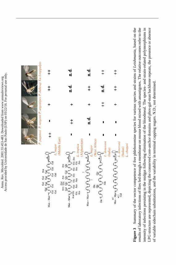
25 Aug 2001 17:28 AR AR135-18-COLOR.tex AR135-18-COLOR.SGM ARv2(2001/05/10)P1: GDL
Fig
ure
3S
umm
ary
ofth
eve
ctor
com
pete
nce
offiv
eph
lebo
tom
ine
spec
ies
for
vario
ussp
ecie
san
dst
rain
sof
Le
ish
ma
nia,
base
don
the
resu
ltsof
labo
rato
ryin
fect
ions
offli
esfe
dth
roug
ha
mem
bran
eon
mou
sebl
ood
mix
edw
itham
astig
otes
.The
rela
tive
outc
omes
refe
rto
the
inte
nsity
ofin
fect
ions
pers
istin
gin
the
mid
gutf
ollo
win
gel
imin
atio
nof
the
bloo
dmea
l.T
hesp
ecie
s-an
dst
rain
-rel
ated
poly
mor
phis
ms
inLP
Gst
ruct
ure
are
repr
esen
ted,
depi
ctin
gth
eco
nser
ved
core
-anc
hor
dom
ains
and
phos
-gal
-man
back
bone
repe
ats,
the
pres
ence
orab
senc
eof
varia
ble
side
chai
nsu
bstit
utio
ns,a
ndth
eva
riabi
lity
inte
rmin
alca
ppin
gsu
gars
.N.D
.,no
tdet
erm
ined
.
Ann
u. R
ev. M
icro
biol
. 200
1.55
:453
-483
. Dow
nloa
ded
from
ww
w.a
nnua
lrev
iew
s.or
g A
cces
s pr
ovid
ed b
y U
nive
rsid
ade
de S
ao P
aulo
(U
SP)
on 0
3/22
/16.
For
per
sona
l use
onl
y.
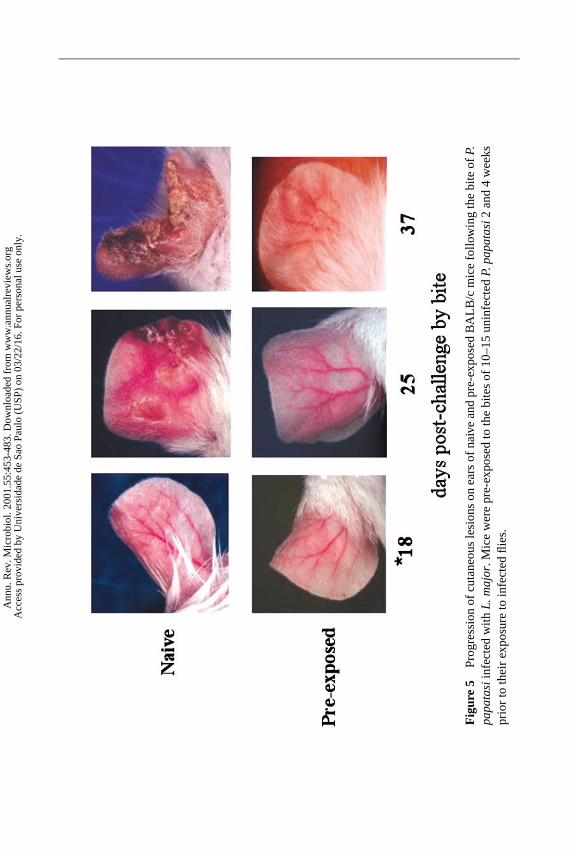
23 Aug 2001 16:36 AR AR135-18-COLOR.tex AR135-18-COLOR.SGM ARv2(2001/05/10)P1: GDL
Fig
ure
5P
rogr
essi
onof
cuta
neou
sle
sion
son
ears
ofna
ive
and
pre-
expo
sed
BA
LB/c
mic
efo
llow
ing
the
bite
ofP.
pa
pa
tasi
infe
cted
with
L.m
ajo
r.M
ice
wer
epr
e-ex
pose
dto
the
bite
sof
10–1
5un
infe
cted
P.p
ap
ata
si2an
d4
wee
kspr
ior
toth
eir
expo
sure
toin
fect
edfli
es.
Ann
u. R
ev. M
icro
biol
. 200
1.55
:453
-483
. Dow
nloa
ded
from
ww
w.a
nnua
lrev
iew
s.or
g A
cces
s pr
ovid
ed b
y U
nive
rsid
ade
de S
ao P
aulo
(U
SP)
on 0
3/22
/16.
For
per
sona
l use
onl
y.

P1: FRK
August 17, 2001 16:57 Annual Reviews AR135-FM
Annual Review of MicrobiologyVolume 55, 2001
CONTENTS
FRONTISPIECE, John L. Ingraham xii
LEARNING TO FLY FISH, John L. Ingraham 1
ROLES OFTHIOL-REDOX PATHWAYS IN BACTERIA, Daniel Ritz andJon Beckwith 21
BACTERIAL GLIDING MOTILITY : MULTIPLE MECHANISMSFORCELL MOVEMENT OVERSURFACES, Mark J. McBride 49
TOXIC SHOCK SYNDROME AND BACTERIAL SUPERANTIGENS:AN UPDATE, John K. McCormick, Jeremy M. Yarwood, andPatrick M. Schlievert 77
BIG BACTERIA, Heide N. Schulz and Bo Barker Jørgensen 105
NONREPLICATINGPERSISTENCE OFMYCOBACTERIUM TUBERCULOSIS,Lawrence G. Wayne and Charles D. Sohaskey 139
QUORUM SENSING IN BACTERIA, Melissa B. Miller and Bonnie L. Bassler 165
ADVANCES IN THE BACTERIOLOGY OF THECOLIFORM GROUP: THEIRSUITABILITY AS MARKERS OFMICROBIAL WATER SAFETY,H. Leclerc, D. A. A. Mossel, S. C. Edberg, and C. B. Struijk 201
BIOLOGICAL WEAPONS—A PRIMER FORMICROBIOLOGISTS,Robert J. Hawley and Edward M. Eitzen, Jr. 235
VIRUSES ANDINTERFERONS, Ganes C. Sen 255
PHAGES OFDAIRY BACTERIA, Harald Brussow 283
BACTERIAL FATTY ACID BIOSYNTHESIS: TARGETS FORANTIBACTERIAL DRUG DISCOVERY, John W. Campbell andJohn E. Cronan, Jr. 305
NOVEL THIOLS OFPROKARYOTES, Robert C. Fahey 333
A COMMUNITY OF ANTS, FUNGI, AND BACTERIA: A MULTILATERALAPPROACH TOSTUDYING SYMBIOSIS, Cameron R. Currie 357
HOMOLOGY-DEPENDENTGENE SILENCING MECHANISMSIN FUNGI, Carlo Cogoni 381
INTERACTION OFBACTERIAL PATHOGENS WITHPOLARIZEDEPITHELIUM, B. I. Kazmierczak, K. Mostov, and J. N. Engel 407
BACTERIOPHAGETHERAPY, William C. Summers 437
vi
Ann
u. R
ev. M
icro
biol
. 200
1.55
:453
-483
. Dow
nloa
ded
from
ww
w.a
nnua
lrev
iew
s.or
g A
cces
s pr
ovid
ed b
y U
nive
rsid
ade
de S
ao P
aulo
(U
SP)
on 0
3/22
/16.
For
per
sona
l use
onl
y.

P1: FRK
August 17, 2001 16:57 Annual Reviews AR135-FM
CONTENTS vii
MOLECULAR ASPECTS OFPARASITE-VECTOR ANDVECTOR-HOSTINTERACTIONS INLEISHMANIASIS, David Sacks and Shaden Kamhawi 453
AMMONIA -OXIDIZING BACTERIA: A MODEL FORMOLECULARMICROBIAL ECOLOGY, George A. Kowalchuk and John R. Stephen 485
IMMUNE CHECKPOINTS INVIRAL LATENCY, Stella Redpath, Ana Angulo,Nicholas R. J. Gascoigne, and Peter Ghazal 531
RECOMBINATION AND THE POPULATION STRUCTURES OFBACTERIALPATHOGENS, Edward J. Feil and Brian G. Spratt 561
PERIPLASMIC STRESS ANDECF SIGMA FACTORS, Tracy L. Raivioand Thomas J. Silhavy 591
HYDROPHOBINS: MULTIPURPOSEPROTEINS, Han A. B. Wosten 625
ANTHRAX, Michele Mock and Agnes Fouet 647
ANTIGENIC VARIATION AT THE INFECTEDRED CELL SURFACEIN MALARIA , Sue Kyes, Paul Horrocks, and Chris Newbold 673
HORIZONTAL GENE TRANSFER INPROKARYOTES: QUANTIFICATIONAND CLASSIFICATION, Eugene V. Koonin, Kira S. Makarova, andL. Aravind 709
ASPECTS OFFUNGAL PATHOGENESIS INHUMANS, Jo-Anne H. van Burikand Paul T. Magee 743
INDEXESSubject Index 773Cumulative Index of Contributing Authors, Volumes 51–55 807Cumulative Index of Chapter Titles, Volumes 51–55 810
ERRATAAn online log of corrections toAnnual Review of Microbiologychapters(if any have yet been occasioned, 1997 to the present) may be found athttp://micro.AnnualReviews.org
Ann
u. R
ev. M
icro
biol
. 200
1.55
:453
-483
. Dow
nloa
ded
from
ww
w.a
nnua
lrev
iew
s.or
g A
cces
s pr
ovid
ed b
y U
nive
rsid
ade
de S
ao P
aulo
(U
SP)
on 0
3/22
/16.
For
per
sona
l use
onl
y.