Embed Size (px)
Citation preview
Host-Parasite Interaction of Resistant Sugarbeet and Heterodera schachtiP
M. H. Yu and A. E. Steele 2
Abstract: T h e host-parasite relationships between Heterodera schachtii Schm. and the nematode-resistant diploid Beta vulgaris L. line '51501' were examined via serial sections of secondary rootlets. Second-stage larvae penetrated sugarbeet roots and migrated up to 1.95 m m before establishing pe rmanen t feeding sites. Most sedentary larvae were oriented parallel to the root axis or in various diagonal or folded positions in the cortex. Nematodes adopted no definite orientat ion with regard to the root apex. Nematode feeding st imulated formation of mult inucleate syncytia in host tissues. Syncytia were 0.3-1.1 m m in length, up to 90 Fm × 150 #m in c r o s s section. Root diameters were enlarged close to feeding sites. Usually nematodes deteriorated con- comitant with necrosis of syncytia, and dead nematodes frequently appeared macerated or flat- tened and deformed. Most nematodes did not develop to maturity" in the resistant host tissues, Cavities left by collapse of syncytia were filled by growth of parenchymatous tissue. Key words: Beta vulgaris L., cyst, histopathology, necrosis, nematode resistance, syncytium.
Sugarbeet (Beta vulgaris L.) is the prin- cipal host for the sugarbeet cyst nematode, Heterodera schachtii Schm. Selection for a true-breeding genotype of B. vulgaris re- sistant to H. schachtii within sugarbeet cul- tivars has been unsuccessful (1,2,8). How- ever, the three wild species--B, procumbens Chr. Sm., B. webbiana Moq., and B. patel- laris Moq.--in the section Patellares are highly resistant to H. schachtii (7). B. pro- cumbens has shown the highest degree of resistance (6). Nematode resistance has been transferred from B. procumbens into sugar- beet genome by interspecific hybridization (14).
Previous studies have described syncytial development in roots of sugarbeet suscepti- ble to the cyst nematode (9,15). Litt le in- formation is available, however, on the his- topathology of H. schachtii infection in the resistance cultivars of sugarbeet. In this study we describe histopathology and nema- tode development in a resistant diploid sugarbeet infected with H. schachtii.
MA T E R I AL S AND M E T H O D S
Sugarbeet nematodes used in this study were collected from a H. schachtii-infested field near Chualar, California, and were in-
Received for publication 22 October 1980. 1Contribution of the U. S. Agricultural Research Station,
Agricultural Research, Science and Education Administration, U. S. Department of Agriculture, Salinas, CA 93915. Mention of a trademark or proprietary product does not constitute a guarantee or warranty of the product by the USDA and does not imply its approval to the exclusion of other products that may also be suitable.
2Respectively, Research Geneticist and Zoologist, USDA SEA AR, P. O. Box 5098, Salinas, CA 93915. T h e authors are indebted to Susan F. Gilliam for her contribution in preparing the slides used.
206
creased for inoculum on sugarbeets in green- house pot cultures. Mature brown cysts were selected and treated with a hatching solu- tion (17) in 20-cm pans and maintained at 27 C in an incubator. Most larvae used for inoculunl hatched within 5 d.
T h e nematode-resistant diploid sugar- beet line '51501' is a progeny of B. vulgaris × B. procumbens hybrids (18). Seeds were germinated in steam-sterilized sand and seedlings transplanted at the two-leaf stage to a luminum foil cylinders (6 X 17.5 cm) containing soil with 40 H. schachtii cysts (estimated to have 4,000 larvae hatched). Forty-five days after transplanting the seed- lings, the external surfaces of roots were ex- amined for the presence of white females. Plants with fewer than five females were re- planted and inoculated two addit ional times each with 2,500 larvae. Six weeks after the third inoculat ion those plants (approxi- mately 5 months old) still not support ing five females were selected as resistant plants.
T h e secondary roots were removed from the tap roots and the selected resistant plants transplanted to 450-g styrofoam cups containing two parts sterilized clay-loam soil and one part sand mixture. Plants were maintained in an incubator at 27 C for a 16-hour photoperiod. Three days after trans- planting, 2,000 active larvae were pipet ted to the soil immediately around each sugar- beet plant. Prel iminary studies showed that few H. schachtii larvae penetrate secondary roots within 1-3 d after inoculation. T o obtain numbers of infected roots sufficient for histological examination, we exposed roots to larvae for 4 d. T h e plants were then
Heterodera schachtii and Resistant Sugarheet: Yu, Steele 207
removed from the cups and the roots thoroughly washed to remove soil. T h e plants were t ransplanted into clean con- tainers filled with sterilized soil for fur ther development of the nematodes.
Segments f rom infected secondary roots were excised for histological examina t ion at 4-5-d intervals up to 31 d after inoculation. Tissues were killed, fixed, and stored in Nawaschin type Craft I I I fixative (13) at room temperatures. Root segments were embedded in Pa rap la s t+ embedding me- dium, and 10-12-/~m longitudinal and trans- verse sections were cut with a rotary micro- tome. Serial sections were moun ted on slides, stained with a modified hematoxylin- safranin-fast green staining schedule, and examined microscopically. Results of these examinat ions were compared with the his- topathology of susceptible sugarbeet de- scribed in previous studies (15,16).
R E S U L T S
Entry and development of H. schachtii in resistant host tissues: Examina t ion of whole, unstained roots revealed longitu- dinal to ovoid necrotic lesions at larval penetra t ion sites. Often lesions were located on slightly swollen areas of the roots. Wi th in 4 d of inoculation, numerous larvae had penetrated into the sugarbeet roots and migrated into tile cortex. Some larvae quickly established a posit ion for feeding while others migrated a short distance in tile cortex leaving tunnels of broken cells. Invasion courts containing nematodes ranged up to 1.95 m m in length. In several cases, invasion courts extended for some distance from the nematode feeding sites. Some root cavities did not contain nema- todes.
Nematode penetra t ion and migrat ion within the roots was intracellular. Most larvae had completely penetra ted the root tissue, others only partially. Larvae fre- quent ly traversed a root t ip adjacent to the root cap and posit ioned themselves less than 0.5 m m from the root tip. Broken cells of the invasion tracts as near as 0.6 m m to the nematode were often necrotic.
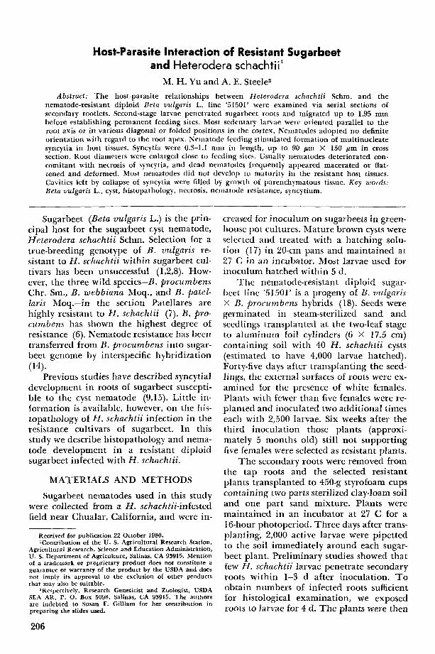
Upon reaching their feeding positions most larvae became oriented parallel to the root axis, bu t some were in diagonal posi-
tions within the cortex (Fig. 1). Some larvae were posit ioned entirely wi th in endodermal cell layers, while tails of other larvae were partially extended three or four cell tiers into the cortex or were exposed on the root surface. T h e head regions of feeding nema- todes were wi thin or close to the endodermal cells and were oriented toward ttle vascular system (Figs. 2, 3).
Larvae frequently adopted folded posi- tions within the cortex, and two or more sections of a single larva were observed in the same plane (Figs. 4, 5). Thus a t tempts to determine the number of nematodes in a part icular root area required the examina- tion of serial sections.
T e n days alter inoculation, male larvae were observed in root sections with their bodies coiled within the old larval cuticle (Figs. 6, 7). Fourth- and early fifth-stage males were coiled into two or three folds within the third-stage cuticle. Several male larvae appeared to have penetra ted only shallowly in tile cortex. Males were not found ill tissues after 20 d.
T h e first indicat ion of larval deteriora- tion was observed 10 d after inoculation. Deteriorated nematodes usually appeared macerated and devoid of recognizable or- gans or contained deformed organs. In transverse sections, dead larvae commonly showed cuticular indentat ions or infoldings (Figs. 8-10). After 20 d or longer, the sclerotized cephalic f ramework of the nema- tode frequently retained its original shape. T h e remaining anter ior por t ion of the nematode contained little or no internal contents and was part ial ly flattened, whereas the posterior third of the nematode occa- sionally contained obscure, disorganized, or deformed reproduct ive systems. T h e length of measurable larval remains ranged be- tween 340 and 470 ~m. Judging f rom cuticular markings that were detectable, several female nematodes developed to the fourth larval stage.
Histopathological reaction of resistant sugarbeets: Within 4 d of infection, phase i l luminat ion showed crystalline cytoplasmic granules in cells fed upon by nematodes (Fig. 11). Thereaf ter , the affected ceils and nuclei enlarged, cytoplasm increased in den- sity, and cytoplasmic granules became more hyperchromat ic (Figs. 5, 12, 1.~). Syncytia
I
208 Journal of Nematology, Volume 13, No. 2, April 1981
f ,
Heterodera schachti i and Resistant Sugarbeet: Yu, Steele 209
typically developed within the stele and in- corporated cells of the pericycle, proto- phloem, and interfascicular parenchyma. Occasionally syncytia were ini t iated at the centripetal boundary of cortical cells and extended into the stele.
Each syncytium extended from the initial cell to neighboring cells near the outer lay- ers of the vascular cylinder. T h e developing syncytium spread longitudinal ly in both directions, by gradual dissolution of cell walls and coalescence of cytoplasm, and merged as one continuous mul t inucleate cytoplasmic uni t (Figs. 12, 13). Differences in the size and staining of syncytial nuclei were observed. Syncytial cytoplasm eventu- ally became turbid and heavily stained. T h e nuclei frequently showed different reticular structures (Fig. 13). Syncytia usu- ally at tained m a x i m u m size adjacent to the nematode feeding points about 10 d after inoculation. T h e m a x i m u m width of syn- cytia near feeding sites was as large as 90 t~m at the edge of pericycle and 150 ~tm from the pericycle to tile center of tile stele. Aftcr 14 d, syncytia ranged from 0.3 to 1.1 m m in length. Cell walls close to nematodes and vascular elements were often diffuse and prominent ly stained (Figs. 5, 7-10, 12-14).
T h e shapes of syncytia were longi tudinal and resembled an asymmetrical spindle, broader at one end and gradually tapered to the other. In a root segment parasitized by a single nematode, the syncytium displaced xylem elements in a l imited sector of the root. Ti le presence of discontinuous cell walls indicated that syncytial complexes were formed by the coalescence of cytoplasm from adjacent cells. In mul t ip le infections, adjacent syncytia resulted in extensive vas- cular damage. Single infections in the same root region frequently induced syncytia that formed discrete units.
T h e enlarged mult ip le syncytia had a
I I I I I \ \ \ \ \
secondary effect of enlarging roots near tile feeding sites of tile nematodes. Less fre- quently, localized enlargement of roots was caused by repeated division of pericyle cells. Swellings extended 60-90 /~m beyond the 300-ttm mean root diameter.
Deter iorat ion of syncytia was observed within 10 d after inoculation. By this time, some necrotic syncytia had separated f rom vascular tissues leaving large cavities de- void of plant cells (Figs. 13-14). Thereaf te r necrosis became progressively more severe, result ing in total collapse of the syncytium (Fig. 15), thereby leaving extensive cavities in the roots. Necrotic syncytia in samples taken 25 d after inoculat ion were usually, but not always, associated with deter iorated larvae. After 30 d, many larvae had degen- erated to the point that they were difficult to identify as nematodes. Rejuvenated par- enchymatous tissues invaded spaces left by the receding syncytial walls (Fig. 16).
DISCUSSION
Second-stage larvae that entered the roots of resistant sugarbeets showed great vari- abili ty in establishment of feeding positions. Some larvae wandered within the cortex changing direction at acute angles thereby causing extensive injury in the invaded tissues. Rarely, larvae only part ial ly pene- trated the root before becoming sedentary. Steele (16) repor ted that some larvae fre- quent ly do not completely enter roots but remain at tached with posteriors external to the root systems.
T h e terminal feeding posit ion and ori- entat ion were not the same for all larvae. For example, in a 2.5-mm root segment where five nematodes were arranged tan- demly in the cortex of a root, at least one was oriented in a reverse direction. This was in agreement with previous research by
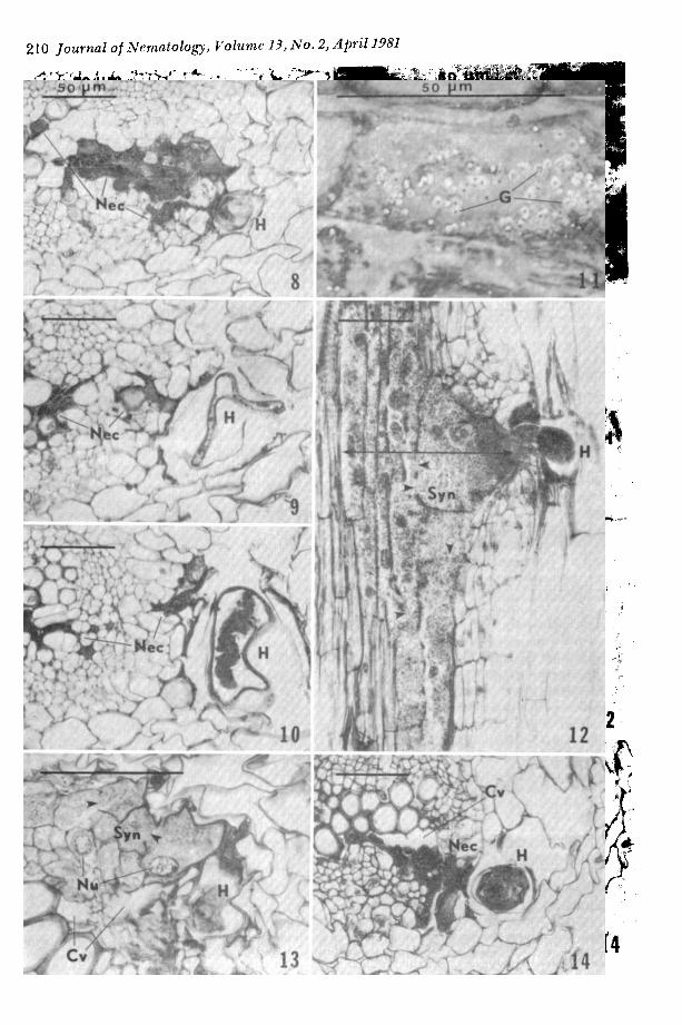
Figs. 1-7. 1) Diagonal penetration of Heterodera schachtii (H) inside the cortex (Cx) of sugarbeet root, 4 d after inoculation. 2,3) Bending of the head region (arrows) of H. schachtii toward the root vascular ele- ments, shown in longitudinal and transverse sections, 10 d. Note the cluster of cytoplasmic granules (G) in Fig. 3. 4,5) Multiple infection of sugarbeet root by H. schachtii (Ha, Hb, and Hc); one nematode (Hb) in folded position showing two and three separate sections, respectively, l0 d. Note the sloughing (S) of cortical tissues, a normal event in sugarbeet (4), and hyperchromatic cytoplasm and nucleus (Nu) in the initial cell of syncytium (Syn), about 20 #m from the anterior tip of a nematode (Ha) in Fig. 5. 6,7) Male larva coiled within cast cuticle (arrows). shown in longitudinal and transverse sections, 14 d. Note the invasion court (IC) of nematode in Fig. 6, and the cast cuticle surrounding the coiled male larvae in Fig. 7 indicating that the fourth molt has occurred. Scale bar = 50 #m.
210 Journal of Nematology, Volume 13, No. 2, April 1981
Heterodera schachtii and Resistant Sugarbeet: Yu, Steele 211
' , , . . . . 5o
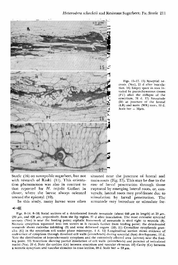
Figs. 15-17. 15) Syncytial ne- crosis (Nec), 25 d after inocula- tion. 16) Empty space in root in- vaded by parenchymatous tissues (PT) after the collapse of the syncytium, 31 d. 17) Nematode (H) at juncture of the lateral (LR) and main (MR) roots, 10 d. Scale bar = 50#m.
Steele (16) on susceptible sugarbeet, but not with research of Raski (11). This orienta- tion phenomenon was also in contrast to tlaat reported for H. tri[olii Goffart in clover, where the larvae always oriented toward the epicotyl (10).
In this study, many larvae were often
~ l IIIII ~, \ \ \ \ \
situated near the juncture of lateral and main roots (Fig. 17). This may be due to the ease of larval penetrat ion through tissue ruptured by emerging lateral roots, or, con- versely, lateral roots may proliferate due to st imulation by larval penetrat ion. T h e n~nlatotte may introduce or stimulate for-
Figs. 8-14. 8-10) Serial sections of a deteriorated female nematode (about 460 #m in length) at 20 #m, 230 #m, and 420 #m, respectively, from the lip region, 31 d after inoculation. The most extensive syncytial necrosis (Nee) is near the feeding point; cephalic framework of nematode is sited right to necrosis (8). Necrotic syncytium separated into two sectors as it extemls farther from feeding point; the deteriorated nematode shows cuticular infolding (9) and some deformed organs (10). 11) Crystalline cytoplasmic gran- ules (G) in the syncytium cell under phase microscopy. 4 d. 12) I,ongitudinal section shows evidence of coalescence of cytoplasm through dissolved cell walls (arrowheads) during syncytial (Syn) development, 14 d. Note the distribution of hyperchromatic cytoplasm and ~he extensively affected area (arrows) near the feed- ing point. 13) Syncytium showing partial dissolution of cell walls (arrowheads) and presence of reticulated nuclei (Nu). 20 d. Note the cavities (Cv) between syncytium and vascular elements. 14) Cavity (Cv) between a necrotic syncytium and vascular elements in cross section, 10 d. Scale bar = 50 #m.
212 Journal o I Nematology, Volume 13, No. 2, April 1981
m a t i o n or r e d i s t r i b u t i o n of g rowth-promot- ing hormones in its host, thereby i n d u c i n g fo rma t ion of new roots (4). F r a n k l i n (5) descr ibed how lateral roots are formed to take the place of those i nvaded by H. schachtii.
In i t ia l ly , syncytia in resis tant sugarbeet appeared to be s imi lar to tha t i n d u c e d ill suscept ible sugarbeet . Previous research (15) ind ica ted roots of susceptible sugar- beets had more m u l t i p l e in fec t ion syncytial complexes.
De te r io ra t ing syncytia left spaces i n to which r e juvena t ed p a r e n c h y m a cells ex- panded , thereby sepa ra t ing syncytia in to several isolated fragments. F requen t ly , one f ragment was ad jacen t to the center of the stele and the o ther nea r the de te r iora ted n e m a t o d e (Figs. 8-10, 16). Because con- t i nuous larval infec t ions d id no t occur in this study, c o n t i n u o u s fo rma t ion of syncytia was no t possible and histological damage in roots of resis tant p lan t s was even tua l ly re- pa i red by new parenchyma.
Once the nematodes establ ished a perma- n e n t feeding site, no fu r the r m i g r a t i o n oc- curred. Deter iora ted larvae were always as- sociated wi th advanced necrosis of syncytia. W h e t h e r larvae died as a resul t of the in- ab i l i ty of degene ra t i ng syncytia to supply nu t r i en t s , or syncytia became necrot ic as a resul t of the cessation of larval feeding, could no t be ascertained. I n Meloidogyne sp. infect ions of tomato, host necrosis var ied inversely wi th larval growth (3). Ross (12) a t t r i b u t e d the i n d u c t i o n of a hypersensi t ive reac t ion in res is tant soybean roo t tissue to the secretions of the soybean cyst nematode . Studies of susceptible sugarbeet (9,15) have shown that syncytia usua l ly d id no t become necrot ic u n t i l after the n e m a t o d e comple ted its life cycle. T h i s suggests tha t collapse of syncytia follows, and is perhaps the resul t of, cessation of feeding.
L I T E R A T U R E C I T E D
1. Curtis, G. 1970. Resistance of sugarbeet to the cyst-nematode Heterodera schachtii Sch. Ann. Appl. Biol. 66:169-177.
2. Doney, D. L., and E. D. Whitney. 1969. Screen- ing sugarbeet for resistance to Heterodera schachtii Sch. J. Am. Soc. Sugar Beet Technol. 15:546-552.
3. Dropkin, V. H. 1969. The necrotic reaction of tomatoes and other hosts resistant to Meloidogyne: reversal by temperature. Phytopathology 59:1632- 1637.
4. Dropkin, V. H. 1969. Cellular responses of plants to nematode infections. Ann. Rev. Phytopath. 7:101-122.
5. Franklin, M. T. 1951. The cyst-forming species of Heterodera. Comm. Agr. Bur., Farnham Royal, Bucks, England.
6. Golden, A. M. 1958. Interrelationships of cer- tain Beta species and Heterodera schachtii, the sugar-beet nematode. Plant Dis. Rep. 42:1157-1162.
7. Golden, A. M. 1959. Susceptibility of several Beta species to the sugar-beet nematode (Heterodera schachtii) and root-knot nematodes (Meloidogyne spp.). J. Am. Soc. Sugar Beet Technol.. 10:444-447.
8. Heijbroek, W. 1977. Partial resistance of sugar- beet to beet cyst eelworm (Heterodera schachtii Schm.). Euphytica 26:257-262.
9. Jatala, P., and H. J. Jensen. 1976. Histopath- ology of Beta vulgaris to individual and concomitant infections by Meloidogyne hapla and Heterodera schachtii. J. Nematol. 8:336-341.
10. Mankau, R., and M. B. Linford. 1960. Host- parasite relationships of the clover cyst nematode, Heterodera trifolii Goffart. IlL Agric. Ext. Stn. Bull. No. 667:1-50.
11. Raski, D. J. 1950. The life history and morphology of the sugar beet nematode, Heterodera schachtii Schm. Phytopathology 40:155-151.
12. Ross, J. P. 1958. Host-parasite relationship of the soybean cyst nematode in resistant soybean roots. Phytopa thology 48:578-579.
13. Sass, J. E. 1958. Botanical microtechnique. 3d ed. Iowa State University Press, Ames, Iowa.
14. Savitsky, H. 1975. Hybridization between Beta vulgaris and B. procumbens and transmission of nematode (Heterodera schachtii) resistance to sugarbeet. Can. J. Genet. Cytol. 17:197-209.
15. Steele, A. E. 1971. Morphological changes in roots of sugarbeet and tomato infected with Heterodera schachtii Schmidt 1871. J. Am. Soc. Sugar Beet Technol. 16:561-567.
16. Steele, A. E. 1971. Orientation and develop- ment of Heterodera schachtii larvae on tomato and sugarbeet roots. J. Nematol. 5:424-426.
17. Whitney, E. D., and D. L. Doney. 1970. Large scale hatching, disinfestation, and storage of Heterodera schachtii larvae. Phytopathology 60:1191- 1194.
18. Yu, M. H. 1978. Meiotic behavior of a disomic nematode-resistant sugarbeet. Crop Sci. 18:615-618.