Embed Size (px)
Citation preview
Molecular approaches for bacterial azoreductases
Montira Leelakriangsak
Department of Science, Faculty of Science and Technology, Prince of Songkla
University, Pattani campus, Pattani, Thailand 94000
Keywords: azoreductase, azo dyes, molecular techniques, recombinant strain
Corresponding author
Email address: [email protected]
Abstract
Azo dyes are the dominant types of synthetic dyes, widely used in textiles,
foods, leather, printing, tattooing, cosmetics, and pharmaceutical industries. Many
microorganisms are able to decolorize azo dyes, and there is increasing interest in
biological waste treatment methods. Bacterial azoreductases can cleave azo linkages
(-N=N-) in azo dyes, forming aromatic amines. This review mainly focuses on
employing molecular approaches, including gene manipulation and recombinant
strains, to study bacterial azoreductases. The construction of the recombinant protein
by cloning and the overexpression of azoreductase is described. The mechanisms and
function of bacterial azoreductases can be studied by other molecular techniques
discussed in this review, such as RT-PCR, southern blot analysis, western blot
analysis, zymography, and mutagenesis in order to understand bacterial azoreductase
properties, function and application. In addition, understanding the regulation of
azoreductase gene expression will lead to the systematic use of gene manipulation in
bacterial strains for new strategies in future waste remediation technologies.
1. Introduction
1
Synthetic dyes are defined as colored substances which are resistant to fading
upon exposure to light, water, sweat, many chemicals and microbial attack (Robinson
et al., 2001, Saratale et al., 2011). Due to their chemical structure, many dyes are
difficult to decolorize (Stolz, 2001). They are classified as acidic, basic, disperse, azo,
diazo, anthroquinone based and metal complex dyes (Robinson et al., 2001). Azo dye
compounds, the most used synthetic dyes, account for approximately half of the dyes
used in the textile industry. They are the most common synthetic colorants released
into the environment (Baiocchi et al., 2002, Saratale et al., 2011).
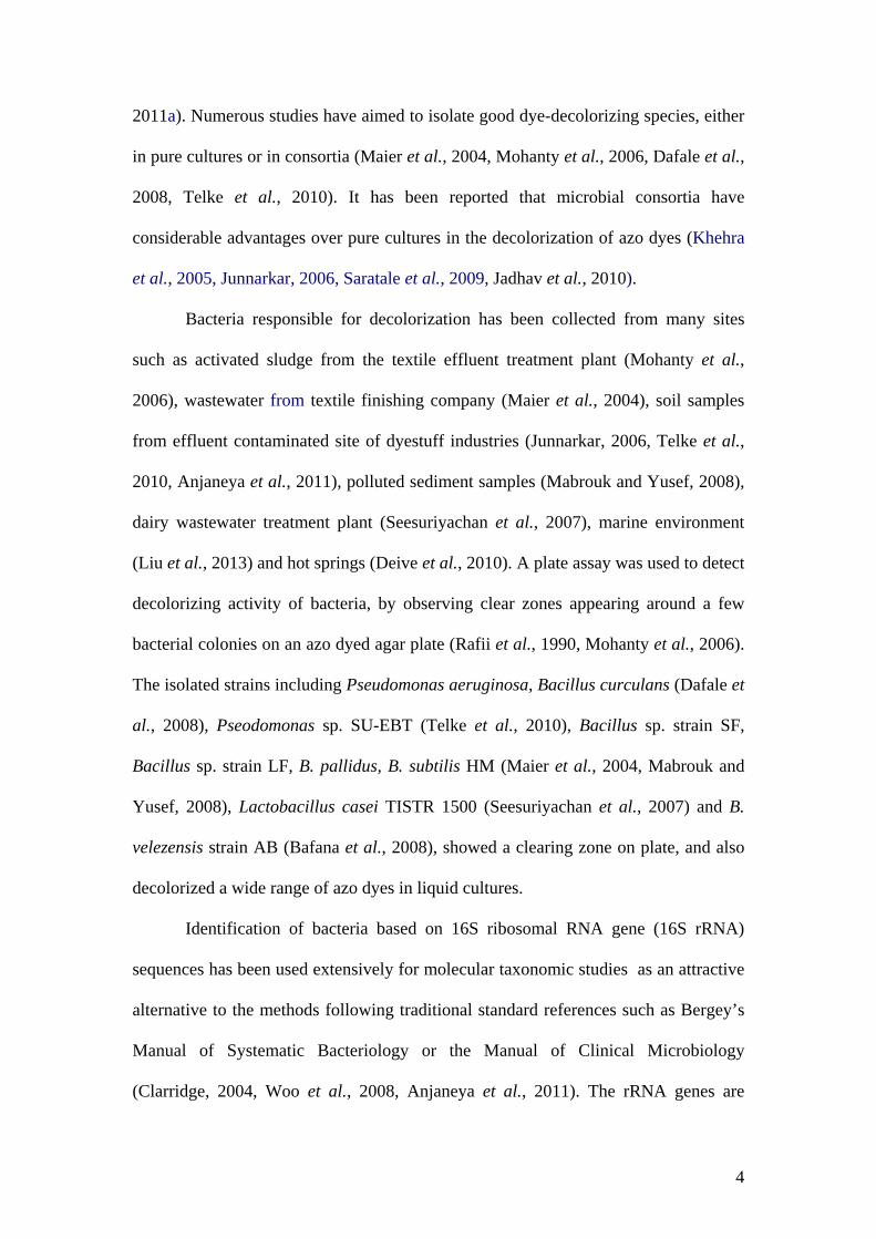
Azo dyes are characterized by the presence of one or more azo group (-N=N-)
that are chromophores, associated with aromatic and other groups such as hydroxyls (-
OH), Chloro (-Cl), methyl (-CH3), nitro (-NO2), amino (-NH3), carboxyl (-COOH)
and sulfonic groups (-SO3H), which give various types of azo dyes (Fig. 1) (Stolz,
2001, Forgacs et al., 2004, Saratale et al., 2011). They are widely used in textiles,
foods, industrial, printing, tattooing, cosmetics and for clinical purposes (Suzuki et al.,
2001, Bin et al., 2004, Chen et al., 2005). These dyes are usually recalcitrant to
conventional wastewater treatment (Forgacs et al., 2004). Several physico-chemical
methods like adsorption, electrocoagulation, chemical treatment, photocatalysis,
oxidation and ion pair extractions, have been adopted and proven to be costly and to
produce large amounts of sludge (Robinson et al., 2001, Forgacs et al., 2004, Saratale
et al., 2011). More studies focus on biological treatment methods (Supaka et al., 2004,
Jadhav et al., 2007, Pandey et al., 2007, Mabrouk and Yusef, 2008, Gopinath et al.,
2009, Parshetti et al., 2010).
A wide range of microorganisms including bacteria (Song et al., 2003, Dafale
et al., 2008, Jadhav et al., 2010, Telke et al., 2010), yeast, (Jadhav and Govindwar,
2006, Jadhav et al., 2007, Tastan et al., 2010), fungi (Gou et al., 2009, Kaushik and
2
Malik, 2009) and algae (Daneshvar et al., 2007), are able to reduce azo compounds to
non-colored products or even to completely mineralize them (Stolz, 2001, Chen et al.,
2004, Mohanty et al., 2006, Ooi et al., 2007). Various microorganisms are able to
metabolize azo dyes by biosorption and biodegradation, involving enzymatic
mechanisms such as those associated with lignin peroxidases, manganese peroxidases,
laccases and azoreductases, (Stolz, 2001, Jadhav et al., 2007, Bafana et al., 2008,
Kaushik and Malik, 2009, Mendes et al., 2011, Saratale et al., 2011). Therefore, the
biological degradation and use of microbial or enzymatic treatment methods for
removal of these dyes have potential important advantages: less sludge, environmental
friendliness and cost competitiveness (Stolz, 2001, Forgacs et al., 2004, Pandey et al.,
2007, Saratale et al., 2011).
There are several reviews on treatment of waste effluents containing synthetic
dyes by physicochemical and microbiological methods, including bacterial
decolorization of azo dyes (Robinson et al., 2001, Stolz, 2001, Forgacs et al., 2004,
Pandey et al., 2007, Saratale et al., 2011). However, the current review focuses on
bacterial azoreductases and their characterization by molecular biology approaches, in
relation to wastewater treatment. In addition, gene manipulation and the recombinant
strains with higher biodegradation capacity are included, because they can
significantly benefit the future technologies for dye removal.
2. Decolorization of azo dyes by bacterial azoreductases
2.1 Isolation and identification of dye decolorizing bacterium
In recent years, there has been an increasing interest in the use of biological
systems, especially bacteria, for the treatment of wastewaters containing dyes (Kumar
et al., 2006, Dafale et al., 2008, Sandhya et al., 2008, Liu et al., 2009b, Mendes et al.,
3
2011a). Numerous studies have aimed to isolate good dye-decolorizing species, either
in pure cultures or in consortia (Maier et al., 2004, Mohanty et al., 2006, Dafale et al.,
2008, Telke et al., 2010). It has been reported that microbial consortia have
considerable advantages over pure cultures in the decolorization of azo dyes (Khehra
et al., 2005, Junnarkar, 2006, Saratale et al., 2009, Jadhav et al., 2010).
Bacteria responsible for decolorization has been collected from many sites
such as activated sludge from the textile effluent treatment plant (Mohanty et al.,
2006), wastewater from textile finishing company (Maier et al., 2004), soil samples
from effluent contaminated site of dyestuff industries (Junnarkar, 2006, Telke et al.,
2010, Anjaneya et al., 2011), polluted sediment samples (Mabrouk and Yusef, 2008),
dairy wastewater treatment plant (Seesuriyachan et al., 2007), marine environment
(Liu et al., 2013) and hot springs (Deive et al., 2010). A plate assay was used to detect
decolorizing activity of bacteria, by observing clear zones appearing around a few
bacterial colonies on an azo dyed agar plate (Rafii et al., 1990, Mohanty et al., 2006).
The isolated strains including Pseudomonas aeruginosa, Bacillus curculans (Dafale et
al., 2008), Pseodomonas sp. SU-EBT (Telke et al., 2010), Bacillus sp. strain SF,
Bacillus sp. strain LF, B. pallidus, B. subtilis HM (Maier et al., 2004, Mabrouk and
Yusef, 2008), Lactobacillus casei TISTR 1500 (Seesuriyachan et al., 2007) and B.
velezensis strain AB (Bafana et al., 2008), showed a clearing zone on plate, and also
decolorized a wide range of azo dyes in liquid cultures.
Identification of bacteria based on 16S ribosomal RNA gene (16S rRNA)
sequences has been used extensively for molecular taxonomic studies as an attractive
alternative to the methods following traditional standard references such as Bergey’s
Manual of Systematic Bacteriology or the Manual of Clinical Microbiology
(Clarridge, 2004, Woo et al., 2008, Anjaneya et al., 2011). The rRNA genes are
4
highly conserved (least variable) DNA in all cells (Boye et al., 1999). The 16S rRNA
gene is now most commonly used for bacterial taxonomic purposes (Tortoli, 2003,
Clarridge, 2004, Seesuriyachan et al., 2007, Woo et al., 2008). Using 16S rRNA
sequencing, bacterial identification is more robust, reproducible, accurate and less
subjective test results (Clarridge, 2004, Woo et al., 2008).
2.2 Decolorization mechanism
Many microorganisms such as bacteria, fungi and yeast have been found to be
able to decolorize azo dyes by bioadsorption or degradation (Mabrouk and Yusef,
2008, Gou et al., 2009). A new fungal isolate, Penicillium sp. QQ could aerobically
decolorize Reactive Brilliant Red X-3B by bioadsorption rather than biodegradation
due to the adsorption of azo dyes by many functional groups located on the surface of
microbial cells (Gou et al., 2009). Mabrouk and Yusef (2008) showed that the
decolorization of Fast Red was achieved by B. subtilis HM due to degradation rather
than adsorption as indicated by the uncolored biomass.
Bacterial decolorization has been associated with various oxidoreductive
enzymes, including laccase, azoreductase and NADH-DCIP reductase (Stolz, 2001,
Kalme et al., 2007, Parshetti et al., 2010, Telke et al., 2010, Kolekar et al., 2013). In
addition, the location of the reaction can be either intracellular or extracellular
(Pandey et al., 2007, Seesuriyachan et al., 2007). This reaction may involve different
mechanisms such as enzymes by direct enzymatic azo dye reduction, low molecular
weight redox mediators, electron donor from the respiratory chain or a combination of
these (Pandey et al., 2007). The significance of oxidoreductive enzymes in
decolorization of Congo Red was examined by Telke and coworkers. The
observations demonstrated that laccase from Pseudomonas sp. SU-EBT was the key
enzyme responsible for Congo Red docolorization (Telke et al., 2010). To elucidate
5
the RO16 decolorization mechanism, oxidative and reductive enzyme was observed
after decolorization by bacterial consortium. The study showed that the laccase and
azoreductase are involved in RO16 biodegradation (Jadhav et al., 2010). The
significant increase in the enzyme activities of azoreductase and NADH-DCIP
reductase were observed by Kocuria rosea MTCC 1532 suggesting its involvement in
decolorization of methyl orange (Parshetti et al., 2010). The proposed pathway for
degradation of methyl orange by K. rosea is reported. Since azoreductases require the
additional of expensive cofactors as electron donors for the reductive reaction
resulting in aromatic amines which are potential toxic (Stolz, 2001, Chen et al., 2004,
Mohanty et al., 2006, Ooi et al., 2007), recently study from Mendes et al., (2011b)
has constructed co-expressing strain (azoreductase and laccase gene) which leads to
maximize decolorization and detoxification of azo dye-containing wastewater.
Azoreductases are involved in the degradation of azo dyes and also found in
intestinal microflora for activation of azo prodrugs in the treatment of inflammatory
bowel disease (IBD) (Ryan et al., 2010a, Wang et al., 2010). Azoreducases have been
shown to reduce azo compounds via a Ping Pong Bi Bi mechanism (Nakanishi et al.,
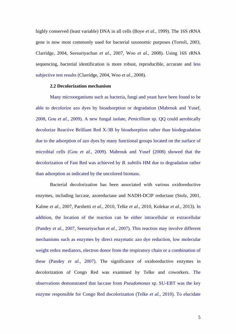
2001, Liu et al., 2008a, Wang et al., 2010, Mendes et al., 2011a). The proposed
mechanism for azo compound reduction requires two cycles of NAD(P)H-dependent
reduction, which reduces the azo substrate to a hydrazine in the first cycle and reduces
the hydrazine to two amines in the second cycle (Fig. 2) (Blumel et al., 2002, Deller
et al., 2008, Ryan et al., 2010a, Ryan et al., 2010b, Wang et al., 2010). The detection
of hydrazine intermediate by mass spectrometry has supported the mechanism (Bin et
al., 2004). By deducted amino acid sequence alignment, the NADH binding motif
(GXGXXG) have been found in azoreductases from B. cereus ATCC 10987, B.
6
anthracis Ames, Geobacillus sp. OY1-2, and Bacillus sp. OY1-2 (Suzuki et al., 2001,
Bin et al., 2004).
In summary, azoreductases have been classified into three major groups based
on structure, flavin dependency and dinucleotide preference: Group I, the polymeric
flavin-dependent NADH-preferred azoreductases; Group II, the polymeric falvin-
dependent NADPH-preferred azoreductases and Group III, the monomeric flavin-free
NAD(P)H-preferred azoreductases (Seesuriyachan et al., 2007, Chen et al., 2010,
Stingley et al., 2010, Feng et al., 2012).
3. Decolorization of azo dyes by recombinant azoreductases
3.1 Cloning and overexpression of azoreductase
The use of molecular tool has become increasingly integrated into
understanding enzyme biochemical properties and characterization. Researchers have
utilized a gene cloning method as a tool in producing recombinant strains for
decolorizing dyes more efficiently. Recombinant strain is also carried out to study
azoreductase activity and its mechanism (Chen et al., 2005, Deller et al., 2006, Ito et
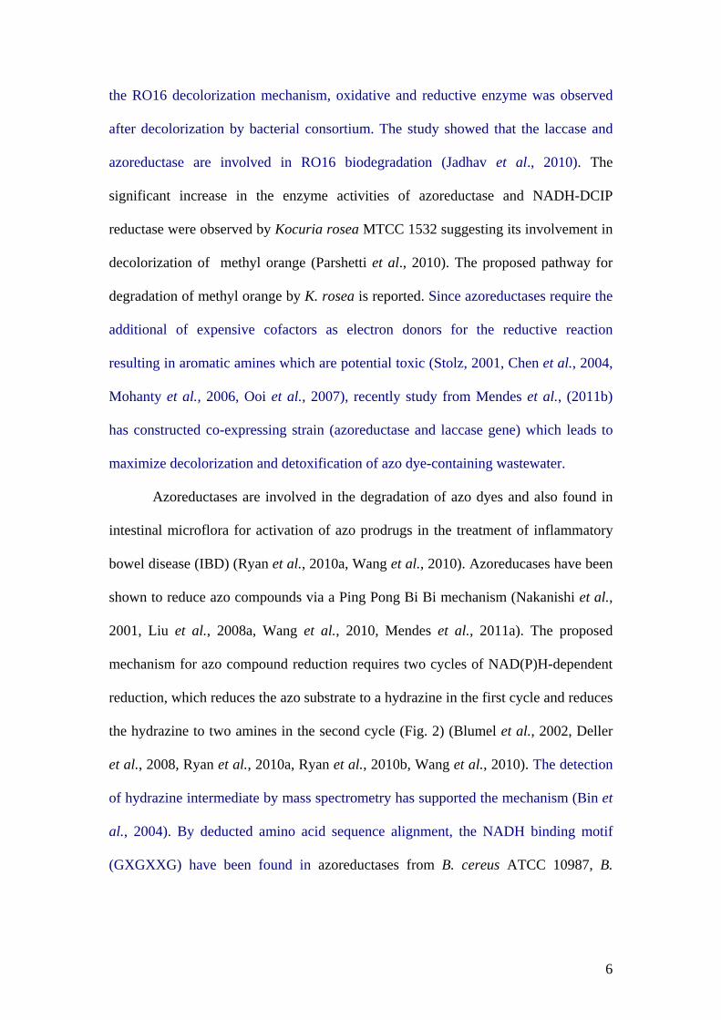
al., 2008, Chen et al., 2010, Ryan et al., 2010a). Construction of recombinant
expression vector is shown in Fig. 3. Recently, genes coding for aerobic azoreductase
have been cloned from Escherichia coli (Nakanishi et al., 2001, Liu et al., 2009a),
Bacillus sp. OY1-2 (Suzuki et al., 2001), B. subtilis (Deller et al., 2006, Nishiya and
Yamamoto, 2007), Enterococcus faecalis (Chen et al., 2004), E. faecium (Macwana et
al., 2010), Staphylococcus aureus (Chen et al., 2005), Rhodobacter sphaeroides (Bin
et al., 2004), Xenophilus azovorans KF46F (Blumel et al., 2002), Pigmentiphaga
kullae K24 (Chen et al., 2010), Pseudomonas aeruginosa (Wang et al., 2007, Ryan et
al., 2010b) and Geobacillus stearothermophilus (Matsumoto et al., 2010) (Table 1).
7
Many researchers have chosen E. coli as a host to express azoreductases
(Table 1). They cloned the azoreductase gene from potential decolorizing bacteria and
expressed it in E. coli to produce a recombinant E. coli strain followed by protein
purification. The expression of proteins in E. coli has many potential advantages
including the ease of growth and manipulation. Many vectors are available with
different N- and C-terminal tags and many host strains have been developed for
maximizing expression. Several chosen expression vectors are shown in table 1. pET
vector system contains several important elements including a lacI gene which codes
for the lac repressor protein, a T7 promoter which is specific to only T7 RNA
polymerase, multiple cloning site, selectable marker and protease cleavage site (e.g.
thrombin site in pET-15b) in order to remove a tag or other fusion proteins. Many
vectors encode additional optional components such as signal sequences (e.g. pET-
22b) to direct secretion and/or short peptide tags that are added to the N- or C-
terminus of the protein in order to improve expression, solubility, detection and
purification (Appelbaum and Shatzman, 1999). Some expression systems require the
use of specialized host strains which provide regulatory elements. Use of protease-
deficient host strains (e.g. BL21) can sometimes enhance product accumulation by
reducing degradation. The gene on the host cell chromosome usually has an inducible
promoter (e.g. T7-lac operator in pET vectors and PL in pTrx vectors) so that protein
expression can be induced by the addition of the proper inducer such as IPTG or by
shifting the temperature (Appelbaum and Shatzman, 1999). The construction of
expression vectors is generally straightforward by cloning gene sequence encoding
the azoreductase sequence to be expressed into an appropriate vector in the same
reading frame. Expression of recombinant azoreductase can be approached in general
by starting with introducing the recombinant plasmid into the required host cell,
8
growing the host cells and inducing expression, lysing the cells and analyzing by
SDS-PAGE to verify the presence of the protein (Qiagen).
3.2 Characterization of recombinant azoreductase
Molecular cloning of the gene encoding azoreductase enzyme followed by
protein purification is likely to be crucial for further characterization and application
of this enzyme (Suzuki et al., 2001, Wang et al., 2007, Ryan et al., 2010a, Wang et
al., 2010, Mendes et al., 2011a). Physiochemical properties, enzyme characterization
and kinetic studies can be investigated by obtaining purified azoreductase from whole
cell extract from the source organism or recombinant cell extract (Nachiyar and
Rajakumar, 2005, Wang et al., 2007, Gopinath et al., 2009, Punj and John, 2009,
Mendes et al., 2011a, Morrison et al., 2012). Purification from whole cell extracts
from the source organism employs classical purification procedures which require
many steps such as ammonium sulfate precipitation followed by ion exchange and
affinity chromatography methods (Maier et al., 2004, Nachiyar and Rajakumar, 2005,
Punj and John, 2009, Kolekar et al., 2013). However, in most cases recombinant DNA
techniques permit the construction of fusion proteins in which specific affinity tags
are added to the protein sequence of interest (Bin et al., 2004, Wang et al., 2007).
Therefore, the purification of the recombinant fusion proteins is simplified by
employing affinity chromatography methods. In addition, the expression and
purification of recombinant proteins facilitate the production and detailed
characterization of virtually any protein. Native molecular weight of a protein can be
determined by native gel electrophoresis and/or size exclusion chromatography
(Moutaouakkil et al., 2003, Deller et al., 2006, Ooi et al., 2007, Wang et al., 2007).
9
Chen et al. (2010) described the cloning of azoreductase gene azoB from
Pigmentiphaga kullae K24. The recombinant azoreductase expressed in E. coli
exhibited optimal for activity of Orange I at pH 6.0 at temperatures between 37 and
45°C. Both NADH and NADPH can be used as an electron donor but NADPH is
preferred. The gene azrA coding for an azoreductase from Bacillus sp. strain B29 was
characterized (Ooi et al., 2007). The recombinant azoreductase expressed in E. coli
exhibited a broad pH stability between 6 and 10 with an optimal temperature of 60-
80°C. AzrA effectively decolorized Methyl Red, Orange I, Orange II and Red 88. No
enzyme activity was detected for Orange G and New Coccin. In addition, the enzyme
activity of AzrA was oxygen insensitive and required NADH as electron donor for
dye reduction. Similar results have also been described for azoreductase enzyme
activity extracted from B. velezensis and P. aeruginosa (Nachiyar and Rajakumar,
2005, Bafana et al., 2008). Furthermore, a gene encoding NADPH-flavin
azoreductase (Azo1) from the skin bacterium Staphylococcus aureus ATCC 25923
overexpressed in E. coli demonstrated that this azoreductase is able to decolorize a
wide range of structurally complex azo dyes (Chen et al., 2005). The Azo1 cleaved
the model azo dye Methyl Red and sulfonated azo dyes Orange II, Amaranth and
Ponceau. However, no enzyme activity was observed when Orange G was used as
substrate. Recently, the gene encoding an FMN-dependent NADH azoreductase AzrG
from thermophilic Geobacillus stearothermophilus was cloned and expressed in
recombinant E. coli (Matsumoto et al., 2010). The optimal temperature of AzrG was
85°C for Methyl Red degradation and enzyme also showed a wide range of degrading
activity towards several tenacious azo dyes such as Acid Red 88, Orange I and Congo
Red. Therefore, the azoreductases expressed from different organisms are diverse and
vary greatly. The purification and characterization experiments of enzymes were
10
conducted and the results indicated that the enzyme activity differs in substrate
specificity and preferential coenzymes serving as electron donors.
In conclusion, characterization of recombinant azoreductases provide
information for understanding these azoreductases properties such as enzyme stability
and activity, kinetic constants, cofactor requirement, substrate profile, structure and
mechanism (Wang et al., 2007, Ooi et al., 2009, Macwana et al., 2010, Ryan et al.,
2010b, Mendes et al., 2011a). A broad range of substrate specificity and
thermostability are important factors in determining the range of biologically
degradable of azo dyes.
4. The genes encoding azoreductases and its other functions
Many azoreductase genes have been studied (Table 1). Azoreductase activity
in azo dyes decolorization has been extensively examined to elucidate azo dye
reduction mechanism (Chen et al., 2005, Deller et al., 2006, Wang et al., 2007, Ryan
et al., 2010a, Ryan et al., 2010b, Feng et al., 2012). Only few reports have studied the
regulation of azoreductase gene expression (Töwe et al., 2007, Liu et al., 2009a, Ryan
et al., 2010a). An increase of mRNA levels for azoreductase genes (ppazoR1,
ppazoR2 and ppazoR3) from P. aeruginosa in the presence of azo dyes have been
reported (Ryan et al., 2010a). The effect of stressors on E. coli azoreductase gene
azoR transcription was investigated (Liu et al., 2009a). The results showed the
significance induction of azoR transcription in the presence of electrophiles including
2-methylhydroquinine, catechol, menadion and diamide. More significant increases in
azoreductase mRNA levels including azoR1 and azoR2 have been observed in B.
subtilis in the presence of quinones (Töwe et al., 2007). It was reported that azoR1
and azoR2 are negatively regulated by redox-sensing transcription factors YodB and
11
YkvE, respectively (Töwe et al., 2007, Leelakriangsak et al., 2008). Redox-sensing
repressor YodB is a MarR/DUF-24 family repressor that directly senses and responds
to quinone and diamide by thiol-disulfide switch (Leelakriangsak et al., 2008, Chi et
al., 2010). Therefore, azoreductases AzoR1 and AzoR2 not only have azoreductase
activity but also have quinone reductase activity that play a role in bacterial protection
thiol-specific stress (Nishiya and Yamamoto, 2007, Töwe et al., 2007, Leelakriangsak
et al., 2008, Leelakriangsak and Borisut, 2012).
More recently, evidence was presented that azoreductase posses quinone
reductase and nitroreductase activity (Rafii and Cerniglia, 1993, Liu et al., 2008a, Liu
et al., 2009a). The flavin-dependent azoreductases AZR, AzoR from Rhodobacter
sphaeroides and E. coli, respectively, overexpressed in E. coli have quinone reductase
activity by reducing quinone compounds as substrate. Moreover, the quinone
compounds were better substrates for AzoR than the model azo dye substrate Methyl
Red (Liu et al., 2009a). Interestingly, the presence of quinone compound accelerated
the azo dye decolorization of overexpressed azoreductase AZR (Liu et al., 2009b).
Parshetti et al. (2010) observed significant increase in the enzyme activities of
azoreductase and NADH-DCIP reductase over a period of methyl orange
decolorization by K. rosea MTCC 1532. A similar result of an increase in
azoreductase and DCIP reductase activity was also observed when Alishewamella sp.
KMK6 exposed to dyes (Kolekar et al., 2013). Interestingly, a putative azoreductase
gene (so3585) of Shewanella oneidensis is up-regulated in response to a heavy metal
(Mugerfeld et al., 2009). However, the results showed that azo dye reduction is not
the primary function of the SO3585 protein in vivo.
In conclusion, the physiological role of bacterial azoreductases has remained
to be elucidated. Many researchers have investigated the toxicity of azo dyes and their
12
metabolite products (aromatic amines) due to high toxic and potential carcinogenic for
some certain azo dyes or their metabolic intermediates (Stolz, 2001, Kumar et al.,
2006, Stingley et al., 2010, Mendes et al., 2011a, Kolekar et al., 2013). Therefore,
azoreductases may be involved in the detoxification of quinones (Liu et al., 2008a,
Liu et al., 2009a, Ryan et al., 2010a) and enhance bacteria survival (Liu et al., 2008a).
5. Other molecular approaches in studying azoreductases and applications
5.1 RT-PCR
RNA levels of azoreductase genes are determined by the RT-PCR technique.
To evaluate the effects of different stressors on the transcription of azoreductase gene,
cells were cultured in media supplemented with different compounds (Liu et al.,
2009a, Mugerfeld et al., 2009). The results showed that the transcription of azoR gene
of E. coli is induced by 2-methylhydroquinone, catechol, menadione and diamide (Liu
et al., 2009a). AzoR is a quinone reductase providing resistance to thiol-specific stress
caused by electophilic quinones. Similar results also have been described in B. subtilis
(Töwe et al., 2007). RT-PCR approach was also performed by Ryan et al. (2010a) to
study the expression of azoreductase genes during growth on different azo
compounds. Therefore, quinones were proposed to be the primary physiological
substrate for azoreductases (Ryan et al., 2010a). Also the co-transcription of a
putative azoreductase gene in gene cluster of Shewanella oneidensis was determined
by RT-PCR under heavy metal challenge (Mugerfeld et al., 2009). The results
suggested that a putative azoreductase gene responded to heavy metal stress by up-
regulation of operon.
5.2 Southern blot hybridization
13
To look for new azoreductase genes from other bacteria, researchers have
adopted southern blot hybridization techniques (Suzuki et al., 2001, Sugiura et al.,
2006). Southern blot hybridization is a useful approach to search for azoreductase
gene homologs in several bacterial strains. Cloning genes similar to the azoreductase
gene of known species from other bacteria, the DNA database is searched using
TBLASTN software at NCBI. A pair of primers is designed according to the sequence
data of the hypothetical ORF (open reading frames) for amplification of the whole
ORF of azoreductase-like gene in other bacteria by PCR. Sugiura et al. (2006) chose a
hypothetical ORF with lower identity found in bacterial genome due to expectation of
altered substrate specificity. Genomic DNA fragments generated by digestion with
restriction enzymes are separated on agarose gel and are then transferred to a
membrane which later is hybridized with digoxigenin labled PCR products carrying
the whole ORF of the azoreductase homolog bands observed in bacterial strains
indicate that these strains carry azoreductase gene homologs. Therefore, researchers
are able to amplify the DNA fragments carrying azoreductase gene homologs and are
then cloned to an appropriate vector followed by nucleotide sequences. More
azoreductase genes are discovered by this approach.
5.3 Gel electrophoresis and Western blot analysis
Recently Stingley et al, (2010) has adopted western blot analysis to detect
similar proteins in skin bacterial. Polyclonal antibodies against enzyme azoreductase
are obtained by injecting small amounts of purified recombinant azoreductase into an
animal such as a mouse, rabbit, sheep or horse. The sera are collected and used for
western blot analysis. Proteins are extracted from several bacterial cultures and
separated by SDS-PAGE transferred to membranes and then hybridized with
polyclonal antibody. Proteins can be visualized by a variety of techniques including
14
colorimetric detection, chemiluminescence or autoradiography (Pierce). The results
showed the detection of similar proteins in several bacteria (Stingley et al., 2010). The
results indicated that some of human skin bacteria are capable of reducing azo dyes
used in cosmetics, tattoo inks and other products that routinely contact skin which
could potentially lead to the formation of carcinogenic aromatic amines. Moreover,
several reports have demonstrated the application of zymography to detect
azoreductase activity by native polyacrylamide gel (Rafii et al., 1990, Maier et al.,
2004, Pricelius et al., 2007). Whole proteins extracted from different bacteria and/or
purified azoreductase are subjected into native polyacylamide gel. The location of
clear bands on the gels indicates azoreductase activity after staining with azo dye
(Rafii et al., 1990, Maier et al., 2004, Pricelius et al., 2007). This approach can
determine different forms of azoreductase expressed in each bacterium by the
migration of the enzyme bands.
5.4 Mutagenesis approach
To improve biodegradation ability of microbial strain, a random mutagenesis
technique is used to induce mutations in organisms and potential strains are selected
based on their decolorization performance compared to wild type strain (Gopinath et
al., 2009). Mutagens including UV irradiation, ethyl methyl sulfonate (EMS) and
ethidium bromide (EtBr) are used for inducing mutation (Gopinath et al., 2009,
Shafique et al., 2010). Gopinath et al., (2009) found that using EtBr was more
effective than UV irradiation in mutageneis. They selected mutants which showed the
improvement of Congo red degradation and reduction of time requirement for
complete degradation. Site-directed mutagenesis is an important approach to
investigate enzyme mechanism and substrate specificity (Ito et al., 2008, Liu et al.,
2008b, Wang et al., 2010, Feng et al., 2012). Single amino acid substitution of
15
azoreductase reveals substrate binding sites (Liu et al., 2008b, Feng et al., 2012).
Based on sequence and structure analysis of azoreductase, residues that are predicted
to participate in the substrate binding site are chosen for site-directed mutageneis (Ito
et al., 2008, Liu et al., 2008b, Feng et al., 2012). By using primers containing the
corresponding mutations in PCR, the mutants are created. The mutant azorecductases
are expressed and purified followed by azoreductase activity assays. Comparison of
the kinetic parameters of wild type and mutant azoreductase indicate the residue
which may affect the substrate binding and enzyme folding (Ito et al., 2008, Liu et al.,
2008b, Wang et al., 2010, Feng et al., 2012).
6. Conclusion and future recommendations
Recent literature reviewed herein indicates that molecular approaches
including gene cloning, PCR techniques, southern blot hybridization, gel
electrophoresis, western blot analysis and mutagenesis have been extensively
employed to understand bacterial azoreductase properties and function as well as a
search for potential azoreductase genes. In addition, the molecular techniques could
also be used to improve bacterial strains which are capable of accelerating
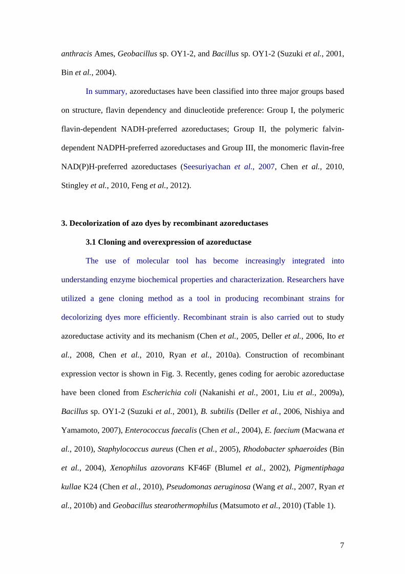
mineralization of the toxic aromatic amines. Scheme of bacterial azoreductase
studying is summarized in Fig 4. However, the physiological role and gene regulation
of azoreductase genes remain to be elucidated. Not surprisingly, few reports indicated
that azoreductases may be involved in detoxification due to the fact that some toxic
aromatic amine intermediates are formed during decolorization. Therefore,
understanding the regulation of azoreductase gene expression will lead to the use of
gene manipulation of bacterial strains systematically with higher biotransformation in
future technologies.
16
References
Anjaneya, O., Souche, S.Y., Santoshkumar, M. and Karegoudar, T.B. 2011.
Decolorization of sulfonated azo dye Metanil Yellow by newly isolated
bacterial strains: Bacillus sp. strain AK1 and Lysinibacillus sp. strain AK2.
Journal of Hazardous Materials. 190, 351-358.
Appelbaum, E.R. and Shatzman, A.R. 1999. Prokaryotic in vivo expression systems.
In Protein Expression, S.J. Higgins and B.D. Hames, editors. The Practical
Approach Series. Oxford University Press, New York, pp. 169-199.
Bafana, A., Chakrabarti, T. and Devi, S.S. 2008. Azoreductase and dye detoxification
activities of Bacillus velezensis strain AB. Applied Microbiology
Biotechnology. 77, 1139-1144.
Baiocchi, C., Brussino, M.C., Pramauro, E., Prevot, A.B., Palmisano, L. and Marci,
G. 2002. Characterization of methyl orange and its photocatalytic degradation
products by HPLC/UV-VIS diode array and atmospheric pressure ionization
quadrupole ion trap mass spectrometry. International Journal of Mass
Spectrometry. 214, 247-256.
Bin, Y., Jiti, Z., Jing, W., Cuihong, D., Hongman, H., Zhiyong, S. and Yongming, B.
2004. Expression and characteristics of the gene encoding azoreductase from
Rhodobacter sphaeroides AS1.1737. FEMS Microbiology Letters. 236, 129-
136.
Blumel, S., Knackmuss, H.J. and Stolz, A. 2002. Molecular cloning and
characterization of the gene coding for the aerobic azoreductase from
Xenophilus azovorans KF46F. Applied Environmental Microbiology. 68,
3948-3955.
17
Boye, K., Hogdall, E. and Borre, M. 1999. Identification of bacteria using two
degenerate 16S rDNA sequencing primers. Microbiological Research. 154, 23-
26.
Chen, H., Wang, R.F. and Cerniglia, C.E. 2004. Molecular cloning, overexpression,
purification, and characterization of an aerobic FMN-dependent azoreductase
from Enterococcus faecalis. Protein Expression & Purification. 34, 302-310.
Chen, H., Hopper, S.L. and Cerniglia, C.E. 2005. Biochemical and molecular
characterization of an azoreductase from Staphylococcus aureus, a tetrameric
NADPH-dependent flavoprotein. Microbiology. 151, 1433-1441.
Chen, H., Feng, J., Kweon, O., Xu, H. and Cerniglia, C.E. 2010. Identification and
molecular characterization of a novel flavin-free NADPH preferred
azoreductase encoded by azoB in Pigmentiphaga kullae K24. BMC
Biochemistry. 11, 13.
Chi, B.K., Albrecht, D., Gronau, K., Becher, D., Hecker, M. and Antelmann, H. 2010.
The redox-sensing regulator YodB senses quinones and diamide via a thiol-
disulfide switch in Bacillus subtilis. Proteomics. 10, 3155-3164.
Clarridge, J.E., 3rd. 2004. Impact of 16S rRNA gene sequence analysis for
identification of bacteria on clinical microbiology and infectious diseases.
Clinical Microbiology Reviews. 17, 840-862.
Dafale, N., Rao, N.N., Meshram, S.U. and Wate, S.R. 2008. Decolorization of azo
dyes and simulated dye bath wastewater using acclimatized microbial
consortium-biostimulation and halo tolerance. Bioresource Technology. 99,
2552-2558.
18
Daneshvar, N., Ayazloo, M., Khataee, A.R. and Pourhassan, M. 2007. Biological
decolorization of dye solution containing Malachite Green by microalgae
Cosmarium sp. Bioresource Technology. 98, 1176-1182.
Deive, F.J., Dominguez, A., Barrio, T., Moscoso, F., Moran, P., Longo, M.A. and
Sanroman, M.A. 2010. Decolorization of dye Reactive Black 5 by newly
isolated thermophilic microorganisms from geothermal sites in Galicia
(Spain). Journal of Hazardous Materials. 182, 735-742.
Deller, S., Macheroux, P. and Sollner, S. 2008. Flavin-dependent quinone reductases.
Cellular and Molecular Life Sciences. 65, 141-160.
Deller, S., Sollner, S., Trenker-El-Toukhy, R., Jelesarov, I., Gubitz, G.M. and
Macheroux, P. 2006. Characterization of a thermostable NADPH:FMN
oxidoreductase from the mesophilic bacterium Bacillus subtilis. Biochemistry.
45, 7083-7091.
Feng, J., Kweon, O., Xu, H., Cerniglia, C.E. and Chen, H. 2012. Probing the NADH-
and Methyl Red-binding site of a FMN-dependent azoreductase (AzoA) from
Enterococcus faecalis. Archives Biochemistry Biophysics. 520, 99-107.
Forgacs, E., Cserhati, T. and Oros, G. 2004. Removal of synthetic dyes from
wastewaters: a review. Environment International. 30, 953-971.
Gopinath, K.P., Murugesan, S., Abraham, J. and Muthukumar, K. 2009. Bacillus sp.
mutant for improved biodegradation of Congo red: random mutagenesis
approach. Bioresource Technology. 100, 6295-6300.
Gou, M., Qu, Y., Zhou, J., Ma, F. and Tan, L. 2009. Azo dye decolorization by a new
fungal isolate, Penicillium sp. QQ and fungal-bacterial cocultures. Journal of
Hazardous Materials. 170, 314-319.
19
Ito, K., Nakanishi, M., Lee, W. C., Zhi, Y., Sasaki, H., Zenno, S., Saigo, K., Kitade,
Y. and Tanokura, M. 2008. Expansion of substrate specificity and catalytic
mechanism of azoreductase by X-ray crystallography and site-directed
mutagenesis. Journal of Biological Chemistry. 283, 13889-13896.
Jadhav, J P. and Govindwar, S.P. 2006. Biotransformation of malachite green by
Saccharomyces cerevisiae MTCC 463. Yeast. 23, 315-323.
Jadhav, J.P., Parshetti, G.K., Kalme, S.D. and Govindwar, S.P. 2007. Decolourization
of azo dye methyl red by Saccharomyces cerevisiae MTCC 463.
Chemosphere. 68, 394-400.
Jadhav, J.P., Kalyani, D.C., Telke, A.A., Phugare, S.S. and Govindwar, S.P. 2010.
Evaluation of the efficacy of a bacterial consortium for the removal of color,
reduction of heavy metals, and toxicity from textile dye effluent. Bioresource
Technology. 101, 165-173.
Junnarkar, N., Murty D.S., Bhatt N.S., Madamwar S.P. 2006. Decolorization of diazo
dye direct red 81 by a novel bacterial consortium. World Journal of
Microbiology Biotechnology. 22, 163-168.
Kalme, S.D., Parshetti, G.K., Jadhav, S.U. and Govindwar, S.P. 2007. Biodegradation
of benzidine based dye Direct Blue-6 by Pseudomonas desmolyticum NCIM
2112. Bioresource Technology. 98, 1405-1410.
Kaushik, P. and Malik, A. 2009. Fungal dye decolourization: recent advances and
future potential. Environment International. 35, 127-141.
Khehra, M.S., Saini, H.S., Sharma, D.K., Chadha, B.S. and Chimni, S.S. 2005.
Decolorization of various azo dyes by bacterial consortium. Dyes and
Pigments. 67, 55-61.
20
Kolekar, Y.M., Konde, P.D., Markad, V.L., Kulkarni, S.V., Chaudhari, A.U. and
Kodam, K.M. 2013. Effective bioremoval and detoxification of textile dye
mixture by Alishewanella sp. KMK6. Applied Microbiology Biotechnology.
97, 881-889.
Kumar, K., Saravana Devi, S., Krishnamurthi, K., Gampawar, S., Mishra, N., Pandya,
G.H. and Chakrabarti, T. 2006. Decolorisation, biodegradation and
detoxification of benzidine based azo dye. Bioresource Technology. 97, 407-
413.
Leelakriangsak, M. and Borisut, S. 2012. Characterization of the decolorizing activity
of azo dyes by Bacillus subtilis. Songklanakarin Journal of Science and
Technology. 34, 509-516.
Leelakriangsak, M., Huyen, N.T., Towe, S., Duy, N.V, Becher, D., Hecker, M.,
Antelmann, H. and Zuber, P. 2008. Regulation of quinone detoxification by
the thiol stress sensing DUF24/MarR-like repressor, YodB in Bacillus subtilis.
Molecular Microbiology. 67, 1108-1124.
Liu, G., Zhou, J., Fu, Q. S. and Wang, J. 2009a. The Escherichia coli azoreductase
AzoR Is involved in resistance to thiol-specific stress caused by electrophilic
quinones. Journal of Bacteriology. 191, 6394-6400.
Liu, G., Zhou, J., Wang, J., Zhou, M., Lu, H. and Jin, R. 2009b. Acceleration of azo
dye decolorization by using quinone reductase activity of azoreductase and
quinone redox mediator. Bioresource Technology. 100, 2791-2795.
Liu, G., Zhou, J., Jin, R., Zhou, M., Wang, J., Lu, H. and Qu, Y. 2008a. Enhancing
survival of Escherichia coli by expression of azoreductase AZR possessing
quinone reductase activity. Applied Microbiology Biotechnology. 80, 409-
416.
21
Liu, G., Zhou, J., Meng, X., Fu, S.Q., Wang, J., Jin, R. and Lv, H. 2013.
Decolorization of azo dyes by marine Shewanella strains under saline
conditions. Applied Microbiology Biotechnology. 97, 4187-4197.
Liu, G., Zhou, J., Wang, J., Yan, B., Li, J., Lu, H., Qu, Y. and Jin, R. 2008b. Site-
directed mutagenesis of substrate binding sites of azoreductase from
Rhodobacter sphaeroides. Biotechnology Letters. 30, 869-875.
Mabrouk, M.E.M. and Yusef, H.H. 2008. Decolorization of Fast Red by Bacillis
subtilis HM. Journal of Applied Sciences Research. 4, 262-269.
Macwana, S.R., Punj, S., Cooper, J., Schwenk, E. and John, G.H. 2010. Identification
and isolation of an azoreductase from Enterococcus faecium. Current Issues in
Molecular Biology. 12, 43-48.
Maier, J., Kandelbauer, A., Erlacher, A., Cavaco-Paulo, A. and Gubitz, G.M. 2004. A
new alkali-thermostable azoreductase from Bacillus sp. strain SF. Applied and
Environmental Microbiology. 70, 837-844.
Matsumoto, K., Mukai, Y., Ogata, D., Shozui, F., Nduko, J.M., Taguchi, S. and Ooi,
T. 2010. Characterization of thermostable FMN-dependent NADH
azoreductase from the moderate thermophile Geobacillus stearothermophilus.
Applied Microbiology Biotechnology. 86, 1431-1438.
Mendes, S., Pereira, L., Batista, C. and Martins, L.O. 2011a. Molecular determinants
of azo reduction activity in the strain Pseudomonas putida MET94. Applied
Microbiology Biotechnology. 92, 393-405.
Mendes, S., Farinha, A., Ramos, C.G., Leitao, J.H., Viegas, C.A. and Martins, L.O.
2011b. Synergistic action of azoreductase and laccase leads to maximal
decolourization and detoxification of model dye-containing wastewaters.
Bioresource Technology. 102, 9852-9859.
22
Mohanty, S., Dafale, N. and Rao, N.N. 2006. Microbial decolorization of reactive
black-5 in a two-stage anaerobic-aerobic reactor using acclimatized activated
textile sludge. Biodegradation. 17, 403-413.
Morrison, J.M., Wright, C.M. and John, G.H. 2012. Identification, Isolation and
characterization of a novel azoreductase from Clostridium perfringens.
Anaerobe. 18, 229-234.
Moutaouakkil, A., Zeroual, Y., Zohra Dzayri, F., Talbi, M., Lee, K. and Blaghen, M.
2003. Purification and partial characterization of azoreductase from
Enterobacter agglomerans. Archives of Biochemistry and Biophysics. 413,
139-146.
Mugerfeld, I., Law, B.A., Wickham, G.S. and Thompson, D.K. 2009. A putative
azoreductase gene is involved in the Shewanella oneidensis response to heavy
metal stress. Applied Microbiology Biotechnology. 82, 1131-1141.
Nachiyar, C.V. and Rajakumar, G.S. 2005. Purification and characterization of an
oxygen insensitive azoreductase from Pseudomonas aeruginosa. Enzyme and
Microbial Technology. 36, 503-509.
Nakanishi, M., Yatome, C., Ishida, N. and Kitade, Y. 2001. Putative ACP
phosphodiesterase gene (acpD) encodes an azoreductase. Journal of Biological
Chemistry. 276, 46394-46399.
Nishiya, Y. and Yamamoto, Y. 2007. Characterization of a
NADH:dichloroindophenol oxidoreductase from Bacillus subtilis. Bioscience
Biotechnology Biochemistry. 71, 611-614.
Ooi, T., Shibata, T., Matsumoto, K., Kinoshita, S. and Taguchi, S. 2009. Comparative
enzymatic analysis of azoreductases from Bacillus sp. B29. Bioscience
Biotechnology Biochemistry. 73, 1209-1211.
23
Ooi, T., Shibata, T., Sato, R., Ohno, H., Kinoshita, S., Thuoc, T.L. and Taguchi, S.
2007. An azoreductase, aerobic NADH-dependent flavoprotein discovered
from Bacillus sp.: functional expression and enzymatic characterization.
Applied Microbiology Biotechnology. 75, 377-386.
Pandey, A., Singh, P. and Iyengar, L. 2007. Bacterial decolorization and degradation
of azo dyes. International Biodeterioration & Biodegradation. 59, 73-84.
Parshetti, G.K., Telke, A.A., Kalyani, D.C. and Govindwar, S.P. 2010. Decolorization
and detoxification of sulfonated azo dye methyl orange by Kocuria rosea
MTCC 1532. Journal of Hazardous Materials. 176, 503-509.
Pricelius, S., Held, C., Murkovic, M., Bozic, M., Kokol, V., Cavaco-Paulo, A. and
Guebitz, G. M. 2007. Enzymatic reduction of azo and indigoid compounds.
Applied Microbiology Biotechnology. 77, 321-327.
Punj, S. and John, G.H. 2009. Purification and Identification of an FMN-dependent
NAD(P)H Azoreductase from Enterococcus faecalis. Current Issues in
Molecular Biology. 11, 59-66.
Rafii, F. and Cerniglia, C.E. 1993. Comparison of the azoreductase and nitroreductase
from Clostridium perfringens. Applied Environmental Microbiology. 59,
1731-1734.
Rafii, F., Franklin, W. and Cerniglia, C.E. 1990. Azoreductase activity of anaerobic
bacteria isolated from human intestinal microflora. Applied Environmental
Microbiology. 56, 2146-2151.
Robinson, T., McMullan, G., Marchant, R. and Nigam, P. 2001. Remediation of dyes
in textile effluent: a critical review on current treatment technologies with a
proposed alternative. Bioresource Technology. 77, 247-255.
24
Ryan, A., Wang, C.J., Laurieri, N., Westwood, I. and Sim, E. 2010a. Reaction
mechanism of azoreductases suggests convergent evolution with quinone
oxidoreductases. Protein Cell. 1, 780-790.
Ryan, A., Laurieri, N., Westwood, I., Wang, C.J., Lowe, E. and Sim, E. 2010b. A
novel mechanism for azoreduction. Journal of Molecular Biology. 400, 24-37.
Sandhya, S., Sarayu, K., Uma, B. and Swaminathan, K. 2008. Decolorizing kinetics of
a recombinant Escherichia coli SS125 strain harboring azoreductase gene
from Bacillus latrosporus RRK1. Bioresource Technology. 99, 2187-2191.
Saratale, R.G., Saratale, G.D., Chang, J.S. and Govindwar, S.P. 2011. Baterial
decolorization and degradation of azo dyes: A review. Journal of the Taiwan
Institute of Chemical Engineers. 42, 138-157.
Saratale, R.G., Saratale, G.D., Kalyani, D.C., Chang, J.S. and Govindwar, S.P. 2009.
Enhanced decolorization and biodegradation of textile azo dye Scarlet R by
using developed microbial consortium-GR. Bioresource Technology. 100,
2493-2500.
Seesuriyachan, P., Takenaka, S., Kuntiya, A., Klayraung, S., Murakami, S. and Aoki,
K. 2007. Metabolism of azo dyes by Lactobacillus casei TISTR 1500 and
effects of various factors on decolorization. Water Research. 41, 985-992.
Shafique, S., Bajwa, R. and Shafique, S. 2010. Molecular characterisation of UV and
chemically induced mutants of Trichoderma reesei FCBP-364. Natural
Product Research. 24, 1438-1448.
Song, Z.Y., Zhou, J.T., Wang, J., Yan, B. and Du, C.H. 2003. Decolorization of azo
dyes by Rhodobacter sphaeroides. Biotechnology Letters. 25, 1815-1818.
25
Stingley, R.L., Zou, W., Heinze, T.M., Chen, H. and Cerniglia, C.E. 2010.
Metabolism of azo dyes by human skin microbiota. Journal of Medical
Microbiology. 59, 108-114.
Stolz, A. 2001. Basic and applied aspects in the microbial degradation of azo dyes.
Applied Microbiology Biotechnology. 56, 69-80.
Sugiura, W., Yoda, T., Matsuba, T., Tanaka, Y. and Suzuki, Y. 2006. Expression and
characterization of the genes encoding azoreductases from Bacillus subtilis
and Geobacillus stearothermophilus. Bioscience Biotechnology Biochemistry.
70, 1655-1665.
Supaka, N., Juntongjin, K., Damronglerd, S., Delia, M.-L. and Strehaiano, P. 2004.
Microbial decolorization of reactive azo dyes in a sequential anaerobic-aerobic
system. Chemical Engineering Journal. 99, 169-176.
Suzuki, Y., Yoda, T., Ruhul, A. and Sugiura, W. 2001. Molecular cloning and
characterization of the gene coding for azoreductase from Bacillus sp. OY1-2
isolated from soil. Journal of Biological Chemistry. 276, 9059-9065.
Tastan, B.E., Ertugrul, S. and Donmez, G. 2010. Effective bioremoval of reactive dye
and heavy metals by Aspergillus versicolor. Bioresource Technology. 101,
870-876.
Telke, A.A., Joshi, S.M., Jadhav, S.U., Tamboli, D.P. and Govindwar, S.P. 2010.
Decolorization and detoxification of Congo red and textile industry effluent by
an isolated bacterium Pseudomonas sp. SU-EBT. Biodegradation. 21, 283-
296.
Tortoli, E. 2003. Impact of genotypic studies on mycobacterial taxonomy: the new
mycobacteria of the 1990s. Clinical Microbiology Reviews. 16, 319-354.
26
Töwe, S., Leelakriangsak, M., Kobayashi, K., Duy, N.V, Hecker, M., Zuber, P. and
Antelmann, H. 2007. The MarR-type repressor MhqR (YkvE) regulates
multiple dioxygenases/glyoxalases and an azoreductase which confer
resistance to 2-methylhydroquinone and catechol in Bacillus subtilis.
Molecular Microbiology. 66, 40-54.
Wang, C.J., Laurieri, N., Abuhammad, A., Lowe, E., Westwood, I., Ryan, A. and
Sim, E. 2010. Role of tyrosine 131 in the active site of paAzoR1, an
azoreductase with specificity for the inflammatory bowel disease prodrug
balsalazide. Acta Crystallographica Section F: Structural Biology and
Crystallization Communications. 66, 2-7.
Wang, C.J., Hagemeier, C., Rahman, N., Lowe, E., Noble, M., Coughtrie, M., Sim, E.
and Westwood, I. 2007. Molecular cloning, characterisation and ligand-bound
structure of an azoreductase from Pseudomonas aeruginosa. Journal of
Molecular Biology. 373, 1213-1228.
Woo, P.C., Lau, S.K., Teng, J.L., Tse, H. and Yuen, K.Y. 2008. Then and now: use of
16S rDNA gene sequencing for bacterial identification and discovery of novel
bacteria in clinical microbiology laboratories. Clinical Microbiology and
Infection. 14, 908-934.
27
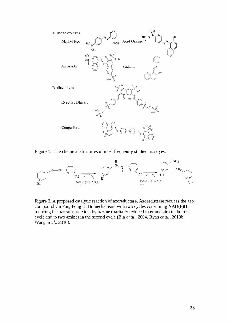
Figure 1. The chemical structures of most frequently studied azo dyes.
R1
N N
R2 R1
N N
R2
H
H
NAD(P)H+ H+
NAD(P)+
R1
NH2
NH2
R2NAD(P)H+ H+
NAD(P)+
Figure 2. A proposed catalytic reaction of azoreductase. Azoreductase reduces the azo compound via Ping Pong Bi Bi mechanism, with two cycles consuming NAD(P)H, reducing the azo substrate to a hydrazine (partially reduced intermediate) in the first cycle and to two amines in the second cycle (Bin et al., 2004, Ryan et al., 2010b, Wang et al., 2010).
28
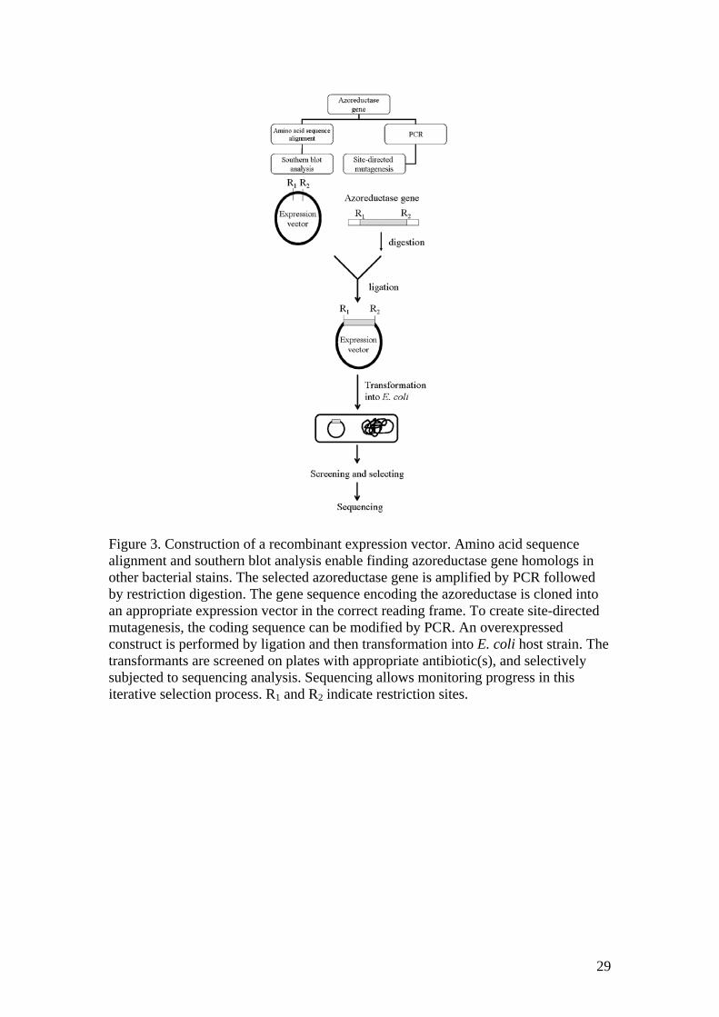
Figure 3. Construction of a recombinant expression vector. Amino acid sequence alignment and southern blot analysis enable finding azoreductase gene homologs in other bacterial stains. The selected azoreductase gene is amplified by PCR followed by restriction digestion. The gene sequence encoding the azoreductase is cloned into an appropriate expression vector in the correct reading frame. To create site-directed mutagenesis, the coding sequence can be modified by PCR. An overexpressed construct is performed by ligation and then transformation into E. coli host strain. The transformants are screened on plates with appropriate antibiotic(s), and selectively subjected to sequencing analysis. Sequencing allows monitoring progress in this iterative selection process. R1 and R2 indicate restriction sites.
29
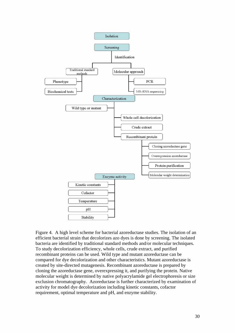
Figure 4. A high level scheme for bacterial azoreductase studies. The isolation of an efficient bacterial strain that decolorizes azo dyes is done by screening. The isolated bacteria are identified by traditional standard methods and/or molecular techniques. To study decolorization efficiency, whole cells, crude extract, and purified recombinant proteins can be used. Wild type and mutant azoreductase can be compared for dye decolorization and other characteristics. Mutant azoreductase is created by site-directed mutagenesis. Recombinant azoreductase is prepared by cloning the azoreductase gene, overexpressing it, and purifying the protein. Native molecular weight is determined by native polyacrylamide gel electrophoresis or size exclusion chromatography. Azoreductase is further characterized by examination of activity for model dye decolorization including kinetic constants, cofactor requirement, optimal temperature and pH, and enzyme stability.
30
31
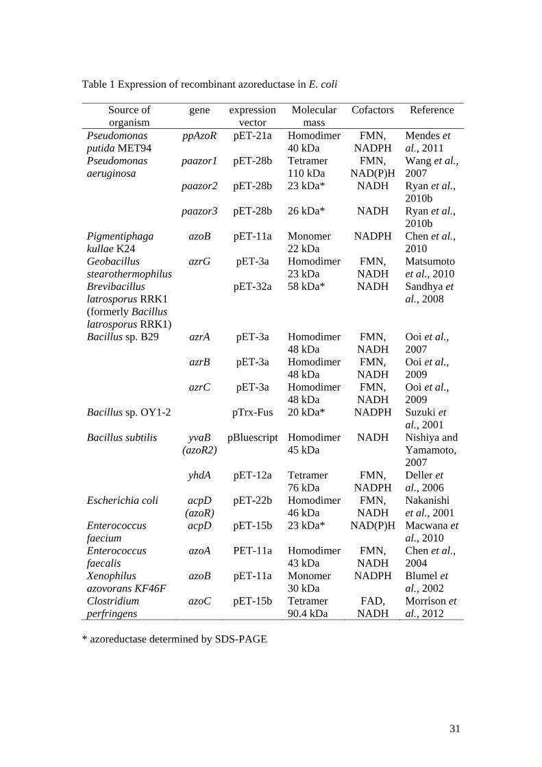
Table 1 Expression of recombinant azoreductase in E. coli
Source of organism
gene expression vector
Molecular mass
Cofactors Reference
Pseudomonas putida MET94
ppAzoR pET-21a Homodimer 40 kDa
FMN, NADPH
Mendes et al., 2011
Pseudomonas aeruginosa
paazor1 pET-28b Tetramer 110 kDa
FMN, NAD(P)H
Wang et al., 2007
paazor2 pET-28b 23 kDa* NADH Ryan et al., 2010b
paazor3 pET-28b 26 kDa* NADH Ryan et al., 2010b
Pigmentiphaga kullae K24
azoB pET-11a Monomer 22 kDa
NADPH Chen et al., 2010
Geobacillus stearothermophilus
azrG pET-3a Homodimer 23 kDa
FMN, NADH
Matsumoto et al., 2010
Brevibacillus latrosporus RRK1 (formerly Bacillus latrosporus RRK1)
pET-32a 58 kDa* NADH Sandhya et al., 2008
Bacillus sp. B29 azrA pET-3a Homodimer 48 kDa
FMN, NADH
Ooi et al., 2007
azrB pET-3a Homodimer 48 kDa
FMN, NADH
Ooi et al., 2009
azrC pET-3a Homodimer 48 kDa
FMN, NADH
Ooi et al., 2009
Bacillus sp. OY1-2 pTrx-Fus 20 kDa* NADPH Suzuki et al., 2001
Bacillus subtilis yvaB (azoR2)
pBluescript Homodimer 45 kDa
NADH Nishiya and Yamamoto, 2007
yhdA pET-12a Tetramer 76 kDa
FMN, NADPH
Deller et al., 2006
Escherichia coli acpD (azoR)
pET-22b Homodimer 46 kDa
FMN, NADH
Nakanishi et al., 2001
Enterococcus faecium
acpD pET-15b 23 kDa* NAD(P)H Macwana et al., 2010
Enterococcus faecalis
azoA PET-11a Homodimer 43 kDa
FMN, NADH
Chen et al., 2004
Xenophilus azovorans KF46F
azoB pET-11a Monomer 30 kDa
NADPH Blumel et al., 2002
Clostridium perfringens
azoC pET-15b Tetramer 90.4 kDa
FAD, NADH
Morrison et al., 2012
* azoreductase determined by SDS-PAGE